Embed Size (px)
Citation preview
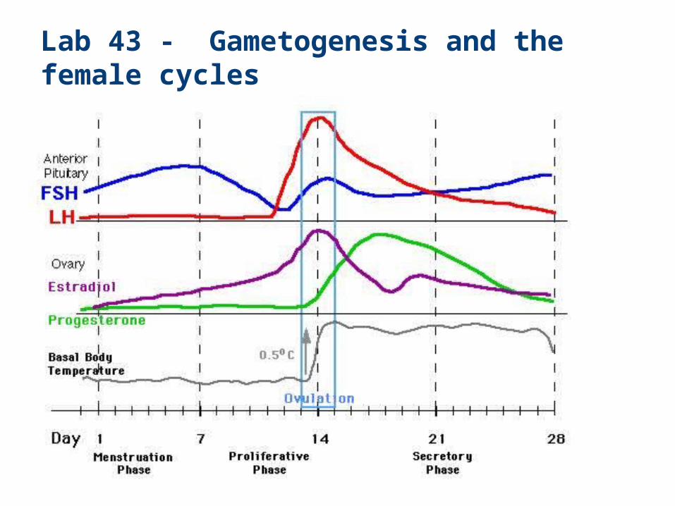
Lab 43 - Gametogenesis and the female cycles
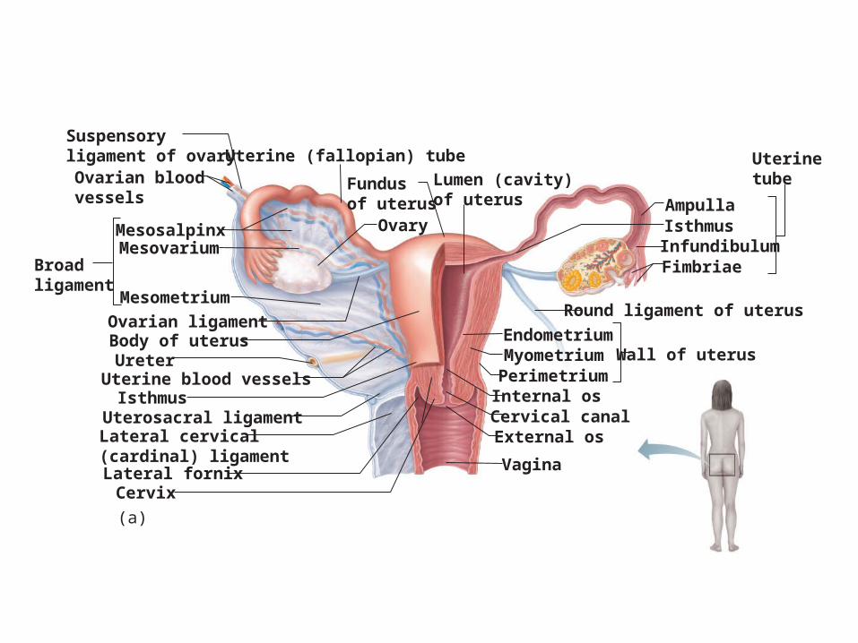
Vagina
External osCervical canalInternal os
Wall of uterusPerimetriumMyometriumEndometrium
Round ligament of uterus
Uterinetube
InfundibulumFimbriae
IsthmusAmpulla
Lumen (cavity)of uterus
Suspensoryligament of ovary Uterine (fallopian) tubeOvarian bloodvessels
MesosalpinxMesovarium
Broadligament
Mesometrium
Ovary
Ovarian ligamentBody of uterusUreterUterine blood vesselsIsthmusUterosacral ligamentLateral cervical(cardinal) ligamentLateral fornixCervix
(a)
Fundusof uterus

Ovaries
• Follicle
• Immature egg (oocyte) surrounded by
• Follicle cells (one cell layer thick)
• Granulosa cells (when more than one layer is present)
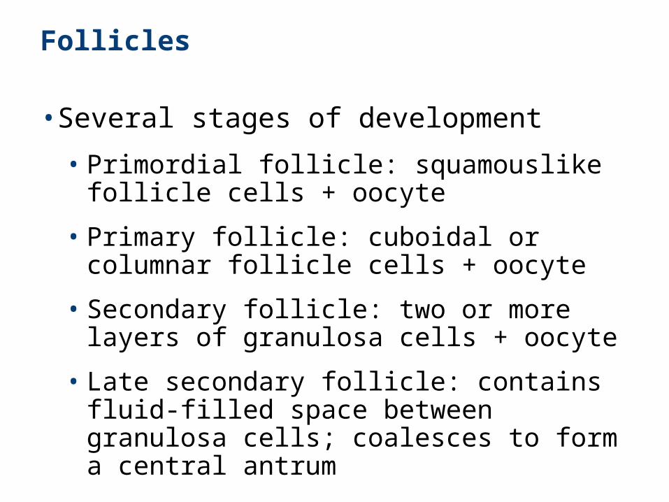
Follicles
• Several stages of development
• Primordial follicle: squamouslike follicle cells + oocyte
• Primary follicle: cuboidal or columnar follicle cells + oocyte
• Secondary follicle: two or more layers of granulosa cells + oocyte
• Late secondary follicle: contains fluid-filled space between granulosa cells; coalesces to form a central antrum
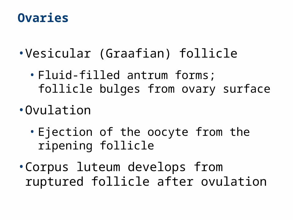
Ovaries
• Vesicular (Graafian) follicle
• Fluid-filled antrum forms; follicle bulges from ovary surface
• Ovulation
• Ejection of the oocyte from the ripening follicle
• Corpus luteum develops from ruptured follicle after ovulation
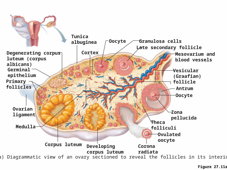
Figure 27.11a
Medulla
Tunicaalbuginea
Germinalepithelium
Cortex
Oocyte Granulosa cellsLate secondary follicle
Antrum
Primaryfollicles
Oocyte
Zonapellucida
Thecafolliculi
Ovulatedoocyte
Mesovarium andblood vessels
Vesicular(Graafian)follicle
Coronaradiata
Developingcorpus luteum
Corpus luteum
Ovarianligament
Degenerating corpusluteum (corpus albicans)
(a) Diagrammatic view of an ovary sectioned to reveal the follicles in its interior
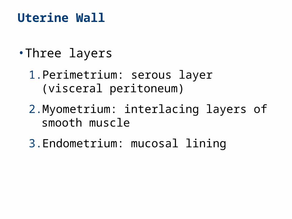
Uterine Wall
• Three layers
1. Perimetrium: serous layer (visceral peritoneum)
2. Myometrium: interlacing layers of smooth muscle
3. Endometrium: mucosal lining
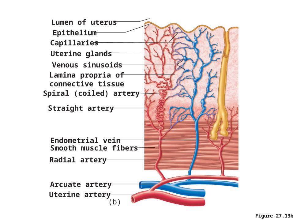
Figure 27.13b
Lumen of uterus
Uterine glands
Smooth muscle fibers
Straight artery
Radial artery
Arcuate arteryUterine artery
Endometrial vein
Capillaries
Venous sinusoids
Epithelium
Spiral (coiled) artery
Lamina propria ofconnective tissue
(b)
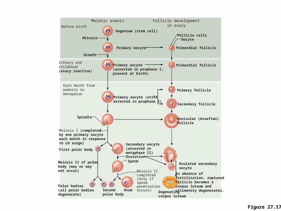
Figure 27.17
Meiotic events Follicle developmentin ovaryBefore birth
Infancy andchildhood(ovary inactive)
Primary oocyte
Primary oocyte (stillarrested in prophase I)
Vesicular (Graafian)follicle
Primary follicle
Primordial follicle
Primordial follicle
Oocyte
Ovulated secondaryoocyte
In absence offertilization, ruptured follicle becomes a corpus luteum andultimately degenerates.Degenating
corpus luteum
Secondary follicle
Primary oocyte(arrested in prophase I;present at birth)
Oogonium (stem cell)
Each month frompuberty to menopause
Meiosis I (completed by one primary oocyte each month in response to LH surge)
First polar body
Mitosis
Growth
Meiosis II of polarbody (may or may not occur)
Polar bodies(all polar bodiesdegenerate)
OvumSecondpolar body
Meiosis IIcompleted(only if spermpenetration occurs)
SpermOvulation
Secondary oocyte(arrested in metaphase II)
Follicle cells
Spindle

Ovarian Cycle
• Monthly series of events associated with the maturation of an egg
• Two consecutive phases (in a 28-day cycle)
• Follicular phase: period of follicle growth (days 1–14)
• Ovulation occurs midcycle
• Luteal phase: period of corpus luteum activity (days 14–28)
Follicular Phase
• Primordial follicle becomes primary follicle
1. The primordial follicle is activated
• Squamouslike cells become cuboidal
2. Follicle enlarges to become a primary (1) follicle
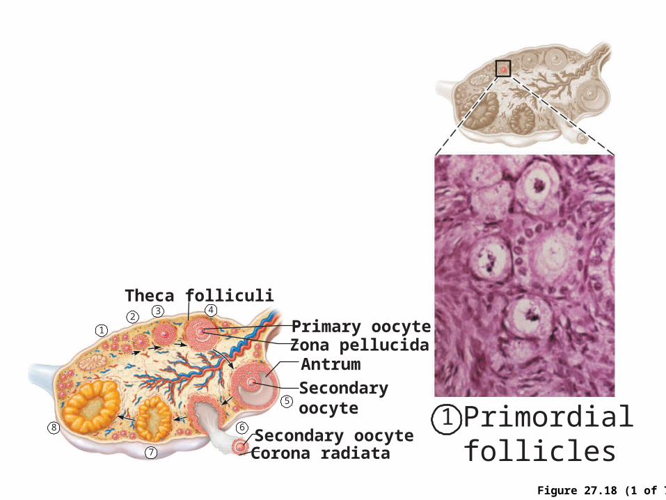
Figure 27.18 (1 of 7)
Theca folliculi
Primary oocyteZona pellucidaAntrum
Secondaryoocyte
Secondary oocyteCorona radiata
12
3 4
5
6
7
8 Primordialfollicles
1
Luteal Phase
• Ruptured follicle collapses
• Granulosa cells and internal thecal cells form corpus luteum
• Corpus luteum secretes progesterone and estrogen
Establishing the Ovarian Cycle
• During childhood, ovaries grow and secrete small amounts of estrogens that inhibit the hypothalamic release of GnRH
• As puberty nears, GnRH is released; FSH and LH are released by the pituitary, and act on the ovaries
• These events continue until an adult cyclic pattern is achieved and menarche occurs
Establishing the Ovarian Cycle
• At puberty
• Leptin from adipose tissue decreases the estrogen inhibition
• GnRH, FSH, and LH are released
• In about four years, an adult cyclic pattern is achieved and menarche occurs
Hormonal Interactions During a 28-Day Ovarian Cycle
• Day 1: GnRH release of FSH and LH
• FSH and LH growth of several follicles, and estrogen release
• estrogen levels
• Inhibit the release of FSH and LH
• Stimulate synthesis and storage of FSH and LH
• Enhance further estrogen output
Hormonal Interactions During a 28-Day Ovarian Cycle
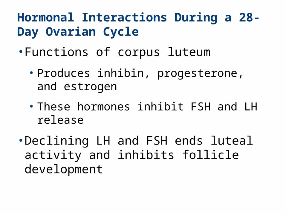
• Functions of corpus luteum
• Produces inhibin, progesterone, and estrogen
• These hormones inhibit FSH and LH release
• Declining LH and FSH ends luteal activity and inhibits follicle development
Hormonal Interactions During a 28-Day Ovarian Cycle
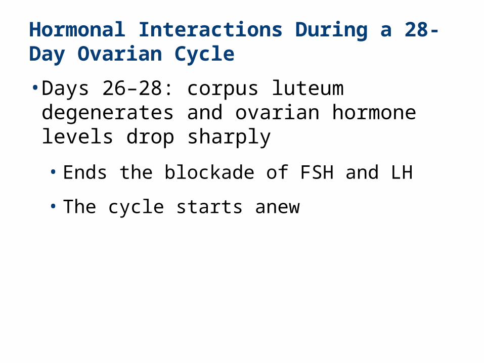
• Days 26–28: corpus luteum degenerates and ovarian hormone levels drop sharply
• Ends the blockade of FSH and LH
• The cycle starts anew
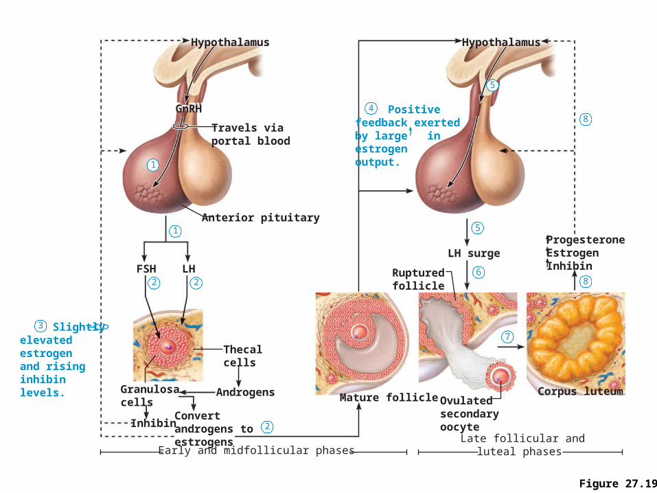
Figure 27.19
Hypothalamus
Late follicular andluteal phases
1
1
2 2
2
3
4
5
5
6
8
8
7 Slightlyelevated estrogen and rising inhibin levels.
Positivefeedback exerted by large inestrogen output.
Mature follicleCorpus luteum
Ovulatedsecondaryoocyte
Rupturedfollicle
LH surgeProgesteroneEstrogenInhibin
Hypothalamus
Early and midfollicular phases
Travels viaportal blood
Granulosacells
Inhibin
Androgens
Convertandrogens toestrogens
Thecalcells
Anterior pituitary
GnRH
FSH LH
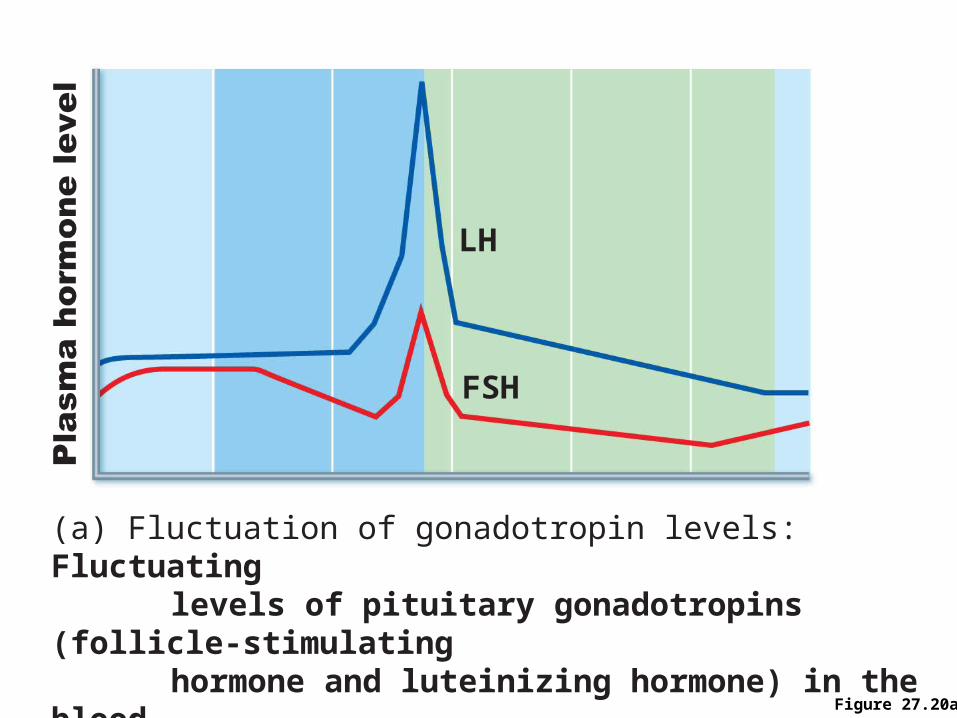
Figure 27.20a
(a) Fluctuation of gonadotropin levels: Fluctuating levels of pituitary gonadotropins (follicle-stimulating hormone and luteinizing hormone) in the blood regulate the events of the ovarian cycle.
FSH
LH
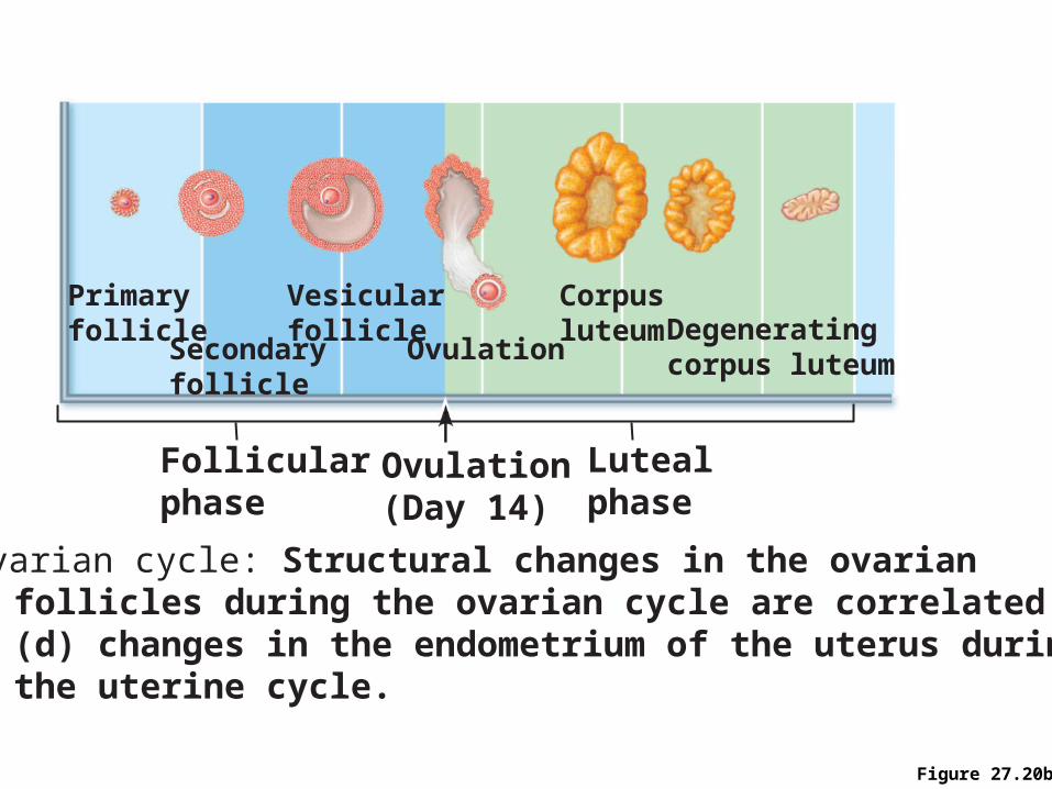
Figure 27.20b
(b) Ovarian cycle: Structural changes in the ovarian follicles during the ovarian cycle are correlated with (d) changes in the endometrium of the uterus during the uterine cycle.
Primaryfollicle
Secondaryfollicle
Vesicularfollicle
Ovulation
Corpusluteum Degenerating
corpus luteum
Follicularphase
Ovulation(Day 14)
Lutealphase
Uterine (Menstrual) Cycle
• Cyclic changes in endometrium in response to ovarian hormones
• Three phases
• Days 1–5: menstrual phase
• Days 6–14: proliferative (preovulatory) phase
• Days 15–28: secretory (postovulatory) phase (constant 14-day length)
Uterine Cycle
• Menstrual phase
• Ovarian hormones are at their lowest levels
• Gonadotropins are beginning to rise
• Stratum functionalis is shed and the menstrual flow occurs
Uterine Cycle
• Proliferative phase
• Estrogen levels prompt generation of new functional layer and increased synthesis of progesterone receptors in endometrium
• Glands enlarge and spiral arteries increase in number
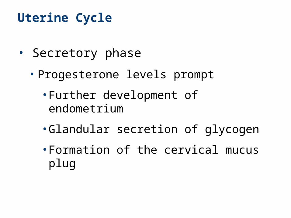
Uterine Cycle
• Secretory phase
• Progesterone levels prompt
• Further development of endometrium
• Glandular secretion of glycogen
• Formation of the cervical mucus plug
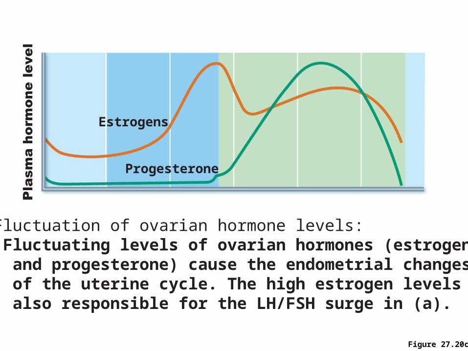
Figure 27.20c
(c) Fluctuation of ovarian hormone levels: Fluctuating levels of ovarian hormones (estrogens and progesterone) cause the endometrial changes of the uterine cycle. The high estrogen levels are also responsible for the LH/FSH surge in (a).
Progesterone
Estrogens
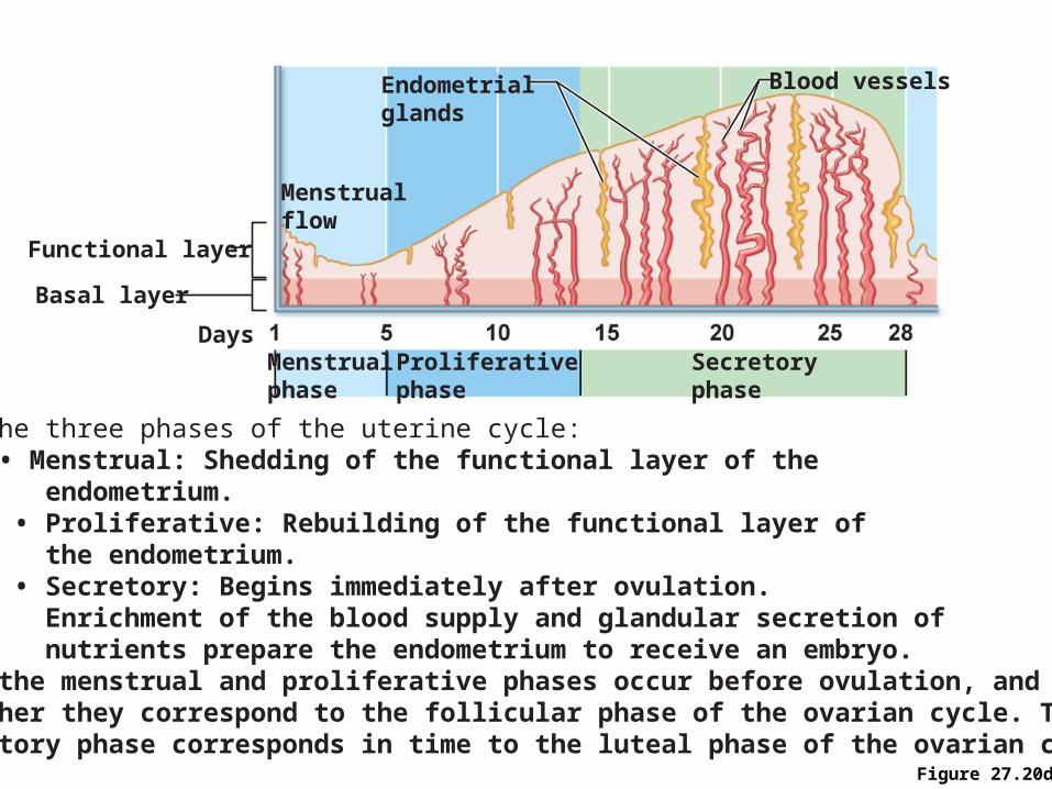
Figure 27.20d
(d) The three phases of the uterine cycle: • Menstrual: Shedding of the functional layer of the endometrium. • Proliferative: Rebuilding of the functional layer of the endometrium. • Secretory: Begins immediately after ovulation. Enrichment of the blood supply and glandular secretion of nutrients prepare the endometrium to receive an embryo.Both the menstrual and proliferative phases occur before ovulation, and together they correspond to the follicular phase of the ovarian cycle. Thesecretory phase corresponds in time to the luteal phase of the ovarian cycle.
Menstrualphase
Menstrualflow
Endometrialglands
Blood vessels
Functional layer
Basal layer
Proliferativephase
Secretoryphase
Days