Embed Size (px)
Citation preview

Oral histology
Lab 5
Cementum and periodontal ligament
Slide 1 A – dentin B - epithelial rests C – cementoblasts D - cementum
E - resting lines F - periodontal region
Formation of Cementum
Dentin (A) lies along the right margin of the field, the periodontal region
occupies the left side. Note the islands of epithelial cells (B) near the
center of the field. These islands are called epithelial rests (of Malassez).
As the epithelial root sheath breaks up, connective tissue of the dental
follicle (sac) comes into contact with the dentin. In these regions multi
potential cells of the dental follicle differentiate into cementoblasts (C)

adjacent to the dentin. They deposit a ground substance around collagen
fibers of fibroblast origin. The osteoid-like mixture of ground substance
with collagen fibers is called cementoid. Mineralization converts
cementoid to cementum.
Cementum (D) in this image contains several basophilic "resting" lines
(E) that reflect periods when formation slows down or stops and then
starts again. Resting lines are incremental lines formed by fiber-free
ground substance. No cells are trapped in this cementum so it is classified
as acellular cementum - the first type to be formed. It is also referred to as
primary cementum.

Slide 2 A - epithelial rests B – cementoblasts C – cementum
D - cementoid E - periodontal region F – dentin G – osteocytes
H – osteoblasts J - alveolar bone K - primary cementum
L - it is acellular
Epithelial rests (A) appear as a broken string of small dense
basophilic structures. Identify cementoblasts (B) whose dark nuclei
stand out along the thin line of light pink staining material between
the cementum (C) and the cementoblasts. This lighter layer is
cementoid (D) indicating active formation of cementum.
Slide 3
A - periodontal region B - epithelial rests C - primary cementum D - dentin E - Sharpey's fibers F - principal fibers
Slide 4
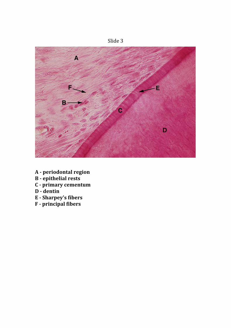
Primary Cementum
This is a ground section of a tooth. From the bottom of the field up
identify the following layers: dentin with dentinal tubules (A), Tomes'
granular layer (B), primary (acellular) cementum (C). Note that primary
cementum is a relatively clear layer, containing no cells (cementocytes).

Slide 5 Cementocyte A - resting lines
B – canalculi.
C-cementocyte lacuna

Slide 6
Overlapping at C-E Junction
A - cementum
B - enamel margin
C – dentin
Enamel Pearl in Section
Slide 7 Enamel pearl
Enamel pearls are localized masses of enamel that develop ectopically, typically over the root surface (A) in close proximity to the cemento-enamel junction (B)

Slide 8
Principal fibers
A - labial mucosa B - labial gingival C - gingival fibers D - alveolar crest
fibers E - oblique fibers F - alveolar bone G - enamel space

Slide 9
Horizontal Fibers of the PDL
Horizontal principal fibers (A) are seen in this field. Note they pass
horizontally from cementum (B) to the alveolar bone (C) just below its
crest. This initial group of horizontal fibers was once referred to as
"cervical fibers". Note that Sharpey's fibers (D) can be seen inserting into
the cemntum on the right, and the alveolar bone on the left.


![Clinical outcome of periodontal regenerative therapy using ... · on systemic health [2]. In the treatment of periodontitis, ... cementum, and periodontal ligament attachment to a](https://img.pdfslide.net/doc/110x75/5ed57ee1276f2405802693ed/clinical-outcome-of-periodontal-regenerative-therapy-using-on-systemic-health.jpg)