Embed Size (px)
Citation preview
Lab on a Chip
COMMUNICATION
Cite this: Lab Chip, 2015, 15, 4273
Received 9th August 2015,Accepted 11th September 2015
DOI: 10.1039/c5lc00953g
www.rsc.org/loc
Microfluidic multiplexed partitioning enablesflexible and effective utilization of magneticsensor arrays
Daniel J. B. Bechstein,a Elaine Ng,b Jung-Rok Lee,a Stephanie G. Cone,c
Richard S. Gaster,bd Sebastian J. Osterfeld,e Drew A. Hall,fg James A. Weaver,f
Robert J. Wilsonh and Shan X. Wang*fh
We demonstrate microfluidic partitioning of a giant
magnetoresistive sensor array into individually addressable
compartments that enhances its effective use. Using different
samples and reagents in each compartment enables measuring of
cross-reactive species and wide dynamic ranges on a single chip.
This compartmentalization technique motivates the employment
of high density sensor arrays for highly parallelized measurements
in lab-on-a-chip devices.
Following the decade old trend of “Moore's Law”,1 featuresizes in integrated circuits (ICs) continue to shrink. The samearguments of technological advances and economics supportshrinking sensor size and integrating more sensors persilicon chip, with the goal of large-scale sensor arrays that fea-ture highly parallelized multiplexing, high speed, and loweroverall costs.
Giant magnetoresistive (GMR) sensors are one of the mostpromising sensor technologies for protein diagnostics.2
Arrays of 64 GMR sensors have been demonstrated to per-form multiplexed protein detection on a single chip,3 andcurrent research targets an even larger number of sensors perarray.4 However, large-scale sensor arrays' utility and bioassayflexibility is limited by concentration mismatch and dynamicrange constraints as well as inter-assay cross-reactivitybetween biological analytes and reagents from differentassays.5 Thus, only a subset of protein assays of interest canbe performed in parallel on a single sensor array chip with-out exceeding cross-reactivity or dynamic range constraints.The dynamic range of sensors can be improved through
optimized particle selection, sensor design, and readout cir-cuitry, which reduces the constraints on concentrationranges.3,6–9 The cross-reactivity between analytes andreagents is a more stringent constraint for accurate readoutthat cannot be solved by increased sensor electronic perfor-mance, but needs to be taken care of at its source – the bio-chemical binding stage early in the detection workflow. Anti-body cross-reactivity5,10,11 and aberrant protein–proteininteractions12 are two undesired sources of assay cross-reac-tivity. This cross-reactivity becomes a problem in sensor-based multiplex assays where the goal is to integrate manytests onto a small sensing region, and leads to challengesand issues with assay reliability. Common approaches dealwith cross-reactivity similar to well-based assays, where eachassay is performed in a separate well. This separation pre-vents cross-reactivity by design: the cross-reactive assays areseparated onto different chips, ultimately requiring anincreased number of sensor chips rather than parallel pro-cessing on a single chip. However, this multi-chip separationapproach defeats the purpose of scaling up sensor arrays toaccommodate more sensors, as the number of potentialcross-reactive reagent pairs increases quadratically with thenumber of assays performed in parallel.
While cross-reactivity is already problematic in the current64 sensor array, larger-scale sensor arrays will only exacerbatethe problem. To enable the effective use of large-scale sensorchips, it is necessary to separate cross-reactive species on thesensor chip. This separation requires technology that caninterface and segregate the sensor at the sensor pitch lengthscale. Microfluidic technology has been demonstrated inter-facing a single GMR sensor,13,14 therefore it is a particularlypromising approach in partitioning the sensor surface intodifferent compartments and addressing individual sensorcompartments with reagents. A diverse range of microfluidicimmunoassays have been implemented using paper, polydi-methylsiloxane (PDMS) and other materials.2,15–18 Of thesetechnologies, PDMS chips are especially useful for interfacingsensors14 and enable automation and miniaturization.16,19
Lab Chip, 2015, 15, 4273–4276 | 4273This journal is © The Royal Society of Chemistry 2015
aMechanical Engineering, Stanford University, Stanford, CA 94305, USAb Bioengineering, Stanford University, Stanford, CA 94305, USAc Biomedical Engineering, University of North Carolina, Chapel Hill, NC 27514,
USAdMedicine, Harvard University, Cambridge, MA 02138, USAeMagArray Inc., Milpitas, CA 95035, USAf Electrical Engineering, Stanford University, Stanford, CA 94305, USA.
E-mail: [email protected] Electrical and Computer Engineering, UCSD, La Jolla, CA 92093, USAhMaterial Science, Stanford University, Stanford, CA 94305, USA
Publ
ishe
d on
14
Sept
embe
r 20
15. D
ownl
oade
d by
Uni
vers
ity o
f C
alif
orni
a -
San
Die
go o
n 11
/11/
2015
20:
33:5
7.
View Article OnlineView Journal | View Issue
4274 | Lab Chip, 2015, 15, 4273–4276 This journal is © The Royal Society of Chemistry 2015
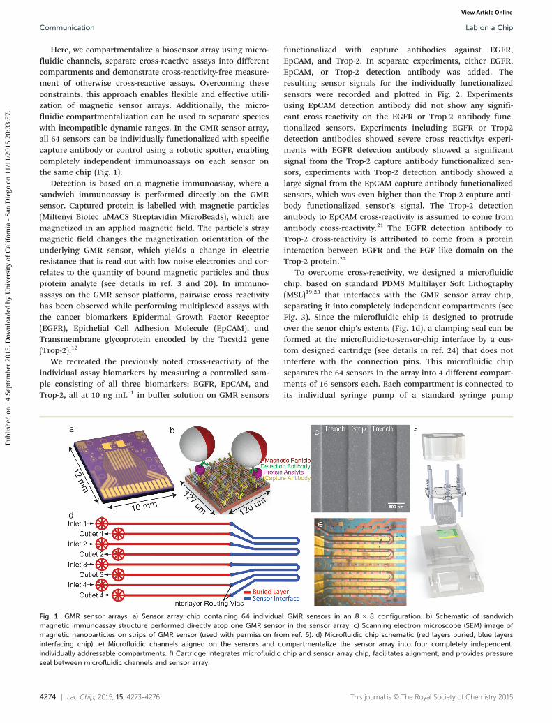
Here, we compartmentalize a biosensor array using micro-fluidic channels, separate cross-reactive assays into differentcompartments and demonstrate cross-reactivity-free measure-ment of otherwise cross-reactive assays. Overcoming theseconstraints, this approach enables flexible and effective utili-zation of magnetic sensor arrays. Additionally, the micro-fluidic compartmentalization can be used to separate specieswith incompatible dynamic ranges. In the GMR sensor array,all 64 sensors can be individually functionalized with specificcapture antibody or control using a robotic spotter, enablingcompletely independent immunoassays on each sensor onthe same chip (Fig. 1).
Detection is based on a magnetic immunoassay, where asandwich immunoassay is performed directly on the GMRsensor. Captured protein is labelled with magnetic particles(Miltenyi Biotec μMACS Streptavidin MicroBeads), which aremagnetized in an applied magnetic field. The particle's straymagnetic field changes the magnetization orientation of theunderlying GMR sensor, which yields a change in electricresistance that is read out with low noise electronics and cor-relates to the quantity of bound magnetic particles and thusprotein analyte (see details in ref. 3 and 20). In immuno-assays on the GMR sensor platform, pairwise cross reactivityhas been observed while performing multiplexed assays withthe cancer biomarkers Epidermal Growth Factor Receptor(EGFR), Epithelial Cell Adhesion Molecule (EpCAM), andTransmembrane glycoprotein encoded by the Tacstd2 gene(Trop-2).12
We recreated the previously noted cross-reactivity of theindividual assay biomarkers by measuring a controlled sam-ple consisting of all three biomarkers: EGFR, EpCAM, andTrop-2, all at 10 ng mL−1 in buffer solution on GMR sensors
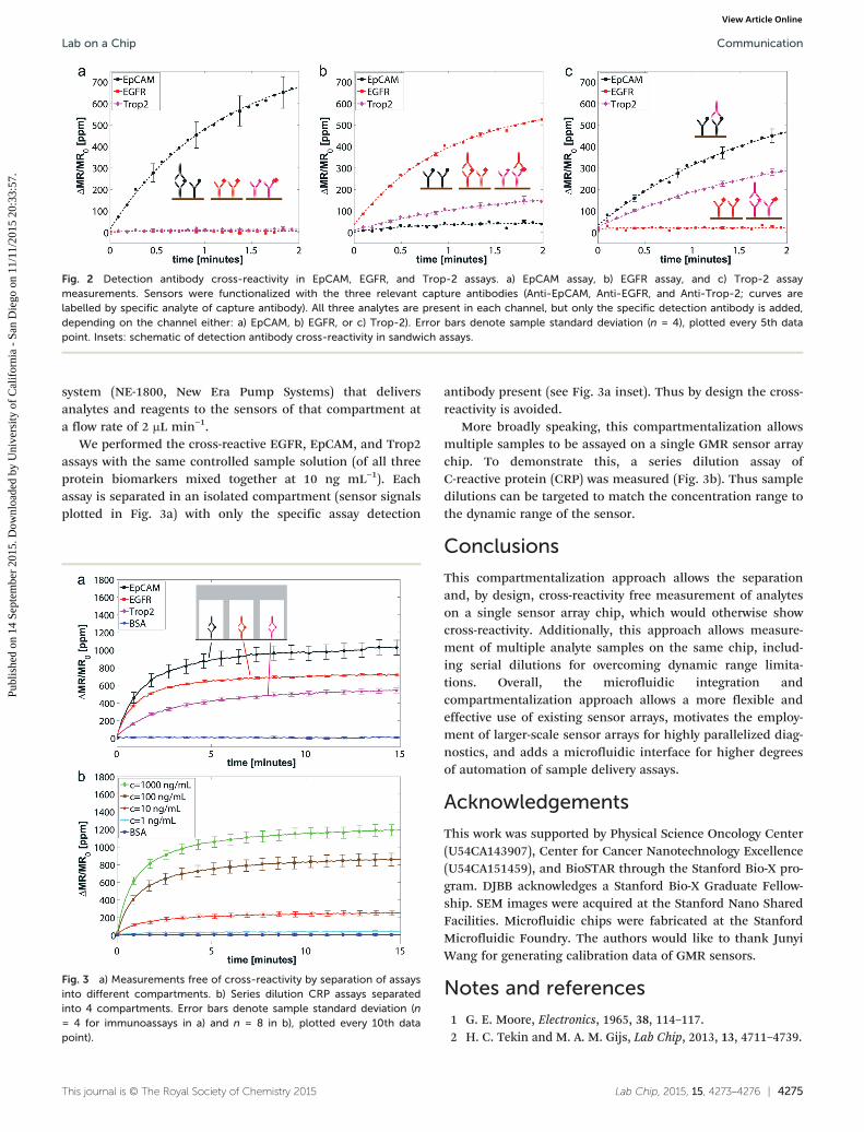
functionalized with capture antibodies against EGFR,EpCAM, and Trop-2. In separate experiments, either EGFR,EpCAM, or Trop-2 detection antibody was added. Theresulting sensor signals for the individually functionalizedsensors were recorded and plotted in Fig. 2. Experimentsusing EpCAM detection antibody did not show any signifi-cant cross-reactivity on the EGFR or Trop-2 antibody func-tionalized sensors. Experiments including EGFR or Trop2detection antibodies showed severe cross reactivity: experi-ments with EGFR detection antibody showed a significantsignal from the Trop-2 capture antibody functionalized sen-sors, experiments with Trop-2 detection antibody showed alarge signal from the EpCAM capture antibody functionalizedsensors, which was even higher than the Trop-2 capture anti-body functionalized sensor's signal. The Trop-2 detectionantibody to EpCAM cross-reactivity is assumed to come fromantibody cross-reactivity.21 The EGFR detection antibody toTrop-2 cross-reactivity is attributed to come from a proteininteraction between EGFR and the EGF like domain on theTrop-2 protein.22
To overcome cross-reactivity, we designed a microfluidicchip, based on standard PDMS Multilayer Soft Lithography(MSL)19,23 that interfaces with the GMR sensor array chip,separating it into completely independent compartments (seeFig. 3). Since the microfluidic chip is designed to protrudeover the senor chip's extents (Fig. 1d), a clamping seal can beformed at the microfluidic-to-sensor-chip interface by a cus-tom designed cartridge (see details in ref. 24) that does notinterfere with the connection pins. This microfluidic chipseparates the 64 sensors in the array into 4 different compart-ments of 16 sensors each. Each compartment is connected toits individual syringe pump of a standard syringe pump
Fig. 1 GMR sensor arrays. a) Sensor array chip containing 64 individual GMR sensors in an 8 × 8 configuration. b) Schematic of sandwichmagnetic immunoassay structure performed directly atop one GMR sensor in the sensor array. c) Scanning electron microscope (SEM) image ofmagnetic nanoparticles on strips of GMR sensor (used with permission from ref. 6). d) Microfluidic chip schematic (red layers buried, blue layersinterfacing chip). e) Microfluidic channels aligned on the sensors and compartmentalize the sensor array into four completely independent,individually addressable compartments. f) Cartridge integrates microfluidic chip and sensor array chip, facilitates alignment, and provides pressureseal between microfluidic channels and sensor array.
Lab on a ChipCommunication
Publ
ishe
d on
14
Sept
embe
r 20
15. D
ownl
oade
d by
Uni
vers
ity o
f C
alif
orni
a -
San
Die
go o
n 11
/11/
2015
20:
33:5
7.
View Article Online
Lab Chip, 2015, 15, 4273–4276 | 4275This journal is © The Royal Society of Chemistry 2015
system (NE-1800, New Era Pump Systems) that deliversanalytes and reagents to the sensors of that compartment ata flow rate of 2 μL min−1.
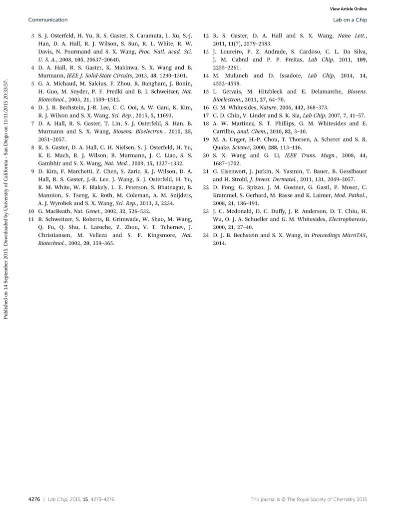
We performed the cross-reactive EGFR, EpCAM, and Trop2assays with the same controlled sample solution (of all threeprotein biomarkers mixed together at 10 ng mL−1). Eachassay is separated in an isolated compartment (sensor signalsplotted in Fig. 3a) with only the specific assay detection
antibody present (see Fig. 3a inset). Thus by design the cross-reactivity is avoided.
More broadly speaking, this compartmentalization allowsmultiple samples to be assayed on a single GMR sensor arraychip. To demonstrate this, a series dilution assay ofC-reactive protein (CRP) was measured (Fig. 3b). Thus sampledilutions can be targeted to match the concentration range tothe dynamic range of the sensor.
Conclusions
This compartmentalization approach allows the separationand, by design, cross-reactivity free measurement of analyteson a single sensor array chip, which would otherwise showcross-reactivity. Additionally, this approach allows measure-ment of multiple analyte samples on the same chip, includ-ing serial dilutions for overcoming dynamic range limita-tions. Overall, the microfluidic integration andcompartmentalization approach allows a more flexible andeffective use of existing sensor arrays, motivates the employ-ment of larger-scale sensor arrays for highly parallelized diag-nostics, and adds a microfluidic interface for higher degreesof automation of sample delivery assays.
Acknowledgements
This work was supported by Physical Science Oncology Center(U54CA143907), Center for Cancer Nanotechnology Excellence(U54CA151459), and BioSTAR through the Stanford Bio-X pro-gram. DJBB acknowledges a Stanford Bio-X Graduate Fellow-ship. SEM images were acquired at the Stanford Nano SharedFacilities. Microfluidic chips were fabricated at the StanfordMicrofluidic Foundry. The authors would like to thank JunyiWang for generating calibration data of GMR sensors.
Notes and references
1 G. E. Moore, Electronics, 1965, 38, 114–117.2 H. C. Tekin and M. A. M. Gijs, Lab Chip, 2013, 13, 4711–4739.
Fig. 2 Detection antibody cross-reactivity in EpCAM, EGFR, and Trop-2 assays. a) EpCAM assay, b) EGFR assay, and c) Trop-2 assaymeasurements. Sensors were functionalized with the three relevant capture antibodies (Anti-EpCAM, Anti-EGFR, and Anti-Trop-2; curves arelabelled by specific analyte of capture antibody). All three analytes are present in each channel, but only the specific detection antibody is added,depending on the channel either: a) EpCAM, b) EGFR, or c) Trop-2). Error bars denote sample standard deviation (n = 4), plotted every 5th datapoint. Insets: schematic of detection antibody cross-reactivity in sandwich assays.
Fig. 3 a) Measurements free of cross-reactivity by separation of assaysinto different compartments. b) Series dilution CRP assays separatedinto 4 compartments. Error bars denote sample standard deviation (n= 4 for immunoassays in a) and n = 8 in b), plotted every 10th datapoint).
Lab on a Chip Communication
Publ
ishe
d on
14
Sept
embe
r 20
15. D
ownl
oade
d by
Uni
vers
ity o
f C
alif
orni
a -
San
Die
go o
n 11
/11/
2015
20:
33:5
7.
View Article Online
4276 | Lab Chip, 2015, 15, 4273–4276 This journal is © The Royal Society of Chemistry 2015
3 S. J. Osterfeld, H. Yu, R. S. Gaster, S. Caramuta, L. Xu, S.-J.Han, D. A. Hall, R. J. Wilson, S. Sun, R. L. White, R. W.Davis, N. Pourmand and S. X. Wang, Proc. Natl. Acad. Sci.U. S. A., 2008, 105, 20637–20640.
4 D. A. Hall, R. S. Gaster, K. Makinwa, S. X. Wang and B.Murmann, IEEE J. Solid-State Circuits, 2013, 48, 1290–1301.
5 G. A. Michaud, M. Salcius, F. Zhou, R. Bangham, J. Bonin,H. Guo, M. Snyder, P. F. Predki and B. I. Schweitzer, Nat.Biotechnol., 2003, 21, 1509–1512.
6 D. J. B. Bechstein, J.-R. Lee, C. C. Ooi, A. W. Gani, K. Kim,R. J. Wilson and S. X. Wang, Sci. Rep., 2015, 5, 11693.
7 D. A. Hall, R. S. Gaster, T. Lin, S. J. Osterfeld, S. Han, B.Murmann and S. X. Wang, Biosens. Bioelectron., 2010, 25,2051–2057.
8 R. S. Gaster, D. A. Hall, C. H. Nielsen, S. J. Osterfeld, H. Yu,K. E. Mach, R. J. Wilson, B. Murmann, J. C. Liao, S. S.Gambhir and S. X. Wang, Nat. Med., 2009, 15, 1327–1332.
9 D. Kim, F. Marchetti, Z. Chen, S. Zaric, R. J. Wilson, D. A.Hall, R. S. Gaster, J.-R. Lee, J. Wang, S. J. Osterfeld, H. Yu,R. M. White, W. F. Blakely, L. E. Peterson, S. Bhatnagar, B.Mannion, S. Tseng, K. Roth, M. Coleman, A. M. Snijders,A. J. Wyrobek and S. X. Wang, Sci. Rep., 2013, 3, 2234.
10 G. MacBeath, Nat. Genet., 2002, 32, 526–532.11 B. Schweitzer, S. Roberts, B. Grimwade, W. Shao, M. Wang,
Q. Fu, Q. Shu, I. Laroche, Z. Zhou, V. T. Tchernev, J.Christiansen, M. Velleca and S. F. Kingsmore, Nat.Biotechnol., 2002, 20, 359–365.
12 R. S. Gaster, D. A. Hall and S. X. Wang, Nano Lett.,2011, 11(7), 2579–2583.
13 J. Loureiro, P. Z. Andrade, S. Cardoso, C. L. Da Silva,J. M. Cabral and P. P. Freitas, Lab Chip, 2011, 109,2255–2261.
14 M. Muluneh and D. Issadore, Lab Chip, 2014, 14,4552–4558.
15 L. Gervais, M. Hitzbleck and E. Delamarche, Biosens.Bioelectron., 2011, 27, 64–70.
16 G. M. Whitesides, Nature, 2006, 442, 368–373.17 C. D. Chin, V. Linder and S. K. Sia, Lab Chip, 2007, 7, 41–57.18 A. W. Martinez, S. T. Phillips, G. M. Whitesides and E.
Carrilho, Anal. Chem., 2010, 82, 3–10.19 M. A. Unger, H.-P. Chou, T. Thorsen, A. Scherer and S. R.
Quake, Science, 2000, 288, 113–116.20 S. X. Wang and G. Li, IEEE Trans. Magn., 2008, 44,
1687–1702.21 G. Eisenwort, J. Jurkin, N. Yasmin, T. Bauer, B. Gesslbauer
and H. Strobl, J. Invest. Dermatol., 2011, 131, 2049–2057.22 D. Fong, G. Spizzo, J. M. Gostner, G. Gastl, P. Moser, C.
Krammel, S. Gerhard, M. Rasse and K. Laimer, Mod. Pathol.,2008, 21, 186–191.
23 J. C. Mcdonald, D. C. Duffy, J. R. Anderson, D. T. Chiu, H.Wu, O. J. A. Schueller and G. M. Whitesides, Electrophoresis,2000, 21, 27–40.
24 D. J. B. Bechstein and S. X. Wang, in Proceedings MicroTAS,2014.
Lab on a ChipCommunication
Publ
ishe
d on
14
Sept
embe
r 20
15. D
ownl
oade
d by
Uni
vers
ity o
f C
alif
orni
a -
San
Die
go o
n 11
/11/
2015
20:
33:5
7.
View Article Online