Embed Size (px)
Citation preview

Cite this: Lab Chip, 2013, 13, 2464
Recent advances in microfluidic techniques for single-cell biophysical characterization
Received 18th March 2013,Accepted 16th April 2013
DOI: 10.1039/c3lc50355k
www.rsc.org/loc
Yi Zheng,ab John Nguyen,a Yuan Weia and Yu Sun*abc
Biophysical (mechanical and electrical) properties of living cells have been proven to play important roles in
the regulation of various biological activities at the molecular and cellular level, and can serve as promising
label-free markers of cells’ physiological states. In the past two decades, a number of research tools have
been developed for understanding the association between the biophysical property changes of biological
cells and human diseases; however, technical challenges of realizing high-throughput, robust and easy-to-
perform measurements on single-cell biophysical properties have yet to be solved. In this paper, we review
emerging tools enabled by microfluidic technologies for single-cell biophysical characterization. Different
techniques are compared. The technical details, advantages, and limitations of various microfluidic devices
are discussed.
Introduction
The cell, the basic functional unit of living organisms,maintains and senses the physiological environment withinthe organism both chemically and physically.1–4 The uniquebiochemical and biophysical properties enable a cell to fulfillits specific functions and adapt to its surrounding environ-ment. Physiological changes within the cells are accompaniedby chemical and physical modifications and reorganization.Thus, pathological cells can be identified biochemically and/orbiophysically. Biochemical properties of pathological cellshave been under intensive study, and many biochemicalmarkers have been developed to identify target cells out of aheterogeneous population.5,6 The biophysical properties ofcells also play important roles in various biological processesand are involved in the regulation of gene expression,differentiation, migration, and metabolic activities.2,7
However, although research on the physical properties of cellshas provided strong evidence about the capability of physicalproperties as potential markers for identifying cell types, mostbiophysics research was limited to proof-of-concept demon-strations. The lack of clinical relevance is due to the very lowmeasurement throughput and tedious operation procedures ofconventional techniques for measuring the biophysical prop-erties of cells.4,7–9 Compared to conventional techniques, theadvantages of small sample volume, integration capability,
biocompatibility and fast response make microfluidic tech-nologies attractive for studying cells. Recent decades havewitnessed significant advances of microfluidic technologiesfor biochemical characterization of cells.10,11 Microfluidics isextending its way into the characterization of single-cellbiophysical properties.12–14
In this review, we summarize existing microfluidic tech-nologies for characterizing biophysical properties of individualcells (i.e., mechanical and electrical properties). Within eachsection, we first provide an introduction of a technique. Wethen classify various techniques based on their workingmechanisms and discuss their advantages and limitations.Finally, we present perspectives on the challenges fordiagnostic applications and future directions of microfluidictechnologies for single-cell biophysical characterization. Thisreview is focused on discussing techniques for characterizingsingle-cell mechanical and electrical properties. Microfluidictechniques for studying the roles of mechanical and electricalstimuli in the regulation of differentiation, migration, andmetabolic activities have been reviewed elsewhere.3,4,13
Mechanical characterization techniques
The association between cell deformability and humandiseases has been of interest since the 1960s.15,16 Thedeformability of nucleated cells is determined by themembrane, the cytoskeletal network (actin filaments, inter-mediate filaments, and microtubules), and its interaction withthe nucleus, while the deformability of red blood cells (RBCs)is determined by the membrane skeleton network and theinteraction between the membrane skeleton and membraneintegral proteins.4,17,18 Physiological and pathological changes
aDepartment of Mechanical and Industrial Engineering, University of Toronto,
Toronto, ON, Canada. E-mail: [email protected]; Fax: 1-416-978-7753;
Tel: 1-416-946-0549bInstitute of Biomaterials and Biomedical Engineering, University of Toronto,
Toronto, ON, CanadacDepartment of Electrical and Computer Engineering, University of Toronto, Toronto,
ON, Canada
Lab on a Chip
CRITICAL REVIEW
2464 | Lab Chip, 2013, 13, 2464–2483 This journal is � The Royal Society of Chemistry 2013
Publ
ishe
d on
17
Apr
il 20
13. D
ownl
oade
d by
Uni
vers
ity o
f T
oron
to o
n 04
/06/
2013
17:
16:2
1.
View Article OnlineView Journal | View Issue

can alter the cytoskeleton composition, reorganize the networkstructure, and change the protein density. As a result, celldeformability can be used as an intrinsic marker foridentifying pathological conditions.8,19,20 For example,deformability is known to play a crucial role in the mobilityof cancerous cells;4,21 and a decrease in RBC deformability hasbeen proven to be relevant in several human diseases (seeTable 1).
Compared to existing experimental tools, such as atomicforce microscopy, micropipette aspiration, optical tweezers,and magnetic tweezers, which are all difficult to use and havea low testing speed, microfluidics offers the potential for high-throughput mechanical measurement of single cells.4,10,12,13
For characterizing the mechanical properties of a cell, the cellmust be deformed. Thus, we classify microfluidic technologiesfor cell deformability measurements according to the mechan-ical stimuli used to deform the cell. Table 2 summarizes thesetechniques and their working mechanisms, cell samples, keytechnologies and observations, and reported throughput. Forthose where throughput was not reported, the total number oftested cells is listed in Table 2 in order to provide a sense ofthe devices’ throughput.
Structure-induced deformation (constriction channels)
Constriction channels, which are marginally smaller than thediameters of tested cells, provide an efficient method togenerate mechanical stimuli. Cells driven through a constric-tion channel are squeezed by the walls of the constrictionchannel. Multiple parameters, such as transit time, elongationand recovery time, in association with cell deformability canbe quantified. Moreover, constriction channels can be easilyfabricated with standard microfabrication techniques and areable to provide an environment to mimic in vivo capillaries.With the use of high-speed imaging or electrical impedancemeasurements, constriction-channel devices are capable ofachieving a higher throughput than most other deformabilitymeasurement approaches. Due to these merits, constrictionchannels have been used to measure the deformability ofRBCs,22,23,39–44 leukocytes45 and cancer cells.46,47
Due to the human capillary-like environment and thephysiological relevance of RBC deformability, RBCs are mostlystudied in the majority of existing constriction channel-baseddevices. The first demonstration of microfluidic constrictionchannels for RBC deformability characterization was reportedin 2003.22 In this study, constriction channels with variousdiameters were used to study the deformability changes
between healthy RBCs and malaria parasite-infected RBCs atdifferent stages (early ring stage, early trophozoite, latetrophozoite, and schizont). They found that the deformabilityof malaria-infected RBCs decreases as the parasite progressesfrom the early ring stage to a schizont, while healthy RBCs areexceptionally deformable and are even able to travel throughthe constriction channels blocked by infected RBCs. Inaddition to RBC deformability, white blood cell (WBC)deformability was also studied using microfluidic constrictionchannels. Rosenbluth et al. demonstrated the clinical rele-vance of their microfluidic constriction channel device insepsis and leukostasis.45 The reported device consists of anetwork of 64 constriction channels. High-speed imaging wasused for measuring cell transit time. They used patientsamples to show that cell transit time increased for diseasedsamples compared to control samples (Fig. 1(a)). In anotherstudy, breast cancer cell lines (MCF-7 and MCF-10A) wereflown through a constriction channel to distinguish non-malignant and malignant cells.46 According to the reportedresults, transit velocity is not significantly affected by celltypes, and the transit time difference is mainly determined bythe entry time. Different cell types can be distinguished on thebasis of scatter plots of cell volume and entry time.
Besides imaging, microfluidic constriction channels canalso be used together with other measurement techniques toachieve multiple parameter measurements for cell typeclassification. We recently developed a microfluidic device(Fig. 1(b)), which combines a constriction channel andimpedance measurements.39 Detection involves only electricalsignals; hence, it enables a throughput higher than 100 cellss21. The multiple parameters quantified as mechanical andelectrical signatures include transit time, impedance ampli-tude ratio, and impedance phase increase. Histogramscompiled from 84 073 adult RBCs and 82 253 neonatal RBCsreveal different biophysical properties across samples andbetween the adult and neonatal RBC populations. Bow et al.demonstrated a deformability-based RBC testing devicecombining constriction channels and fluorescence measure-ment. They showed a population-based correlation betweenbiochemical properties, such as cell surface markers, andmechanical deformability.23 They also showed experimentallythat the entrance geometry of the constriction channel has asignificant impact on RBC transit time, and developed adissipative particle dynamics model to interpret the parasiticeffect on RBC deformability. An optical stretcher was alsointroduced to enhance the sensitivity and reliability of aconstriction channel-based microfluidic device.40 Scatter plots
Table 1 RBC deformability decrease under pathological conditions
Disease Possible genetic and molecular cause Reference
Malaria Interruption of cytoskeleton network by parasite protein 17, 22–25Hereditary spherocytosis Low cytoskeleton density 17, 26–28Sickle cell disease Polymerized and deoxygenated hemoglobin 29, 30Sepsis Altered spectrin interaction 31–33Diabetes Not clear 34–36Stored RBCs Depletion of ATP and Nitric oxide 37, 38
This journal is � The Royal Society of Chemistry 2013 Lab Chip, 2013, 13, 2464–2483 | 2465
Lab on a Chip Critical Review
Publ
ishe
d on
17
Apr
il 20
13. D
ownl
oade
d by
Uni
vers
ity o
f T
oron
to o
n 04
/06/
2013
17:
16:2
1.
View Article Online

Table 2 Microfluidic devices for single-cell mechanical characterization
Techniques Targeted cells Summary Throughputa Ref.
Structure-induced Constriction channelsof differentcross-sectional areas
Malaria infected RBCs Uninfected RBCs can readilytravel through the constrictionchannel. RBCs become lessdeformable as the stage ofmalaria infection progresses.
N/A 22
Cuneiform constrictionchannel array +fluorescence
Malaria infected RBCs Entrance geometry of theconstriction channel has asignificant impact on RBCtransit time. Deformabilitymeasurements of ring stage P.falciparum-infected and uninfectedRBCs show significant overlap.
y100cells min21
23
Constriction channel +impedance measurement
Adult and neonatal RBCs Characterization of multipleparameters (transit time, impedanceamplitude and impedance phase)improves the classification accuracy.
100–150cells s21
39
Constriction channel +optical pressure
RBCs from healthy donorsand leukemia patients
Recovery time of RBCs fromhealthy donors and leukemiapatients was found to besignificantly different.
N = 140 40
Constriction channel array Blood cells from AMLand ALL patients, andHL-60 cell lines.
Transit time of WBCs fromAML patients with leukostasissymptoms is significantly higherthan the WBCs from AML patientswithout leukostasis symptomsand ALL patients.
50–100cells min21
45
Constriction channel MCF-7 and MCF-10A The different transit time ismainly contributed by the longerentry time of MCF-7.
N # 100 46
Constriction channel +impedance measurement
MC-3T3 and MLO-Y4;EMT6 and EMT6/AR1.0
Cells with comparable sizes(EMT6 and EMT6/AR1.0) wereclassified based on their differentdeformability and electrical properties.
y1 cells s21 47
Constriction channel +volume measurement
HeLa Controlled Hela cells have longertransit time than Hela cells treatedwith latrunculin A and cytochalasin B.
800 cells min21 48
Constriction channel +deformable membrane
MCF-7 and MCF-10A MCF-7 and MCF-10A can bedistinguished on the basisof transit time.
N = 150–200 49
Fluid-induced Inertial focusing +hydrodynamic stretching
Leukocytes and malignantcells in pleural effusions;pluripotent stem cells
The prediction of disease statesin patients with cancer and immuneactivation achieved a sensitivity of91% and a specificity of 86%.
y2000 cells s21 19
Straight microchannel RBCs and fixed RBCs Effects of diamide and glutaraldehydeon RBC dynamics were examined byvisual observation of cells whenflowing through a microchannel.
N/A 50
Hyperbolic convergingmicrochannel
RBCs The results prove that hyperbolicshape is more efficient in deformingRBCs.
N/A 51
Shear stress +resistance measuring
RBCs and fixed RBCs The resistance signal generatedby integrated electrodes is correlatedto the deformation of RBCs.
N = 72 83
Electroporation-induced
Microfluidic channel +high electrical field
MCF-10A and MCF-7 More malignant and metastatic celltypes exhibit more significant swelling.
5 cells s21 61
Microfluidic channel +high electrical field
Controlled mouse RBCs,mouse RBCs withdeficienciesof ankyrin, b-adducinand Tmod1
Time taken for completeelectroporation-induced lysis can becorrelated to the defects in thecytoskeleton network.
2000–3000cells/sample
63
Optical stretcher Microfluidic channel +laser beam
RBCs; BALB 3T3 Optical deformability can be usedto differentiate cells by measuringthe differences in elastic response.
N/A 67
Microfluidic channel +laser beam
MCF-10A, MCF-7 andmodMCF-7; MDA-MB-231;BALB/3T3 and SV-T2
Cancer cell lines are significantlymore deformable than normal cells.
N/A 70
Microfluidic channel +laser beam +microcouplers
RBCs A two-axis active microcouplercapable of generating substantialdisplacement for accurate alignmentof buried optical fibers was developed.
N/A 71
2466 | Lab Chip, 2013, 13, 2464–2483 This journal is � The Royal Society of Chemistry 2013
Critical Review Lab on a Chip
Publ
ishe
d on
17
Apr
il 20
13. D
ownl
oade
d by
Uni
vers
ity o
f T
oron
to o
n 04
/06/
2013
17:
16:2
1.
View Article Online

compiled from transit time, elongation, and recovery timemeasured by this device proved effective for the discriminationof RBCs from normal donors and leukemia patients.
Other applications of microfluidic constriction channelsinclude the use of wedge-shaped constriction channels tomeasure the surface area and volume of a large population ofRBCs,41–43 and a microfluidic manometer to measure thepressure drop due to the presence of a cell in the constrictionregion, which correlates with the stiffness of the cell, byobserving the displacement of the downstream fluid–fluidinterface.44 Despite the advantages of the constriction channeltechnique, cell volume and adhesion between the cellmembrane and channel walls are coupled with cell deform-ability. Consequently, a longer transit time does not necessa-rily mean lower deformability since larger and stickier cellscan also lead to a longer transit time. Efforts have been madeto take the cell volume/size effect into account. For example,Adamo et al. reported a microfluidic device for probing bothcell volume and transit time. Comparisons of cell transit timewere made among cells with a similar volume48. Theydemonstrated that Hela cells in the control group have alonger transit time than Hela cells treated with latrunculin Aand cytochalasin B. The size of the constriction channel canalso be adjusted on demand according to targeted cell size(MCF-7 and MCF-10A).49 However, there exists no techniquethat is capable of characterizing the adhesion (or friction)between the cell membrane and channel walls. In addition,since the diameter of the constriction channel is smaller thanthe diameter of the targeted cells, the channel is susceptible toclogging. Using an array of constriction channels appears to bea potential solution to the clogging issue.
Fluid-induced deformation
RBCs are highly deformable and can easily deform under fluidshear stress inside blood vessels. Having micrometer dimen-sions, which are comparable with in vivo capillaries, micro-fluidic channels provide an ideal tool for investigating RBC
deformability. Compared to constriction channels, the micro-fluidic channels used to generate shear stress are larger thanRBCs. Thus, RBCs are deformed by fluid shear stress insteadof the channel’s confinement structures. The deformationindex (DI) or stretch ratio quantified via high-speed imaginghas been used as a measure of RBC deformability.36,50,51
Forsyth et al. studied the deformability and dynamic behaviorof chemically ‘‘stiffened’’ RBCs using a simple straightmicrofluidic channel, revealing three different types of motiondue to the increased shear rate in the microfluidic channel:stretching, tumbling, and recoiling.50 Besides straight chan-nels, a hyperbolic converging microchannel was also devel-oped for assessing RBC deformability by measuringextensional flow-induced deformation.51 The results con-firmed that extensional flow is more efficient in causing RBCdeformation. Shear stress generated in microfluidic channelswas also used to measure the dynamics of shear-inducedadenosine triphosphate (ATP) release from RBCs. In ref. 52,RBCs were driven through a microfluidic channel with a cross-sectional area of 20 6 20 mm, while the amount of ATPreleased was measured by counting the photons emitted froma standard luciferase-ATP bioluminescent reaction. Celldeformation and dynamics were quantified simultaneouslyusing high-speed imaging.
Although shear stress proves effective for investigating RBCdeformability, the low magnitude of shear stress is typicallynot able to deform other types of cells. Gossett et al. recentlyreported a hydrodynamic-stretching microfluidic device foridentifying malignant cells in a human pleural fluid samplewith a measurement speed of y2000 cells s21 (see Fig. 1(c)).19
Cells are focused towards a narrow streamline near the centerof the microfluidic channel and delivered to a junction of twoorthogonal channels at a high flow rate, where the cellsundergo mechanical stretching. Meanwhile, cell deformationsare captured using a high-speed camera and images are thenanalyzed to extract the cell volume and deformation index (DI).The results revealed that cancerous cells in pleural fluid have
Table 2 (Continued)
Techniques Targeted cells Summary Throughputa Ref.
Microfluidic channel +laser beam
Healthy and cancerousoral squamous cells
Cancer cells showed a higher meandeformability and increased variance.
N = 200–300 74
DEP-induced Microfabricatedelectrodes
CHO-K1 and U937 Cell deformation was interpreted intomechanical properties using ananalytical model.
N = 15 77
Microfabricatedelectrodes
SiHa and ME180 A FEM model was developed to extractthe Young’s modulus of cells.
N = 14 78
ITO electrodes +microchamber array
Healthy andspherocytosis RBCs
RBCs from patients with spherocytosisare less deformable than RBCs ofhealthy volunteers.
N = 200–300for each cell type
79
Aspiration-induced
Funnel chain +pressure attenuator
Healthy andmalaria RBCs
Malaria infected RBCs were provenless deformable than normal RBCs.
N = y100 foreach cell type
81
Microfluidic pipette +impedance measurement
MC-3T3 MC-3T3 was characterized byimpedance spectroscopy andmicropipette aspiration.
N = 18 82
a Throughput: For those references that explicitly reported speed/throughput, speed/throughput is listed directly in the table. For thosereferences where speed/throughput was not explicitly reported, we calculated speed/throughput using the data plots (e.g., histograms)presented in those references
This journal is � The Royal Society of Chemistry 2013 Lab Chip, 2013, 13, 2464–2483 | 2467
Lab on a Chip Critical Review
Publ
ishe
d on
17
Apr
il 20
13. D
ownl
oade
d by
Uni
vers
ity o
f T
oron
to o
n 04
/06/
2013
17:
16:2
1.
View Article Online

larger volumes and are more deformable (higher DI values)than benign cells. This approach achieved both a high testingspeed and larger deformation of tested cells. The authorspredicted disease states in patients with cancer and immuneactivation with a sensitivity of 91% and a specificity of 86%.
In fluid-induced deformation-based microfluidic devices,channel dimensions are larger than cell diameters. DI is usedas the deformability indicator and is not affected by theadhesion between cell membrane and channel walls (vs.constriction channel). However, since cells of different sizes
may experience different forces due to non-uniformity in theshear stress and hydrodynamic pressure within the micro-channel, DI is still associated with cell volume. Morespecifically, shear stress is highest near channel walls and isalmost zero in the center of the channel. As a result, RBCs withlarger volumes experience higher shear stress on the edgescausing larger deformation compared to cells with smallervolumes. Within the hydrodynamic-stretching microfluidicdevice, the highest compressive pressure appears at the centerof the junction where the cells are stretched, which causes
Fig. 1 (a) A network of bifurcating microfluidic channels for blood cell deformability measurement. The transit time of the individual cells are measured using a high-speed camera. Reproduced with permission from ref. 45. (b) A microfluidic system for electrical and mechanical characterization of RBCs at a speed of 100–150 cellss21. The transit time is obtained from electrical impedance data captured during RBCs flowing through the constriction channel. Reproduced with permission fromref. 39. (c) Hydrodynamic stretching microfluidic device. Cells are focused to the center lines of the channels by inertial force and stretched by fluid pressure. Celldeformation is measured by analyzing images recorded by a high-speed camera. Reproduced with permission from ref. 19. (d) Electroporation-induced RBC lysis.Time-lapse images of RBCs are recorded by a high-speed camera as the cells flow through the channel, while a constant DC voltage is applied. Cell lysis time iscorrelated with RBC deformability. Reproduced with permission from ref. 63.
2468 | Lab Chip, 2013, 13, 2464–2483 This journal is � The Royal Society of Chemistry 2013
Critical Review Lab on a Chip
Publ
ishe
d on
17
Apr
il 20
13. D
ownl
oade
d by
Uni
vers
ity o
f T
oron
to o
n 04
/06/
2013
17:
16:2
1.
View Article Online

cells with different volumes or shapes to be exposed under adifferent pressure environment. To address this issue, cellvolume is measured as an independent parameter and used asa reference for cell-type classification.19
A limitation of fluid-induced deformation-based microflui-dic devices is the use of high-speed imaging (tens of kHz) formeasuring DI. High-speed cameras are often costly and mustbe used on a microscope, making the overall microfluidicsystem bulky. Furthermore, since real-time image data transferfrom a high-speed camera to the computer hard drive istechnically difficult to achieve, images are stored in thecamera’s on-board memory for later off-line transfer. Sincestate-of-the-art high-speed cameras typically have only a fewGB of on-board memory, after recording for a few seconds, theexperiment must be stopped for image data transfer.Processing massive amounts of image data also coststremendous computational effort and time. Advances inhigh-speed cameras and hardware-based image processingtechniques, such as FPGA-based techniques, will possibly beable to solve the data transfer problem. Additionally, opto-microfluidics, microfluidic devices integrated with opticalcomponents, also seems a promising way to tackle thisproblem. Integrating on-chip lenses and CMOS chips53–56
can achieve real-time performance and in the meantime,eliminate the need for the use of bulky microscopes.
Electroporation-induced deformation
Electroporation is a technique used for introducing foreignmolecules, such as DNA and proteins, into cells. The conceptof electroporation capitalizes on the relatively weak nature ofthe phospholipid bilayer’s hydrophobic/hydrophilic interac-tions and its ability to spontaneously reassemble after adisturbance.57 Thus, a voltage shock can disrupt areas of themembrane temporarily, allowing polar molecules to pass. Themembrane then can re-seal and leave the cell intact.57–59
Swelling or expansion in cell size accompanies the cell’selectrical property changes,60 which is caused by the influx ofmolecules through the open pores in the cell membrane (vs.dielectric force in DEP-induced deformation). Recently, a fewstudies correlated electroporation-induced cell deformationand lysis to cell deformability changes. Bao et al.61 developedmicrofluidic electroporative flow cytometry to study thedeformability of single cells. A constant DC voltage wasestablished across the microchannel, which concentrated theelectric field to yield repeatable cell exposure to uniformelectrical fields. When cells were flown through the micro-channel, the swelling of cells was recorded by imaging andquantified as a deformability indicator. Deformability changesof breast cancer cell lines61 and the expansion of the nuclei ofcirculating tumor cells (CTCs) were tested using the system.62
The capability to detect RBCs with deficiencies of thecytoskeletal protein network was also demonstrated (seeFig. 1(d)).63 The achieved throughput was about 5 cells s21.
It is notable that electroporation efficiency is also dependenton cell volume. Under the same uniform electrical field, theeffective voltage across the cell membrane is a function of celldiameter.64 Hence, cells with larger diameters are exposed tohigher voltages and are more easily porated. In addition, theplasma membrane of different cell types (e.g., metastatic
tumor cells vs. non-metastatic tumor cells) can possessdifferent poration properties. In other words, some cell typesmay be more susceptible to electrical fields than others. Thus,based on electroporation-induced expansion or swelling alone,it can be inaccurate to conclude that cell deformation iscaused by cell deformability rather than the membrane’sporation susceptibility to electrical voltages.
Optical stretcher
Optical trapping was discovered in 197065 when radiationpressure from laser light was found to be able to accelerateand trap micron-sized dielectric particles. Light interacts witha particle by imparting some of its momentum onto it, thusexerting a force on the particle. If the particle is not centeredon the optical axis of the beam, a restoring force is exerted onthe particle to keep it on the optical axis. Optical stretchersutilize two slightly divergent Gaussian beams to trap an objectin the middle. This concept was used to deform and measurethe stress profiles of erythrocytes, which led to the develop-ment of the first optical stretcher device reported in 2000.66
The setup was then demonstrated to stretch BALB 3T3fibroblasts and measure their viscoelastic properties.67
A typical optical stretcher system consists of a microchannelfor loading cells into the testing region and two laser fiberslocated on the sides of the passageway (see Fig. 2(a)).68 Cellsflowing through the microfluidic channels are serially trappedand deformed with the two counter propagating divergentlaser beams.69,70 For trapping and stretching cells in theoptical stretcher, the alignment of the fibers is crucial. In orderto improve alignment, a platform capable of adjusting fiberpositions was developed71 using pneumatically driven manip-ulators. The femtosecond laser technique has been utilized forthe fabrication of optical stretcher-based microfluidicdevices.72,73 By direct writing of both optical waveguides andmicrofluidic channels, the alignment problem can be miti-gated significantly. The deformability of human cancer celllines,69,70 red blood cells71 and patient oral squamous cells74
was characterized using optical stretchers.
DEP-induced deformation
When polarized in an electric field, biological cells willexperience a dielectric force, which is well known as a DEPforce.75,76 Electro-deformation devices utilize the DEP force fortrapping and generating mechanical stimuli to quantify thedeformability of individual cells.77,78 The Young’s modulus oftested cells can be extracted with either analytical models ornumerical simulation. For improving the throughput ofelectro-deformation devices, a single-cell microchamber arraydevice79 was proposed. The device is able to trap individualRBCs in a large array of micro-wells integrated with DEPelectrodes. ITO electrodes also allow the correlation of RBCdeformation with cell surface and cytosolic characteristics(Fig. 2(b)). However, due to the time-consuming procedure ofcell trapping, the overall throughput of this device is stillrather limited. On the other hand, the complex physicalphenomena involved in electro-deformation and unknown cellelectrical properties pose difficulties in extracting forcesexperienced by an electro-deformed cell.75,77,78 Since the DEPforce is determined by the dielectrical properties of cells, DEP-
This journal is � The Royal Society of Chemistry 2013 Lab Chip, 2013, 13, 2464–2483 | 2469
Lab on a Chip Critical Review
Publ
ishe
d on
17
Apr
il 20
13. D
ownl
oade
d by
Uni
vers
ity o
f T
oron
to o
n 04
/06/
2013
17:
16:2
1.
View Article Online

based techniques can also be used to electrically characterizecells, which is discussed in the electrorotation section underelectrical characterization.
Aspiration-induced deformation
Pipette aspiration is a conventional technique for studying themechanical properties of single cells.80 The mathematicalmodels in conventional pipette aspiration have been adoptedin microfluidic characterization of cells. Fig. 2(c) shows amicrofluidic pipette aspiration device.81 Single cells areinfused into a microfluidic channel and deformed through aseries of funnel-shaped constrictions. Malaria-infected RBCswere tested using both membrane cortical tension andthreshold pressure as readouts. We developed a microfluidicdevice for single-cell electrical and mechanical characteriza-tion using impedance spectroscopy and micropipette aspira-tion.82 Cellular deformation was recorded as a function ofincreasing pressure, while cellular impedance was measured
via two Ag/AgCl electrodes inserted into the culture medium.With pipette aspiration and equivalent circuit models, bothmechanical properties and dielectric properties of single cellswere quantified. In addition to throughput limitation, therectangle-like cross-section in microfluidic pipette aspirationdevices can cause concerns about the validity of applyingconventional pipette aspiration models.
Electrical characterization techniques
Besides mechanical deformability, electrical properties of cellsare also important physical properties, serving as the basis ofcounting, trapping, focusing, separating, and the characteriza-tion of single cells.84,85 Early work on cell electrical measure-ments dates back to the 1910s,86–88 when approximatehemoglobin conductivity inside RBCs was reported. The singleshell model proposed by Pauly and Schwan in the 1950s laid
Fig. 2 (a) An optical stretcher. Cells travel along a flow channel integrated with two opposing laser beams. Cells are trapped and deformed optically, and celldeformation images are recorded. Reproduced with permission from ref. 68. (b) A single-cell microchamber array for electro-deformation. An array of microchambersintegrated with DEP electrodes is used to trap individual RBCs and apply a DEP force. Reproduced with permission from ref. 79. (c) Microfluidic pipette aspiration ofcells using a funnel channel chain. Reproduced with permission from ref. 81.
2470 | Lab Chip, 2013, 13, 2464–2483 This journal is � The Royal Society of Chemistry 2013
Critical Review Lab on a Chip
Publ
ishe
d on
17
Apr
il 20
13. D
ownl
oade
d by
Uni
vers
ity o
f T
oron
to o
n 04
/06/
2013
17:
16:2
1.
View Article Online

the foundation for interpreting the electrical properties ofcells, where the cell is modeled as a spherical cytoplasmsurrounded by a thin dielectric membrane.89,90 Generally, theelectrical properties of a plasma membrane are affected by themembrane morphology, lipid bilayer composition and thick-ness, and embedded proteins.91–93 Electrical properties of thecytoplasm are influenced by the intracellular structures andphysiological conditions (e.g., nucleus-to-cytoplasm ratio andion concentrations inside the cell).94–96 Early techniques wereonly capable of measuring the average electrical properties of acell population. The patch-clamp technique, developed in 1976is capable of electrical characterization of single cells.97
However, a patch-clamp typically takes tens of minutes to testone cell. Because of the capability of manipulating micro-scaled objects and integrating microelectrodes on-chip,microfluidic techniques have gained momentum in single-cellelectrical characterization in recent years. Table 3 summariesand classifies microfluidic techniques for the electricalcharacterization of single cells.
Microfluidic Coulter counter
The Coulter counter monitors the DC resistance of a smallorifice as microparticles pass through. Since the cell mem-brane acts as an insulating layer at DC, the presence of the cellalters the resistance of the orifice by replacing the conductiveliquid. Well established models are available for correlatingDC resistance changes to cell volumes. Several designs haverecently been proposed to improve the microfluidic Coultercounter’s performance. For instance, two-dimensional sheathflow focusing was demonstrated to overcome the cloggingissue;98 and throughput of microfluidic Coulter counters wasimproved using multiple-orifice designs.99
One challenge for microfluidic Coulter counters lies in theselection of the electrode material. Ag/AgCl non-polarizableelectrodes, which work well in the conventional Coultercounter, are an ideal choice. Nonetheless, integrating Ag/AgCl electrodes into microfluidic channels is complex, and Ag/AgCl electrodes have a limited lifetime.100,101 Although othermetal electrodes can be more readily integrated into micro-fluidic channels, the electrical double layers formed betweenthe interface of electrodes and the liquid, which are mainlycapacitive, pose difficulties in applying DC signals. Methodsfor minimizing the electrical double layer effect include themodification of the electrode surface roughness in order toincrease the surface area102 and the utilization of polyelec-trolytic salt bridges (PSBEs)103 or polyelectrolyte gel electrodes(PGEs).104 Most recently, a DC impedance-based microcyt-ometer device integrating PGEs was reported for CTC celldetection. Sheath flow is used for focusing cells and prevent-ing cell adhesion to chambers and channels (see Fig. 3(a)).CTCs were successfully detected in blood samples from breastcancer patients.105 Besides the electrode design that demandsspecial design consideration, the model used in the conven-tional Coulter counter to calculate particle volumes is not
directly applicable to microfluidic Coulter counter devices, dueto the different electrode configuration and channel geome-try.106,107 A microfluidic Coulter counter is limited to countingand sizing single cells, but incapable of characterizing theirelectrical properties.
Electrorotation
When a cell/particle is placed in a non-uniform electrical field,a force is exerted on the induced dipole and can cause the cellto either move (DEP) or rotate (ROT).75,108 Maxwell’s mixturetheory is commonly used to interpret DEP and ROT data, forassociating the complex permittivity of the suspension to thecomplex permittivity of the particle/cell.75,108 The DEP forceand ROT torque are proportional to the real and imaginaryparts of the Clausius–Mossotti factor, respectively.9 AlthoughDEP and ROT are closely related, ROT is more suitable forsingle-cell electrical characterization since in ROT, the cellsare only rotated at a certain position in the electric field. Thus,the amplitude of the electric field remains unchanged, whichis suitable for fitting the rotation spectra to determineintrinsic electrical properties of single cells (e.g., specificmembrane capacitance, cytoplasm conductivity and cytoplasmpermittivity). Differently, forces in DEP are determined by theelectric field gradient. A constant electric field gradient istechnically challenging to achieve. Furthermore, the rotationrate is only determined by the rotation force (constant as a cellrotates) and the viscosity of the suspension medium in ROT,which can be readily measured, whereas the DEP force (varyingas a cell moves) is difficult to quantify. DEP was demonstratedto electrically characterize cells by curve fitting of theClausius–Mossotti factor spectra or cell count spectra.However, since the spectra are not from the measurementsof the same cells, only average electrical properties of a cellpopulation can be obtained.109–111 More details on DEP andROT models can be found in other reviews.9,112
The most popular ROT setup uses four electrodes connectedto sine waves in phase quadrature.91,113 Cells are placed in thecenter of the four electrodes (e.g., using laser tweezers).Rotation spectra are obtained by measuring the rotation speedat each frequency over a frequency range of 1 kHz to around100 MHz. By fitting the rotation spectra, specific membranecapacitance, cytoplasm conductivity and cytoplasm permittiv-ity values are determined.92,114,115 Fig. 3(b) illustrates theworking mechanism of a microfluidic electro-rotationdevice.108 The ROT technique has been used to test a numberof cell types, such as white blood cells and human cancercells.91,92,114–116 Electrorotation is a slow technique. It takesapproximately 30 min to test a single cell.113,117 It is alsodifficult to achieve efficient rotation in a high conductivityphysiological buffer. Hence, electrical properties of the testedcell may have already been altered when immersed in the lowconductivity sucrose buffer. Nevertheless, electrorotation isthe only method capable of extracting inherent electricalproperties of cells, such as membrane permittivity andcytoplasm conductivity. The methodology and electricalmodels can be instrumental for the development of newtechniques for higher throughput.
This journal is � The Royal Society of Chemistry 2013 Lab Chip, 2013, 13, 2464–2483 | 2471
Lab on a Chip Critical Review
Publ
ishe
d on
17
Apr
il 20
13. D
ownl
oade
d by
Uni
vers
ity o
f T
oron
to o
n 04
/06/
2013
17:
16:2
1.
View Article Online

Tab
le3
Mic
roflu
idic
dev
ices
for
sin
gle
-cel
lel
ectr
ical
char
acte
riza
tio
n
Tec
hn
iqu
esT
arge
ted
cell
sFr
equ
ency
Sum
mar
yT
hro
ugh
puta
Ref
.
Cou
lter
cou
nte
rLi
quid
aper
ture
25m
mpa
rtic
les
50kH
zA
liqu
idap
ertu
red
efin
edby
two-
dim
ensi
onal
shea
thfl
owpr
even
tscl
oggi
ng
and
prov
ides
ah
igh
erac
cura
cy.
N/A
98
Mu
ltip
lech
ann
els
40m
mpa
rtic
les
and
17–2
3m
mpo
llen
DC
Cou
lter
cou
nte
ru
sin
gm
ult
iple
chan
nel
sim
prov
edef
fici
ency
by30
0%.
N/A
99
Plat
inu
mbl
ack
elec
trop
late
del
ectr
odes
5.8
and
10m
mpa
rtic
les
and
bloo
dce
lls
100
Hz–
7M
Hz
Plat
inu
mbl
ack
elec
trop
late
del
ectr
odes
red
uce
dth
eel
ectr
ical
dou
ble
laye
ref
fect
byin
crea
sin
gsu
rfac
ear
ea.
N/A
102
Poly
elec
trol
ytic
salt
brid
ge-b
ased
elec
trod
es9.
95an
d5.
7m
mpa
rtic
les;
RB
Cs
and
WB
Cs
DC
RB
Cs
and
WB
Cs
wer
ed
isti
ngu
ish
edu
sin
gpo
lyel
ectr
olyt
icsa
ltbr
idge
-ba
sed
elec
trod
es.
1000
cell
ss2
110
3
Poly
elec
trol
ytic
gel
elec
trod
es7.
2,10
.0an
d15
.0m
mpa
rtic
les
and
RB
Cs
DC
Poly
elec
trol
ytic
gel
elec
trod
esw
ere
dev
elop
edto
red
uce
the
elec
tric
ald
oubl
ela
yer
effe
ct.
N/A
104
Tu
nab
lese
nsi
tivi
tyE.
coli
50kH
zA
mu
lti-
laye
red
mic
rofl
uid
icd
evic
ew
ith
tun
able
det
ecti
onvo
lum
een
able
da
hig
her
sen
siti
vity
and
dyn
amic
ran
ge.
N=
hu
nd
red
s13
5
Poly
elec
trol
ytic
gel
elec
trod
es+
focu
sin
gC
TC
sD
CC
TC
sw
ere
succ
essf
ull
yd
etec
ted
inbl
ood
sam
ples
from
24ou
tof
24br
east
can
cer
pati
ents
.
N/A
105
Ele
ctro
rota
tion
Fou
rel
ectr
odes
inph
ase
quad
ratu
reW
BC
s10
kHz–
100
MH
zLe
uko
cyte
subp
opu
lati
ons
wer
eid
enti
fied
base
don
thei
rel
ectr
ical
prop
erti
es.
N=y
220
91
Fou
rel
ectr
odes
inph
ase
quad
ratu
reD
S19
10kH
z–10
0M
Hz
Spec
ific
mem
bran
eca
paci
tan
ceis
det
erm
ined
byth
eco
mpl
exit
yof
surf
ace
feat
ure
s.
N=y
200
92
Fou
rel
ectr
odes
inph
ase
quad
ratu
reM
CF/
neo
,M
CF/
HE
R2-
11,
MC
F/H
ER
2-18
10kH
z–10
0M
Hz
Spec
ific
mem
bran
eca
paci
tan
ceof
vari
ous
cell
lin
esw
asre
port
ed.
N=
8911
3
Fou
rel
ectr
odes
inph
ase
quad
ratu
reM
DA
231,
T-ly
mph
ocyt
esan
dR
BC
s1
kHz–
1G
Hz
Spec
ific
mem
bran
eca
paci
tan
ce,
cyto
plas
mco
nd
uct
ivit
y,an
dcy
topl
asm
perm
itti
vity
valu
esw
ere
repo
rted
.
N=
6011
5
m-E
ISM
icro
pill
ars
HeL
a1
Hz–
100
kHz
Aci
rcu
itm
odel
was
dev
elop
edfo
rel
ectr
ical
com
pon
ent
calc
ula
tion
.N
/A11
8
Hyd
rau
lic
trap
pin
gH
eLa
300
kHz
Surf
acta
nt
and
bact
eria
lpo
re-f
orm
ing
toxi
nef
fect
sw
ere
inve
stig
ated
usi
ng
impe
dan
cem
onit
orin
g.
N/A
119
Ver
tica
lh
ole
MC
F-7,
MC
F-10
A,
MC
F-M
B-2
31an
dM
DA
-MB
-435
10kH
z–3
MH
zIm
ped
ance
spec
tra
wer
esh
own
tobe
sign
ific
antl
yd
iffe
ren
tbe
twee
nth
en
orm
alce
llli
nes
and
each
ofth
eca
nce
rce
llli
nes
.
7–10
cell
s/ty
pe12
0
Para
llel
late
ral
trap
pin
gh
oles
686L
Nan
d68
6LN
-M4e
40H
z–10
MH
zT
he
phas
epa
rtof
impe
dan
ces
cou
ldbe
use
dto
dif
fere
nti
ate
the
poor
lym
etas
tati
cce
llli
ne
from
the
hig
hly
met
asta
tic
cell
lin
e.
N=
129
121
DE
Ptr
appi
ng
HeL
a1
Hz–
100
kHz
AD
EP
forc
ew
asu
sed
totr
apce
lls.
N/A
122
Ver
tica
lh
ole
L929
1H
z–1
MH
zA
cult
ure
ofL9
29ce
lls
and
the
toxi
city
effe
cton
impe
dan
cem
easu
rem
ent
are
mon
itor
ed.
N/A
124
Ver
tica
lh
ole
Arp
e-19
1kH
zT
he
subt
oxic
effe
ctw
asm
easu
red
bym
onit
orin
gim
ped
ance
sign
als
over
tim
e.N
/A12
5
2472 | Lab Chip, 2013, 13, 2464–2483 This journal is � The Royal Society of Chemistry 2013
Critical Review Lab on a Chip
Publ
ishe
d on
17
Apr
il 20
13. D
ownl
oade
d by
Uni
vers
ity o
f T
oron
to o
n 04
/06/
2013
17:
16:2
1.
View Article Online

Tab
le3
(Con
tinue
d) Tec
hn
iqu
esT
arge
ted
cell
sFr
equ
ency
Sum
mar
yT
hro
ugh
puta
Ref
.
IFC
Cop
lan
arel
ectr
odes
Th
ree
type
sof
alga
e32
7kH
zan
d6.
03M
Hz
Th
ree
popu
lati
ons
ofal
gae
wer
ed
isti
ngu
ish
edon
the
basi
sof
impe
dan
cem
easu
rem
ent.
N#
3392
128
Cop
lan
arel
ectr
odes
Bab
esia
bovi
s-in
fect
edR
BC
s8.
7M
Hz
Th
ere
alpa
rtan
dim
agin
ary
part
ofth
eim
ped
ance
sign
alw
ere
use
dfo
rce
llty
pecl
assi
fica
tion
.
N=
5900
136
Para
llel
faci
ng
elec
trod
esR
BC
s,gh
ost
RB
Cs
and
fixe
dR
BC
s60
2kH
zan
d10
MH
zC
ontr
olle
dR
BC
s,gh
ost
RB
Cs
and
fixe
dR
BC
sw
ere
dis
tin
guis
hed
usi
ng
impe
dan
ceop
acit
y.
1000
cell
sm
in2
112
7
Para
llel
faci
ng
elec
trod
esW
BC
s57
3kH
zan
d1.
7M
Hz
Mic
rofl
uid
icim
ped
ance
flow
cyto
met
ryw
asin
corp
orat
edw
ith
flu
ores
cen
ced
etec
tion
.y
100
cell
ss2
112
9
Para
llel
faci
ng
elec
trod
esM
acro
phag
e,M
CF-
7,R
N22
,bl
ood
cell
s;ba
cter
iaan
dye
ast
0.5
MH
z–15
MH
zM
acro
phag
ed
iffe
ren
tiat
ion
,ce
llvi
abil
ity,
bloo
dce
lls,
and
RN
22w
ith
alte
red
mem
bran
epo
ten
tial
and
inte
rcel
lula
rca
lciu
mco
nce
ntr
atio
nw
ere
dis
tin
guis
hed
.
200–
2000
0ce
lls
min
21
130
Para
llel
faci
ng
elec
trod
esR
BC
san
dW
BC
s57
3kH
zan
d1.
7M
Hz
Th
efu
nct
ion
sof
bloo
dd
ilu
tion
,R
BC
sly
sis,
and
hem
oglo
bin
det
ecti
onw
ere
inte
grat
ed.
N/A
131
Para
llel
faci
ng
elec
trod
es3T
3-L1
,A
dip
ocyt
ean
dm
onoc
yte,
Jurk
atce
ll,
Yea
stce
ll62
4kH
zan
d1–
15M
Hz
Var
iou
sce
llli
nes
,h
um
anm
onoc
ytes
and
invi
tro-
dif
fere
nti
ated
den
dri
tic
cell
san
dm
acro
phag
es,
viab
lean
dap
opto
tic
Jurk
atce
lls
wer
ed
iscr
imin
ated
.Y
east
cell
grow
thw
asal
som
onit
ored
usi
ng
impe
dan
cem
easu
rem
ent.
200–
1000
cell
sm
in2
113
7
Para
llel
faci
ng
elec
trod
es+
hyd
rody
nam
icfo
cus
E.co
lian
d1,
2m
mbe
ads
503
kHz
Afo
cusi
ng
tech
niq
ue
mit
igat
edth
ecl
oggi
ng
issu
ean
din
crea
sed
sen
siti
vity
.N
/A13
8
Liqu
idel
ectr
odes
+D
EP
focu
sin
gY
east
cell
s50
0kH
zan
d15
MH
zD
EP
was
appl
ied
tore
du
cem
easu
rem
ent
vari
atio
ns
byfo
cusi
ng
part
icle
sin
the
mid
dle
ofth
ech
ann
el.
N/A
133
Ele
ctro
phys
iolo
gica
lcy
tom
etry
Plu
ripo
ten
tst
emce
lls
N/A
Clu
ster
sof
un
dif
fere
nti
ated
hu
man
-in
duc
edpl
uri
pote
nt
stem
cell
s(i
PSC
)w
ere
iden
tifi
edfr
omiP
SC-d
eriv
edca
rdio
myo
cyte
(iPS
C-C
M)
clu
ster
s.
N=
4513
4
Con
stri
ctio
nch
ann
el+
7fr
equ
enci
esm
easu
rem
ent
AM
L-2
and
HL-
601
kHz–
400
kHz
Spec
ific
mem
bran
eca
paci
tan
cean
dcy
topl
asm
con
du
ctiv
ity
valu
esw
ere
det
erm
ined
ata
spee
dof
5–10
cell
ss2
1.
5–10
cell
ss2
113
9
aT
hro
ugh
put:
For
thos
ere
fere
nce
sth
atex
plic
itly
repo
rted
spee
d/t
hro
ugh
put,
spee
d/t
hro
ugh
put
isli
sted
dir
ectl
yin
the
tabl
e.Fo
rth
ose
refe
ren
ces
wh
ere
spee
d/t
hro
ugh
put
was
not
expl
icit
lyre
port
ed,
we
calc
ula
ted
spee
d/t
hro
ugh
put
usi
ng
the
dat
apl
ots
(e.g
.,h
isto
gram
s)pr
esen
ted
inth
ose
refe
ren
ces
This journal is � The Royal Society of Chemistry 2013 Lab Chip, 2013, 13, 2464–2483 | 2473
Lab on a Chip Critical Review
Publ
ishe
d on
17
Apr
il 20
13. D
ownl
oade
d by
Uni
vers
ity o
f T
oron
to o
n 04
/06/
2013
17:
16:2
1.
View Article Online

Microelectrical impedance spectroscopy (m-EIS)
Microelectrical impedance spectroscopy (m-EIS) is a techniquewherein a frequency-dependent excitation signal is appliedacross a trapped cell to measure the corresponding currentresponse. Various cell trapping mechanisms incorporated withmicrofabricated electrodes have been proposed, such ashydrodynamic traps,118,119 negative pressure traps,120,121 andDEP traps.122
Jang et al. developed a microfluidic device which utilizesmicropillars within a microfluidic channel to physicallycapture and measure the impedance of a single humancervical epithelioid carcinoma (HeLa) cell using EIS.118
Hydraulic trapping devices were also demonstrated whereimpedance measurements were accomplished by recordingthe current from two electrode pairs, one empty (reference)
and one containing a cell. The effect of surfactant andbacterial pore-forming toxins on HeLa cells was monitoredcontinuously.119 To minimize current leakage, caused by thecurrent undesirably obviating the targeted cell through the lowresistivity medium, Cho et al. developed an array of planarmicro-holes for positioning cells and forming direct contactbetween cells and electrodes (see Fig. 3(c)).121 Li et al. used avertical sub-micrometer opening with embedded recordingelectrodes. The device was capable of reducing the serialresistance, while keeping the seal resistance for high sensitiv-ity measurements.123 Since single cells can be cultured righton the vertical holes, the capability of monitoring the dynamicchanges of singe cell electrical properties over a period of timeis a merit of the vertical hole-based technique.124,125
Fig. 3 (a) A schematic illustration of a PGE-based DC impedance cell counter. Ionic flows between the PGEs under low DC bias are interrupted when a cell passesthrough, causing a DC impedance change. The sheath flow is used for focusing cells and preventing cell adhesion to chambers and channels. Reproduced withpermission from ref. 105. (b) Electrorotation. A cell is placed in the center of four micro-electrodes connected in phase quadrature (90u difference) and rotated by theDEP forces generated by the rotating electrical field. Rotation spectra (rotation rate vs. frequency) are used to extract the electrical properties of the cells. Reproducedwith permission from ref. 108. (c) A schematic representation of the m-EIS system showing four impedance analysis sites positioned along the two cell flow channels.The close-up view illustrates a single cell trapped inside a cell trap. The trapped cell forms a tight seal with the two integrated electrodes for impedance measurement.Reproduced with permission from ref. 121.
2474 | Lab Chip, 2013, 13, 2464–2483 This journal is � The Royal Society of Chemistry 2013
Critical Review Lab on a Chip
Publ
ishe
d on
17
Apr
il 20
13. D
ownl
oade
d by
Uni
vers
ity o
f T
oron
to o
n 04
/06/
2013
17:
16:2
1.
View Article Online
There are two major limitations of the m-EIS technique.Firstly, since the trapping and releasing process is timeconsuming, and the measurement of impedance spectra alsotakes time, the throughput of m-EIS is low. Only tens of cellswere tested in reported studies. Secondly, although thecapacitance and resistance of tested cells can be determinedby interpreting impedance spectra with electrical models,these parameters are strongly affected by electrode size, thecell trapping mechanism, cell volume and the interaction withother cells (vs. size-independent parameters, such as specificmembrane capacitance and cytoplasm conductivity).
Impedance flow cytometry (IFC)
Although the Coulter counter has become a widely usedtechnique in clinical instruments (e.g., hematology analyzers),the conventional Coulter counter is only capable of classifyingcell types that have distinct volume differences. Some of themost recent commercial hematology analyzers adopted bothDC and RF measurements. The ratio of the RF signal to the DCsignal is defined as opacity, which is used as a volumeindependent electrical signature of the tested cells. Along withthe DC signal (volume signature), a WBC differentials countcan be achieved. This technique was recently demonstrated onmicrofluidic devices. In addition to WBC count, microfluidicflow cytometry has also been used for analyzing a variety ofparticles.126–130
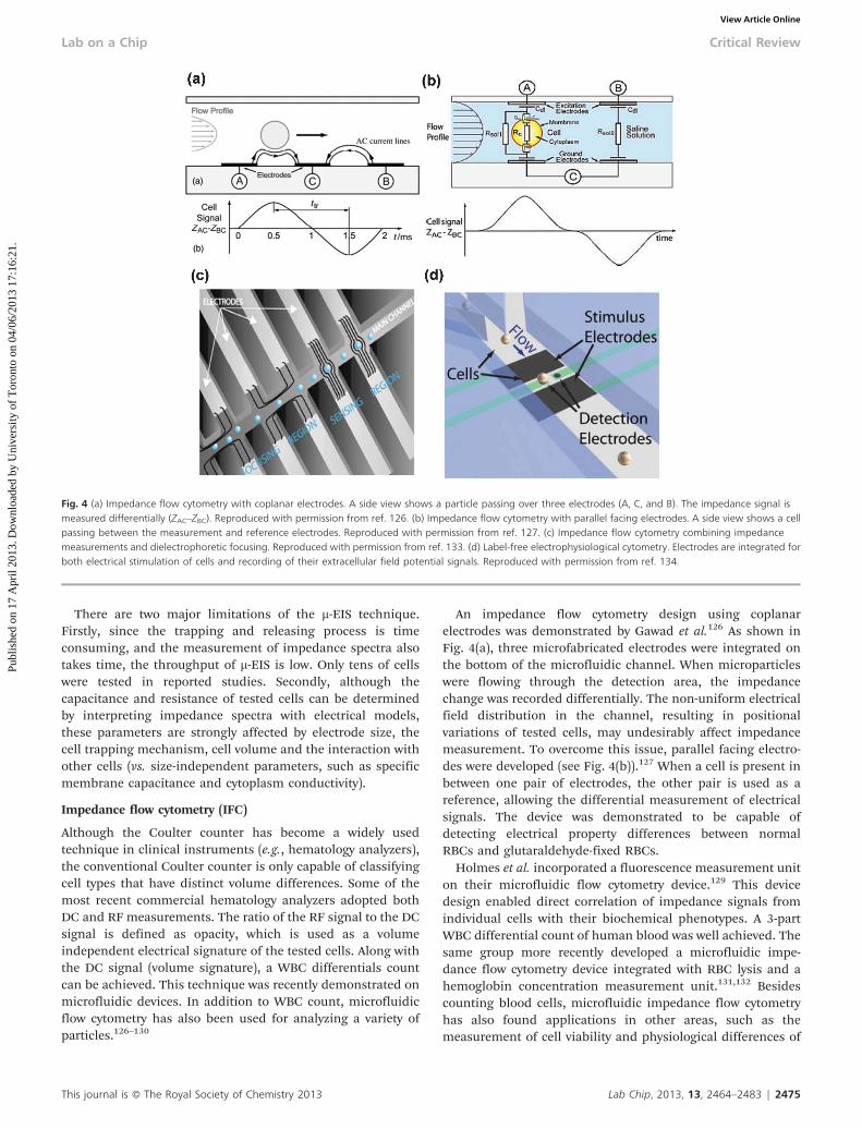
An impedance flow cytometry design using coplanarelectrodes was demonstrated by Gawad et al.126 As shown inFig. 4(a), three microfabricated electrodes were integrated onthe bottom of the microfluidic channel. When microparticleswere flowing through the detection area, the impedancechange was recorded differentially. The non-uniform electricalfield distribution in the channel, resulting in positionalvariations of tested cells, may undesirably affect impedancemeasurement. To overcome this issue, parallel facing electro-des were developed (see Fig. 4(b)).127 When a cell is present inbetween one pair of electrodes, the other pair is used as areference, allowing the differential measurement of electricalsignals. The device was demonstrated to be capable ofdetecting electrical property differences between normalRBCs and glutaraldehyde-fixed RBCs.
Holmes et al. incorporated a fluorescence measurement uniton their microfluidic flow cytometry device.129 This devicedesign enabled direct correlation of impedance signals fromindividual cells with their biochemical phenotypes. A 3-partWBC differential count of human blood was well achieved. Thesame group more recently developed a microfluidic impe-dance flow cytometry device integrated with RBC lysis and ahemoglobin concentration measurement unit.131,132 Besidescounting blood cells, microfluidic impedance flow cytometryhas also found applications in other areas, such as themeasurement of cell viability and physiological differences of
Fig. 4 (a) Impedance flow cytometry with coplanar electrodes. A side view shows a particle passing over three electrodes (A, C, and B). The impedance signal ismeasured differentially (ZAC–ZBC). Reproduced with permission from ref. 126. (b) Impedance flow cytometry with parallel facing electrodes. A side view shows a cellpassing between the measurement and reference electrodes. Reproduced with permission from ref. 127. (c) Impedance flow cytometry combining impedancemeasurements and dielectrophoretic focusing. Reproduced with permission from ref. 133. (d) Label-free electrophysiological cytometry. Electrodes are integrated forboth electrical stimulation of cells and recording of their extracellular field potential signals. Reproduced with permission from ref. 134.
This journal is � The Royal Society of Chemistry 2013 Lab Chip, 2013, 13, 2464–2483 | 2475
Lab on a Chip Critical Review
Publ
ishe
d on
17
Apr
il 20
13. D
ownl
oade
d by
Uni
vers
ity o
f T
oron
to o
n 04
/06/
2013
17:
16:2
1.
View Article Online

cells.130 More details on impedance flow cytometry can befound elsewhere.112,130 In addition to coplanar and parallelfacing electrode designs, Mernier et al. demonstrated ‘‘liquidelectrodes’’ to discriminate living and dead yeast cells (seeFig. 4(c)). The larger electrodes recessed in lateral channelsallow measurements at low frequencies (down to 1 kHz), andthe same ‘‘liquid electrodes’’ can also be used for DEPfocussing of the particles.133 Recently, the concept of label-free electrophysiological cytometry was reported. This techni-que utilizes the nature of electrically-excitable cells (uponactivation by sufficient transmembrane electric fields, differ-ent cell types generate different extracellular field potentialsignals) to distinguish different cell types. As shown inFig. 4(d), the device is integrated with electrodes for bothelectrical stimulation and recording of extracellular fieldpotential signals from suspended cells in flow.134
Undifferentiated human-induced pluripotent stem cells(iPSC) and iPSC-derived cardiomyocyte (iPSC-CM) cells weredistinguished.
Other physical characterization techniques
While clinical applications have been mostly driving thedevelopment of microfluidic tools for single-cell biophysicalcharacterization, these new tools are also enabling biologiststo gain insights into the cell development process. Forinstance, the microfluidic device reported in ref. 140 for thefirst time enabled the measurement of the mass of micro-particles. When particles suspended in liquid flow throughnanomechanical resonators, the resulting frequency shift isused to determine the mass of the particles with extraordinarysensitivity (10221 g). Combined with microfluidic control, the‘instantaneous’ growth rates of individual cells were deter-mined,141 and it was found that heavier cells grew faster thanlighter cells. Using this technology, the density of single livingcells was also measured.33 As shown in Fig. 5(a), thesuspended microfluidic resonator consists of a siliconcantilever and an embedded microfluidic channel. When cellsflow through the microfluidic channel, the changes inresonance frequency are proportional to the buoyant mass ofthe cell. In order to measure the density of the cell, the
Fig. 5 (a) Suspended microchannel resonator for single-cell density measurement. The buoyant mass of the cell was recorded in two fluids of different densities. Thered fluid is less dense than the cell, while the blue fluid has a higher density than the cell. From the resonance frequency shift, absolute mass and density aredetermined. Reproduced with permission from ref. 33. (b) A cell stretching device containing a micropost array membrane to modulate equibiaxial cell stretching,while simultaneously measuring live-cell subcellular contractile response. A vacuum could be drawn in the evacuation chamber to stretch the micropost array over theviewing aperture. Reproduced with permission from ref. 152. (c) A microfluidic device with four independent branches for cell adhesion measurement. Taperedchannels are used to explore a range of shear stresses along the channel. The colored rectangles are the visual fields of different shear stress zones. Reproduced withpermission from ref. 158.
2476 | Lab Chip, 2013, 13, 2464–2483 This journal is � The Royal Society of Chemistry 2013
Critical Review Lab on a Chip
Publ
ishe
d on
17
Apr
il 20
13. D
ownl
oade
d by
Uni
vers
ity o
f T
oron
to o
n 04
/06/
2013
17:
16:2
1.
View Article Online

buoyant mass of the targeted single cells was recorded in twofluids of different densities. The absolute mass and densitywere then determined and were used to distinguish malaria-infected RBCs, RBCs from patients with sickle cell disease andthalassemia, and drug-treated leukemia cells. The microfluidicsystem has a testing speed of y500 cells h21.
Compressive forces can be applied directly to cells througha mechanism similar to bulging elastomeric valves.142 Ingeneral, such devices consist of multiple layers and a thinelastomeric membrane separating the flow and controlchannels. The cells within the flow channels are compressedwhen a pneumatic pressure is applied to deflect the thinmembrane. Microfluidic devices based on the compressionhave been used to monitor cell viability and induce mechan-ical cell lysis of adherent breast cancer cells (MCF-7).143 Amore recent study by Kim et al. has shown that under excessivedeformation of the deflecting membrane, small morphologicalchanges known as ‘‘bulges’’ occur on the cellular membrane asa result of local detachment of the cytoskeleton lipid bilayer.In addition, differences in the uniformity of bulge distributionon the cell’s periphery can be used as a physical indicator todistinguish different cells.144 A similar technology can also beused for viscoelastic characterization of cells of a smallpopulation in suspension. Du et al. estimated global viscoe-lastic time constants of the HL-60 cell line and 3T3 fibroblastsby observing the recovery time upon the removal of thecompression force.145 This design can also be extended to themechanical characterization of other specimens, such asbiofilms.146 Alternatively, hydrostatic pressure can apply auniform compressive force without physical contact.147
Micropost arrays have been used to measure the tractionforce generated by a cell at the adhesion sites.148 The forceapplied by the cell to the substrate was calculated down to thelevel of a single adhesion site, based on the visualization ofdisplacements and locations of the focal adhesions.148 Tanet al. used microfabricated microposts to determine mechan-ical cell–substrate interactions. By varying the geometry of themicroposts, cell adhesion was controlled, and the resultsrevealed that the magnitude of the traction force generated bythe cell was regulated by its morphology.149 Schoen et al.investigated the effect of elastic substrate warping on forcemeasurements. A correction factor was analytically andexperimentally determined, which allows for a more accuratecomparison of absolute forces between different pillargeometries and designs.150 In order to separately study thecellular response to external forces applied to a cell, and theinternal forces generated by the cell, magnetic micropostscontaining cobalt nanowires that can be actively manipulatedwere developed.151 Fu and co-workers devised a strategyinvolving an array of microposts integrated onto a stretchableelastomeric membrane, enabling simultaneous stretching andcontractile response monitoring of vascular smooth musclecells152 (see Fig. 5(b)).
Due to the micro-scale dimensions of microfluidic chan-nels, microchannels are suitable for generating fluid shearforces to study the substrate adhesion strength of cells. Several
studies have quantified the adhesion strength of mammalianfibroblasts.153–155 These microfluidic devices share the com-monalities of using microchannels with a protein coating toalter cell adhesion, which is tested under shearing fluid flow.Channels with varying widths were also developed to quantifycell adhesion strength.155,156 Using a similar tapered design,Gutierrez et al. demonstrated that activated neutrophils had agreater adhesion strength than their non-activated counter-parts,157 and Rupprecht et al. used a tapered channel toquantify the adhesion strength of human breasts cancercells158 (see Fig. 5(c)).
Outlooks
For clinical applications, cell samples from pleural fluid, urineand blood are highly heterogeneous, and the number of cellsof interest is usually low (e.g., CTC cells). Thus, low-throughputtechniques that are only able to test tens or hundreds of cellshave little clinical relevance. In order to obtain statisticallymeaningful data, testing a large number of cells is necessary.Several techniques are able to provide reasonable throughput.For example, the constriction-channel device reported in ref.39 achieved 100–150 cells s21 and is capable of performingmulti-parameter measurements. Since the measurements arecompletely electrical, by refining experimental parameters, afurther increase in throughput could be feasibly achievable.The hydrodynamic stretching microfluidic device reported inref. 19 has proven its clinical relevance and can provide atesting speed of approximately 2000 cells s21. Although high-speed image recording and image analysis are a bottleneck,rapid advances in the imaging industry and hardware-basedimage processing could further enhance the clinical relevanceof the microfluidic deformability cytometry technique.159
Besides throughput, one challenge for microfluidic celldeformability measurements presently lies in the weakcorrelation between cell deformability and the widely usedbiochemical markers. An intrinsic connection between patho-logical cell deformability changes and their biochemical/molecular changes must be well established before celldeformability can practically be accepted as a clinical marker.
Cell deformability changes in response to environmentalfactors also needs to be better understood. After cells areharvested from the incubating environment or retrieved fromthe human body, the deformability of the cells starts tochange. Many environmental parameters, such as tempera-ture, CO2 concentration, and osmotic pressure, can have aninfluence on the deformability change. Thus, for cell deform-ability testing, testing conditions should be maintained asconsistently as possible, and all cells should be tested within astrictly defined period of time. Establishing a standard testingprotocol is necessary as microfluidic cell deformability testingmoves towards clinical applications.
As for electrical measurements, microfluidic impedanceflow cytometry appears promising since the conventionalCoulter counter is a well-accepted technology. The DC/RF
This journal is � The Royal Society of Chemistry 2013 Lab Chip, 2013, 13, 2464–2483 | 2477
Lab on a Chip Critical Review
Publ
ishe
d on
17
Apr
il 20
13. D
ownl
oade
d by
Uni
vers
ity o
f T
oron
to o
n 04
/06/
2013
17:
16:2
1.
View Article Online

measurement technique has proven effective for clinical WBCdifferential count and is used in some commercial hematologyanalyzers. Thus, clinical acceptance and commercial successof microfluidic impedance flow cytometry could be achievable.As a precursor, instruments based on a miniaturized Coultercounter have entered the market. For instance, Chempaq(UNITECH, Denmark) is a portable hematology analyzercapable of 3-part WBC differential count, RBC count, andhemoglobin concentration measurements. Finger prick bloodis loaded onto a disposable cassette, in which an aperture isplaced. Only DC measurement is conducted by the instrument,and a 3-part WBC differential count is based on themodification of cell volume from chemical treatment.
Microfluidic impedance flow cytometry has proven morepowerful than the microfluidic Coulter counter. Microfluidicimpedance flow cytometry has been demonstrated to distin-guish cells based on their physiological and pathologicalconditions, such as viability and membrane potentialchange.130 Although it is known that electrical properties ofcells can possibly be used for distinguishing cell types (e.g.,benign and malignant tumor cells), the establishment of theconnections between cell physiological changes and theirelectrical properties has not gained as much interest as celldeformability research, likely due to the lack of powerful toolsfor cellular electrical characterization. Among the techniquesdiscussed earlier in this review, only electrorotation is capableof quantifying a cell’s intrinsic (size and shape-independent)electrical properties (e.g., specific membrane capacitance andcytoplasm conductivity). However, electrorotation’s testingthroughput is too low to generate meaningful results fordiagnostic applications. Microfluidic impedance flow cytome-try127,130–132 that uses opacity (ratio of high-frequency ampli-tude/low-frequency amplitude) as the indicator of a cell’selectrical properties can offer a high throughput. Opacity is acombined effect of the cell membrane and cytoplasm. A singleRF frequency must be selected carefully so that differentelectrical properties of the cell membrane and/or cytoplasmcan be well reflected in the opacity value. It is difficult tocorrelate electrical properties to physiological changes of cellssince the opacity value can only reflect the electrical propertychanges of the cells. It cannot tell you what has caused thedifference in opacity values and how the electrical propertieshave changed. In order to tackle this challenge, we recentlyreported a microfluidic device, which is capable of quantifyinginherent electrical properties (specific membrane capacitanceand cytoplasm conductivity) of single cells at a reasonabletesting speed of 5–10 cells s21 (vs. minutes per cell usingexisting techniques, such as electrorotation).139 Impedanceprofiles at seven frequencies were measured simultaneouslywhen cells were aspirated through a constriction channel.Based on the geometrical and electrical models, specificmembrane capacitance and cytoplasm conductivity values ofover 6000 HL-60 and AML-2 cells were quantified.
In summary, we highlighted the recent developments ofmicrofluidics-based techniques for the characterization ofbiophysical properties of single cells. Compared with conven-
tional single-cell biophysical characterization tools, microflui-dic devices have demonstrated great potential in realizingmulti-parameter measurements on single cells at a highertesting speed. Biophysical measurements as a label-freeapproach can potentially be used for disease prescreening.For example, if biophysical measurements on cells indicatepossible diseases, further clinical examinations can be done toevaluate the disease condition. Biophysical measurements canalso be incorporated with a cell sorting unit to collect cellshaving different physical properties for further biochemicalassaying.
While the throughput, sensitivity, and specificity of micro-fluidic biophysical characterization devices still await improve-ment, biophysical measurements on single cells already proveuseful to provide bolstering information to biochemicalmeasurements. Several microfluidic systems combining bio-physical and biochemical measurements have been developedto capture multi-parameter information of the targetcells.23,160,161 In the foreseeable future, closer correlationsbetween biophysical and biochemical properties of cells willbe established, and advances in microfluidics-based biophy-sical measurement techniques will move closer to practicalapplications and enable more discoveries in biology research.Meanwhile, as microfluidic devices for single-cell biophysicalcharacterization are more integrated, compact, and easy-to-use, they can potentially become clinically accepted point-of-care tools.
Acknowledgements
Financial support from the Natural Sciences and EngineeringResearch Council of Canada (NSERC) through an NSERCSteacie Fellowship, from the University of Toronto through aConnaught Innovation Award, and from the Canada ResearchChairs Program is acknowledged.
References
1 J. El-Ali, P. K. Sorger and K. F. Jensen, Cells on chips,Nature, 2006, 442, 403–411.
2 A. Fritsch, M. Hockel, T. Kiessling, K. D. Nnetu, F. Wetzel,M. Zink and J. A. Kas, Are biomechanical changesnecessary for tumour progression?, Nat. Phys., 2010, 6,730–732.
3 C. Moraes, Y. Sun and C. A. Simmons, (Micro)managingthe mechanical microenvironment, Integr. Biol., 2011, 3,959–971.
4 S. Suresh, Biomechanics and biophysics of cancer cells,Acta Biomater., 2007, 3, 413–438.
5 J. Chen, J. Li and Y. Sun, Microfluidic approaches forcancer cell detection, characterization, and separation,Lab Chip, 2012, 12, 1753–1767.
6 S. I. Kim and H. I. Jung, Circulating Tumor Cells:Detection Methods and Potential Clinical Application inBreast Cancer, J. Breast Cancer, 2010, 13, 125–131.
2478 | Lab Chip, 2013, 13, 2464–2483 This journal is � The Royal Society of Chemistry 2013
Critical Review Lab on a Chip
Publ
ishe
d on
17
Apr
il 20
13. D
ownl
oade
d by
Uni
vers
ity o
f T
oron
to o
n 04
/06/
2013
17:
16:2
1.
View Article Online

7 G. Y. H. Lee and C. T. Lim, Biomechanics approaches tostudying human diseases, Trends Biotechnol., 2007, 25,111–118.
8 S. E. Cross, Y. S. Jin, J. Rao and J. K. Gimzewski,Nanomechanical analysis of cells from cancer patients,Nat. Nanotechnol., 2007, 2, 780–783.
9 H. Morgan, T. Sun, D. Holmes, S. Gawad and N. G. Green,Single cell dielectric spectroscopy, J. Phys. D: Appl. Phys.,2007, 40, 61–70.
10 R. N. Zare and S. Kim, Microfluidic Platforms for Single-Cell Analysis, Annu. Rev. Biomed. Eng., 2010, 12, 187–201.
11 G. M. Whitesides, The origins and the future of micro-fluidics, Nature, 2006, 442, 368–373.
12 S. A. Vanapalli, M. H. G. Duits and F. Mugele,Microfluidics as a functional tool for cell mechanics,Biomicrofluidics, 2009, 3, 012006.
13 D. H. Kim, P. K. Wong, J. Park, A. Levchenko and Y. Sun,Microengineered Platforms for Cell Mechanobiology,Annu. Rev. Biomed. Eng., 2009, 11, 203–233.
14 Y. Zheng and Y. Sun, Microfluidic devices for mechanicalcharacterisation of single cells in suspension, Micro NanoLett., 2011, 6, 327–331.
15 A. W. L. Jay, Viscoelastic properties of the human redblood cell membrane. I. Deformation, volume loss, andrupture of red cells in micropipettes, Biophys. J., 1973, 13,1166–1182.
16 R. Skalak and Branemar Pi, Deformation of Red BloodCells in Capillaries, Science, 1969, 164, 177.
17 M. Diez-Silva, M. Dao, J. Y. Han, C. T. Lim and S. Suresh,Shape and Biomechanical Characteristics of Human RedBlood Cells in Health and Disease, MRS Bull., 2010, 35,382–388.
18 M. Makale, Cellular mechanobiology and cancer metas-tasis, Birth Defects Res., Part C, 2007, 81, 329–343.
19 D. R. Gossett, H. T. K. Tse, S. A. Lee, Y. Ying, A.G. Lindgren, O. O. Yang, J. Rao, A. T. Clark and D. DiCarlo, Hydrodynamic stretching of single cells for largepopulation mechanical phenotyping, Proc. Natl. Acad. Sci.U. S. A., 2012, 109, 7630–7635.
20 D. Di Carlo, A Mechanical Biomarker of Cell State inMedicine, JALA, 2012, 17, 32–42.
21 F. Lautenschlager, S. Paschke, S. Schinkinger, A. Bruel,M. Beil and J. Guck, The regulatory role of cell mechanicsfor migration of differentiating myeloid cells, Proc. Natl.Acad. Sci. U. S. A., 2009, 106, 15696–15701.
22 J. P. Shelby, J. White, K. Ganesan, P. K. Rathod and D.T. Chiu, Amicrofluidic model for single-cell capillaryobstruction by Plasmodium falciparum infected erythro-cytes, Proc. Natl. Acad. Sci. U. S. A., 2003, 100,14618–14622.
23 H. Bow, I. V. Pivkin, M. Diez-Silva, S. J. Goldfless, M. Dao,J. C. Niles, S. Suresh and J. Y. Han, A microfabricateddeformability-based flow cytometer with application tomalaria, Lab Chip, 2011, 11, 1065–1073.
24 J. P. Mills, M. Diez-Silva, D. J. Quinn, M. Dao, M. J. Lang,K. S. W. Tan, C. T. Lim, G. Milon, P. H. David,O. Mercereau-Puijalon, S. Bonnefoy and S. Suresh, Effectof plasmodial RESA protein on deformability of humanred blood cells harboring Plasmodium falciparum, Proc.Natl. Acad. Sci. U. S. A., 2007, 104, 9213–9217.
25 Y. K. Park, M. Diez-Silva, G. Popescu, G. Lykotrafitis, W.S. Choi, M. S. Feld and S. Suresh, Refractive index mapsand membrane dynamics of human red blood cellsparasitized by Plasmodium falciparum, Proc. Natl. Acad.Sci. U. S. A., 2008, 105, 13730–13735.
26 S. Eber and S. E. Lux, Hereditary spherocytosis-Defects inproteins that connect the membrane skeleton to the lipidbilayer, Semin. Hematol., 2004, 41, 118–141.
27 S. Perrotta, P. G. Gallagher and N. Mohandas, Hereditaryspherocytosis, Lancet, 2008, 372, 1411–1426.
28 L. D. Walensky, M. Narla and S. E. Lux, Blood: principlesand practice of hematology, Lippincott Williams andWilkins, Philadelphia, 2003.
29 S. K. Ballas and E. D. Smith, Red-Blood-Cell Changesduring the Evolution of the Sickle-Cell Painful Crisis,Blood, 1992, 79, 2154–2163.
30 G. A. Barabino, M. O. Platt and D. K. Kaul, Sickle CellBiomechanics, Annu. Rev. Biomed. Eng., 2010, 12, 345–367.
31 O. K. Baskurt, D. Gelmont and H. J. Meiselman, Red bloodcell deformability in sepsis, Am. J. Respir. Crit. Care Med.,1998, 157, 421–427.
32 T. C. Hurd, K. S. Dasmahapatra, B. F. Rush and G.W. Machiedo, Red Blood-Cell Deformability in Humanand Experimental Sepsis, Arch. Surg., 1988, 123, 217–220.
33 W. H. Grover, A. K. Bryan, M. Diez-Silva, S. Suresh, J.M. Higgins and S. R. Manalis, Measuring single-celldensity, Proc. Natl. Acad. Sci. U. S. A., 2011, 108,10992–10996.
34 C. D. Brown, H. S. Ghali, Z. H. Zhao, L. L. Thomas and E.A. Friedman, Association of reduced red blood celldeformability and diabetic nephropathy, Kidney Int.,2005, 67, 295–300.
35 S. Keymel, C. Heiss, P. Kleinbongard, M. Kelm andT. Lauer, Impaired Red Blood Cell Deformability inPatients with Coronary Artery Disease and DiabetesMellitus, Horm. Metab. Res., 2011, 43, 760–765.
36 K. Tsukada, E. Sekizuka, C. Oshio and H. Minamitani,Direct measurement of erythrocyte deformability indiabetes mellitus with a transparent microchannel capil-lary model and high-speed video camera system,Microvasc. Res., 2001, 61, 231–239.
37 E. Bennett-Guerrero, T. H. Veldman, A. Doctor, M.J. Telen, T. L. Ortel, T. S. Reid, M. A. Mulherin, H. Zhu,R. D. Buck, R. M. Califf and T. J. McMahon, Evolution ofadverse changes in stored RBCs, Proc. Natl. Acad. Sci. U. S.A., 2007, 104, 17063–17068.
38 J. Bonaventura, Clinical implications of the loss ofvasoactive nitric oxide during red blood cell storage,Proc. Natl. Acad. Sci. U. S. A., 2007, 104, 19165–19166.
39 Y. Zheng, E. Shojaei-Baghini, A. Azad, C. Wang andY. Sun, High-throughput biophysical measurement ofhuman red blood cells, Lab Chip, 2012, 12, 2560–2567.
40 W. G. Lee, H. Bang, H. Yun, J. Lee, J. Park, J. K. Kim,S. Chung, K. Cho, C. Chung, D. C. Han and J. K. Chang,On-chip erythrocyte deformability test under opticalpressure, Lab Chip, 2007, 7, 516–519.
41 S. C. Gifford, M. G. Frank, J. Derganc, C. Gabel, R.H. Austin, T. Yoshida and M. W. Bitensky, Parallelmicrochannel-based measurements of individual erythro-cyte areas and volumes, Biophys. J., 2003, 84, 623–633.
This journal is � The Royal Society of Chemistry 2013 Lab Chip, 2013, 13, 2464–2483 | 2479
Lab on a Chip Critical Review
Publ
ishe
d on
17
Apr
il 20
13. D
ownl
oade
d by
Uni
vers
ity o
f T
oron
to o
n 04
/06/
2013
17:
16:2
1.
View Article Online

42 S. C. Gifford, J. Derganc, S. S. Shevkoplyas, T. Yoshida andM. W. Bitensky, A detailed study of time-dependentchanges in human red blood cells: from reticulocytematuration to erythrocyte senescence, Br. J. Haematol.,2006, 135, 395–404.
43 T. Herricks, M. Antia and P. K. Rathod, Deformabilitylimits of Plasmodium falciparum-infected red blood cells,Cell. Microbiol., 2009, 11, 1340–1353.
44 M. Abkarian, M. Faivre and H. A. Stone, High-speedmicrofluidic differential manometer for cellular-scalehydrodynamics, Proc. Natl. Acad. Sci. U. S. A., 2006, 103,538–542.
45 M. J. Rosenbluth, W. A. Lam and D. A. Fletcher, Analyzingcell mechanics in hematologic diseases with microfluidicbiophysical flow cytometry, Lab Chip, 2008, 8, 1062–1070.
46 H. W. Hou, Q. S. Li, G. Y. H. Lee, A. P. Kumar, C. N. Ongand C. T. Lim, Deformability study of breast cancer cellsusing microfluidics, Biomed. Microdevices, 2009, 11,557–564.
47 J. Chen, Y. Zheng, Q. Y. Tan, E. Shojaei-Baghini, Y.L. Zhang, J. Li, P. Prasad, L. D. You, X. Y. Wu and Y. Sun,Classification of cell types using a microfluidic device formechanical and electrical measurement on single cells,Lab Chip, 2011, 11, 3174–3181.
48 A. Adamo, A. Sharei, L. Adamo, B. Lee, S. Mao and K.F. Jensen, Microfluidics-based assessment of cell deform-ability, Anal. Chem., 2012, 84, 6438–6443.
49 G. Guan, P. C. Y. Chen, W. K. Peng, A. A. Bhagat, C. J. Ongand J. Han, Real-time control of a microfluidic channelfor size-independent deformability cytometry, J.Micromech. Microeng., 2012, 22, 105037.
50 A. M. Forsyth, J. D. Wan, W. D. Ristenpart and H. A. Stone,The dynamic behavior of chemically "stiffened" red bloodcells in microchannel flows, Microvasc. Res., 2010, 80,37–43.
51 S. S. Lee, Y. Yim, K. H. Ahn and S. J. Lee, Extensional flow-based assessment of red blood cell deformability usinghyperbolic converging microchannel, Biomed. Microdevices,2009, 11, 1021–1027.
52 A. M. Forsyth, J. Wan, P. D. Owrutsky, M. Abkarian and H.A. Stone, Multiscale approach to link red blood celldynamics, shear viscosity, and ATP release, Proc. Natl.Acad. Sci. U. S. A., 2011, 108, 10986–10991.
53 W. Bishara, T. W. Su, A. F. Coskun and A. Ozcan, Lensfreeon-chip microscopy over a wide field-of-view using pixelsuper-resolution, Opt. Express, 2010, 18, 11181–11191.
54 X. Q. Cui, L. M. Lee, X. Heng, W. W. Zhong, P.W. Sternberg, D. Psaltis and C. H. Yang, Lensless high-resolution on-chip optofluidic microscopes forCaenorhabditis elegans and cell imaging, Proc. Natl.Acad. Sci. U. S. A., 2008, 105, 10670–10675.
55 A. Greenbaum, W. Luo, T. W. Su, Z. Gorocs, L. Xue, S.O. Isikman, A. F. Coskun, O. Mudanyali and A. Ozcan,Imaging without lenses: achievements and remainingchallenges of wide-field on-chip microscopy, Nat.Methods, 2012, 9, 889–895.
56 S. O. Isikman, W. Bishara, H. Y. Zhu and A. Ozcan,Optofluidic Tomography on a Chip, Appl. Phys. Lett., 2011,98, 161109.
57 W. K. Purves, Life: The Science of Biology, SinauerAssociates, 6th edn, 2001.
58 J. Kim, I. Hwang, D. Britain, T. D. Chung, Y. Sun and D.H. Kim, Microfluidic approaches for gene delivery andgene therapy, Lab Chip, 2011, 11, 3941–3948.
59 S. Movahed and D. Li, Microfluidics cell electroporation,Microfluid. Nanofluid., 2011, 10, 703–734.
60 E. Ferret, C. Evrard, A. Foucal and P. Gervais, Volumechanges of isolated human K562 leukemia cells inducedby electric field pulses, Biotechnol. Bioeng., 2000, 67,520–528.
61 N. Bao, Y. H. Zhan and C. Lu, Microfluidic electroporativeflow cytometry for studying single-cell biomechanics,Anal. Chem., 2008, 80, 7714–7719.
62 N. Bao, T. T. Le, J. X. Cheng and C. Lu, Microfluidicelectroporation of tumor and blood cells: observation ofnucleus expansion and implications on selective analysisand purging of circulating tumor cells, Integr. Biol., 2010,2, 113–120.
63 N. Bao, G. C. Kodippili, K. M. Giger, V. M. Fowler, P.S. Low and C. Lu, Single-cell electrical lysis of erythrocytesdetects deficiencies in the cytoskeletal protein network,Lab Chip, 2011, 11, 3053–3056.
64 B. E. Henslee, A. Morss, X. Hu, G. P. Lafyatis and L. J. Lee,Electroporation Dependence on Cell Size: OpticalTweezers Study, Anal. Chem., 2011, 83, 3998–4003.
65 A. Ashkin, Acceleration and Trapping of Particles byRadiation Pressure, Phys. Rev. Lett., 1970, 24, 156.
66 J. Guck, R. Ananthakrishnan, T. J. Moon, C. C. Cunninghamand J. Kas, Optical deformability of soft biologicaldielectrics, Phys. Rev. Lett., 2000, 84, 5451–5454.
67 J. Guck, R. Ananthakrishnan, H. Mahmood, T. J. Moon, C.C. Cunningham and J. Kas, The optical stretcher: A novellaser tool to micromanipulate cells, Biophys. J., 2001, 81,767–784.
68 E. Jonietz, MECHANICS: The forces of cancer, Nature,2012, 491, S56–S57.
69 B. Lincoln, H. M. Erickson, S. Schinkinger, F. Wottawah,D. Mitchell, S. Ulvick, C. Bilby and J. Guck, Deformability-based flow cytometry, Cytometry, Part B, 2004, 59A,203–209.
70 J. Guck, S. Schinkinger, B. Lincoln, F. Wottawah, S. Ebert,M. Romeyke, D. Lenz, H. M. Erickson, R. Ananthakrishnan,D. Mitchell, J. Kas, S. Ulvick and C. Bilby, Opticaldeformability as an inherent cell marker for testingmalignant transformation and metastatic competence,Biophys. J., 2005, 88, 3689–3698.
71 C. W. Lai, S. K. Hsiung, C. L. Yeh, A. Chiou and G. B. Lee,A cell delivery and pre-positioning system utilizingmicrofluidic devices for dual-beam optical trap-and-stretch, Sens. Actuators, B, 2008, 135, 388–397.
72 N. Bellini, K. C. Vishnubhatla, F. Bragheri, L. Ferrara,P. Minzioni, R. Ramponi, I. Cristiani and R. Osellame,Femtosecond laser fabricated monolithic chipfor opticaltrapping and stretching of single cells, Opt. Express, 2010,18, 4679–4688.
73 F. Bragheri, L. Ferrara, N. Bellini, K. C. Vishnubhatla,P. Minzioni, R. Ramponi, R. Osellame and I. Cristiani,Optofluidic chip for single cell trapping and stretchingfabricated by a femtosecond laser, J. Biophotonics, 2010, 3,234–243.
74 T. W. Remmerbach, F. Wottawah, J. Dietrich, B. Lincoln,C. Wittekind and J. Guck, Oral cancer diagnosis by
2480 | Lab Chip, 2013, 13, 2464–2483 This journal is � The Royal Society of Chemistry 2013
Critical Review Lab on a Chip
Publ
ishe
d on
17
Apr
il 20
13. D
ownl
oade
d by
Uni
vers
ity o
f T
oron
to o
n 04
/06/
2013
17:
16:2
1.
View Article Online

mechanical phenotyping, Cancer Res., 2009, 69,1728–1732.
75 J. Voldman, Electrical forces for microscale cell manip-ulation, Annu. Rev. Biomed. Eng., 2006, 8, 425–454.
76 R. Pethig, Dielectrophoresis: Status of the theory, technol-ogy, and applications (vol 4, 022811, 2010), Biomicrofluidics,2010, 4, 022811.
77 L. A. MacQueen, M. D. Buschmann and M. R. Wertheimer,Mechanical properties of mammalian cells in suspensionmeasured by electro-deformation, J. Micromech. Microeng.,2010, 20, 065007.
78 J. Chen, M. Abdelgawad, L. M. Yu, N. Shakiba, W.Y. Chien, Z. Lu, W. R. Geddie, M. A. S. Jewett andY. Sun, Electrodeformation for single cell mechanicalcharacterization, J. Micromech. Microeng., 2011, 21,054012.
79 I. Doh, W. C. Lee, Y. H. Cho, A. P. Pisano and F.A. Kuypers, Deformation measurement of individual cellsin large populations using a single-cell microchamberarray chip, Applied Physics Letters, 2012, 100, 173702.
80 R. M. Hochmuth, Micropipette aspiration of living cells, J.Biomech., 2000, 33, 15–22.
81 Q. Guo, S. J. Reiling, P. Rohrbach and H. S. Ma,Microfluidic biomechanical assay for red blood cellsparasitized by Plasmodium falciparum, Lab Chip, 2012,12, 1143–1150.
82 J. Chen, Y. Zheng, Q. Y. Tan, Y. L. Zhang, J. Li, W.R. Geddie, M. A. S. Jewett and Y. Sun, A microfluidicdevice for simultaneous electrical and mechanical mea-surements on single cells, Biomicrofluidics, 2011, 5,014113.
83 Y. Katsumoto, K. Tatsumi, T. Doi and K. Nakabe,Electrical classification of single red blood cell deform-ability in high-shear microchannel flows, Int. J. Heat FluidFlow, 2010, 31, 985–995.
84 J. Nilsson, M. Evander, B. Hammarstrom and T. Laurell,Review of cell and particle trapping in microfluidicsystems, Anal. Chim. Acta, 2009, 649, 141–157.
85 D. R. Gossett, W. M. Weaver, A. J. Mach, S. C. Hur, H. T.K. Tse, W. Lee, H. Amini and D. Di Carlo, Label-free cellseparation and sorting in microfluidic systems, Anal.Bioanal. Chem., 2010, 397, 3249–3267.
86 R. Hoeber, Eine Methode die elektrische Leitfaehigkeit imInnern von Zellen zu messen, Pfluegers Arch., 1910, 133,237–259.
87 R. Hoeber, Ein zweites Verfahren die Leitfaehigkeit imInnern von Zellen zu messen, Pfluegers Arch., 1912, 148,189–221.
88 R. Hoeber, Messungen der inneren Leitfaehigkeit vonZellen III, Pfluegers Arch., 1913, 150, 15–45.
89 H. P. Schwan, Electrical properties of tissue and cellsuspensions, Adv. Biol. Med. Phys., 1957, 5, 147–209.
90 H. P. Schwan, Electrode polarization impedance andmeasurements in biological materials, Ann. N. Y. Acad.Sci., 1968, 148, 191–209.
91 J. Yang, Y. Huang, X. J. Wang, X. B. Wang, F. F. Becker andP. R. C. Gascoyne, Dielectric properties of humanleukocyte subpopulations determined by electrorotationas a cell separation criterion, Biophys. J., 1999, 76,3307–3314.
92 X. B. Wang, Y. Huang, P. R. C. Gascoyne, F. F. Becker,R. Holzel and R. Pethig, Changes in Friend MurineErythroleukemia Cell-Membranes during Induced-Differentiation Determined by Electrorotation, Biochim.Biophys. Acta, Biomembr., 1994, 1193, 330–344.
93 D. Zimmermann, A. Zhou, M. Kiesel, K. Feldbauer,U. Terpitz, W. Haase, T. Schneider-Hohendorf, E. Bambergand V. L. Sukhorukov, Effects on capacitance by over-expression of membrane proteins, Biochem. Biophys. Res.Commun., 2008, 369, 1022–1026.
94 Y. Huang, X. B. Wang, R. Holzel, F. F. Becker and P. R.C. Gascoyne, Electrorotational Studies of the CytoplasmicDielectric-Properties of Friend Murine Erythroleukemia-Cells, Phys. Med. Biol., 1995, 40, 1789–1806.
95 L. Duncan, H. Shelmerdine, M. P. Hughes, H. M. Coley,Y. Hubner and F. H. Labeed, Dielectrophoretic analysis ofchanges in cytoplasmic ion levels due to ion channelblocker action reveals underlying differences betweendrug-sensitive and multidrug-resistant leukaemic cells,Phys. Med. Biol., 2008, 53, N1–N7.
96 R. Holzel, Non-invasive determination of bacterial singlecell properties by electrorotation, Biochim. Biophys. Acta,Mol. Cell Res., 1999, 1450, 53–60.
97 E. Neher and B. Sakmann, Single-Channel CurrentsRecorded from Membrane of Denervated Frog Muscle-Fibers, Nature, 1976, 260, 799–802.
98 J. H. Nieuwenhuis, F. Kohl, J. Bastemeijer, P. M. Sarro andM. J. Vellekoop, Integrated Coulter counter based on2-dimensional liquid aperture control, Sens. Actuators, B,2004, 102, 44–50.
99 J. Zhe, A. Jagtiani, P. Dutta, J. Hu and J. Carletta, Amicromachined high throughput Coulter counter forbioparticle detection and counting, J. Micromech.Microeng., 2007, 17, 304–313.
100 M. W. Shinwari, D. Zhitomirsky, I. A. Deen, P.R. Selvaganapathy, M. J. Deen and D. Landheer,Microfabricated Reference Electrodes and their BiosensingApplications, Sensors, 2010, 10, 1679–1715.
101 J. H. Zhou, K. N. Ren, Y. Z. Zheng, J. Su, Y. H. Zhao, D. Ryanand H. K. Wu, Fabrication of a microfluidic Ag/AgClreference electrode and its application for portable anddisposable electrochemical microchips, Electrophoresis,2010, 31, 3083–3089.
102 S. Y. Zheng, M. Liu and Y. C. Tai, Micro coulter counterswith platinum black electroplated electrodes for humanblood cell sensing, Biomed. Microdevices, 2008, 10,221–231.
103 H. G. Chun, T. D. Chung and H. C. Kim, Cytometry andvelocimetry on a microfluidic chip using polyelectrolyticsalt bridges, Anal. Chem., 2005, 77, 2490–2495.
104 K. B. Kim, H. Chun, H. C. Kim and T. D. Chun, Red bloodcell quantification microfluidic chip using polyelectrolyticgel electrodes, Electrophoresis, 2009, 30, 1464–1469.
105 H. Choi, K. B. Kim, C. S. Jeon, I. Hwang, S. A. Lee, H.K. Kim, H. C. Kim and T. D. Chung, A label-free DCimpedance-based microcytometer for circulating rarecancer cell counting, Lab Chip, 2013, 13, 970–977.
106 R. W. Deblois and C. P. Bean, Counting and Sizing ofSubmicron Particles by Resistive Pulse Technique, Rev.Sci. Instrum., 1970, 41, 909.
This journal is � The Royal Society of Chemistry 2013 Lab Chip, 2013, 13, 2464–2483 | 2481
Lab on a Chip Critical Review
Publ
ishe
d on
17
Apr
il 20
13. D
ownl
oade
d by
Uni
vers
ity o
f T
oron
to o
n 04
/06/
2013
17:
16:2
1.
View Article Online

107 O. A. Saleh and L. L. Sohn, Quantitative sensing ofnanoscale colloids using a microchip Coulter counter,Rev. Sci. Instrum., 2001, 72, 4449–4451.
108 E. G. Cen, C. Dalton, Y. L. Li, S. Adamia, L. M. Pilarski andK. V. I. S. Kaler, A combined dielectrophoresis, travelingwave dielectrophoresis and electrorotation microchip forthe manipulation and characterization of human malig-nant cells, J. Microbiol. Methods, 2004, 58, 387–401.
109 L. Wu, L. Lanry Yung and K. Lim, Dielectrophoreticcapture voltage spectrum for measurement of dielectricproperties and separation of cancer cells, Biomicrofluidics,2012, 6, 014113.
110 D. M. Vykoukal, P. R. C. Gascoyne and J. Vykoukal,Dielectric characterization of complete mononuclear andpolymorphonuclear blood cell subpopulations for label-free discrimination, Integr. Biol., 2009, 1, 477–484.
111 L. M. Broche, F. H. Labeed and M. P. Hughes, Extractionof dielectric properties of multiple populations fromdielectrophoretic collection spectrum data, Phys. Med.Biol., 2005, 50, 2267–2274.
112 T. Sun and H. Morgan, Single-cell microfluidic impedancecytometry: a review, Microfluid. Nanofluid., 2010, 8,423–443.
113 M. Cristofanilli, G. De Gasperis, L. S. Zhang, M. C. Hung,P. R. C. Gascoyne and G. N. Hortobagyi, Automatedelectrorotation to reveal dielectric variations related toHER-2/neu overexpression in MCF-7 sublines, Clin. CancerResearch, 2002, 8, 615–619.
114 H. Ying, R. Holzel, R. Pethig and X. B. Wang, Differencesin the Ac Electrodynamics of Viable and Nonviable Yeast-Cells Determined through Combined Dielectrophoresisand Electrorotation Studies, Phys. Med. Biol., 1992, 37,1499–1517.
115 F. F. Becker, X. B. Wang, Y. Huang, R. Pethig, J. Vykoukaland P. R. C. Gascoyne, Separation of Human Breast-Cancer Cells from Blood by Differential Dielectric Affinity,Proc. Natl. Acad. Sci. U. S. A., 1995, 92, 860–864.
116 X. B. Wang, Y. Huang, P. R. C. Gascoyne and F. F. Becker,Dielectrophoretic manipulation of particles, IEEE Trans.Ind. Appl., 1997, 33, 660–669.
117 G. De Gasperis, X. B. Wang, J. Yang, F. F. Becker and P. R.C. Gascoyne, Automated electrorotation: dielectric char-acterization of living cells by real-time motion estimation,Meas. Sci. Technol., 1998, 9, 518–529.
118 L. S. Jang and M. H. Wang, Microfluidic device for cellcapture and impedance measurement, Biomed.Microdevices, 2007, 9, 737–743.
119 D. Malleo, J. T. Nevill, L. P. Lee and H. Morgan,Continuous differential impedance spectroscopy of singlecells, Microfluid. Nanofluid., 2010, 9, 191–198.
120 K. H. Han, A. Han and A. B. Frazier, Microsystems forisolation and electrophysiological analysis of breastcancer cells from blood, Biosens. Bioelectron., 2006, 21,1907–1914.
121 Y. Cho, H. S. Kim, A. B. Frazier, Z. G. Chen, D. M. Shinand A. Han, Whole-Cell Impedance Analysis for Highlyand Poorly Metastatic Cancer Cells, J. Microelectromech.Syst., 2009, 18, 808–817.
122 L.-S. Kung-Chieh Lan, Integration of single-cell trappingand impedance measurement utilizing microwell electro-des, Biosens. Bioelectron., 2010, 26(5), 2025–2031.
123 S. Li and L. W. Lin, A single cell electrophysiologicalanalysis device with embedded electrode, Sens. Actuators,A, 2007, 134, 20–26.
124 S. B. Cho and H. Thielecke, Micro hole-based cell chipwith impedance spectroscopy, Biosens. Bioelectron., 2007,22, 1764–1768.
125 C. M. Kurz, H. Buth, A. Sossalla, V. Vermeersch,V. Toncheva, P. Dubruel, E. Schacht and H. Thielecke,Chip-based impedance measurement on single cells formonitoring sub-toxic effects on cell membranes, Biosens.Bioelectron., 2011, 26, 3405–3412.
126 S. Gawad, L. Schild and P. Renaud, Micromachinedimpedance spectroscopy flow cytometer for cell analysisand particle sizing, Lab Chip, 2001, 1, 76–82.
127 K. Cheung, S. Gawad and P. Renaud, Impedance spectro-scopy flow cytometry: On-chip label-free cell differentia-tion, Cytometry, Part A, 2005, 65A, 124–132.
128 G. Benazzi, D. Holmes, T. Sun, M. C. Mowlem andH. Morgan, Discrimination and analysis of phytoplanktonusing a microfluidic cytometer, IET Nanobiotechnol., 2007,1, 94–101.
129 D. Holmes, D. Pettigrew, C. H. Reccius, J. D. Gwyer, C. vanBerkel, J. Holloway, D. E. Davies and H. Morgan,Leukocyte analysis and differentiation using high speedmicrofluidic single cell impedance cytometry, Lab Chip,2009, 9, 2881–2889.
130 K. C. Cheung, M. Di Berardino, G. Schade-Kampmann,M. Hebeisen, A. Pierzchalski, J. Bocsi, A. Mittag andA. Tarnok, Microfluidic Impedance-Based FlowCytometry, Cytometry, Part A, 2010, 77A, 648–666.
131 X. J. Han, C. van Berkel, J. Gwyer, L. Capretto andH. Morgan, Microfluidic Lysis of Human Blood forLeukocyte Analysis Using Single Cell ImpedanceCytometry, Anal. Chem., 2012, 84, 1070–1075.
132 C. van Berkel, J. D. Gwyer, S. Deane, N. Green, J. Holloway,V. Hollis and H. Morgan, Integrated systems for rapidpoint of care (PoC) blood cell analysis, Lab Chip, 2011, 11,1249–1255.
133 G. Mernier, E. Duqi and P. Renaud, Characterization of anovel impedance cytometer design and its integrationwith lateral focusing by dielectrophoresis, Lab Chip, 2012,12, 4344–4349.
134 F. B. Myers, C. K. Zarins, O. J. Abilez and L. P. Lee, Label-free electrophysiological cytometry for stem cellderivedcardiomyocyte clusters, Lab Chip, 2013, 13, 220–228.
135 J. Riordon, M. Mirzaei and M. Godin, Microfluidic cellvolume sensor with tunable sensitivity, Lab Chip, 2012, 12,3016–3019.
136 C. Kuttel, E. Nascimento, N. Demierre, T. Silva,T. Braschler, P. Renaud and A. G. Oliva, Label-freedetection of Babesia bovis infected red blood cells usingimpedance spectroscopy on a microfabricated flowcytometer, Acta Trop., 2007, 102, 63–68.
137 G. Schade-Kampmann, A. Huwiler, M. Hebeisen,T. Hessler and M. Di Berardino, On-chip non-invasiveand label-free cell discrimination by impedance spectro-scopy, Cell Proliferation, 2008, 41, 830–840.
138 C. Bernabini, D. Holmes and H. Morgan, Micro-impe-dance cytometry for detection and analysis of micron-sized particles and bacteria, Lab Chip, 2011, 11, 407–412.
2482 | Lab Chip, 2013, 13, 2464–2483 This journal is � The Royal Society of Chemistry 2013
Critical Review Lab on a Chip
Publ
ishe
d on
17
Apr
il 20
13. D
ownl
oade
d by
Uni
vers
ity o
f T
oron
to o
n 04
/06/
2013
17:
16:2
1.
View Article Online

139 Y. Zheng, E. Shojaei-Baghini, C. Wang and Y. Sun,Microfluidic characterization of specific membrane capa-citance and cytoplasm conductivity of single cells, Biosens.Bioelectron., 2012, 42, 496–502.
140 T. P. Burg, M. Godin, S. M. Knudsen, W. Shen, G. Carlson,J. S. Foster, K. Babcock and S. R. Manalis, Weighing ofbiomolecules, single cells and single nanoparticles influid, Nature, 2007, 446, 1066–1069.
141 M. Godin, F. F. Delgado, S. M. Son, W. H. Grover, A.K. Bryan, A. Tzur, P. Jorgensen, K. Payer, A. D. Grossman,M. W. Kirschner and S. R. Manalis, Using buoyant mass tomeasure the growth of single cells, Nat. Methods, 2010, 7,387–U70.
142 M. A. Unger, H. P. Chou, T. Thorsen, A. Scherer and S.R. Quake, Monolithic microfabricated valves and pumpsby multilayer soft lithography, Science, 2000, 288,113–116.
143 Y. C. Kim, J. H. Kang, S. J. Park, E. S. Yoon and J. K. Park,Microfluidic biomechanical device for compressive cellstimulationand lysis, Sens. Actuators, B, 2007, 128,108–116.
144 Y. C. Kim, S. J. Park and J. K. Park, Biomechanical analysisof cancerous and normal cells based on bulge generationin a microfluidic device, Analyst, 2008, 133, 1432–1439.
145 G. S. Du, A. Ravetto, Q. Fang and J. M. J. den Toonder, Celltypes can be distinguished by measuring their viscoelasticrecovery times using a micro-fluidic device, Biomed.Microdevices, 2011, 13, 29–40.
146 D. N. Hohne, J. G. Younger and M. J. Solomon, FlexibleMicrof luidic Device for Mechanical PropertyCharacterization of Soft Viscoelastic Solids Such asBacterial Biofilms, Langmuir, 2009, 25, 7743–7751.
147 L. A. G. Lin, A. Q. Liu, Y. F. Yu, C. Zhang, C. S. Lim, S.H. Ng, P. H. Yap and H. J. Gao, Cell compressibilitystudies utilizing noncontact hydrostatic pressure mea-surements on single living cells in a microchamber, Appl.Physics Letters, 2008, 92.
148 N. Q. Balaban, U. S. Schwarz, D. Riveline, P. Goichberg,G. Tzur, I. Sabanay, D. Mahalu, S. Safran, A. Bershadsky,L. Addadi and B. Geiger, Force and focal adhesionassembly: a close relationship studied using elasticmicropatterned substrates, Nat. Cell Biol., 2001, 3,466–472.
149 J. L. Tan, J. Tien, D. M. Pirone, D. S. Gray, K. Bhadrirajuand C. S. Chen, Cells lying on a bed of microneedles: Anapproach to isolate mechanical force, Proc. Natl. Acad. Sci.U. S. A., 2003, 100, 1484–1489.
150 I. Schoen, W. Hu, E. Klotzsch and V. Vogel, Probing CellularTraction Forces by Micropillar Arrays: Contribution of
Substrate Warping to Pillar Deflection, Nano Lett., 2010,10, 1823–1830.
151 N. J. Sniadecki, A. Anguelouch, M. T. Yang, C. M. Lamb,Z. Liu, S. B. Kirschner, Y. Liu, D. H. Reich and C. S. Chen,Magnetic microposts as an approach to apply forces toliving cells, Proc. Natl. Acad. Sci. U. S. A., 2007, 104,14553–14558.
152 R. H. W. Lam, Y. B. Sun, W. Q. Chen and J. P. Fu,Elastomeric microposts integrated into microfluidics forflow-mediated endothelial mechanotransduction analy-sis, Lab Chip, 2012, 12, 1865–1873.
153 C. Christophis, M. Grunze and A. Rosenhahn,Quantification of the adhesion strength of fibroblastcells on ethylene glycol terminated self-assembled mono-layers by a microfluidic shear force assay, Phys. Chem.Chem. Phys., 2010, 12, 4498–4504.
154 K. V. Christ, K. B. Williamson, K. S. Masters and K.T. Turner, Measurement of single-cell adhesion strengthusing a microfluidic assay, Biomed. Microdevices, 2010, 12,443–455.
155 H. Lu, L. Y. Koo, W. C. M. Wang, D. A. Lauffenburger, L.G. Griffith and K. F. Jensen, Microfluidic shear devices forquantitative analysis of cell adhesion, Anal. Chem., 2004,76, 5257–5264.
156 S. Usami, H. H. Chen, Y. H. Zhao, S. Chien and R. Skalak,Design and Construction of a Linear Shear-Stress FlowChamber, Ann. Biomed. Eng., 1993, 21, 77–83.
157 E. Gutierrez and A. Groisman, Quantitative measurementsof the strength of adhesion of human neutrophils to asubstratum in a microfluidic device, Anal. Chem., 2007,79, 2249–2258.
158 P. Rupprecht, L. Gole, J. P. Rieu, C. Vezy, R. Ferrigno, H.C. Mertani and C. Riviere, A tapered channel microfluidicdevice for comprehensive cell adhesion analysis, usingmeasurements of detachment kinetics and shear stress-dependent motion, Biomicrofluidics, 2012, 6.
159 K. Goda, A. Ayazi, D. R. Gossett, J. Sadasivam, C.K. Lonappan, E. Sollier, A. M. Fard, S. C. Hur, J. Adam,C. Murray, C. Wang, N. Brackbill, D. Di Carlo and B. Jalali,High-throughput single-microparticle imaging flow ana-lyzer, Proc. Natl. Acad. Sci. U. S. A., 2012, 109,11630–11635.
160 D. Barat, D. Spencer, G. Benazzi, M. C. Mowlem andH. Morgan, Simultaneous high speed optical and impe-dance analysis of single particles with a microfluidiccytometer, Lab Chip, 2012, 12, 118–126.
161 D. Holmes and H. Morgan, Single Cell ImpedanceCytometry for Identification and Counting of CD4T-Cells in Human Blood Using Impedance Labels, Anal.Chem., 2010, 82, 1455–1461.
This journal is � The Royal Society of Chemistry 2013 Lab Chip, 2013, 13, 2464–2483 | 2483
Lab on a Chip Critical Review
Publ
ishe
d on
17
Apr
il 20
13. D
ownl
oade
d by
Uni
vers
ity o
f T
oron
to o
n 04
/06/
2013
17:
16:2
1.
View Article Online