Embed Size (px)
Citation preview
Label-Free and Real-Time Detection of Tuberculosis in Human UrineSamples Using a Nanophotonic Point-of-Care PlatformPatricia Ramirez-Priego,† Daan Martens,‡,§ Ayssar A. Elamin,∥ Pieterjan Soetaert,⊥ Wim Van Roy,#
Rita Vos,# Birgit Anton,∇ Ronny Bockstaele,⊥ Holger Becker,∇ Mahavir Singh,∥ Peter Bienstman,‡,§
and Laura M. Lechuga*,†
†Nanobiosensors and Bioanalytical Applications Group, Catalan Institute of Nanoscience and Nanotechnology (ICN2), CSIC, BISTand CIBER-BBN, Campus UAB, Bellaterra, 08193 Barcelona, Spain‡Photonics Research Group, Ghent University/imec, Technologiepark-Zwijnaarde 15, 9052 Ghent, Belgium§Center for Nano- and Biophotonics, Ghent University, Technologiepark-Zwijnaarde 15, 9052 Ghent, Belgium∥LIONEX Diagnostics and Therapeutics GmbH, Salzdahlumer Str. 196, Building 1A, 38126 Braunschweig, Germany⊥Trinean NV, Dulle-Grietlaan 17/3, 9050 Ghent, Belgium#imec, Kapeldreef 75, 3001 Leuven, Belgium∇microfluidic ChipShop GmbH, Stockholmer Str. 20, 07747 Jena, Germany
*S Supporting Information
ABSTRACT: Tuberculosis (TB) is the leading global causeof death from a single infectious agent. Registered incidencerates are low, especially in low-resource countries with weakhealth systems, due to the disadvantages of current diagnostictechniques. A major effort is directed to develop a point-of-care (POC) platform to reduce TB deaths with a prompt andreliable low-cost technique. In the frame of the EuropeanPOCKET Project, a novel POC platform for the direct andnoninvasive detection of TB in human urine was developed.The photonic sensor chip is integrated in a disposablecartridge and is based on a highly sensitive Mach−ZehnderInterferometer (MZI) transducer combined with an on-chipspectral filter. The required elements for the readout areintegrated in an instrument prototype, which allows real-time monitoring and data processing. In this work, the novel POCplatform has been employed for the direct detection of lipoarabinomannan (LAM), a lipopolysaccharide found in themycobacterium cell wall. After the optimization of several parameters, a limit of detection of 475 pg/mL (27.14 pM) wasachieved using a direct immunoassay in undiluted human urine in less than 15 min. A final validation of the technique wasperformed using 20 clinical samples from TB patients and healthy donors, allowing the detection of TB in people regardless ofHIV coinfection. The results show excellent correlation to those obtained with standard techniques. These promising resultsdemonstrate the high sensitivity, specificity and applicability of our novel POC platform, which could be used during routinecheck-ups in developing countries.
KEYWORDS: point-of-care, nanophotonic biosensor, LAM, biomarker, urine, diagnosis, tuberculosis
Tuberculosis (TB) is a re-emerging infectious diseasecaused by Mycobacterium tuberculosis affecting millions of
people worldwide. According to the 2017 World HealthOrganization (WHO) report,1 TB infected 10.4 millionpeople, resulting in 1.7 million deaths with more than 95%of these cases occurring in developing countries. TB isconsidered the leading fatal infectious agent, surpassinghuman immunodeficiency virus (HIV) and acquired immunedeficiency syndrome (AIDS).1 Two types of TB have beenidentified: active and latent. In both cases, the bacterium ispresent in the human body. In active TB, the bacterium isconstantly multiplying, invading diverse organs with a high
infective potential. On the other hand, latent TB isasymptomatic and the bacterium is inactive without the abilityto infect others.Traditional methods for the detection of active TB include
culture testing as the current reference standard and sputumsmear microscopy. However, both of them are imprecise, slowand require qualified personnel and laboratory equipment.1−3
Since 2010, WHO began to recommend the use of a new
Received: May 10, 2018Accepted: September 30, 2018Published: September 30, 2018
Article
pubs.acs.org/acssensorsCite This: ACS Sens. 2018, 3, 2079−2086
© 2018 American Chemical Society 2079 DOI: 10.1021/acssensors.8b00393ACS Sens. 2018, 3, 2079−2086
Dow
nloa
ded
via
UN
IV G
EN
T o
n N
ovem
ber
15, 2
018
at 0
8:59
:53
(UT
C).
Se
e ht
tps:
//pub
s.ac
s.or
g/sh
arin
ggui
delin
es f
or o
ptio
ns o
n ho
w to
legi
timat
ely
shar
e pu
blis
hed
artic
les.
molecular assay, GeneXpert (Cepheid, US), based on nucleicacid amplification for the detection of M. tuberculosis and forthe determination of its resistance to rifampicin, the commonantibiotic employed to treat several infectious diseases,including TB.1,4 Although promising at the beginning,GeneXpert has not improved the TB screening capabilities ascompared to conventional tests; also it incurs infrastructurelimitations in developing countries, as it needs a controlledtemperature environment, with the addition of the instrumen-tation, reagents, and cartridges costs.1,5 Therefore, there is stillan urgent need to develop a highly sensitive and specific point-of-care (POC) platform for the rapid diagnosis of TB in low-resource countries; a platform that should be cost efficient,user-friendly, noninvasive and requiring minimal biosafetystandards.6−8
Several M. tuberculosis secreted antigens have been describedas biomarkers in TB, such as lipoarabinomannan (LAM),antigen 85 complex (Ag85), ESAT-6, CFP-10, MPT64, and 38kDa antigen.7,9−12 These antigens can be used for the directdetection of active TB in different biological samples otherthan sputum, such as urine and plasma. Of these, urine ispotentially the best candidate for a POC analysis as it can bereadily obtained from children and adults, can be easily stored,has a low infection risk during sample collection, and does notrequire a laboratory with a high biosafety level.7,13,14
LAM is a widely known and structurally important 17.5 kDalipopolysaccharide found in mycobacterial cell walls.13,15−17
When M. tuberculosis is lysed by the host immune system, it isfiltered by the kidneys and LAM can then be detected in theurine.16 Since no human glycosidases are known for thedegradation of LAM, an antigen with glycosidic linkages, it is akey potential biomarker in urine.14 Several studies haveconfirmed the presence of LAM antigens in the urine ofpatients with active TB13,18 and have found that the amount ofLAM in urine is correlated with the bacterial burden metabolicactivity and rate of degradation of the bacteria, and henceallows for a semiquantitative assessment of the infection. It hasalso been found that LAM antigens play a key role inphagocyte evasion,18 modulation of the host response duringinfection,19 inhibition of mycobacterial antigen processing,18
production of tumor necrosis factor,18 and growth andpathogenesis of M. tuberculosis.20 Detection of LAM in urinehas several advantages in comparison to the conventionaltechniques. There are ELISA and lateral flow testscommercially available for the detection of LAM in urine15,21
with adequate diagnostic specificity of 98−99%,22 butunsatisfactory low sensitivity for TB patients who do nothave HIV. These techniques achieve higher sensitivity (61−67%) for people with less than 50 cluster of differentiation four(CD4) cells/μL, who are seriously ill HIV-positive patients,than for patients only infected with TB (4−8%).15,22 Takinginto account that TB is the leading cause of death of HIV-positive people with more than 40% of incidents worldwide1
and that the current methods are not able to detect TB inpeople without HIV,21 effort is necessary to develop newmethodologies for TB diagnosis in patients with and withoutHIV in order to reduce the number of TB cases and deaths,especially in developing countries.We present the optimization and validation of a novel POC
platform for the direct and noninvasive detection of TB inhuman urine. This new POC platform was developed in theframe of a European project (www.pocket-proj.eu), and thecomplete description of the new instrument can be found in ref
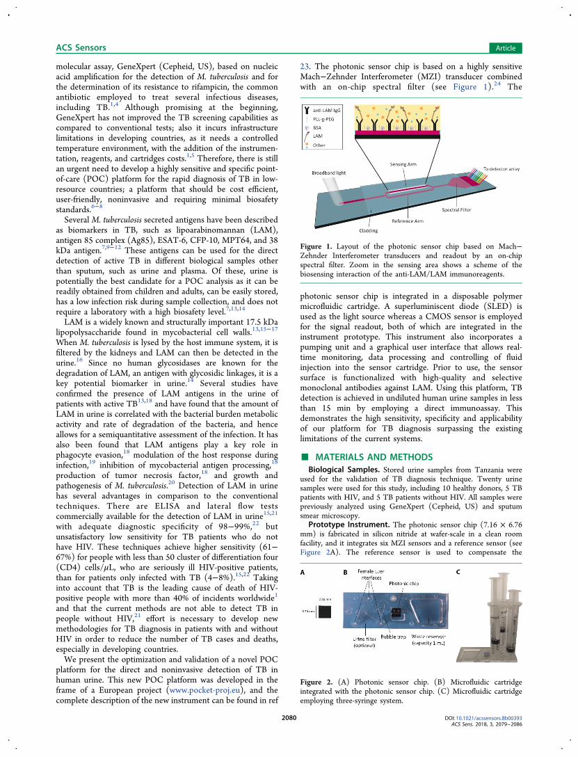
23. The photonic sensor chip is based on a highly sensitiveMach−Zehnder Interferometer (MZI) transducer combinedwith an on-chip spectral filter (see Figure 1).24 The
photonic sensor chip is integrated in a disposable polymermicrofluidic cartridge. A superluminiscent diode (SLED) isused as the light source whereas a CMOS sensor is employedfor the signal readout, both of which are integrated in theinstrument prototype. This instrument also incorporates apumping unit and a graphical user interface that allows real-time monitoring, data processing and controlling of fluidinjection into the sensor cartridge. Prior to use, the sensorsurface is functionalized with high-quality and selectivemonoclonal antibodies against LAM. Using this platform, TBdetection is achieved in undiluted human urine samples in lessthan 15 min by employing a direct immunoassay. Thisdemonstrates the high sensitivity, specificity and applicabilityof our platform for TB diagnosis surpassing the existinglimitations of the current systems.
■ MATERIALS AND METHODSBiological Samples. Stored urine samples from Tanzania were
used for the validation of TB diagnosis technique. Twenty urinesamples were used for this study, including 10 healthy donors, 5 TBpatients with HIV, and 5 TB patients without HIV. All samples werepreviously analyzed using GeneXpert (Cepheid, US) and sputumsmear microscopy.
Prototype Instrument. The photonic sensor chip (7.16 × 6.76mm) is fabricated in silicon nitride at wafer-scale in a clean roomfacility, and it integrates six MZI sensors and a reference sensor (seeFigure 2A). The reference sensor is used to compensate the
Figure 1. Layout of the photonic sensor chip based on Mach−Zehnder Interferometer transducers and readout by an on-chipspectral filter. Zoom in the sensing area shows a scheme of thebiosensing interaction of the anti-LAM/LAM immunoreagents.
Figure 2. (A) Photonic sensor chip. (B) Microfluidic cartridgeintegrated with the photonic sensor chip. (C) Microfluidic cartridgeemploying three-syringe system.
ACS Sensors Article
DOI: 10.1021/acssensors.8b00393ACS Sens. 2018, 3, 2079−2086
2080
dispersion of the grating couplers, the light source, and the on-chipspectral filter, resulting in an accuracy increase of the response.A complete description of the new POC instrument can be found
in ref 23. Briefly, a SLED (850 nm) broadband light source is coupledto each sensor simultaneously using a separated grating coupler. Theoutput of each MZI sensor is connected to an arrayed waveguidegrating (AWG) with 30 channels which work as on-chip spectral filter.Other separated grating couplers are used to couple the light from the30 output channels of the AWG to a CMOS camera which monitorsthe intensity of the different spectral channels simultaneously.The photonic sensor chip was placed in a disposable cartridge
(75.5 × 25.5 mm) which is fabricated with cyclic olefin copolymer(COC) as shown in Figure 2B. The input and output grating couplersare left exposed to avoid variations in the optical pathways. Amicrofluidic channel delivers the sample onto the MZI sensing arms.A waste reservoir able to handle a maximum volume of 1 mL isconnected at the end of the fluidic path. The cartridge includes threefemale Luer interfaces to facilitate connections with standard syringesemployed for the injection of urine samples and the required buffers.The fluidic system includes a special bubble trap to avoid undesiredair bubbles, and an optional urine filter (5 μm pore size).The complete instrument (22 × 22 × 49.30 cm) contains the
optical system, the pumping units and a touch screen as a userinterface, as shown in Figure S-1. The sensors are evaluated bymeasuring wavelength shifts due to surface refractive index changes.The sensorgram of each sensor is graphically displayed in real-time
providing the user with immediate feedback of the patient samplediagnosis. Additionally, the interface enables the control of the pumpspeed and time of injection for each syringe and bubble trapmonitoring.Due to the volume limitations of the cartridge waste reservoir and
the three-syringe configuration in the final POC configuration (Figure2C), we decided to modify the instrument with an in-flow system inorder to perform its complete analytical characterization at thelaboratory level, prior to the evaluation of the final POC instrument.To include this in-flow system, we have added (i) a syringe pump(NewEra, US) with adjustable pumping speed, (ii) a two-positionvalve (VICI, US) that allows sequential loading of the sample loopand injection into the cartridge, and (iii) three Teflon caps to provideconnections to the cartridge female Luer interfaces. One of these capsallows the flow of the solution in the cartridge, and the other two forma vacuum to avoid air bubbles. The evaluation of real samples with thenew POC platform was performed using both the in-flow and thethree-syringe configuration.Antibody Immobilization and Direct Immunoassay. For the
detection of LAM, a monoclonal IgG antibody against LAM (anti-LAM) was developed within the framework of the project byLIONEX GmbH (Germany). The antibody was diluted in PBS 10mM and immobilized by physical adsorption onto the surface of thephotonic sensor chip, followed by a blocking step to avoid nonspecificadsorptions. Milli-Q water was used as the running buffer at a flowrate of 10 μL/min.Once the immobilization process was completed, an immunoassay
was performed which allows the direct detection of LAM from thesample. The running buffer was changed to PBS 10 mM. In order toobtain a complete calibration curve, different LAM concentrations (1,10, 50, 100, 250, 500, 750, and 1000 ng/mL) were employed. Anadditive assay was performed by flowing successive dilutions ofincreasing LAM concentrations.
■ RESULTS AND DISCUSSIONEvaluation of the Bulk Sensitivity. The estimation of the
bulk sensitivity of the photonic sensor chip to changes in therefractive indices of a solution over its sensing area provides apreliminary evaluation of the performance and reproducibilityof our device and the prototype instrument.In order to analyze the performance of the photonic sensor
chip, different solutions of PBS (5, 10, 25, 50, 75, and 100mM), which do not chemically modify the sensor surface, were
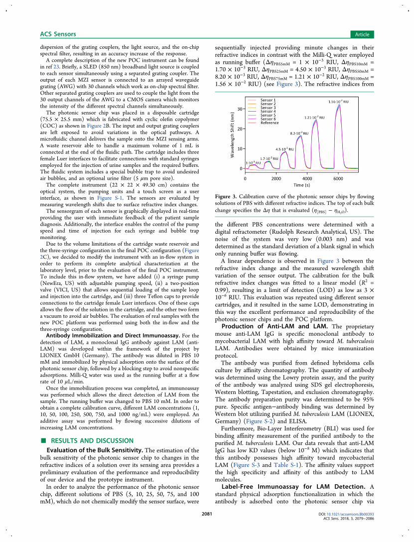
sequentially injected providing minute changes in theirrefractive indices in contrast with the Milli-Q water employedas running buffer (ΔηPBS5mM = 1 × 10⁻³ RIU, ΔηPBS10mM =1.70 × 10⁻³ RIU, ΔηPBS25mM = 4.50 × 10⁻³ RIU, ΔηPBS50mM =8.20 × 10⁻³ RIU, ΔηPBS75mM = 1.21 × 10⁻² RIU, ΔηPBS100mM =1.56 × 10⁻² RIU) (see Figure 3). The refractive indices from
the different PBS concentrations were determined with adigital refractometer (Rudolph Research Analytical, US). Thenoise of the system was very low (0.003 nm) and wasdetermined as the standard deviation of a blank signal in whichonly running buffer was flowing.A linear dependence is observed in Figure 3 between the
refractive index change and the measured wavelength shiftvariation of the sensor output. The calibration for the bulkrefractive index changes was fitted to a linear model (R2 =0.99), resulting in a limit of detection (LOD) as low as 3 ×10⁻⁶ RIU. This evaluation was repeated using different sensorcartridges, and it resulted in the same LOD, demonstrating inthis way the excellent performance and reproducibility of thephotonic sensor chips and the POC platform.
Production of Anti-LAM and LAM. The proprietarymouse anti-LAM IgG is specific monoclonal antibody tomycobacterial LAM with high affinity toward M. tuberculosisLAM. Antibodies were obtained by mice immunizationprotocol.The antibody was purified from defined hybridoma cells
culture by affinity chromatography. The quantity of antibodywas determined using the Lowry protein assay, and the purityof the antibody was analyzed using SDS gel electrophoresis,Western blotting, Tapestation, and exclusion chromatography.The antibody preparation purity was determined to be 95%pure. Specific antigen−antibody binding was determined byWestern blot utilizing purified M. tuberculosis LAM (LIONEX,Germany) (Figure S-2) and ELISA.Furthermore, Bio-Layer Interferometry (BLI) was used for
binding affinity measurement of the purified antibody to thepurified M. tuberculosis LAM. Our data reveals that anti-LAMIgG has low KD values (below 10−8 M) which indicates thatthis antibody possesses high affinity toward mycobacterialLAM (Figure S-3 and Table S-1). The affinity values supportthe high specificity and affinity of this antibody to LAMmolecules.
Label-Free Immunoassay for LAM Detection. Astandard physical adsorption functionalization in which theantibody is adsorbed onto the photonic sensor chip via
Figure 3. Calibration curve of the photonic sensor chips by flowingsolutions of PBS with different refractive indices. The top of each bulkchange specifies the Δη that is evaluated (η[PBS] − ηH2O).
ACS Sensors Article
DOI: 10.1021/acssensors.8b00393ACS Sens. 2018, 3, 2079−2086
2081
intermolecular forces, such as electrostatic and hydrophobicinteractions, ionic and hydrogen bonds, van der Waals forces,or a combination of those, was selected for our new diagnosticplatform.25−27 The biofunctionalization protocol based onphysical adsorption involves two main steps: the noncovalentand random immobilization of the antibodies and the blockingof the remaining free sites on the sensor surface to preventnonspecific adsorptions.To pursue the biosensing evaluation, an optimization with
buffer is advised before using complex matrices, such as urine.The evaluation in buffer will allow us to study the performanceof the biosensor platform for the specific TB biomarkerwithout the interference of other proteins or compounds alsopresent in urine.Different immobilization parameters must be optimized for
LAM detection in buffer. In order to provide a good anti-LAMdensity with enough accessibility to LAM and to guarantee asuitable surface coverage for minimizing nonspecific adsorp-tions, various concentrations of the antibody and BSA, asblocking agent, were tested to improve the immobilizationperformance. Each antibody concentration, ranging from 10 to100 μg/mL, was evaluated at fixed conditions for pump speed(10 μL/min) and time (30 min). The surface coverage wasanalyzed from the immobilization signal. Immobilizationsignals showed an increasing tendency with increasingantibody concentrations (data not shown). As antibodyconcentrations above 50 μg/mL showed a similar signal, thismeans that the sensor surface was almost covered with 50 μg/mL of the antibody. After the antibody immobilization, thesame procedure was followed for the blocking step usingdifferent concentrations of BSA (from 1 to 10 mg/mL) andanalyzing the detection signal of a nonspecific protein (datanot shown). A PBS 10 mM solution with 10 mg/mL of BSAwas the one that blocks the remaining free sensor surface areasand avoids nonspecific adsorptions.Using the optimized protocol, an antibody solution (50 μg/
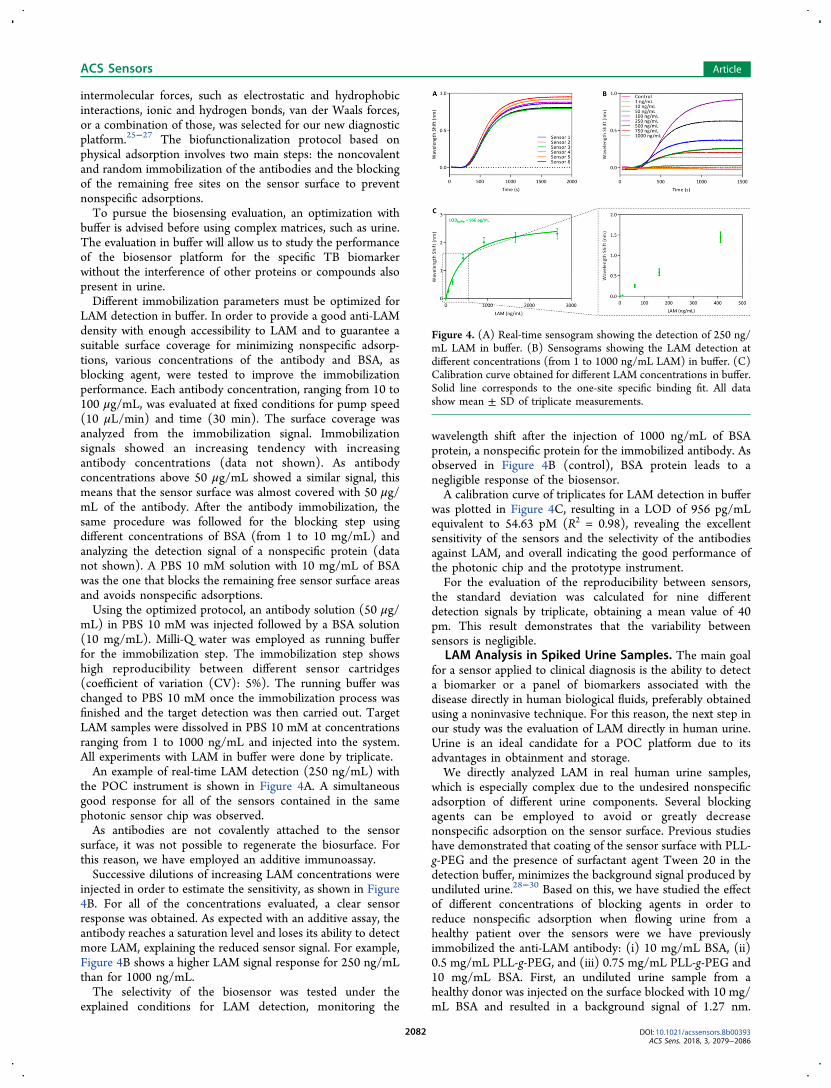
mL) in PBS 10 mM was injected followed by a BSA solution(10 mg/mL). Milli-Q water was employed as running bufferfor the immobilization step. The immobilization step showshigh reproducibility between different sensor cartridges(coefficient of variation (CV): 5%). The running buffer waschanged to PBS 10 mM once the immobilization process wasfinished and the target detection was then carried out. TargetLAM samples were dissolved in PBS 10 mM at concentrationsranging from 1 to 1000 ng/mL and injected into the system.All experiments with LAM in buffer were done by triplicate.An example of real-time LAM detection (250 ng/mL) with
the POC instrument is shown in Figure 4A. A simultaneousgood response for all of the sensors contained in the samephotonic sensor chip was observed.As antibodies are not covalently attached to the sensor
surface, it was not possible to regenerate the biosurface. Forthis reason, we have employed an additive immunoassay.Successive dilutions of increasing LAM concentrations were
injected in order to estimate the sensitivity, as shown in Figure4B. For all of the concentrations evaluated, a clear sensorresponse was obtained. As expected with an additive assay, theantibody reaches a saturation level and loses its ability to detectmore LAM, explaining the reduced sensor signal. For example,Figure 4B shows a higher LAM signal response for 250 ng/mLthan for 1000 ng/mL.The selectivity of the biosensor was tested under the
explained conditions for LAM detection, monitoring the
wavelength shift after the injection of 1000 ng/mL of BSAprotein, a nonspecific protein for the immobilized antibody. Asobserved in Figure 4B (control), BSA protein leads to anegligible response of the biosensor.A calibration curve of triplicates for LAM detection in buffer
was plotted in Figure 4C, resulting in a LOD of 956 pg/mLequivalent to 54.63 pM (R2 = 0.98), revealing the excellentsensitivity of the sensors and the selectivity of the antibodiesagainst LAM, and overall indicating the good performance ofthe photonic chip and the prototype instrument.For the evaluation of the reproducibility between sensors,
the standard deviation was calculated for nine differentdetection signals by triplicate, obtaining a mean value of 40pm. This result demonstrates that the variability betweensensors is negligible.
LAM Analysis in Spiked Urine Samples. The main goalfor a sensor applied to clinical diagnosis is the ability to detecta biomarker or a panel of biomarkers associated with thedisease directly in human biological fluids, preferably obtainedusing a noninvasive technique. For this reason, the next step inour study was the evaluation of LAM directly in human urine.Urine is an ideal candidate for a POC platform due to itsadvantages in obtainment and storage.We directly analyzed LAM in real human urine samples,
which is especially complex due to the undesired nonspecificadsorption of different urine components. Several blockingagents can be employed to avoid or greatly decreasenonspecific adsorption on the sensor surface. Previous studieshave demonstrated that coating of the sensor surface with PLL-g-PEG and the presence of surfactant agent Tween 20 in thedetection buffer, minimizes the background signal produced byundiluted urine.28−30 Based on this, we have studied the effectof different concentrations of blocking agents in order toreduce nonspecific adsorption when flowing urine from ahealthy patient over the sensors were we have previouslyimmobilized the anti-LAM antibody: (i) 10 mg/mL BSA, (ii)0.5 mg/mL PLL-g-PEG, and (iii) 0.75 mg/mL PLL-g-PEG and10 mg/mL BSA. First, an undiluted urine sample from ahealthy donor was injected on the surface blocked with 10 mg/mL BSA and resulted in a background signal of 1.27 nm.
Figure 4. (A) Real-time sensogram showing the detection of 250 ng/mL LAM in buffer. (B) Sensograms showing the LAM detection atdifferent concentrations (from 1 to 1000 ng/mL LAM) in buffer. (C)Calibration curve obtained for different LAM concentrations in buffer.Solid line corresponds to the one-site specific binding fit. All datashow mean ± SD of triplicate measurements.
ACS Sensors Article
DOI: 10.1021/acssensors.8b00393ACS Sens. 2018, 3, 2079−2086
2082
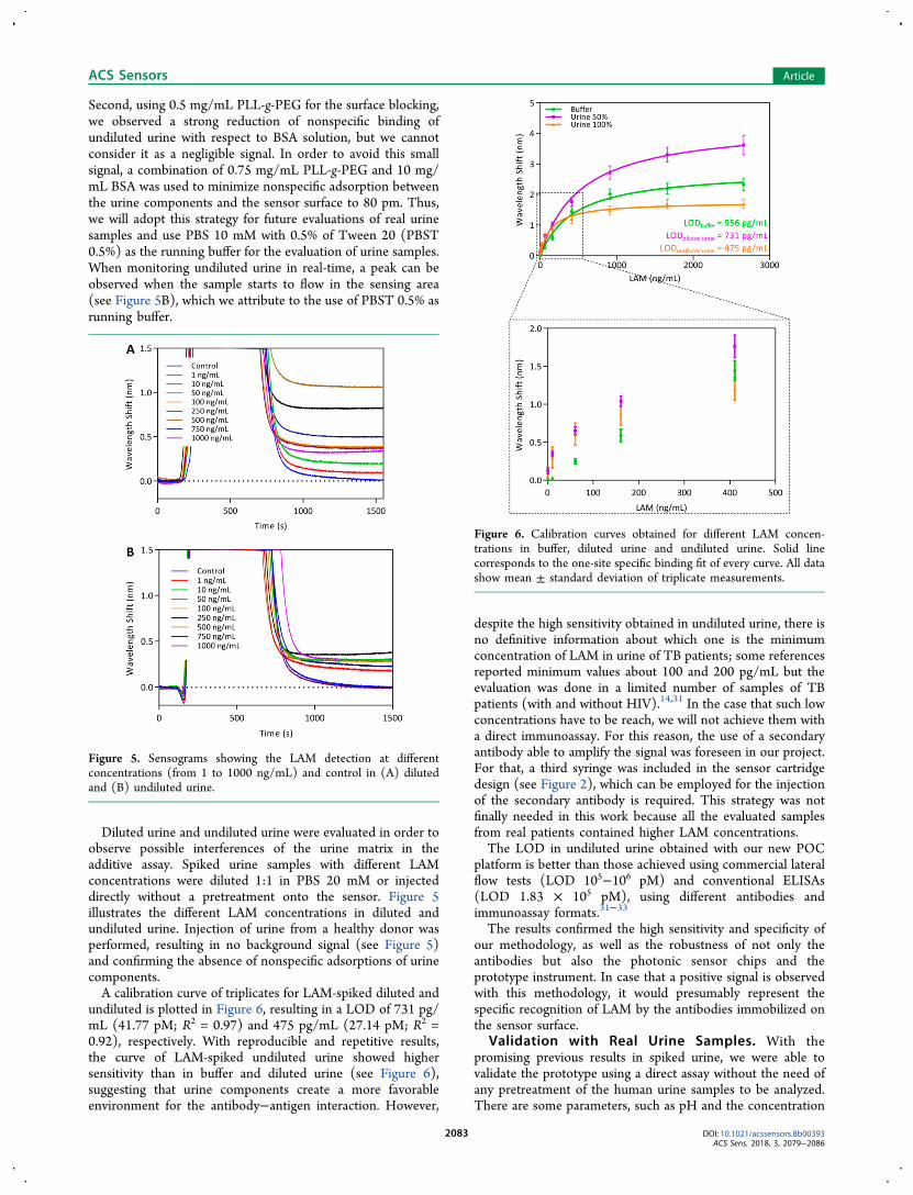
Second, using 0.5 mg/mL PLL-g-PEG for the surface blocking,we observed a strong reduction of nonspecific binding ofundiluted urine with respect to BSA solution, but we cannotconsider it as a negligible signal. In order to avoid this smallsignal, a combination of 0.75 mg/mL PLL-g-PEG and 10 mg/mL BSA was used to minimize nonspecific adsorption betweenthe urine components and the sensor surface to 80 pm. Thus,we will adopt this strategy for future evaluations of real urinesamples and use PBS 10 mM with 0.5% of Tween 20 (PBST0.5%) as the running buffer for the evaluation of urine samples.When monitoring undiluted urine in real-time, a peak can beobserved when the sample starts to flow in the sensing area(see Figure 5B), which we attribute to the use of PBST 0.5% asrunning buffer.
Diluted urine and undiluted urine were evaluated in order toobserve possible interferences of the urine matrix in theadditive assay. Spiked urine samples with different LAMconcentrations were diluted 1:1 in PBS 20 mM or injecteddirectly without a pretreatment onto the sensor. Figure 5illustrates the different LAM concentrations in diluted andundiluted urine. Injection of urine from a healthy donor wasperformed, resulting in no background signal (see Figure 5)and confirming the absence of nonspecific adsorptions of urinecomponents.A calibration curve of triplicates for LAM-spiked diluted and
undiluted is plotted in Figure 6, resulting in a LOD of 731 pg/mL (41.77 pM; R2 = 0.97) and 475 pg/mL (27.14 pM; R2 =0.92), respectively. With reproducible and repetitive results,the curve of LAM-spiked undiluted urine showed highersensitivity than in buffer and diluted urine (see Figure 6),suggesting that urine components create a more favorableenvironment for the antibody−antigen interaction. However,
despite the high sensitivity obtained in undiluted urine, there isno definitive information about which one is the minimumconcentration of LAM in urine of TB patients; some referencesreported minimum values about 100 and 200 pg/mL but theevaluation was done in a limited number of samples of TBpatients (with and without HIV).14,31 In the case that such lowconcentrations have to be reach, we will not achieve them witha direct immunoassay. For this reason, the use of a secondaryantibody able to amplify the signal was foreseen in our project.For that, a third syringe was included in the sensor cartridgedesign (see Figure 2), which can be employed for the injectionof the secondary antibody is required. This strategy was notfinally needed in this work because all the evaluated samplesfrom real patients contained higher LAM concentrations.The LOD in undiluted urine obtained with our new POC
platform is better than those achieved using commercial lateralflow tests (LOD 105−106 pM) and conventional ELISAs(LOD 1.83 × 10⁵ pM), using different antibodies andimmunoassay formats.31−33
The results confirmed the high sensitivity and specificity ofour methodology, as well as the robustness of not only theantibodies but also the photonic sensor chips and theprototype instrument. In case that a positive signal is observedwith this methodology, it would presumably represent thespecific recognition of LAM by the antibodies immobilized onthe sensor surface.
Validation with Real Urine Samples. With thepromising previous results in spiked urine, we were able tovalidate the prototype using a direct assay without the need ofany pretreatment of the human urine samples to be analyzed.There are some parameters, such as pH and the concentration
Figure 5. Sensograms showing the LAM detection at differentconcentrations (from 1 to 1000 ng/mL) and control in (A) dilutedand (B) undiluted urine.
Figure 6. Calibration curves obtained for different LAM concen-trations in buffer, diluted urine and undiluted urine. Solid linecorresponds to the one-site specific binding fit of every curve. All datashow mean ± standard deviation of triplicate measurements.
ACS Sensors Article
DOI: 10.1021/acssensors.8b00393ACS Sens. 2018, 3, 2079−2086
2083
of different urine components, which can strongly varybetween patients and which can have an effect in theevaluation.With the optimal conditions previously selected for the
evaluation of undiluted urine, we applied the POC platform tostudy the presence of LAM in the urine of 10 patients withconfirmed TB from Tanzania and 10 healthy patients fromTanzania and Spain. The Tanzania samples were collectedfrom hospitals and directly observed treatment short course(DOTS) centers,34 while the Spain samples were donated byvolunteers.Nontreated and undiluted real urine samples were directly
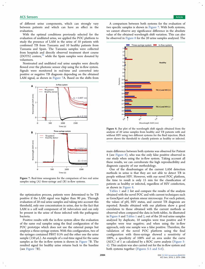
flowed over the photonic sensor chip using the in-flow system.Signals were monitored in real-time and converted intopositive or negative TB diagnosis depending on the obtainedLAM signal, as shown in Figure 7A. Based on the shifts from
the optimization process, patients were determined to be TBpositive if the LAM signal was higher than 90 pm. Throughevaluation of 20 real urine samples and taking into account thisthreshold, only one concentration in urine, due to the fact thatLAM is a cell wall component of M. tuberculosis and can onlybe present in the urine of those infected with the pathogenicbacteria.Positive results with the in-flow system allow the evaluation
of the same real samples using the final configuration of thePOC prototype which does not use the external pumps butemploys a three-syringe system. With this configuration, two ofthe syringes contained PBST 0.5% and the other one the urinesample (150 μL). An example of a real-time signal for the samesamples as for the in-flow system is shown in Figure 7B. Theresulted signal for healthy urine returns back to the baseline(see Figure 7B).
A comparison between both systems for the evaluation oftwo specific samples is shown in Figure 7. With both systems,we cannot observe any significance difference in the absolutevalue of the obtained wavelength shift variation. This can alsobe observed in Figure 8 for the 20 urine samples analyzed. The
main difference between both systems was observed for Patient4 (see Figure 8), who was the only false positive observed inour study when using the in-flow system. Taking account allthese results, we can corroborate the high reproducibility anddiagnosis capacity of our methodology.One of the disadvantages of the current LAM detection
methods in urine is that they are not able to detect TB inpeople without HIV. However, with our novel POC platform,the time to result is only 15 min for the classification ofpatients as healthy or infected, regardless of HIV coinfection,as shown in Figure 8.Tables 1 and 2 list and compare the results of the analysis
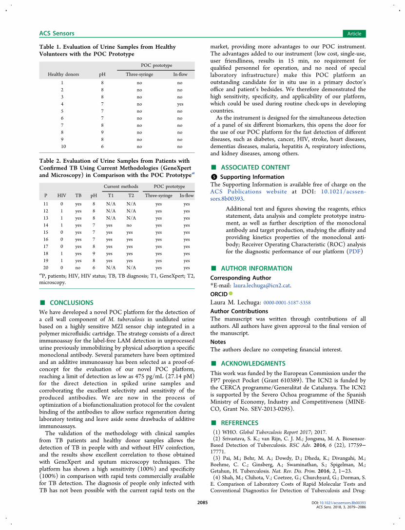
obtained with the novel POC and with current techniques suchas GeneXpert and sputum smear microscopy. For each patient,the values of pH, HIV status, and current TB diagnosis arereported. Results obtained with our platform show a goodcorrelation to those obtained with the current methods asobserved when compared the data in both tables. As illustratedin Figure 8 and Tables 1 and 2, out of the 20 real urine samplesanalyzed by duplicate, 10 samples were true positive and 9samples were true negative; and when using the in-flowapproach, only one sample was a false positive. Therefore, thevalidation of the novel POC platform using the finalconfiguration with three-syringe showed a sensitivity of100%, a specificity of 100% and an area under the curve(AUC) of 1 as calculated by a ROC curve analysis (Figure S-4). This analysis was also carried out for the in-flow system andboth systems together (Figures S-5 and S-6).
Figure 7. Real-time sensograms for the comparison of two real urinesamples using (A) three-syringe and (B) in-flow systems.
Figure 8. Bar plot of the wavelength shift signals obtained from theanalysis of 20 urine samples from healthy and TB patients with andwithout HIV using two different systems for the fluid injection. Blackarrow shows the threshold to classify patients as healthy or infected.
ACS Sensors Article
DOI: 10.1021/acssensors.8b00393ACS Sens. 2018, 3, 2079−2086
2084
■ CONCLUSIONSWe have developed a novel POC platform for the detection ofa cell wall component of M. tuberculosis in undiluted urinebased on a highly sensitive MZI sensor chip integrated in apolymer microfluidic cartridge. The strategy consists of a directimmunoassay for the label-free LAM detection in unprocessedurine previously immobilizing by physical adsorption a specificmonoclonal antibody. Several parameters have been optimizedand an additive immunoassay has been selected as a proof-of-concept for the evaluation of our novel POC platform,reaching a limit of detection as low as 475 pg/mL (27.14 pM)for the direct detection in spiked urine samples andcorroborating the excellent selectivity and sensitivity of theproduced antibodies. We are now in the process ofoptimization of a biofunctionalization protocol for the covalentbinding of the antibodies to allow surface regeneration duringlaboratory testing and leave aside some drawbacks of additiveimmunoassays.The validation of the methodology with clinical samples
from TB patients and healthy donor samples allows thedetection of TB in people with and without HIV coinfection,and the results show excellent correlation to those obtainedwith GeneXpert and sputum microscopy techniques. Theplatform has shown a high sensitivity (100%) and specificity(100%) in comparison with rapid tests commercially availablefor TB detection. The diagnosis of people only infected withTB has not been possible with the current rapid tests on the
market, providing more advantages to our POC instrument.The advantages added to our instrument (low cost, single-use,user friendliness, results in 15 min, no requirement forqualified personnel for operation, and no need of speciallaboratory infrastructure) make this POC platform anoutstanding candidate for in situ use in a primary doctor’soffice and patient’s bedsides. We therefore demonstrated thehigh sensitivity, specificity, and applicability of our platform,which could be used during routine check-ups in developingcountries.As the instrument is designed for the simultaneous detection
of a panel of six different biomarkers, this opens the door forthe use of our POC platform for the fast detection of differentdiseases, such as diabetes, cancer, HIV, stroke, heart diseases,dementias diseases, malaria, hepatitis A, respiratory infections,and kidney diseases, among others.
■ ASSOCIATED CONTENT*S Supporting InformationThe Supporting Information is available free of charge on theACS Publications website at DOI: 10.1021/acssen-sors.8b00393.
Additional text and figures showing the reagents, ethicsstatement, data analysis and complete prototype instru-ment, as well as further description of the monoclonalantibody and target production, studying the affinity andproviding kinetics properties of the monoclonal anti-body; Receiver Operating Characteristic (ROC) analysisfor the diagnostic performance of our platform (PDF)
■ AUTHOR INFORMATIONCorresponding Author*E-mail: [email protected] M. Lechuga: 0000-0001-5187-5358Author ContributionsThe manuscript was written through contributions of allauthors. All authors have given approval to the final version ofthe manuscript.NotesThe authors declare no competing financial interest.
■ ACKNOWLEDGMENTSThis work was funded by the European Commission under theFP7 project Pocket (Grant 610389). The ICN2 is funded bythe CERCA programme/Generalitat de Catalunya. The ICN2is supported by the Severo Ochoa programme of the SpanishMinistry of Economy, Industry and Competitiveness (MINE-CO, Grant No. SEV-2013-0295).
■ REFERENCES(1) WHO. Global Tuberculosis Report 2017; 2017.(2) Srivastava, S. K.; van Rijn, C. J. M.; Jongsma, M. A. Biosensor-Based Detection of Tuberculosis. RSC Adv. 2016, 6 (22), 17759−17771.(3) Pai, M.; Behr, M. A.; Dowdy, D.; Dheda, K.; Divangahi, M.;Boehme, C. C.; Ginsberg, A.; Swaminathan, S.; Spigelman, M.;Getahun, H. Tuberculosis. Nat. Rev. Dis. Prim. 2016, 2, 1−23.(4) Shah, M.; Chihota, V.; Coetzee, G.; Churchyard, G.; Dorman, S.E. Comparison of Laboratory Costs of Rapid Molecular Tests andConventional Diagnostics for Detection of Tuberculosis and Drug-
Table 1. Evaluation of Urine Samples from HealthyVolunteers with the POC Prototype
POC prototype
Healthy donors pH Three-syringe In-flow
1 8 no no2 8 no no3 8 no no4 7 no yes5 7 no no6 7 no no7 8 no no8 9 no no9 8 no no10 6 no no
Table 2. Evaluation of Urine Samples from Patients withConfirmed TB Using Current Methodologies (GeneXpertand Microscopy) in Comparison with the POC Prototypea
Current methods POC prototype
P HIV TB pH T1 T2 Three-syringe In-flow
11 0 yes 8 N/A N/A yes yes12 1 yes 8 N/A N/A yes yes13 1 yes 8 N/A N/A yes yes14 1 yes 7 yes no yes yes15 0 yes 7 yes yes yes yes16 0 yes 7 yes yes yes yes17 0 yes 8 yes yes yes yes18 1 yes 9 yes yes yes yes19 1 yes 8 yes yes yes yes20 0 no 6 N/A N/A yes yes
aP, patients; HIV, HIV status; TB, TB diagnosis; T1, GeneXpert; T2,microscopy.
ACS Sensors Article
DOI: 10.1021/acssensors.8b00393ACS Sens. 2018, 3, 2079−2086
2085
Resistant Tuberculosis in South Africa. BMC Infect. Dis. 2013, 13 (1),1−8.(5) Callaway, E. Improved Diagnostics Fail to Halt the Rise ofTuberculosis. Nature 2017, 551 (7681), 424−425.(6) Vinuelas-Bayon, J.; Vitoria, M. A.; Samper, S. Rapid Diagnosis ofTuberculosis. Detection of Drug Resistance Mechanisms. Enferme-dades Infecc. y Microbiol. Clin. (English ed.) 2017, 35 (8), 520−528.(7) Goletti, D.; Petruccioli, E.; Joosten, S. A.; Ottenhoff, T. H. M.Tuberculosis Biomarkers: From Diagnosis to Protection. Infect. Dis.Rep. 2016, 8 (2), 24−32.(8) Dheda, K.; Barry, C. E.; Maartens, G. Tuberculosis. Lancet 2016,387 (10024), 1211−1226.(9) Bekmurzayeva, A.; Sypabekova, M.; Kanayeva, D. TuberculosisDiagnosis Using Immunodominant, Secreted Antigens of Mycobacte-rium Tuberculosis. Tuberculosis 2013, 93 (4), 381−388.(10) Woldeyohannes, D.; Sisay, S.; Mengistu, B.; Kassa, H. DirectlyObserved Treatment Short-Course (DOTS) for Treatment of NewTuberculosis Cases in Somali Regional State, Eastern Ethiopia: TenYears Retrospective Study. BMC Res. Notes 2015, 8 (1), 1−7.(11) Shete, P. B.; Ravindran, R.; Chang, E.; Worodria, W.; Chaisson,L. H.; Andama, A.; Davis, J. L.; Luciw, P. A.; Huang, L.; Khan, I. H.Evaluation of Antibody Responses to Panels of M. TuberculosisAntigens as a Screening Tool for Active Tuberculosis in Uganda. PLoSOne 2017, 12 (8), 1−12.(12) Montoya, A.; March, C.; Montagut, Y.; Moreno, M.; Manclus,J.; Arnau, A.; Jimenez, Y.; Jaramillo, M.; Marin, P.; Torres, R. A HighFundamental Frequency (HFF)-Based QCM Immunosensor forTuberculosis Detection. Curr. Top. Med. Chem. 2017, 17 (14),1623−1630.(13) Peter, J.; Green, C.; Hoelscher, M.; Mwaba, P.; Zumla, A.;Dheda, K. Urine for the Diagnosis of Tuberculosis: CurrentApproaches, Clinical Applicability, and New Developments. Curr.Opin. Pulm. Med. 2010, 16 (3), 262−270.(14) Hamasur, B.; Bruchfeld, J.; Haile, M.; Pawlowski, A.; Bjorvatn,B.; Kallenius, G.; Svenson, S. B. Rapid Diagnosis of Tuberculosis byDetection of Mycobacterial Lipoarabinomannan in Urine. J. Microbiol.Methods 2001, 45 (1), 41−52.(15) World Health Organization. The Use of Lateral Flow UrineLipoarabinomannan Assay (LF-LAM) for the Diagnosis and Screening ofActive Tuberculosis in People Living with HIV: Policy; 2015; pp 1−74.(16) Agha, M. A.; El-Helbawy, R. H.; El-Helbawy, N. G.; El-Sheak,N. M. Utility of Quantitative Analysis of Urine Lipoarabinomannan inthe Diagnosis of Tuberculosis. Egypt. J. Chest Dis. Tuberc. 2013, 62(3), 401−407.(17) Minion, J.; Leung, E.; Talbot, E.; Dheda, K.; Pai, M.; Menzies,D. Diagnosing Tuberculosis with Urine Lipoarabinomannan: System-atic Review and Meta-Analysis. Eur. Respir. J. 2011, 38 (6), 1398−1405.(18) Mukundan, H.; Price, D. N.; Goertz, M.; Parthasarathi, R.;Montano, G. A.; Kumar, S.; Scholfield, M. R.; Anderson, A. S.;Gnanakaran, S.; Iyer, S. Understanding the Interaction of Lip-oarabinomannan with Membrane Mimetic Architectures. Tuberculosis2012, 92 (1), 38−47.(19) Mishra, A. K.; Driessen, N. N.; Appelmelk, B. J.; Besra, G. S.Lipoarabinomannan and Related Glycoconjugates: Structure, Bio-genesis and Role in Mycobacterium Tuberculosis Physiology andHost-Pathogen Interaction. FEMS Microbiol. Rev. 2011, 35 (6),1126−1157.(20) Fukuda, T.; Matsumura, T.; Ato, M.; Hamasaki, M.; Nishiuchi,Y.; Murakami, Y.; Maeda, Y.; Yoshimori, T.; Matsumoto, S.;Kobayashi, K. Critical Roles for Lipomannan and Lipoarabinomannanin Cell Wall Integrity of Mycobacteria and Pathogenesis ofTuberculosis. mBio 2013, 4 (1), 8−10.(21) Paris, L.; Magni, R.; Zaidi, F.; Araujo, R.; Saini, N.; Harpole,M.; Coronel, J.; Kirwan, D. E.; Steinberg, H.; Gilman, R. H. UrineLipoarabinomannan Glycan in HIV-Negative Patients with Pulmo-nary Tuberculosis Correlates with Disease Severity. Sci. Transl. Med.2017, 9, 1−12.
(22) Lawn, S. D. Point-of-Care Detection of Lipoarabinomannan(LAM) in Urine for Diagnosis of HIV-Associated Tuberculosis: AState of the Art Review. BMC Infect. Dis. 2012, 12 (1), 1−12.(23) Martens, D.; Ramirez-Priego, P.; Murib, M. S.; Elamin, A. A.;Gonzalez-Guerrero, A. B.; Stehr, M.; Jonas, F.; Anton, B.; Hlawatsch,N.; Soetaert, P. A Low-Cost Integrated Biosensing Platform Based onSiN Nanophotonics for Biomarker Detection in Urine. Anal. Methods2018, 10 (25), 3066−3073.(24) Gonzalez-Guerrero, A. B.; Maldonado, J.; Herranz, S.; Lechuga,L. M. Trends in Photonic Lab-on-Chip Interferometric Biosensors forPoint-of-Care Diagnostics. Anal. Methods 2016, 8 (48), 8380−8394.(25) Welch, N. G.; Scoble, J. A.; Muir, B. W.; Pigram, P. J.Orientation and Characterization of Immobilized Antibodies forImproved Immunoassays (Review). Biointerphases 2017, 12 (2),02D301.(26) Kim, D.; Herr, A. E. Protein Immobilization Techniques forMicrofluidic Assays. Biomicrofluidics 2013, 7 (4), 041501.(27) Trilling, A. K.; Beekwilder, J.; Zuilhof, H. Antibody Orientationon Biosensor Surfaces: A Minireview. Analyst 2013, 138 (6), 1619−1627.(28) Soler, M.; Estevez, M.-C.; Villar-Vazquez, R.; Casal, J. I.;Lechuga, L. M. Label-Free Nanoplasmonic Sensing of Tumor-Associate Autoantibodies for Early Diagnosis of Colorectal Cancer.Anal. Chim. Acta 2016, 930, 31−38.(29) Gutierrez-Mejía, F. A.; van Ijzendoorn, L. J.; Prins, M. W. J.Surfactants Modify the Torsion Properties of Proteins: A SingleMolecule Study. New Biotechnol. 2015, 32 (5), 441−449.(30) Soler, M.; Estevez, M.-C.; Alvarez, M.; Otte, M. A.; Sepulveda,B.; Lechuga, L. M. Direct Detection of Protein Biomarkers in HumanFluids Using Site-Specific Antibody Immobilization Strategies. Sensors2014, 14 (2), 2239−2258.(31) Mukundan, H.; Kumar, S.; Price, D. N.; Ray, S. M.; Lee, Y. J.;Min, S.; Eum, S.; Kubicek-Sutherland, J.; Resnick, J. M.; Grace, W. K.Rapid Detection of Mycobacterium Tuberculosis Biomarkers in aSandwich Immunoassay Format Using a Waveguide-Based OpticalBiosensor. Tuberculosis 2012, 92 (5), 407−416.(32) Shah, M.; Variava, E.; Holmes, C. B.; Coppin, A.; Golub, J. E.;McCallum, J.; Wong, M.; Luke, B.; Martin, D. J.; Chaisson, R. E.Diagnostic Accuracy of a Urine Lipoarabinomannan Test forTuberculosis in Hospitalized Patients in a High HIV PrevalenceSetting. JAIDS, J. Acquired Immune Defic. Syndr. 2009, 52 (2), 145−151.(33) Shah, M.; Martinson, N. A.; Chaisson, R. E.; Martin, D. J.;Variava, E.; Dorman, S. E. Quantitative Analysis of a Urine-BasedAssay for Detection of Lipoarabinomannan in Patients withTuberculosis. J. Clin. Microbiol. 2010, 48 (8), 2972−2974.(34) WHO. World Health Organization. What Is DOTS (DirectlyObserved Treatment, Short Course)? SEARO, 2017.
ACS Sensors Article
DOI: 10.1021/acssensors.8b00393ACS Sens. 2018, 3, 2079−2086
2086