Embed Size (px)
Citation preview

Available online at www.sciencedirect.com
Label-free optical imaging of no
nfluorescent molecules bystimulated radiationWei MinImaging contrasts other than fluorescence are highly desirable
for label-free detection and interrogation of nonfluorescent
molecular species inside live cells, tissues, and organisms. The
recently developed stimulated Raman scattering (SRS) and
stimulated emission microscopy techniques provide sensitive
and specific contrast mechanisms for nonfluorescent species,
by employing the light amplification aspect of stimulated
radiation. Compared to their spontaneous counterparts,
stimulated radiation can enhance the imaging performance
significantly, making the previously ‘dark’ molecules
observable. Here we review and summarize the underlying
principles of this emerging class of molecular imaging
techniques.
Address
Department of Chemistry, Columbia University, New York, NY 10027,
United States
Corresponding author: Min, Wei ([email protected])
Current Opinion in Chemical Biology 2011, 15:831–837
This review comes from a themed issue on
Molecular Imaging
Edited by Alanna Schepartz and Ruben L Gonzalez, Jr.
Available online 4th November 2011
1367-5931/$ – see front matter
# 2011 Elsevier Ltd. All rights reserved.
DOI 10.1016/j.cbpa.2011.10.005
IntroductionFluorescence microscopy is currently the most popular
and powerful imaging technique used in biological stu-
dies [1]. This is mainly because of the unprecedented
sensitivity offered by background-free fluorescence
detection, and the exquisite labeling specificity thanks
to genetically encoded fluorescent proteins [2–4], exogen-
ous fluorophore dyes, and nanocrystals [5]. As such,
various versatile fluorescence-based imaging techniques,
such as confocal laser scanning [1], two-photon excited
fluorescence [6], single-molecule microscopy [7,8], and
super resolution imaging [9,10], have flourished and
greatly advanced modern life sciences.
However, fluorescence microscopy faces two fundamen-
tal limitations. First, many intracellular small molecules
such as metabolites, signaling peptides, neurotransmit-
ters, and drugs are intrinsically nonfluorescent. It is no
longer feasible to label these small molecules with fluor-
escent labels, because the exogenous labels are larger
www.sciencedirect.com
than the sizes of the small molecules of interest. Second,
many intracellular chromophores such as hemoglobin and
cytochromes absorb light but do not fluoresce efficiently,
due to their fast nonradiative decay rates from excited
states. Imaging the distributions of nonfluorescent small
molecules and chromophores with high sensitivity using
novel optical contrasts is a nontrivial task.
Hence, optical imaging methods other than fluorescence
will be highly desirable in biomedical sciences and appli-
cations. To this end, two different but related optical
imaging techniques have been recently developed to cope
with the two challenges: stimulated Raman scattering
microscopy for imaging small chemical species, and stimu-
lated emission microscopy for imaging chromophores with
nondetectable fluorescence. Spectroscopically, both of
them take advantage of the light amplification aspect of
stimulated radiation which includes both the scattering and
the emission processes. By directly interrogating
vibrational and electronic energy levels of molecules,
respectively, they offer contrast mechanisms for label-free
molecular imaging with high sensitivity and specificity.
Stimulated Raman scattering microscopyRaman spectroscopy, which disperses and detects inelastic
scattering of incident photons off Raman-active molecular
vibrations, offers label-free contrast based on characteristic
vibrational frequencies for all major biochemical species,
such as lipids, water, DNA, and proteins, as well as a variety
of small molecules such as drugs or metabolites [11].
Figure 1 summarizes various Raman interactions. Unfor-
tunately, the spontaneous Raman cross-sections are typi-
cally 10–12 orders of magnitudes smaller than the
absorption/fluorescence cross-sections. As a result, very
long acquisition times are often required for spontaneous
Raman microscopy [11]. In addition, the auto-fluorescence
background of biological specimen often overwhelms the
feeble spontaneous Raman signal from the target species.
Coherent anti-Stokes Raman scattering (CARS), which
makes use of the coherent amplification excited by joint
pump and Stokes beams, drastically increases the other-
wise weak Raman scattering signal [12–14]. CARS has
been applied to fast imaging of biological samples since
1999 [15�]. However, CARS microscopy suffers from spec-
tral distortion, limited sensitivity arising from an unwanted
nonresonant background, nonlinear concentration depen-
dence, and coherent image artifacts [12–14,15�]. Special
CARS derivatives exist to alleviate some of these difficul-
ties, but they normally involve much increased complexity
of instrumentation and data extraction.
Current Opinion in Chemical Biology 2011, 15:831–837

832 Molecular Imaging
Figure 1
ωp
ωs
ωp
ωs
ωp
ωp
ωas
ωs
Ω
(a) SpontaneousRaman
(b) StimulatedRaman (SRS)
(c) Coherent anti-StokesRaman (CARS)
electronicexcited state
virtual states
vibrational state
ground state
Current Opinion in Chemical Biology
Energy diagram of various Raman interactions. (a) Spontaneous Raman scattering. Pump field is inelastically scattered off molecular vibrations,
generating new and red-shifted field components at the Stokes frequencies vs = vp � V. (b) Stimulated Raman scattering (SRS). Both Pump and
Stokes frequencies are provided to illuminate the sample. When the frequency difference vp � vs matches a molecular vibration, V, stimulated
excitation of the vibration transition occurs. (c) Coherent anti-Stokes Raman scattering (CARS). CARS is a four-wave mixing process generating a new
field at the anti-Stokes frequency vas = 2vp � vs. When the energy difference between vp and vs matches a molecular vibration, the scattering
process is resonantly enhanced.
Historically, the stimulated Raman scattering (SRS)
effect was discovered immediately after the laser was
invented [16�,17,18�]. Since then, stimulated Raman
spectroscopy has been performed on various molecular
systems [19–21]. In particular, femtosecond stimulated
Raman spectroscopy provides vibrational structural infor-
mation of chromophores with both high temporal and
spectral resolution [22]. In terms of imaging applications,
SRS as a contrast mechanism for microscopy was first
reported in 2007 using multiplex detection combined
with a femtosecond amplified laser system [23�]. How-
ever, the amplified laser system is not suitable for bio-
imaging because of the excessive peak power and the low
repetition rate. This problem was overcome by using
narrow-band high-repetition-rate picosecond pulse trains
and high-frequency modulation transfer, which yielded
superior sensitivity and fast imaging speed. This new,
rapid, and sensitive SRS microscopy was first demon-
strated by Xie and coworkers in 2008 [24��], followed
immediately by independent reports from two other
groups [25,26].
As opposed to CARS which probes vibrational coherence
by detecting the anti-Stokes radiation, SRS probes the
excited vibrational population through a gain or loss
detection of the incident beams [12]. As shown in
Figure 2, when the energy difference (V) between the
input pump and Stokes beams is tuned into a vibrational
resonance frequency, V! vv, the rate of the vibrational
excitation will be greatly accelerated compared to that in
Current Opinion in Chemical Biology 2011, 15:831–837
spontaneous Raman by a factor of rstim.Raman/rspon.Ra-
man = nprobe + 1, where nprobe is the (normally large) num-
ber of photons in the optical mode of the probe beam
[14,18]. As required by energy conservation, each quan-
tum of the vibrational excitation is accompanied by one
photon being annihilated from the pump beam and
simultaneously a photon being created into the Stokes
beam. The resulting intensity loss in the pump beam and
the intensity gain in the Stokes beam, which is called
stimulated Raman loss and stimulated Raman gain,
respectively, are the optical signals of SRS.
In a typical SRS bio-imaging setup, two temporally syn-
chronized ultrafast pump and Stokes pulse trains are
spatially combined and focused collinearly onto a common
focal spot. Before the sample, the intensity of the Stokes
beam is modulated at a high frequency f (>5 MHz), while
the pump beam is originally un-modulated. After interact-
ing with the sample, only the intensity of the pump beam is
collected and detected by a photodiode. The readout of the
photodiode is then demodulated by a lock-in amplifier to
extract the modulation depth at the frequency f . When the
Stokes beam is blocked, the pump beam maintains its
intensity after passing through the sample; when the
Stokes beam is unblocked, the pump beam experiences
stimulated Raman loss in the vibrationally resonant con-
dition. Hence, a temporal modulation of the Stokes beam
at f would give rise to a modulation of the transmitted
pump beam intensity, at the same frequency f . Such a high-
frequency modulation signal can be sensitively detected
www.sciencedirect.com
Label-free optical imaging Min 833
Figure 2
Photodiode
Filter
Lock-inamplifier
Modulator
(b)
Input laser pulses
Modulated Stokes
Output laser pulses
time
time
(c)
Stimulated Raman loss
time
Pump
Stokes
Pum
p
Sto
kes
(a)
Pumpphotons
Stokesphotons
Light-molecule
interaction
Ramanloss
Ramangain
Pum
p
Sto
kes
virtual state virtual state
Ω
Current Opinion in Chemical Biology
Principle of SRS microscopy. (a) Energy diagram of SRS when the energy difference between the synchronized Pump and Stokes beams is resonant
with the vibrational level, of a particular chemical bond. As a result of the joint action between the Pump and Stokes beams, the vibrational excitation is
greatly accelerated compared to that of spontaneous Raman. Meanwhile, optical energy will transfer from the Pump beam to the Stokes beam to
power the vibrational excitation and to fulfill energy conservation. Consequently, the Stokes beam and the Pump beam exhibit stimulated Raman gain
and stimulated Raman loss, respectively. (b) SRS microscope. A fast modulation scheme is utilized to remove slow laser noise. The intensity of the
Stokes beam is modulated at �10 MHz by a modulator, and the transmitted Pump beam is detected by a photodiode and then demodulated by a lock-
in amplifier. (c) Temporal modulation of the input and output Pump and Stokes pulse trains. The stimulated Raman loss signal is highlighted as a
periodic intensity modulation.
above the laser intensity noise. With the amount of the
modulation transfer being registered for each pixel, a three-
dimensional (3D) image is then constructed by scanning
the combined pump/Stokes laser beams across the sample
point-by-point with a scanning microscope.
It is the optical amplification of the vibrational excitation
rate that lies in the orders of magnitude enhancement of
the SRS imaging speed over that of spontaneous Raman
microscopy. A 5 mM methanol solution (�3 � 105 C–H
bonds within the focal volume) gives a stimulated Raman
loss signal of about DISRS/Ip � 7 � 10�8 [24]. With a
known sRaman � 10�29 cm2 for one C–H bond, the total
spontaneous Raman scattering cross-sections of 3 � 105
bonds will add up to a cross-section of 3 � 10�24 cm2.
Given a laser waist area of 10�9 cm2, one would expect to
www.sciencedirect.com
produce a relative spontaneous Raman signal of DIspon.Ra-
man/Ip = (3 � 10�24 cm2)/(10�9 cm2)–3 � 10�15. There-
fore, rstim.Raman/rspon.Raman is estimated to be 7 � 10�8/
3 � 10�15–107, an enhancement large enough for appli-
cations such as in vivo video-rate SRS microscopy of live
animals becomes feasible [27].
As a chemical imaging technique superseding CARS, SRS
microscopy exhibits a number of favorable properties
[28��]. First, SRS is free from the nonresonant back-
ground. This is so because, in the absence of a vibrational
eigenstate that could hold the population, energy simply
cannot transfer from the pump beam to the Stokes beam,
as required by energy conservation. Second, without the
interference effect from the background, the SRS spec-
trum is identical to that of spontaneous Raman scattering.
Current Opinion in Chemical Biology 2011, 15:831–837

834 Molecular Imaging
Figure 3
(c) (d)
SRS SRS 2950 cm-1
(a)In
tens
ity(b)
CARS
Unsaturated lipidSaturated lipid
10μm 10μm
(e)2800 2900
all cellular lipids omega-3 fatty acid
Raman shift [cm-1]3000 3100
OH
OCH3
Current Opinion in Chemical Biology
SRS imaging in cells, tissues, organisms and humans. (a) Saturated and unsaturated fatty acid images of a lung cancer cell incubated with v�3 fatty
acids, adapted from Ref. [24��]. The strong peak at 3015 cm�1 is characteristic of unsaturated fatty acids. (b) Lipid (green) and protein (blue) images of
a sebaceous gland of mouse skin showing lipid-rich gland cells and adipocytes as well as protein-rich structures such as hairs and collagen. Overlaid
are micro-capillaries due to red blood cells (magenta). Simultaneous (c) CARS and (d) SRS lipid images of a live worm, C. elegans. Whereas SRS
specifically probes lipid contributions, CARS is complicated by the nonresonant background. (e) SRS image of the stratum corneum on a human skin
surface tuned into the CH3 stretching of proteins (2950 cm�1).
Adapted from Ref. [27].
Third, the detection sensitivity of SRS is much higher
than that of CARS microscopy. Thanks to the high-
frequency modulation at a high f , SRS can detect DIp/
Ip of 10�8 within 1 s. Fourth, the strict linear concen-
tration dependence of SRS permits straightforward and
reliable quantification. Fifth, as SRS is automatically
phase matched, there exists a well-defined point spread
function that can be used for image deconvolution.
Having achieved label-free vibrational specificity, unpre-
cedented imaging speed and superb detection sensitivity,
SRS has opened up a wide range of chemical imaging
applications by targeting various vibrational bands, as
highlighted in Figure 3. For example, unlike CARS
microscopy, SRS only probes the lipid contribution from
the intestine and the hypodermal skin of Caenorhabditiselegans without the nonresonant background contribution
Current Opinion in Chemical Biology 2011, 15:831–837
from other tissues, representing a major advantage for invivo lipid analysis [29,30]. When combined with powerful
genetic manipulations (RNA interference, in particular)
of this model organism, the underlying genes responsible
for lipid storage and distribution can be screened and
identified in a high-throughput manner [29].
Stimulated emission microscopyStimulated emission was first proposed by Einstein in
1917 and later confirmed experimentally in 1928. An atom
or molecule in its excited electronic state can be stimu-
lated down to the ground state by an incident photon with
the appropriate frequency, resulting in the creation of a
new coherent photon identical to the original incident
one in all physical aspects including energy, polarization,
and phase. Population de-excitation of the molecular
excited state and the optical amplification of the incident
www.sciencedirect.com
Label-free optical imaging Min 835
Figure 4
gtCP cjBlue
10-5
10-4
2μm
15μm
2μm
(a) (b)
(c)
S0
S1E
xcita
tion
Stim
ulat
ion
Non
-rad
iativ
e de
cay
Current Opinion in Chemical Biology
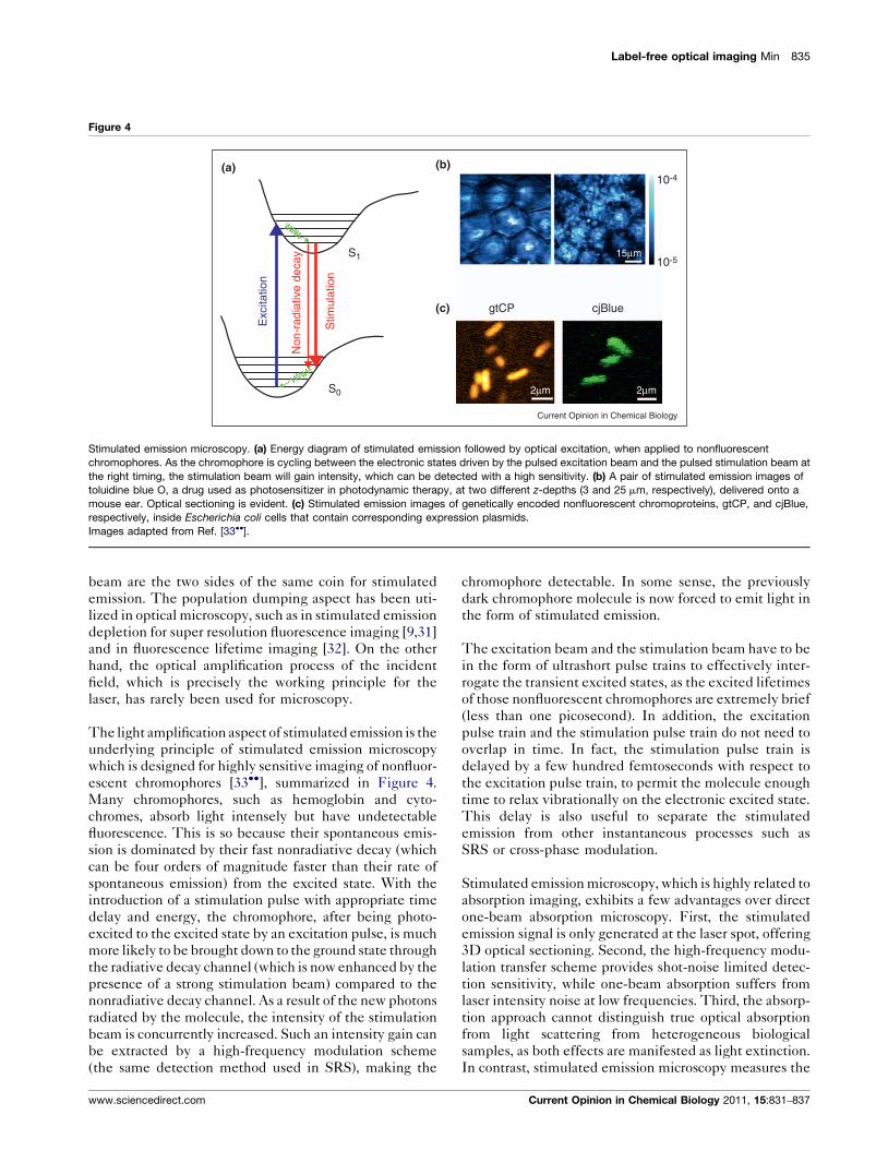
Stimulated emission microscopy. (a) Energy diagram of stimulated emission followed by optical excitation, when applied to nonfluorescent
chromophores. As the chromophore is cycling between the electronic states driven by the pulsed excitation beam and the pulsed stimulation beam at
the right timing, the stimulation beam will gain intensity, which can be detected with a high sensitivity. (b) A pair of stimulated emission images of
toluidine blue O, a drug used as photosensitizer in photodynamic therapy, at two different z-depths (3 and 25 mm, respectively), delivered onto a
mouse ear. Optical sectioning is evident. (c) Stimulated emission images of genetically encoded nonfluorescent chromoproteins, gtCP, and cjBlue,
respectively, inside Escherichia coli cells that contain corresponding expression plasmids.
Images adapted from Ref. [33��].
beam are the two sides of the same coin for stimulated
emission. The population dumping aspect has been uti-
lized in optical microscopy, such as in stimulated emission
depletion for super resolution fluorescence imaging [9,31]
and in fluorescence lifetime imaging [32]. On the other
hand, the optical amplification process of the incident
field, which is precisely the working principle for the
laser, has rarely been used for microscopy.
The light amplification aspect of stimulated emission is the
underlying principle of stimulated emission microscopy
which is designed for highly sensitive imaging of nonfluor-
escent chromophores [33��], summarized in Figure 4.
Many chromophores, such as hemoglobin and cyto-
chromes, absorb light intensely but have undetectable
fluorescence. This is so because their spontaneous emis-
sion is dominated by their fast nonradiative decay (which
can be four orders of magnitude faster than their rate of
spontaneous emission) from the excited state. With the
introduction of a stimulation pulse with appropriate time
delay and energy, the chromophore, after being photo-
excited to the excited state by an excitation pulse, is much
more likely to be brought down to the ground state through
the radiative decay channel (which is now enhanced by the
presence of a strong stimulation beam) compared to the
nonradiative decay channel. As a result of the new photons
radiated by the molecule, the intensity of the stimulation
beam is concurrently increased. Such an intensity gain can
be extracted by a high-frequency modulation scheme
(the same detection method used in SRS), making the
www.sciencedirect.com
chromophore detectable. In some sense, the previously
dark chromophore molecule is now forced to emit light in
the form of stimulated emission.
The excitation beam and the stimulation beam have to be
in the form of ultrashort pulse trains to effectively inter-
rogate the transient excited states, as the excited lifetimes
of those nonfluorescent chromophores are extremely brief
(less than one picosecond). In addition, the excitation
pulse train and the stimulation pulse train do not need to
overlap in time. In fact, the stimulation pulse train is
delayed by a few hundred femtoseconds with respect to
the excitation pulse train, to permit the molecule enough
time to relax vibrationally on the electronic excited state.
This delay is also useful to separate the stimulated
emission from other instantaneous processes such as
SRS or cross-phase modulation.
Stimulated emission microscopy, which is highly related to
absorption imaging, exhibits a few advantages over direct
one-beam absorption microscopy. First, the stimulated
emission signal is only generated at the laser spot, offering
3D optical sectioning. Second, the high-frequency modu-
lation transfer scheme provides shot-noise limited detec-
tion sensitivity, while one-beam absorption suffers from
laser intensity noise at low frequencies. Third, the absorp-
tion approach cannot distinguish true optical absorption
from light scattering from heterogeneous biological
samples, as both effects are manifested as light extinction.
In contrast, stimulated emission microscopy measures the
Current Opinion in Chemical Biology 2011, 15:831–837

836 Molecular Imaging
response of the stimulation beam intensity only at the
modulation frequency, filtering out the sample scattering
effect at low frequencies.
Concluding remarksIt is constructive to summarize the common strategy
employed by both stimulated Raman scattering micro-
scopy and stimulated emission microscopy. When the
spontaneous Raman scattering is too weak for typical
vibrational oscillators, stimulated Raman is used to sig-
nificantly boost the vibrational excitation rate, imaging
speed, and sensitivity. Likewise, when the spontaneous
emission (i.e. fluorescence) is too weak for nonfluorescent
chromophores, stimulated emission is used to enhance
the radiative decay rate. Hence, stimulated radiation can
indeed improve on the imaging performance of its spon-
taneous counterpart.
The detection sensitivity of both stimulated Raman
scattering microscopy and stimulated emission micro-
scopy has approached the shot-noise limit of the incident
laser beam, which arises from stochastic arrivals of
photons at the detector. This poses a fundamental chal-
lenge as to how to further improve the sensitivity to study
even more dilute molecular species. We envision that the
principles and techniques from quantum optics, such as
entangled photons and squeezed light, might be bor-
rowed to create a ‘quantum jump’ for the detection
sensitivity beyond the shot-noise limit.
While the chemical biology strategies of fluorescence
labeling have become increasingly sophisticated
[34,35], many more molecular species cannot or should
not be labeled in biomedical applications. To this end,
stimulated Raman scattering microscopy and stimulated
emission microscopy represent an emerging direction for
nonfluorescent optical imaging. Exciting applications in
various areas are expected for many years to come.
AcknowledgementsWe are grateful to X. Sunney Xie and the members of his group who havecontributed to the development and applications of stimulated Ramanscattering microscopy and stimulated emission microscopy, in particular,C.W. Freudiger, S. Lu, B.G. Saar, S. Chong, G.R. Holtom, M. Roeffaers, X.Zhang and R. Roy. In addition, we wish to thank M.C. Wang, J.X. Kang andG. Ruvkun for collaboration on lipid biology.
References and recommended readingPapers of particular interest, published within the period of review,have been highlighted as:
� of special interest�� of outstanding interest
1. Pawley JB (Ed): Handbook of Biological Confocal Microscopy, edn3. Springer; 2006.
2. Chalfie M, Tu Y, Euskirchen G, Ward WW, Prasher DC: Greenfluorescent protein as a marker for gene expression. Science1994, 263:802-805.
3. Tsien RY: The green fluorescent protein. Annu Rev Biochem1998, 67:509-544.
Current Opinion in Chemical Biology 2011, 15:831–837
4. Zhang J, Campbell RE, Ting AY, Tsien RY: Creating newfluorescent probes for cell biology. Nat Rev Mol Biol 2002,3:906-918.
5. Michalet X, Pinaud FF, Bentolila LA, Tsay JM, Doose S, Li JJ,Sundaresan G, Wu AM, Gambhir SS, Weiss S: Quantum dots forlive cells, in vivo imaging, and diagnostics. Science 2005,307:538-544.
6. Denk W, Strickler J, Webb WW: Two-photon laser scanningfluorescence microscopy. Science 1990, 248:73-76.
7. Moerner WE, Orrit M: Illuminating single molecules incondensed matter. Science 1999, 283:1670-1676.
8. Xie XS, Trautman JK: Optical studies of single molecules atroom temperature. Annu Rev Phys Chem 1998, 49:441-480.
9. Hell SW: Far-field optical nanoscopy. Science 2007, 316:1153-1158.
10. Huang B, Babcock H, Zhuang X: Breaking the diffraction barrier:super-resolution imaging of cells. Cell 2010, 143:1047-1058.
11. Turrell G, Corset J: Raman Microscopy: Developments andApplications. San Diego: Academic Press; 1996.
12. Levenson MD, Kano SS: Introduction to Nonlinear LaserSpectroscopy. San Diego: Academic Press; 1988.
13. Clark RJH, Hester RE (Eds): Advances in Nonlinear Spectroscopy,vol 15. New York: John Wiley and Sons Ltd.; 1988.
14. Boyd RW: Nonlinear Optics. London: Academic; 2003.
15.�
Zumbusch A, Holtom GR, Xie XS: Three-dimensional vibrationalimaging by coherent anti-Stokes Raman scattering. Phys RevLett 1999, 82:4142-4145.
The modern version of coherent anti-Stokes Raman scattering micro-scopy is presented.
16.�
Woodbury EJ, Ng WK: Proc Inst Radio Eng1962, 50:2347-2348.The very first report of stimulated Raman scattering effect.
17. Jones WJ, Stoicheff BP: Inverse Raman spectra: inducedabsorption at optical frequencies. Phys Rev Lett 1964,13:657-659.
18.�
Bloembergen N: The stimulated Raman effect. Am J Phys 1967,35:989-1023.
The first systematic theoretical treatment of stimulated Raman scatteringeffect.
19. Owyoung A: Coherent Raman gain spectroscopy using CWlaser sources. IEEE J Quant Electron 1978, QE-14:192-203.
20. Levine BF, Shank CV, Heritage JP: Surface vibrationalspectroscopy using stimulated Raman scattering. IEEE JQuant Electron 1979, QE-15:1418-1432.
21. Levenson MD, Moerner WE, Horne DE: FM spectroscopydetection of stimulated Raman gain. Opt Lett 1983, 8:108-110.
22. Kukura P, McCamant DW, Mathies RA: Femtosecond stimulatedRaman spectroscopy. Annu Rev Phys Chem 2007, 58:461-488.
23.�
Ploetz E, Laimgruber S, Berner S, Zinth W, Gilch P: Femtosecondstimulated Raman microscopy. Appl Phys B 2007, 87:389-393.
Stimulated Raman scattering as a contrast mechanism is demonstratedwith femtosecond amplified laser system together with multiplex detection.
24.��
Freudiger CW, Min W, Saar BG, Lu S, Holtom GR, He C, Tsai JC,Kang JX, Xie XS: Label-free biomedical imaging with highsensitivity by stimulated Raman scattering microscopy.Science 2008, 322:1857-1861.
The first demonstration of stimulated Raman scattering microscopy usingnarrow-band high-repetition-rate picosecond pulse trains and high-fre-quency modulation transfer, which yielded superior sensitivity and fastimaging speed.
25. Nandakumar P, Kovalev A, Volkmer A: Vibrational imaging basedon stimulated Raman scattering microscopy. New J Phys 2009,11:033026-33035.
26. Ozeki Y, Dake F, Kajiyama S, Fukui K, Itoh K: Analysis andexperimental assessment of the sensitivity of stimulatedRaman scattering microscopy. Opt Express 2009,17:3651-3658.
www.sciencedirect.com
Label-free optical imaging Min 837
27. Saar BG, Freudiger CW, Reichman J, Stanley MC, Holtom GR,Xie XS: Video-rate molecular imaging in vivo with stimulatedRaman scattering. Science 2010, 330:1368-1370.
28.��
Min W, Freudiger CW, Lu S, Xie XS: Coherent nonlinear opticalimaging: beyond fluorescence microscopy. Annu Rev PhysChem 2011, 62:507-530.
A comprehensive and technical comparison between CARS and SRSmicroscopy is provided.
29. Wang MC, Min W, Freudiger CW, Ruvkun G, Xie XS: RNAiscreening for fat regulatory genes with SRS microscopy. NatMethods 2011, 8:135-138.
30. Bewersdorf J, Farese RV, Walther TC: A new way to look at fat.Nat Methods 2011, 8:132-133.
31. Hell SW, Wichmann J: Breaking the diffraction resolution limitby stimulated emission: stimulated-emission-depletion
www.sciencedirect.com
fluorescence microscopy. Opt Lett 1994,19:780-782.
32. Dong CY, So PT, French T, Gratton E: Fluorescence lifetimeimaging by asynchronous pump-probe microscopy. Biophys J1995, 69:2234-2242.
33.��
Min W, Lu S, Chong S, Roy R, Holtom GR, Xie XS: Imagingchromophores with undetectable fluorescence by stimulatedemission microscopy. Nature 2009, 461:1105-1109.
Stimulated emission as a contrast mechanism for microscopy is firstdemonstrated.
34. Jing C, Cornish VW: Chemical tags for labeling proteins insideliving cells. Acc Chem Res 2011, 44:784-792.
35. Fernandez-Suarez M, Ting AY: Fluorescent probes for super-resolution imaging in living cells. Nat Rev Mol Cell Biol 2008,9:929-943.
Current Opinion in Chemical Biology 2011, 15:831–837


![10-Molecules and Solids 10.3~10.6.ppt [호환 모드]optics.hanyang.ac.kr/~shsong/10-Molecules and Solids 10.3... · 2016-08-31 · 10.1 Molecular Bonding and Spectra 10.2 Stimulated](https://img.pdfslide.net/doc/110x75/5ec590394f8cfe6e6475cdca/10-molecules-and-solids-103106ppt-eeoe-shsong10-molecules-and-solids.jpg)