Embed Size (px)
Citation preview

Clinical Microbiology Newsletter 33:8,2011 © 2011 Elsevier 0196-4399/00 (see frontmatter) 55
The number of total joint replace-ments being performed in the UnitedStates is rising and is anticipated tocontinue to increase for at least the nexttwo decades. It has been projected that576,000 primary hip arthroplasties and3.48 million primary knee arthroplastieswill be performed in the year 2030 (1).Other types of joint replacement areshoulder, which have been increasingparallel to hip and knee arthroplasty (2),and elbow, which is less commonly per-formed. Most of these procedures havea satisfactory outcome; however, a smallpercentage of patients develop failureof the arthroplasty and require revision.The most common reason for revisionarthroplasty is aseptic loosening of theprosthesis, followed by prosthetic jointinfection (PJI) (3). A correct diagnosisof PJI and distinction between asepticloosening and PJI are crucial, as themanagement of these conditions isdifferent.
Infection after joint implantationhas been reported to occur in 0.8 to1.9% of knee arthroplasties (4), 0.3 to1.7% of hip arthroplasties (4), 0.7% ofshoulder arthroplasties (2), and 3% ofelbow arthroplasties (5). Bacteremia,postoperative wound complications,rheumatoid arthritis, malignancy, andprevious arthroplasty are risk factorsassociated with PJI (6-8). PJIs are asso-ciated with a large economic burden tothe health care system, a high rate ofdissatisfaction (loss of function), and amortality rate of up to 7% compared toprimary arthroplasties (9).
Microorganisms (mostly skin flora)are typically introduced to the prosthe-sis surface at the time of implantationbut may also seed the implant hemato-genously following the implantation(10). Bacteria attach to the foreign sur-face, forming microcolonies that matureinto biofilms, which are communitiesof bacteria encased by an extracellularpolymeric matrix. In biofilms, bacteriaare relatively protected against hostimmunity and antimicrobial treatment.The formation of biofilms not onlyimpacts the treatment of PJI, but alsochallenges diagnosis and renders somebacteria that uncommonly infect nativejoints important PJI pathogens.
Staphylococci (Staphylococcus
aureus and coagulase-negative Staphy-lococcus species) are the most commonorganisms associated with PJI, causingabout 50% of cases, followed by poly-microbial infection (20%), streptococci(9%), gram-negative bacilli (8%), anae-robes (6%), and other microorganisms(6). The microbiology of prostheticshoulder infection differs from that ofhip and knee arthroplasty infection, asPropionibacterium acnes is a com-monly isolated organism in cases ofshoulder arthroplasty infection (11).
DiagnosisWhen evaluating a patient with pros-
thetic joint dysfunction, clinicians haveto determine first if the cause of the dys-function is septic or aseptic. The diag-nosis of PJI can be made clinically in
Laboratory Diagnosis of Prosthetic Joint Infection, Part I*
Eric Gomez, M.D.1 and Robin Patel, M.D.,1,2 1Division of Infectious Diseases, Department of Medicine, 2Division of ClinicalMicrobiology, Department of Laboratory Medicine and Pathology, Mayo Clinic, Rochester, Minnesota
AbstractProsthetic joint infection (PJI), although a rare complication of primary or revision arthroplasty, is reported more frequently
as the number patients undergoing arthroplasty increases. Accurate diagnosis of PJI is essential for adequate management andoutcome. Although multiple tests have been applied, in some cases, differentiation of PJI from aseptic loosening of the prosthesisremains a challenge. Here, we review the current diagnostic laboratory modalities used for the diagnosis PJI. In Part I of this two-part article, components of the preoperative evaluation of the patient and the histology of the intraoperative evaluation is discussed.
Vol. 33, No. 8 www.cmnewsletter.com April 15, 2011
*Editor’s Note: Part II of this article willappear in the May 1, 2011 issue of ClinicalMicrobiolgy Newsletter (Vol. 33, No. 9).
Corresponding Author: Robin Patel, M.D.,Mayo Clinic, 200 First St. SW, Rochester,MN 55905. Tel: 507-538-0579. Fax: 507-284-4272. E-mail: [email protected]
ClinicalMicrobiologyNewsletter
$99

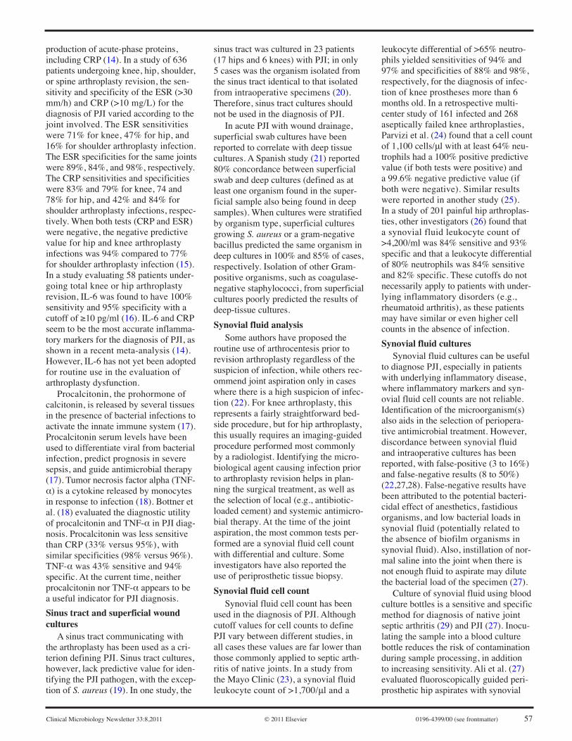
some cases, while in others, the use oflaboratory tests is needed. Once the diag-nosis of PJI has been made, cliniciansdepend on the microbiology laboratoryto identify the causal microorganism(s)and to obtain antimicrobial susceptibili-ties to implement an effective treatmentplan (Fig. 1).
PJI caused by virulent organisms(e.g., S. aureus) may present acutelywith fever, effusion, warmth, erythema,and pain localized to the joint and canusually be easily identified clinically.However, infection with low-virulenceorganisms (e.g., P. acnes and coagulase-negative staphylococci) usually presentsas a subacute or chronic process mani-fested by pain and/or loosening of thehardware without other clinical signsor symptoms of inflammation, makingit difficult for the clinician to distinguishthis type of PJI from aseptic arthroplastyfailure. In such cases, laboratory testsare used to rule out infection (4).
When evaluating the performance ofa test used in the diagnosis of PJI, spe-cial attention should be paid to the PJIdefinition used in the evaluation, as thisaffects sensitivity and specificity. Cur-rently, there is no universal definitionof PJI, but most studies consider PJI tobe present when one of the followingcriteria is met: (i) visible purulence inthe synovial fluid or surrounding theprosthesis, (ii) acute inflammation onhistopathological examination of peri-prosthetic tissue sections, (iii) a sinustract communicating with the pros-thesis, and (iv) growth of the sameorganism in more than one culture ofsynovial fluid or periprosthetic tissueor in significant quantity from theimplant itself (4).
Preoperative EvaluationBlood tests
A complete blood cell count has poor
sensitivity for the diagnosis of PJI (12).C-reactive protein (CRP), erythrocytesedimentation rate (ESR), and, morerecently, interleukin 6 (IL-6) are non-invasive tests that may be used in thepreoperative assessment of PJI. Thesetests are affected by noninfectious
conditions, such as neoplasia and con-nective tissue disease (13). CRP is anacute-phase protein produced by theliver that increases with infectious,inflammatory, and neoplastic processes.IL-6 is a cytokine produced by mono-cytes and macrophages that induces
56 0196-4399/00 (see frontmatter) © 2011 Elsevier Clinical Microbiology Newsletter 33:8,2011
Figure 1. Diagnostic algorithm of PJI. WBC, white blood cells.
*Crystal induced inflammatory arthritis, although uncommon inarthroplasty, can present with inflammatory changes.
Clinical Microbiology Newsletter 33:8,2011 © 2011 Elsevier 0196-4399/00 (see frontmatter) 57
production of acute-phase proteins,including CRP (14). In a study of 636patients undergoing knee, hip, shoulder,or spine arthroplasty revision, the sen-sitivity and specificity of the ESR (>30mm/h) and CRP (>10 mg/L) for thediagnosis of PJI varied according to thejoint involved. The ESR sensitivitieswere 71% for knee, 47% for hip, and16% for shoulder arthroplasty infection.The ESR specificities for the same jointswere 89%, 84%, and 98%, respectively.The CRP sensitivities and specificitieswere 83% and 79% for knee, 74 and78% for hip, and 42% and 84% forshoulder arthroplasty infections, respec-tively. When both tests (CRP and ESR)were negative, the negative predictivevalue for hip and knee arthroplastyinfections was 94% compared to 77%for shoulder arthroplasty infection (15).In a study evaluating 58 patients under-going total knee or hip arthroplastyrevision, IL-6 was found to have 100%sensitivity and 95% specificity with acutoff of ≥10 pg/ml (16). IL-6 and CRPseem to be the most accurate inflamma-tory markers for the diagnosis of PJI, asshown in a recent meta-analysis (14).However, IL-6 has not yet been adoptedfor routine use in the evaluation ofarthroplasty dysfunction.
Procalcitonin, the prohormone ofcalcitonin, is released by several tissuesin the presence of bacterial infections toactivate the innate immune system (17).Procalcitonin serum levels have beenused to differentiate viral from bacterialinfection, predict prognosis in severesepsis, and guide antimicrobial therapy(17). Tumor necrosis factor alpha (TNF-α) is a cytokine released by monocytesin response to infection (18). Bottner etal. (18) evaluated the diagnostic utilityof procalcitonin and TNF-α in PJI diag-nosis. Procalcitonin was less sensitivethan CRP (33% versus 95%), withsimilar specificities (98% versus 96%).TNF-α was 43% sensitive and 94%specific. At the current time, neitherprocalcitonin nor TNF-α appears to bea useful indicator for PJI diagnosis.
Sinus tract and superficial woundcultures
A sinus tract communicating withthe arthroplasty has been used as a cri-terion defining PJI. Sinus tract cultures,however, lack predictive value for iden-tifying the PJI pathogen, with the excep-tion of S. aureus (19). In one study, the
sinus tract was cultured in 23 patients(17 hips and 6 knees) with PJI; in only5 cases was the organism isolated fromthe sinus tract identical to that isolatedfrom intraoperative specimens (20).Therefore, sinus tract cultures shouldnot be used in the diagnosis of PJI.
In acute PJI with wound drainage,superficial swab cultures have beenreported to correlate with deep tissuecultures. A Spanish study (21) reported80% concordance between superficialswab and deep cultures (defined as atleast one organism found in the super-ficial sample also being found in deepsamples). When cultures were stratifiedby organism type, superficial culturesgrowing S. aureus or a gram-negativebacillus predicted the same organism indeep cultures in 100% and 85% of cases,respectively. Isolation of other Gram-positive organisms, such as coagulase-negative staphylococci, from superficialcultures poorly predicted the results ofdeep-tissue cultures.
Synovial fluid analysisSome authors have proposed the
routine use of arthrocentesis prior torevision arthroplasty regardless of thesuspicion of infection, while others rec-ommend joint aspiration only in caseswhere there is a high suspicion of infec-tion (22). For knee arthroplasty, thisrepresents a fairly straightforward bed-side procedure, but for hip arthroplasty,this usually requires an imaging-guidedprocedure performed most commonlyby a radiologist. Identifying the micro-biological agent causing infection priorto arthroplasty revision helps in plan-ning the surgical treatment, as well asthe selection of local (e.g., antibiotic-loaded cement) and systemic antimicro-bial therapy. At the time of the jointaspiration, the most common tests per-formed are a synovial fluid cell countwith differential and culture. Someinvestigators have also reported theuse of periprosthetic tissue biopsy.
Synovial fluid cell countSynovial fluid cell count has been
used in the diagnosis of PJI. Althoughcutoff values for cell counts to definePJI vary between different studies, inall cases these values are far lower thanthose commonly applied to septic arth-ritis of native joints. In a study fromthe Mayo Clinic (23), a synovial fluidleukocyte count of >1,700/μl and a
leukocyte differential of >65% neutro-phils yielded sensitivities of 94% and97% and specificities of 88% and 98%,respectively, for the diagnosis of infec-tion of knee prostheses more than 6months old. In a retrospective multi-center study of 161 infected and 268aseptically failed knee arthroplasties,Parvizi et al. (24) found that a cell countof 1,100 cells/μl with at least 64% neu-trophils had a 100% positive predictivevalue (if both tests were positive) anda 99.6% negative predictive value (ifboth were negative). Similar resultswere reported in another study (25).In a study of 201 painful hip arthroplas-ties, other investigators (26) found thata synovial fluid leukocyte count of>4,200/ml was 84% sensitive and 93%specific and that a leukocyte differentialof 80% neutrophils was 84% sensitiveand 82% specific. These cutoffs do notnecessarily apply to patients with under-lying inflammatory disorders (e.g.,rheumatoid arthritis), as these patientsmay have similar or even higher cellcounts in the absence of infection.
Synovial fluid culturesSynovial fluid cultures can be useful
to diagnose PJI, especially in patientswith underlying inflammatory disease,where inflammatory markers and syn-ovial fluid cell counts are not reliable.Identification of the microorganism(s)also aids in the selection of periopera-tive antimicrobial treatment. However,discordance between synovial fluidand intraoperative cultures has beenreported, with false-positive (3 to 16%)and false-negative results (8 to 50%)(22,27,28). False-negative results havebeen attributed to the potential bacteri-cidal effect of anesthetics, fastidiousorganisms, and low bacterial loads insynovial fluid (potentially related tothe absence of biofilm organisms insynovial fluid). Also, instillation of nor-mal saline into the joint when there isnot enough fluid to aspirate may dilutethe bacterial load of the specimen (27).
Culture of synovial fluid using bloodculture bottles is a sensitive and specificmethod for diagnosis of native jointseptic arthritis (29) and PJI (27). Inocu-lating the sample into a blood culturebottle reduces the risk of contaminationduring sample processing, in additionto increasing sensitivity. Ali et al. (27)evaluated fluoroscopically guided peri-prosthetic hip aspirates with synovial

58 0196-4399/00 (see frontmatter) © 2011 Elsevier Clinical Microbiology Newsletter 33:8,2011
fluid culture using blood culture bottles.Synovial fluid culture showed a sensi-tivity and specificity of 82% and 91%,respectively, for the diagnosis of pros-thetic hip infection compared to intra-operative cultures. A similar study byRoberts et al. (30) showed a sensitivityand specificity of 87% and 95%, res-pectively, when synovial fluid culturein blood culture bottles was comparedto periprosthetic tissue cultures per-formed using homogenized tissuesinoculated into blood culture bottles.
Preoperative tissue culturesSeveral investigators have reported
the use of tissue biopsy prior to surgeryfor the diagnosis of PJI. Sadiq et al. (20)reported 70% concordance betweenpreoperative tissue and intraoperativecultures. Negative preoperative withpositive intraoperative tissue cultureswere reported in 10% of cases. Overall,the sensitivity and specificity of corebiopsy were 88 and 91%, respectively,for the diagnosis of PJI. Fink et al. (31)combined preoperative tissue cultureswith histopathology (assessing acuteinflammation) and obtained sensitivity,specificity, and negative predictive val-ues of 100, 98, and 100%, respectivelycompared to intraoperative culturesand/or histopathology.
However, other studies have shownno difference between synovial fluidaspirates and preoperative tissue cultures.In a study of hip arthroplasty infections,Williams et al. (32) reported that syn-ovial fluid aspirate and preoperativetissue cultures had sensitivities (80%versus 83%) and specificities (94% ver-sus 90%) similar to those of intraopera-tive tissue cultures. Meermans et al. (33)found similar results, with sensitivitiesof 83% and 79%, respectively, for aspi-ration and tissue biopsy cultures com-pared to intraoperative tissue cultures.In this study, both tests were 100% spe-cific, and when both tests were com-bined, the sensitivity improved to 90%.Based on the available evidence, preop-erative tissue biopsy and culture do notappear to offer an advantage over lessinvasive synovial fluid cultures.
Intraoperative EvaluationDuring surgical revision of an arthro-
plasty, the surgeon assesses gross evi-dence of infection (i.e., purulence), butthis may be absent in patients with PJI.Several tests can be performed (or initi-
ated) at the time of surgery that can aidin the evaluation of arthroplasty failure.
HistopathologyEvidence of acute inflammation on
frozen sections of periprosthetic tissuesamples correlates with the presenceof PJI. Acute inflammation is definedby the presence of polymorphonuclearleukocytes visualized by high-powerfield (HPF) microscopy. Differentstudies have used different cutoffs, butusually, ≥5 to 10 polymorphonuclearleukocytes per HPF typically indicatesacute inflammation (34,35). Severalinvestigators have shown that histo-pathology has excellent specificity,with variable sensitivity, to detect PJI(Table 1). The low sensitivity may bedue to sampling bias or infection withlow-virulence microorganisms that donot elicit neutrophil infiltration (36).High specificity makes this test usefulas a confirmatory test for PJI in patientswithout underlying inflammatory jointdisease. The accuracy of the test dependson the experience of the pathologist, asthere can be significant interobservervariability.
Frozen-section analysis may also beused at the time of the joint reimplanta-tion to assess the persistence of infection.Bori et al. (37) noted a 100% positivepredictive value for the presence ofa microorganism at the time of jointreimplantation; however, the negativepredictive value was only 74%.
Editor’s Note: Part II of this articlewill appear in the May 1, 2011 issueof CMN (Vol. 33, No. 9).
References
1. Kurtz, S. et al. 2007. Projections ofprimary and revision hip and kneearthroplasty in the United States from2005 to 2030. J. Bone Joint Surg. Am.89:780-785.
2. Bohsali, K.I., M.A.Wirth, and C.A.Rockwood, Jr. 2006. Complications oftotal shoulder arthroplasty. J. Bone JointSurg. Am. 88:2279-2292.
3. Roberts, V.I., C.N. Esler, and W.M.Harper. 2007. A 15-year follow-up studyof 4606 primary total knee replacements.J. Bone Joint Surg. Br. 89:1452-1456.
4. Del Pozo, J.L. and R. Patel. 2009.Clinical practice. Infection associatedwith prosthetic joints. N. Engl. J. Med.361:787-794.
5. Cheung, E.V., R.A. Adams, and B.F.Morrey. 2008. Reimplantation of a totalelbow prosthesis following resection
arthroplasty for infection. J. Bone JointSurg. Am. 90:589-594.
6. Berbari, E.F. et al. 1998. Risk factors forprosthetic joint infection: case-controlstudy. Clin. Infect. Dis. 27:1247-1254.
7. Jamsen, E. et al. 2009. Risk factorsfor infection after knee arthroplasty. Aregister-based analysis of 43,149 cases.J. Bone Joint Surg. Am. 91:38-47.
8. Aslam, S., C. Reitman, and R.O.Darouiche. 2010. Risk factors for sub-sequent diagnosis of prosthetic jointinfection. Infect. Control Hosp.Epidemiol. 31:298-301.
9. Poultsides, L.A., L.L. Liaropoulos, andK.N. Malizos. 2010. The socioeconomicimpact of musculoskeletal infections.J. Bone Joint Surg. Am. 92:e13.
10. Gristina, A. G. and J. Kolkin. 1983.Current concepts review. Total jointreplacement and sepsis. J Bone JointSurg Am. 65:128-134.
11. Piper, K.E. et al. 2009. Microbiologicdiagnosis of prosthetic shoulder infec-tion by use of implant sonication. J.Clin. Microbiol. 47:1878-1884.
12. Tohtz, S.W. et al. 2010. Validity offrozen sections for analysis of peri-prosthetic loosening membranes. Clin.Orthop. Relat. Res. 468:762-768.
13. Levine, S.E. et al. 1993. Diagnoses andstaging. Osteomyelitis and prostheticjoint infections. Clin. Orthop. Relat.Res. 295:77-86.
14. Berbari, E. et al. 2010. Inflammatoryblood laboratory levels as markers ofprosthetic joint infection: a systematicreview and meta-analysis. J. Bone JointSurg. Am. 92:2102-2109.
15. Piper, K.E. et al. 2010. C-reactive pro-tein, erythrocyte sedimentation rate andorthopedic implant infection. PLoS One5:e9358.
16. Di Cesare, P.E. et al. 2005. Seruminterleukin-6 as a marker of peripros-thetic infection following total hip andknee arthroplasty. J. Bone Joint Surg.Am. 87:1921-1927.
17. Gilbert, D.N. 2010. Use of plasmaprocalcitonin levels as an adjunct toclinical microbiology. J. Clin.Microbiol. 48:2325-2329.
18. Bottner, F. et al. 2007. Interleukin-6,procalcitonin and TNF-alpha: markersof peri-prosthetic infection followingtotal joint replacement. J. Bone JointSurg. Br. 89:94-99.
19. Mackowiak, P. A., S.R. Jones, and J.W.Smith. 1978. Diagnostic value of sinus-tract cultures in chronic osteomyelitis.JAMA 239:2772-2775.
20. Sadiq, S. et al. 2005. Application of corebiopsy in revision arthroplasty for deep

Clinical Microbiology Newsletter 33:8,2011 © 2011 Elsevier 0196-4399/00 (see frontmatter) 59
infection. J. Arthroplasty 20:196-201.
21. Cune, J. et al. 2009. A superficial swabculture is useful for microbiologic diag-nosis in acute prosthetic joint infections.Clin. Orthop. Relat. Res. 467:531-535.
22. Lachiewicz, P.F., G.D. Rogers, andH.C. Thomason. 1996. Aspiration ofthe hip joint before revision total hiparthroplasty. Clinical and laboratoryfactors influencing attainment of apositive culture. J. Bone Joint Surg.Am. 78:749-754.
23. Trampuz, A. et al. 2004. Synovial fluidleukocyte count and differential for thediagnosis of prosthetic knee infection.Am. J. Med. 117:556-562.
24. Parvizi, J. et al. 2008. Diagnosis ofinfected total knee: findings of a multi-center database. Clin. Orthop. Relat.Res. 466:2628-2633.
25. Ghanem, E. et al. 2008. Cell count anddifferential of aspirated fluid in thediagnosis of infection at the site of total
knee arthroplasty. J. Bone Joint Surg.Am. 90:1637-1643.
26. Schinsky, M.F. et al. 2008. Perioperativetesting for joint infection in patientsundergoing revision total hip arthro-plasty. J. Bone Joint Surg. Am.90:1869-1875.
27. Ali, F. et al. 2006. Accuracy of jointaspiration for the preoperative diagnosisof infection in total hip arthroplasty.J. Arthroplasty 21:221-226.
28. Fehring, T.K. and B. Cohen. 1996.Aspiration as a guide to sepsis inrevision total hip arthroplasty. J.Arthroplasty 11:543-547.
29. Hughes, J.G. et al. 2001. Culturewith BACTEC Peds Plus/F bottlecompared with conventional methodsfor detection of bacteria in synovialfluid. J. Clin. Microbiol. 39:4468-4471.
30. Roberts, P., A.J. Walters, and D.J.McMinn. 1992. Diagnosing infectionin hip replacements. The use of fine-
needle aspiration and radiometric cul-ture. J. Bone Joint Surg. Br. 74:265-269.
31. Fink, B. et al. 2008. The value ofsynovial biopsy, joint aspiration andC-reactive protein in the diagnosis oflate peri-prosthetic infection of totalknee replacements. J. Bone Joint Surg.Br. 90:874-878.
32. Williams, J.L., P. Norman, and I.Stockley. 2004. The value of hip aspir-ation versus tissue biopsy in diagnosinginfection before exchange hip arthro-plasty surgery. J. Arthroplasty 19:582-586.
33. Meermans, G. and F.S. Haddad. 2010.Is there a role for tissue biopsy in thediagnosis of periprosthetic infection?Clin. Orthop. Relat. Res. 468:1410-1417.
34. Mirra, J.M. et al. 1976. The pathologyof the joint tissues and its clinicalrelevance in prosthesis failure. Clin.Orthop. Relat. Res. 117:221-240.
35. Ko, P.S. et al. 2005. The role of intra-
Table 1. Frozen section/histopathology in diagnosis of PJICutoff (no. of Positive Negative
No. (type) of polymorphonuclear Sensitivity Specificity predictive predictiveStudy (yr) Reference arthroplasty leukocytes/HPF) (%) (%) value (%) value (%)
Fehring et al. (1994) 117 107 No cutoff 18 90 - -
Feldman (1995) 118 33 (knee-hip) 5 100 96 - -
Athanasou et al. (1995) 119 106 (knee-hip) 1 90 96 88 98
Lonner et al. (1996) 120 175 (knee-hip) 5 84 96 70 9810 84 99 89 98
Pace et al. (1997) 121 18 (knee-hip) 5 82 93 82 -
Abdul-Karim (1998) 122 64 (knee-hip) 5 43 97 60 -
Della Valle et al. (1999) 123 64 (knee-hip) 10 25 98 50 95
Banit et al. (2002) 124 121 (knee-hip) 10 67 93 67 93
Musso et al. (2003) 125 45 (knee-hip) 5 50 95 60 92
Wong et al. (2005) 126 33 (knee-hip) 5 93 77 68 9510 86 85 75 92
Ko et al. (2005) 35 40 (knee-hip) 5 67 97 86 91
Nunez et al. (2007) 127 136 (hip) 5 85 87 79 91
Frances Borrego et al. (2007) 128 146 (knee-hip) 10 67 90 81 81
Bori et al. (2007) 37 21 (hip) 5 29 100 100 73.61 71 64 50 82
Kanner et al. (2008) 129 132 (knee-hip) 5 29 95 40 92
Morawietz (2009) 130 147 (knee-hip) 23 73 95 91 84
Tohtz et al. (2010) 12 64 (hip) 2 86 100 100 95

60 0196-4399/00 (see frontmatter) © 2011 Elsevier Clinical Microbiology Newsletter 33:8,2011
operative frozen section in decisionmaking in revision hip and kneearthroplasties in a local communityhospital. J. Arthroplasty 20:189-195.
36. Bori, G. et al. 2010. Interface membrane
is the best sample for histological studyto diagnose prosthetic joint infection.Mod. Pathol. PMID 21131917.
37. Bori, G. et al. 2007. Usefulness of histo-logical analysis for predicting the pre-
sence of microorganisms at the timeof reimplantation after hip resectionarthroplasty for the treatment of infec-tion. J. Bone Joint Surg. Am. 89:1232-1237.