Embed Size (px)
Citation preview
Page 1 of 24
WHO Global Foodborne Infections Network
(formerly WHO Global Salm-Surv)
"A WHO network building capacity to detect, control and prevent foodborne and other enteric infections from farm to table”
Laboratory Protocol: “Serotyping of Shigella spp.”
van der Ploeg, Claudia A. (INPB)
Viñas, María R. (INEI) Terragno, Raquel (INEI) Bruno, Susana B. (INPB) Binsztein, Norma (INEI)
Instituto Nacional de Enfermedades Infecciosas (INEI) Instituto Nacional de Producción de Biológicos (INPB)
ANLIS – “Dr. Carlos G. Malbrán”- Argentina
With acknowledgments for significant technical and editorial contributions to the: WHO Global Foodborne Infections Network Laboratory Sub-Committee
& The U.S. Centers for Disease Control and Prevention’s National Escherichia and Shigella Reference Laboratory

Page 2 of 24
Title: Laboratory protocol: Serotyping of Shigella spp
Protocol Number: 2010GFNLAB003
Effective Date: 30-06-2010 Revision Number:
REVISION HISTORY
HISTORY OF CHANGES
Rev. Level
Sections Changed Description of Change (From—To) Date Approval
0
1

Page 3 of 24
I. PURPOSE
A standardized process for the serotyping of Shigella spp.
II. TEST PRINCIPLES & BACKGROUND Recent phylogenetic studies indicate that the shigellae are not a unique genus; rather they are an immunologic and biochemically distinct lineage of Escherichia coli. However, for medical purposes and to maintain historical consistency, the shigellae have not been merged with E. coli. The genus Shigella is comprised of four species: Shigella dysenteriae (also referred to Group A), Shigella flexneri (also referred to Group B), Shigella boydii (also referred to Group C), & Shigella sonnei (also referred to Group D). Included among these organisms, is the etiologic agent of epidemic dysentery, S. dysenteriae serotype 1. With the exception of S. sonnei, each species may be further divided into serotypes on the basis of reactivity with hyperimmune serum: S. dysenteriae (15 serotypes), S. flexneri (6 serotypes and 2 variants), & S. boydii (20 serotypes).
Shigella group and serotype are identified by slide agglutination using antisera. Both polyvalent and monovalent Shigella antisera are available. Polyvalent antisera, is used to determine group and contains antibodies to multiple serotypes of Shigella. For example, most commercially available polyvalent antisera for Shigella flexneri will react with all serotypes of S. flexneri. Monovalent antisera, is used to determine serotype. For example, S. dysenteriae Type 1 antiserum will only react with isolates of S. dysenteriae Type 1. A final, confirmatory, identification of Shigella spp. is based on immunologic and biochemical profile (Appendix A: Tables 1, 2 & 3). Serotyping results must be interpreted in the context of biochemical data and the serologic and biochemical identification must be consistent prior to reporting. Inconsistent / discordant results must be confirmed by a reference centre. Additional background information on Shigella is provided in Appendix A.
III. RESPONSIBILITIES III.1 Staff Responsibilities
This is a general overview. Each institution must specify what responsibility their staff members will have in the execution of this protocol. Please refer to applicable manuals within the facility/locality for complete set of responsibilities to properly conduct this procedure.
III.2 Specific Safety Requirements and Responsibilities Universal biosafety precautions must always be employed when processing any sample of human origin. Specific regulations will vary by country and may change over time. The reader is advised to consult local authorities. Biosafety level 2 (BSL-2 / RG-2) practices and procedures must be utilized when working clinical isolates of unknown Enterobacteriaceae, non-typhoidal Salmonellae, and Shigella spp. Biosafety level 3 (BSL-3/ RG-3) practices and procedures should be utilized when performing procedures likely to generate aerosols of S. dysenteriae Type 1 or invasive serovars of Salmonella spp. Organism specific information may be obtained at: http://www.phac-aspc.gc.ca/msds-ftss/ Additional safety information may be obtained at: http://www.cdc.gov/OD/ohs/biosfty/bmbl5/bmbl5toc.htm http://www.hse.gov.uk/biosafety/biologagents.pdf

Page 4 of 24
The laboratory director is responsible for insuring staff compliance with all safety requirements. All personnel shall be instructed to report any unsafe conditions to their supervisor or laboratory safety officer. For detailed, institution specific safety requirements and responsibilities, the reader should refer to facility specific safety manuals.
IV. SAMPLE COLLECTION/TRANSPORT/STORAGE
Only pure cultures should be used for serotyping. Serotyping results must be interpreted in conjunction with biochemical testing. Biochemical testing may be performed prior to serotyping or biochemical testing and serotyping may be performed simultaneously; however, a confirmatory identification cannot be made until BOTH biochemical and serological testing is complete.
NOTE: All testing must be performed using pure cultures. To insure accurate results, it is essential to select a single, well isolated colony for biochemical testing (“single colony pick”) and serotyping. Using a single, isolated colony helps to insure that the media will be inoculated with a pure culture of a single bacterial organism. If you are uncertain about the purity of your single colony pick, you should transfer the colony to a selective agar (e.g. MAC) to verify purity and then continue with biochemical and serotyping tests
For additional details describing the identification of Shigella spp., please refer to the WHO GFN Laboratory Protocol: “Biochemical Identification of Salmonella/Shigella Using an Abbreviated Panel of Tests”.
For short term storage and for transport to the laboratory, Shigella cultures may be stored in semi-solid agar at room temperature. For long term preservation and to best maintain isolates for future studies (i.e. PFGE), isolates should be frozen at -70ºC in a cryopreservation medium (see appendix E). Sample Collection/Transport/Storage must be recorded according internal procedures.
V. MATERIALS/SUPPLIES V.I- Materials
- disposable (or wire) microbiological loops
- glass slides (approx.10 x 15 cm)
- micropipettes and tips
- pasteur pipettes and pipette bulbs
- Khan tubes
- timer
- electrical Burner (if wire loops are utilized)
- lamp
- wax pencils V.II- Supplies Media
- tryptic soy agar (TSA)
- tryptic soy broth (TSB)
Page 5 of 24
- blood agar Note: See Appendix F for the formula of individual media. Reagents
- physiological saline solution (NaCl 0.85% w/v)
- 0.5 % formalinized physiological saline solution (0.85 w/v NaCl – 0.5 % formalin v/v)
- Shigella O polyvalent antisera
- Shigella O monovalent antisera
Note: See Appendix F for preparation of solutions. Reference strains According to the antisera to be tested. VI. EQUIPMENT
- incubator 36°C (+/- 1°C)
- refrigerator 4 (+/- 1°C)
Note: For information related to maintenance, troubleshooting and calibration of applicable equipment, refer to internal manuals, SOPs and/or operating manuals.
VII. QUALITY ASSURANCE / QUALITY CONTROL (QA/QC) Note: The following are general QA/QC guidelines. QA/QC requirements may vary based on: institutional policy, national regulations, or accreditation agency (example: ISO or CAP). It is important to consult your institutional QA/QC manual and appropriate regulatory guidelines prior to utilizing this protocol. Each new lot and/or shipment of antisera is tested for positive and negative reactivity using known control strains. Antisera are tested when first used (or first diluted). Routinely used antisera should be quality control tested every six months. Antisera which are less commonly used may be quality control tested prior to use. The quality control test results are recorded on a data sheet (see Appendix C). Most of the media required and listed above are commercially available and should be prepared, controlled and sterilized according to the manufacturer's instructions. If QC of media or sera does not meet expected results, technicians must report the anomaly to their supervisor and the lot should be rejected. Laboratory technician(s) performing the tests should be properly trained in this SOP, in QA and H&S (health and security) aspects and in microbiological techniques. This training should be recorded. Antisera should be checked for reactivity and should produce 3+ or 4+ agglutination when tested against its homologous antigen. No reactivity should be observed with tested with heterologous antigens. VIII. PROCEDURE
VIII.1- Initial culture Inoculate a Tryptic Soy Agar (TSA) slant (or other non-selective agar) with a single, well isolated, colony of Shigella spp. Incubate the slant at 36°C (+/- 1°C) for 18-24 hours.

Page 6 of 24
Note: is advisable to keep the original culture (selective plate or transport slant) until the characterization is finished. VIII.2- Bacterial suspension Procedure note: Conventional serotyping of Shigella spp. is based on an antigen / antibody reaction. The isolate (antigen) is mixed with antisera of known specificities; the pattern of reactivity observed determines subgroup and serotype. Two methods for preparing the bacterial antigen suspension are given below. Many operators find “Option A” faster and simpler to perform; whereas “Option B” may provide a more standardized antigen suspension. It is important to note that antisera producers typically will recommend a method for preparing a bacterial antigen suspension. When specific guidance is provided by the manufacturer; it is important to follow those recommendations. VIII.2 Option (A): Draw a circle approximately 2cm in diameter on to a clean glass slide. Add 20ul of physiological solution to center of the circle. Collect a small amount of bacteria (1uL loop) from the TSA slant and suspend the bacteria in the saline. Note: for carrying out each agglutination reaction, a new suspension should be prepared. VIII.2 Option (B): Using a sterile loop, thoroughly suspend the totality of the bacterial growth on the TSA slant with 0.5 ml of 0.5% formalinized physiological saline solution, mix to insure that there are no visible clumps or particles and then transfer the suspension into a Khan tube. Note: all the agglutinations tests can be done with this working suspension. VIII.3- Auto-agglutination control (negative control) VIII.3.1- Draw a circle approximately 2cm in diameter on to a clean glass slide. Place 20l of antigen suspension and 20l of physiological solution into the circle and mix thoroughly with the microbiological loop or stick. VIII.3.2- Gently tilt the slide back and forth for 1 minute. Observe for agglutination under indirect light. VIII.3.3- If no agglutination is observed proceed to subgroup / serotype determination. If agglutination is observed; the isolate is rough, refer to “Limitations of Procedure” and do not attempt serotyping. VIII.4- Group/serotype determination VIII.4.1- Draw a circle approximately 2cm in diameter on to a clean glass slide. Place 20l of antigen suspension and 20l of antiserum into the circle and mix thoroughly with the microbiological loop or stick.

Page 7 of 24
Procedure Note: Serologic identification of Shigella begins with the use of polyvalent antisera. Polyvalent antisera is used to identify the species (i.e. S. dysenteriae, S. boydii, S. flexneri, or S. sonnei). If agglutination is observed with polyvalent antisera, the isolate is then tested with the individual monospecific serum found in the polyvalent antisera. Isolates should be initially tested with polyvalent antisera that covers the species / serotypes which are most common in the region. VIII.4.2- Holding the slide against a dark background, tilt the slide gently back and forth for up to 1 minute observing for agglutination. Reactions developing after 1 minute are non-specific. VIII.4.3- Record the result on the datasheet (see Appendix B). An homogenous suspension is a negative reaction. Appearance of clumps means a positive reaction. Score and record the results as follows:
4+ All organisms are clumped and the supernatant, or suspending fluid is clear 3+ About 75% agglutination, the supernatant fluid is slightly cloudy 2+ About 50% agglutination, the supernatant fluid is moderately cloudy 1+ About 25% agglutination, the supernatant fluid is cloudy Tr Trace amount of agglutination present Negative No agglutination apparent and suspension remains homogenous.
4+ 3+ 2+ 1+ Negative VIII.4.4- If agglutination is observed with one of the polyvalent antisera, the serotype of the isolate is determined by testing the isolate with monovalent antisera specific to the serotypes / sub-factors found in the polyvalent sera. VIII.5- Presence of capsular antigens In the presence of capsular antigens, heat the bacterial suspension at 100ºC in water bath during 45 min. and repeat the procedure (see also point X.2). VIII.6- Considerations VIII.6.1- All the antisera must be used according to the manufacturer’s instructions. VIII.6.2- For future studies it is important to preserve the initial culture for short periods (i.e. by inoculating semisolid agar) and /or long periods (i.e. by cryopreservation at -70ºC). (For details see Appendix E).

Page 8 of 24
IX. INTERPRETATION OF RESULTS IX.1- Identification of Shigella is based on serologic and phenotypic criteria. As such, serotyping results must be interpreted in conjunction with biochemical test results (Appendix A: Tables 1, 2 & 3). IX.2- All isolates are initially tested with polyvalent antisera, if agglutination with polyvalent antisera is observed, serotype is identified by testing with monovalent antisera. If monovalent antisera is unavailable, agglutination with polyvalent antisera and biochemical profile may be used to obtain a species level identification. IX.3- Agglutination with polyvalent and monovalent antisera is required for serotype level identification. IX.4- Serotype level identification of S. flexneri requires the detection of two antigens: “type” antigens and “group” antigens. These antigens are identified using monovalent antisera and identified as follows:
Symbols in parentheses indicate variably expressed antigens.
*The subserotype 4c was proposed to the International Committee on Systematic Bacteriology Subcommittee on the Taxonomy of Enterobacteriaceae, for its incorporation in the Shigella flexneri scheme (Pryamukhina NS, Khomenko NA -1988); however this subserotype is not been officially recognized.
X. LIMITATIONS OF PROCEDURE X.1- Rough strains: Serotyping should not be attempted on strains which autoagglutinate saline. Rough isolates seldom revert to smooth forms. However, if autoagglutination in saline is observed, an attempt to recover smooth colonies may be made by performing 2-4 serial sub-cultures on enriched media, such as blood agar or Mueller Hinton agar.
Serotype Type antigen Group antigen
1a I 4
1b I 4; 6
2a II 3,4
2b II 7,8
3a III (3,4); 6; 7,8
3b III (3,4); 6
4a IV 3,4
4b IV 6
4c* IV 7,8
5a V 3,4
5b V 7,8
6 VI (4)
Variant X -- 7,8
Variant Y -- 3,4
Page 9 of 24
X.2- Capsular antigens: Occasionally, the presence of capsular antigens may prevent some isolates of Shigella spp. from reacting with polyvalent antisera. The presence of capsular antigens should be considered when isolates which are biochemically typical of Shigella spp., fail to agglutinate (or agglutinate poorly) with Shigella polyvalent antisera. In these situations, an attempt should be made to remove the capsular antigen (by boiling); as discussed in step VIII.5. Note: the capsular antigens are thermolabile. X.3- When conflicting results are observed (example: biochemical profile disagrees with serological profile or epidemiology); three (3) to five (5) additional, well isolated, colonies should be taken from the original plate and tested. X.4.- Due to the presence of conserved antigens, S. sonnei polyvalent antisera can produce cross-reactions with some cultures of S. boydii type 6. In these situations, biochemical profile and the use of Shigella boydii type 6 monovalent antiserum will be necessary to confirm the identification.
XI. REPORTING
A final, confirmatory, identification of Shigella spp. is based on immunologic and biochemical profile. Serotyping results must be interpreted in the context of biochemical data and the serologic and biochemical identification must be consistent prior to reporting. Inconsistent / discordant results must be confirmed by a reference centre.
If biochemical and serological results are in agreement, the isolate may be reported to the extent it was characterized. For example:
If an isolate was serotyped as S. dysenteriae Type 1 and is biochemically consistent with S. dysenteriae Type 1, it may be reported as: Shigella dysenteriae Type 1.
If an isolate was reactive with S. boydii polyvalent antiserum and is biochemically consistent with S. boydii, but not further serotyping was performed, the isolate may be reported as: Shigella boydii. If smooth colonies cannot be recovered from a rough culture, the organism cannot be serotyped. The isolate should be biochemically identified and reported as: “Biochemically consistent with Shigella (species) Comment: This isolate was rough, as such serotyping could not be performed.”
Each institution must establish its own policy regarding how, where, and to whom results are reported.
Policies should also be established to facilitate the expedited reporting of critical results. Critical results may include:
Identification of Shigella dysenteriae Type 1 (etiologic agent of epidemic dysentery).
Unusual or sudden increase in the number of isolates identified.
Unusual or sudden increase in a particular subgroup (or serotype).

Page 10 of 24
XII. REFERENCES
Allison, G., Verma, N. -2000-. Serotype-converting bacteriophages and O-antigen modification in Shigella flexneri; Trends in Microbiology, 8, (17-23). Ansaruzzaman, M., Kibriya, A., Rahman, A., Neogi, P., Faruque, A., Rowe, B., Albert. M. - 1995 - Detection of provisional serovars of Shigella dysenteriae and designation as S. dysenteriae serotypes 14 and 15. J. Clin. Microbiol., 33 (1423-1425) Bopp, Ch., Brenner, F., Wells, J., Strockbine N. -1999- Manual of Clinical Microbiology. Chapter 28. Brenner, D.J. -1984- Recommendation on recent proposals for the classification of shigellae. Int. J. Syst. Bacteriol, 34, (87-88). Brenner, D.J., Krieg, N., Staley, J. Bergey’s Manual of Systematic Bacteriology 2nd Edition, Vol. 2, Part B. 2005, Ewing, W., Edwards, P.. -1986-. Identification of Enterobacteiaceae. 4th edition. Elsevier Science Publishers. Andrews, W.H., Jacobson, A. -1995- FDA Bacteriological Analytical Manual. 8th edition, Chapter 6. Food Directorate´s (Health Canada´s) website at www.hc-sc.gc.ca www.hc-sc.gc.ca/fn-an/res-rech/analy-meth/microbio/volume3/mflp25-eng.php www.hc-sc.gc.ca/fn-an/res-rech/analy-meth/microbio/volume3/mflp26-eng.php Gemski, P., Koeltzow, D. E., Formal, S. B. -1975-.Phage conversion of Shigella flexneri group antigens. Infection and Immunity, 11, (685-691). Gray, L. D. -1995- Manual of Clinical Microbiology. Chapter 33. Houng, H., Sethabutr, O. and Echeverría, P. -1997- A simple polymerase chain reaction technique to detect and differentiate Shigella and enteroinvasive Escherichia coli in human feces. Diagn. Microbiol. Infect. Dis., 28, (19-25). Kaisar A., et al. -2002- Phenotipic and Genotypic Characterization of serologically atypical strains of Shigella flexneri type 4 isolated in Dhaka, Bangladesh. J. Clin. Microbiol, 40, (2490-2497). Le Minor, L., Richard, C. -1993- Méthodes de laboratoire pour l´Identification des Entérobactéries. Institut Pasteur. Liu, B., Knirel, Y. and als. -2008- Structure and genetics of Shigella O antigens. FEMS MIcrobiol. Rev., 32, (627-653). Mikoleit, M. L. –2009- Laboratory Protocol: “Biochemical identification of Salmonella spp. and Shigella spp.” Global Salm Surv. Parsot, C. and Sansonetti, P. Pathogenicity islands and the other mobile virulence elements Chapter 8 (ed. J Kaper, J Hacker). Popoff, M. -2001- Guidelines for the preparation of Salmonella Antisera, 6th Ed., WHO Collaborating Centre for Reference and Research on Salmonella Pasteur Institute. Post, D.E. -1998- Food-Borne Pathogens. Oxoid Monograph Number 5: Escherichia coli, Shigella species.

Page 11 of 24
Pryamukhina NS, Khomenko NA. -1988- Suggestion to supplement Shigella flexneri classification scheme with the subserovar Shigella flexneri 4c: phenotypic characteristics of strains. J. Clin. Microbiol., 26 (1147-1149). Radnedge, L. et al. -1997- Plasmid maintenance functions of the large virulence plasmid of Shigella flexneri. The Journal of Bacteriology, 179, (3670-3675). Simmons, D. A. R. -1971- Immunochemistry of Shigella flexneri O-Antigens: a study of structural and genetic aspects of the biosynthesis of cell-surface antigens. Bacteriological Reviews, 35, (117-148). Simmons, D. A. R., Romanowska, E. -1987- Structure and biology of Shigella flexneri O-antigens. J .Med. Microbiol., 23, (289-302). Woodward, D., Clark, C., Caldeira, R., Ahmed, R, Soule, G., Bryden, L., Tabor, H., Melito, P., Foster, R., Walsh, J., Ng, L., Malcolm, G., Strockbine, N., Rodgers, F. -2005- Identification and characterization of Shigella boydii 20 serovar nov., a new and emerging Shigella serotype. J. Clin. Microbiol., 54 (741-748).
Page 12 of 24
XIII ATTACHMENTS
XIII.1- Appendix A: Background information
XIII.2- Appendix B: Shigella serotyping worksheet
XIII.3- Appendix C: Shigella antisera quality control record data sheet.
XIII.4- Appendix D: Flowchart for Shigella spp. serotyping.
XIII.5- Appendix E: Storage of Shigella spp. cultures.
XIII.6- Appendix F: Preparation of culture media and solutions.

Page 13 of 24
APPENDIX A
BACKGROUND INFORMATION
Introduction The genus Shigella belongs to the family Enterobacteriaceae. The Shigella are Gram-negative rods, 0.3 to 1 µm in diameter and 1 to 6 µm in length, appearing singly, in pairs and in chains, are non-sporeforming, facultatively anaerobic, oxidase negative, and ferment glucose and other carbohydrates without producing gas. By definition, all Shigella spp. are nonmotile and lysine decarboxylase negative. Additionally, the Shigella are Voges-Proskauer negative and methyl-red positive, do not utilize Simmons citrate, nor produce H2S and are arginine dihydrolase and urease negative. The shigellae, with the exception of S. dysenteriae Type 1, are catalase positive. The optimum temperature of growth is 37ºC. The genus Shigella is divided into four species: Shigella dysenteriae (Group A), Shigella flexneri (Group B), Shigella boydii (Group C), and Shigella sonnei (Group D). Each of these species, with the exception of S. sonnei, has several serotypes based on the reactivity with hyperimmune serum. The proportion of each species varies from country to country and from region to region. The hallmark of infection with Shigella is diarrhea with blood, often termed “dysentery.” However, in most cases, Shigella spp causes acute non-bloody diarrhea that cannot be distinguished clinically from diarrhea caused by other enteric pathogens. S. dysenteriae 1, the etiologic agent of epidemic dysentery, differs from other Shigella in four important characteristics: produces a potent cytotoxin (Shiga toxin); causes illness that is more severe, more prolonged, and more frequently fatal than illness caused by other Shigella; resistance to antimicrobials occurs more frequently than in other Shigella; and it causes large, often regional, epidemics, frequently with high attack rates and high fatality rates. The genetic information for Shigella pathogenesis is codified at both the chromosomal and plasmid level. These microorganisms have a megaplasmid of invasion (220kb) and synthesizes Type-III secretion system that allows the insertion of a pore trough which the effector proteins are distributed in the gastrointestinal eucariotic cell. Shigella invades the colonic epithelium causing patchy destruction, which leads to the formation of micro-ulcers and inflammatory exudates, with the subsequent appearance of inflammatory cells (polymorphonuclear leucocytes) and blood in the stool. The only reservoirs for Shigella are humans and primates. Transmission occurs primarily through direct or indirect fecal-oral contact with patients or carriers. Fecally contaminated water and foods also transmit the disease; contamination of foods can also occur by infected food handlers. Shigella has a low infectious dose which facilitates person to-person spread, the ingestion of 10 to 100 cells typically produces disease. The severity of the illness depends on the age and the nutritional condition of the person and it is also dose-related. A definitive diagnosis of Shigella infection can only be made by isolating and identifying the organism from a stool culture. The isolate is also required to determine the antimicrobial sensitivity and additional subtyping. Sensitive molecular techniques to detect Shigella, such as PCR, have been developed and they are practical and rapid screening techniques for monitoring food samples (“Isolation and identification of Shigella spp. from foods”, “Detection of Shigella spp. in foods by the polymerase chain reaction”, published on the Food Directorate`s (Health Canada`s) website at www.hc-sc.gc.ca). There are others PCR protocols that have been developed for the analysis of human samples for identification of Shigella genus and species. It must be considered that the ipaH gene used as target for Shigella in these PCR protocols is also present in enteroinvasive Escherichia coli (EIEC), therefore it is necessary to perform differential testing to complete the characterization (Houng, H, Sethabutr, O. and Echeverria; P. – 1997).
Page 14 of 24
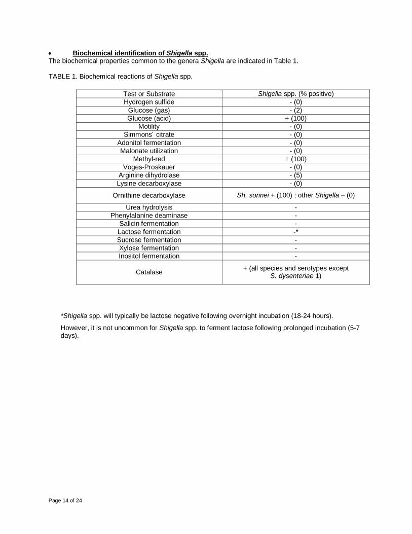
Biochemical identification of Shigella spp. The biochemical properties common to the genera Shigella are indicated in Table 1. TABLE 1. Biochemical reactions of Shigella spp.
Test or Substrate Shigella spp. (% positive) Hydrogen sulfide - (0)
Glucose (gas) - (2) Glucose (acid) + (100)
Motility - (0) Simmons´ citrate - (0)
Adonitol fermentation - (0) Malonate utilization - (0)
Methyl-red + (100) Voges-Proskauer - (0)
Arginine dihydrolase - (5) Lysine decarboxylase - (0)
Ornithine decarboxylase Sh. sonnei + (100) ; other Shigella – (0)
Urea hydrolysis - Phenylalanine deaminase -
Salicin fermentation - Lactose fermentation -* Sucrose fermentation - Xylose fermentation - Inositol fermentation -
Catalase + (all species and serotypes except S. dysenteriae 1)
*Shigella spp. will typically be lactose negative following overnight incubation (18-24 hours).
However, it is not uncommon for Shigella spp. to ferment lactose following prolonged incubation (5-7 days).

Page 15 of 24
TABLE 2. Biochemical properties of S. dysenteriae & S. flexneri Group, serotype
S. dysenteriae (Group A) Indol
Production Ornithine
Decarboxylase Mannitol
fermentation Xylose
fermentation ONPG
1 0% 0% 0% 0% 100% 2 100% 0% 0% 0% 2% 3 0% 0% 0% 0% 25% 4 0% 0% 0% 0% 70% 5 0% 0% 0% 0% 0% 6 0% 0% 0% 0% 71% 7 100% 0% 0% 0% 85% 8 100% 0% 0% 76% 0% 9 0% 0% 0% 0% 50% 10 0% 0% 0% 100% 0% 11 0% 0% 0% 0% 63% 12 0% 0% 0% 0% 54% 13 0% 0% 0% 0% 0% 14 0% 0% 0% 0% 0% 15 0% 0% 0% 0% 0%
S. flexneri (Group B) 1a [I:4] 83% 0% 100% 0% 0%
1b [I:4,6] 33% 0% 100% 0% 0% 2a [II:3,4] 73% 0% 100% 0% 0% 2b [II:7,8] 78% 0% 0% 0% 0%
3a [III:(3,4),6,7,8] 83% 0% 91% 0% 0% 3b [III:(3,4),6] 64% 0% 100% 0% 0%
4a [IV:3,4] 74% 0% 76% 10% n/a 4b [IV:6] 70% 0% 99% 0% n/a
5a [V:(3,4)] 31% 0% 96% 4% 0% 5b [V:7,8] 95% 0% 98% 0% n/a
6 [VI:4], bioserotype Boyd 88 0% 0% 100% 0% n/a 6 [VI:4], bioserotype
Manchester 0% 0% 100% 2% n/a
6 [VI:4], bioserotype Newcastle 0% 0% 0% 0% n/a variant X [-:7,8] 75% 0% 100% 0% 0% variant Y [-:3,4] 30% 0% 96% 0% 5%
Page 16 of 24
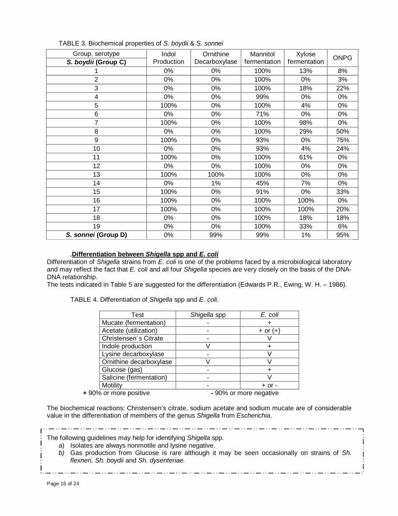
TABLE 3. Biochemical properties of S. boydii & S. sonnei Group, serotype
S. boydii (Group C) Indol
Production Ornithine
Decarboxylase Mannitol
fermentation Xylose
fermentation ONPG
1 0% 0% 100% 13% 8% 2 0% 0% 100% 0% 3% 3 0% 0% 100% 18% 22% 4 0% 0% 99% 0% 0% 5 100% 0% 100% 4% 0% 6 0% 0% 71% 0% 0% 7 100% 0% 100% 98% 0% 8 0% 0% 100% 29% 50% 9 100% 0% 93% 0% 75% 10 0% 0% 93% 4% 24% 11 100% 0% 100% 61% 0% 12 0% 0% 100% 0% 0% 13 100% 100% 100% 0% 0% 14 0% 1% 45% 7% 0% 15 100% 0% 91% 0% 33% 16 100% 0% 100% 100% 0% 17 100% 0% 100% 100% 20% 18 0% 0% 100% 18% 18% 19 0% 0% 100% 33% 6%
S. sonnei (Group D) 0% 99% 99% 1% 95%
.Differentiation between Shigella spp and E. coli Differentiation of Shigella strains from E. coli is one of the problems faced by a microbiological laboratory and may reflect the fact that E. coli and all four Shigella species are very closely on the basis of the DNA-DNA relationship. The tests indicated in Table 5 are suggested for the differentiation (Edwards P.R., Ewing, W. H. – 1986).
TABLE 4. Differentiation of Shigella spp and E. coli.
Test Shigella spp E. coli Mucate (fermentation) - + Acetate (utilization) - + or (+) Christensen´s Citrate - V Indole production V + Lysine decarboxylase - V Ornithine decarboxylase V V Glucose (gas) - + Salicine (fermentation) - V Motility - + or -
+ 90% or more positive - 90% or more negative The biochemical reactions: Christensen’s citrate, sodium acetate and sodium mucate are of considerable value in the differentiation of members of the genus Shigella from Escherichia. The following guidelines may help for identifying Shigella spp.
a) Isolates are always nonmotile and lysine negative. b) Gas production from Glucose is rare although it may be seen occasionally on strains of Sh.
flexneri, Sh. boydii and Sh. dysenteriae.

Page 17 of 24
Serotyping of Shigella spp Serotyping is subtyping method based on the immuno-reactivity of various antigens. Microorganisms produce a variety of antigens: structural components of the cells (cell wall constituents, capsules or envelopes, flagellae, fimbriae); secretion products of the cells (toxins, extracellular enzymes) or antigens contained in the interior of the cells. Chemically the antigens used for such purposes are of two main kinds: proteins and carbohydrates (including mixtures of both components). The Shigellae are by definition non-motile, as such, only the somatic (O) antigens are utilized for the determination of serotype. Flagellar (H) antigens are not expressed. The O antigen consists of repeat units of oligosaccharide, and is part of the lipopolysaccharide (LPS) of the outer membrane of Gram-negative bacteria and contributes to the main antigenic variability on the cell surface. On the basis of O antigen structure and biochemical profile, Shigella spp can be classified into four species: S. dysenteriae, S. flexneri, S. boydii and S. sonnei. Each species (with the exception of S. sonnei) can be further divided into multiple serotypes).
Group A or Shigella dysenteriae: includes 15 serotypes (S. dysenteriae 1 to the 15). Group B or Shigella flexneri: includes 8 serotypes (S. flexneri 1 to the 6, S. flexneri X and S.
flexneri Y) that can be divided into subserotypes according to its antigenic group factors (designated 3,4 – 6 and 7,8). Therefore with all the possible different antigenic combinations, the serotypes 1 to 5 of S. flexneri are subdivided into 11 subserotypes (see table in section IX - Interpretation of Results) (Annual Summary 2005 CDC http://www.cdc.gov/ncidod/dbmd/phlisdata/shigella.htm)
Group C or Shigella boydii: includes 20 serotypes (S. boydii 1 to the 20). Group D or Shigella sonnei: contains only one serotype that may occur in two forms, form I
(smooth) and form II (rough).
₪₪₪₪₪₪₪₪₪₪₪₪₪₪₪₪₪₪₪₪₪₪₪₪₪₪₪₪₪
Shigella flexneri Shigella flexneri serotypes are subdivided into subserotypes (see table in section IX - Interpretation of Results) (Edwards P.R., Ewing, W. H. – 1986; Brenner, D.J., 1984). The O antigen is an important virulence factor, and their diversity within Group B of Shigella is due to the presence of temperate bacteriophages that generates the serotype conversion. Several phages serotype-converting have been isolated and the genes involved in the O antigen modification were identified and characterized. (Allison, G.E., Verma, N.K. - 2000). The serotypes of S. flexneri (with the exception of serotype 6) have some degree of antigenic relatedness between them attributable to a common repeating tetrasaccharide unit, to which -D-glucopyranosyl and O-acetyl residues are added, providing the basis for their type (i.e., I to VI) and group (i.e., 3.4, 6, and 7.8) antigenic factors. The addition of these residues is promoted by the insertion of temperated-bacteriophages. The figure 1 shows the chemical composition of the different serotypes of Shigella flexneri.

Page 18 of 24
FIGURE 1 – Chemical composition of different serotypes of S. flexneri
Fig. 1. Chemical composition of the different serotypes of Shigella flexneri. The basic O-antigen (serotype Y) consists of repeating
units of the N-acetylglucosamine-rhamnose-rhamnose-rhamnose tetrasaccharide. The linkages between each sugar residue are
indicated for serotype Y. Serotypes differ by the addition of either glucosyl or O-acetyl groups to different sugars within the
tetrasaccharide repeat unit via the linkages indicated. Each serotype has one type-specific (Roman numeral) and one or more
group-specific (Arabic numeral) antigenic determinants. Abbreviations: GlcNac, N-acetylglucosamine; Rha, rhamnose. (Allison,
G.E., Verma, N.K. - 2000). ₪₪₪₪₪₪₪₪₪₪₪₪₪₪₪₪₪₪₪₪₪₪₪₪₪₪₪₪₪
The Shigella serogroup and serotype are identified by agglutination tests with polyclonal antibodies, where a positive interaction indicates that the bacterium in study contains antigens that react specifically with the serum antibodies These polyclonal antisera are obtained by hyperimmunization of healthy rabbits with whole cells of Shigella spp. heat inactivated. The antisera are tested for specificity and if it is necessary they are absorbed to remove cross-reacting agglutininins. There are polyvalent and monovalent Shigella antisera. The polyvalent antisera, are those that their antibodies recognize antigens present in the different serotypes of Shigella (i.e. polyvalent antisera for Sh. flexneri, recognizes all the serotypes of this group). The monovalent antisera, only recognize specific epitopes of a serotype (i.e. monovalent antisera for S. flexneri 2) or of a group factor (i.e. monovalent antisera for S. flexneri group factor 7,8). These reagents are used on slide agglutination test, to serotype well defined biochemical isolates. The final identification of the microorganism must be based on the results of biochemical and serological approaches. The commercially available antisera may not able to cover all possible epitopes of the O antigen of Shigella spp. There are probably a multitude of epitopes not covered by the typing scheme currently in use. New serotypes or subserotypes are being isolated from different parts of the world (Kaisar A., et al; 2002 – Ansaruzzaman, M. et al; 1995 – Woodward, D. et al; 2005).

Page 19 of 24
APPENDIX B
SHIGELLA SEROTYPING WORKSHEET
Patient:_________________ Accession Number:_______________ Date Received:________ Date Inoculated:________________ Inoculated By:______________ Serotyping Performed By: ______________ Date:_____________ Lots of the media and solutions employed: ______________ ____________________________ ______________ ____________________________ ______________ ____________________________ ______________
Final Result: _________________________________________________________________ Reported By:_____________________ Approved By: ________________ Date:_________
Antiserum Lot Colony 1 Colony 2 (if tested) Boiled Suspension (if tested )
POLY A
POLY A1
POLY B
POLY C
POLY C1
POLY C2
POLY C3 POLY D
Single factor:_______
Single factor:______
Single factor:______
Single factor:______
Single factor:______
Single factor:______
Single factor:______
Single factor:______
Single factor:______
Single factor:______

Page 20 of 24
APPENDIX C
SHIGELLA ANTISERA QC RECORD DATASHEET
Serotyping Performed By: ______________ Date:_____________ Lots of the media and solutions employed: ______________ ____________________________ ______________ ____________________________ ______________ ____________________________ ______________ Antisera:__________________
* Refers to the auttoaglutination test (positive or negative)
Interpretation: Pass: Antiserum produces 3-4+ agglutination with positive control (< 60 seconds) & the reference strain not autoagglutinate Fail: No reaction / weak reaction with positive control and / or autoagglutination of the reference strain
Manufacturer / Lot # Expiration: Positive Control Strain: Result:
Control of the reference strain:*
Result of QC
Testing: Comments:
Pass / Fail
Pass / Fail
Pass / Fail
Page 21 of 24
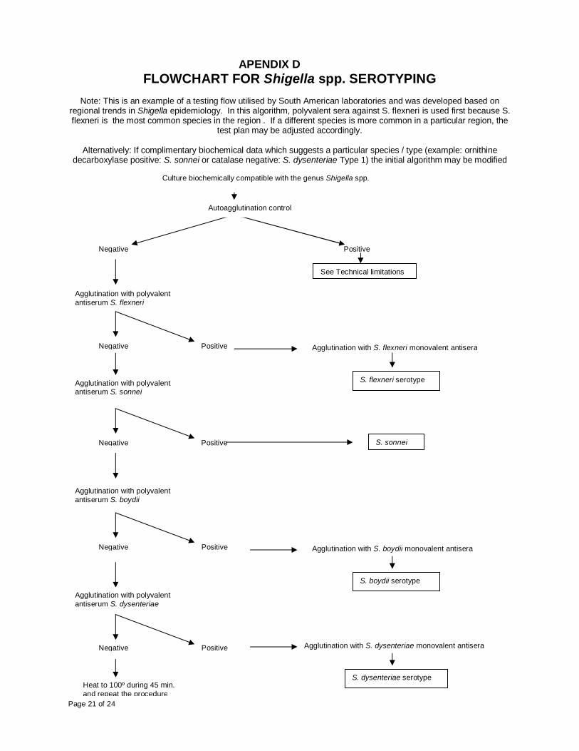
APENDIX D FLOWCHART FOR Shigella spp. SEROTYPING
Note: This is an example of a testing flow utilised by South American laboratories and was developed based on
regional trends in Shigella epidemiology. In this algorithm, polyvalent sera against S. flexneri is used first because S. flexneri is the most common species in the region . If a different species is more common in a particular region, the
test plan may be adjusted accordingly.
Alternatively: If complimentary biochemical data which suggests a particular species / type (example: ornithine decarboxylase positive: S. sonnei or catalase negative: S. dysenteriae Type 1) the initial algorithm may be modified
Culture biochemically compatible with the genus Shigella spp.
Autoagglutination control
Negative Positive
Agglutination with polyvalent antiserum S. flexneri
Negative Positive Agglutination with S. flexneri monovalent antisera
Negative Positive S. sonnei
Negative Positive
Negative Positive
Heat to 100º during 45 min. and repeat the procedure
Agglutination with polyvalent antiserum S. sonnei
Agglutination with polyvalent antiserum S. boydii
Agglutination with polyvalent antiserum S. dysenteriae
S. flexneri serotype
Agglutination with S. boydii monovalent antisera
S. boydii serotype
Agglutination with S. dysenteriae monovalent antisera
S. dysenteriae serotype
See Technical limitations

Page 22 of 24
APPENDIX E
STORAGE OF Shigella spp. CULTURES
(A) Preservation in semisolid agar
The semisolid agar is dispensed in small tubes, sterilized 15 min at 121ºC and allow to cool. Tubes are stab-inoculated. After overnight incubation, the tubes are stored in the dark at room temperature. It is not necessary –at least in countries with temperate climate- to store in the refrigerator. When the stored strains need to be used again, a portion of the culture is transferred to a sterile broth with an inoculating loop. After overnight incubation at 35-37ºC, a loopful of broth cultures is streaked on a nutrient agar, as TSA. (Popoff, 2001, Guidelines for the preparation of Salmonella Antisera, 6th Ed., WHO Collaborating Centre for Reference and Research on Salmonella Pasteur Institute)
(B) Freezing at -70ºC
Freezing can conveniently be done as follows: Add tryptic soy broth (TSB) containing 15% (v/v) glycerol and approximately 30, sterile glass beads (2mm in diameter) to a sterile, freezer-safe (10 x 30 mm) screw-capped vial. Prepare as many vials as needed. Using a fresh, pure, culture, make a heavy suspension of bacteria in the vial. Carefully remove as much of the excess suspension as possible. Label the vial and place the vial in a tray. The tray is then immediately stored at -70ºC. To recover a frozen strain, a vial is removed from the freezer and one bead is picked up using a platinum loop or sterile forceps. The removed bead is transferred to nutrient broth and incubated overnight at 35-37ºC and the vial is immediately returned to to freezer. (Popoff, 2001, Guidelines for the preparation of Salmonella Antisera, 6th Ed., WHO Collaborating Centre for Reference and Research on Salmonella Pasteur Institute)
Page 23 of 24
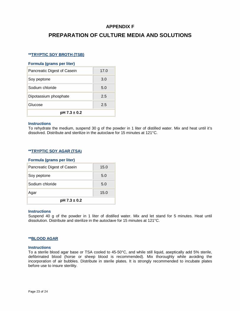
APPENDIX F
PREPARATION OF CULTURE MEDIA AND SOLUTIONS **TRYPTIC SOY BROTH (TSB) Formula (grams per liter)
Pancreatic Digest of Casein 17.0
Soy peptone 3.0
Sodium chloride 5.0
Dipotassium phosphate 2.5
Glucose 2.5
pH 7.3 ± 0.2
Instructions To rehydrate the medium, suspend 30 g of the powder in 1 liter of distilled water. Mix and heat until it’s dissolved. Distribute and sterilize in the autoclave for 15 minutes at 121°C. **TRYPTIC SOY AGAR (TSA) Formula (grams per liter)
Pancreatic Digest of Casein 15.0
Soy peptone 5.0
Sodium chloride 5.0
Agar 15.0
pH 7.3 ± 0.2
Instructions Suspend 40 g of the powder in 1 liter of distilled water. Mix and let stand for 5 minutes. Heat until dissolution. Distribute and sterilize in the autoclave for 15 minutes at 121°C. **BLOOD AGAR Instructions To a sterile blood agar base or TSA cooled to 45-50°C, and while still liquid, aseptically add 5% sterile, defibrinated blood (horse or sheep blood is recommended). Mix thoroughly while avoiding the incorporation of air bubbles. Distribute in sterile plates. It is strongly recommended to incubate plates before use to insure sterility.

Page 24 of 24
**SEMI-SOLID AGAR (FOR STORAGE) Formula (grams per liter)
Beef extract 5.0
Peptone 10.0
NaCl 3.0
Na2HPO4.12H2O 2.0
Agar 10
pH 7.4 ± 0.1 Instructions Suspend the components in 1 liter of distilled water. Let stand for 10 to 15 minutes. Heat to boil. Adjust to pH 7,4 ±0,1. Sterilize in autoclave for 15 minutes at 121°C. **PHYSIOLOGICAL SALINE SOLUTION (NaCl 0,85% w/v) Formula (grams per liter)
NaCl 8,5
Instructions Suspend 8,5 g of the NaCl in 800 ml of distilled water. Complete volume up 1000 ml with distilled water. Distribute and sterilize in the autoclave for 15 minutes at 121°C. **0,5 % FORMALINIZED PHYSIOLOGICAL SALINE SOLUTION (NaCl 0,85 w/v – 0,5 % formalin v/v) Formula (grams per liter)
NaCl 8,5
Formalin 5.0 ml
Instructions Suspend 8,5 g of the NaCl in 995 ml of distilled water. Autoclave for 15 minutes at 121°C. Allow to cool to room temperature. Add 5 ml of formalin. DO NOT AUTOCLAVE AFTER ADDING FORMALIN.