Embed Size (px)
Citation preview

Global Salm-Surv
AA gglloobbaall SSaallmmoonneellllaa ssuurrvveeiillllaannccee aanndd llaabboorraattoorryy ssuuppppoorrtt pprroojjeecctt ooff tthhee WWoorrlldd HHeeaalltthh OOrrggaanniizzaattiioonn
Laboratory Protocols
Level 2 Training Course
Susceptibility testing of Salmonella using disk diffusion
3rd Ed. July. 2002
Edited by: Rene S. Hendriksen (DFVF)

Contents Page 1. Susceptibility testing: Determination of phenotypic resistance..............................................3 2. Disk diffusion - qualitative susceptibility testing ...................................................................5 3. Composition and preparation of culture media and reagents..................................................8 Laboratory record sheets.............................................................................................................9 1. Susceptibility testing: Determination of phenotypic resistance
2
1) Agar diffusion with disk 2) Agar diffusion with E-test 3) MIC-determination using Agar dilution method. Introduction The MIC (Minimal Inhibitory Concentration) of a bacterium to a certain antimicrobial agent gives a quantitative estimate of the susceptibility. MIC is defined as the lowest concentration of antimicrobial agent required to inhibit growth of the organism. The principle is simple: Agar plates, tubes or microtitre trays with two-fold dilutions of antibiotics are inoculated with a standardised inoculum of the bacteria and incubated under standardised conditions following NCCLS guidelines. The next day, the MIC is recorded as the lowest concentration of antimicrobial agent with no visible growth. The MIC informs you about the degree of resistance and might give you important information about the resistance mechanism and the resistance genes involved. MIC-determination performed as agar dilution is regarded as the gold standard for susceptibility testing. Agar diffusion tests are often used as qualitative methods to determine whether a bacterium is resistant, intermediately resistant or susceptible. However, the agar diffusion method can be used for determination of MIC values provided the necessary reference curves for conversion of inhibition zones into MIC values are available. After an agar plate is inoculated with the bacteria, a tablet, disk or paper strip with the antimicrobial agent is placed on the surface. During incubation the antimicrobial agent diffuses into the agar and inhibits growth of the bacteria if susceptible. Diffusion tests are cheap compared to most MIC-determination methods. E-test is a diffusion test, but has been developed to give an approximate MIC-value. Well standardised methods are essential for all kinds of susceptibility testing, since the methods are highly sensitive to variations in several factors, such as size of inoculum, contents and acidity of the growth medium, time and temperature of incubation. The agar diffusion methods are also strongly influenced by factors such as agar depth, diffusion rate of the antimicrobial agent and growth rate of the specific bacteria. The MIC-determination and disk diffusion methods described in this protocol are in accordance with the international recommendations given by the National Committee for Clinical Laboratory Standards (NCCLS). The NCCLS describes how to perform the tests and sets international guidelines for interpretation of the results. It should be noted that the WHO does not recommend any specific method for performance and interpretation of susceptibility tests. Internal quality control should be regularly performed as recommended by NCCLS. References
3

1. National Committee for Clinical Laboratory Standards. Performance standards for antimicrobial
disk susceptibility tests, 7th ed. Approved standard. M2-A7. NCCLS, Wayne, Pennsylvania, 2000.
2. National Committee for Clinical Laboratory Standards. Performance standards for antimicrobial
disk and dilution susceptibility tests for bacteria isolated from animals. Approved standard. M31-A2, NCCLS, Wayne, Pennsylvania, 2001.
3. National Committee for Clinical Laboratory Standards. Methods for dilution antimicrobial
susceptibility tests for bacteria that grow aerobically. 5th ed. Approved standard. M7-A5, NCCLS, Wayne, Pennsylvania, 2000.
4

2. Disk Diffusion – susceptibility testing
Materials Equipment • McFarland standard 0.5 • White paper with black lines • Mixer • Scissors • Forceps • Loops (1 µl and 10 µl) • Bunsen burner • Small sterile cotton swabs • Disk dispenser • Ruler or calipers Media • Sterile normal saline (0.9%), 4 ml volumes in tubes • Mueller-Hinton II agar plates (9 cm and a uniform agar depth of 4 mm) without blood • Disks with antimicrobial agents • Nutrient agar plates (9 cm) Bacterial strains • Salmonella strains on non-selective agar • Strain for quality control: Escherichia coli ATCC 25922
Safety Carry out all procedures in accordance with the local codes of safe practice.
5

Procedure Day 1 Standardisation of inoculum With a loop, touch the top of 3 or 4 individual colonies and transfer to a tube of saline. Emulsify the inoculum on the inside of the tube to avoid lumps. Adjust to McFarland 0.5: Compare turbidity to that in the 0.5 McFarland standard using paper with black lines. Adjust turbidity of inoculum to match that standard. Inoculate agar plate Check purity of Mueller Hinton II agar plates. Swab plate within 15 minutes of preparing the adjusted inoculum: Dip a sterile cotton swab into the inoculum and pulling out slightly, rotate the swab several times against the inside of the tube above the fluid level to remove excess liquid. Streak the swab over the entire surface of the Mueller Hinton II agar plate. Rotate the plate approximately 60o then repeat streaking motion. Rotate 60o again and repeat streaking. Complete inoculation by running the swab around the rim of the agar. Leave the lid of the plate ajar for 5 minutes (no more than 15 minutes) to allow any excess moisture to be absorbed before applying disks. Dispense disks to the agar surface with the Disk dispenser or forceps. Do not move any disks after contact with the agar. Make sure the disk have complete contact with the agar-surface by touching the disk with forceps.
Theory / comments Picking material from more than one colony ensures sufficient numbers of bacterial cells for the test. It is done to minimise the risk of picking bacteria that have lost their resistance and to assure a sufficient number of bacteria. McFarland 0.5 ~ approximately 108 CFU/ml. Standardised inoculum is essential because the zone size of inhibition depends on the growth density, and because the interpretation of the results is based on a confluent lawn of growth (NCCLS/Kirby-Bauer). To avoid further growth before inoculation Homogeneous plating is important to yield reliable results. Moving the disk yields oval zones, which are difficult to read and might give unreliable results.
6

Procedure Check purity of the inoculum: Transfer inoculum from the tube onto a nutrient agar plate using a 10 µl loop. Incubate plates at 35oC for 16 to 18 hours in ambient air. Day 2 Reading plates /interpretation of results Check purity Check that the growth is a confluent lawn. If individual colonies are apparent the inoculum has been too light. Retest the sample. Check that zones are round, not oval Measure the diameter of inhibition zones (to full growth). Interpretation of the results (i.e. categorisation of isolates into susceptible, intermediary or resistant) is done according to NCCLS guidelines. Be aware of special readings for trimethoprim and sulphonamides. In these cases, zones of inhibition are measured up to colonies of normal size (disregard slight growth and measure the more obvious margin). With trimethoprim and the sulfonamides antagonists in the medium may allow some slight growth; therefore, disregard slight growth (20% or less of the lawn of growth), and measure the most obvious margin to determine the zone diameter.
Theory / comments Most microbiological laboratories have their incubators set at 37oC. Incubating disk diffusion agar plates at 37oC is a practical, and by most laboratories’ experience acceptable, deviation from the recommended standard. Dense growth would yield zones that are too small. . Sometimes interaction between drugs in disks placed closely together may produce distortion of inhibition zones (i.e. antagonism, synergism, inhibition and/or induction) not necessarily an oval shape. Such valuable additional information should not be considered in the reading of the inhibition zones but provides important data about the putative mechanism of resistance, bacterial id, etc. The interpretation of zone diameters is given by the NCCLS guidelines. Interpretation is based on a confluent lawn of growth and on regression lines found by contemporary testing of a large number of isolates by both the disk diffusion and a standardised MIC-determination method. The antibiotic trimethoprim and the sulphonamides allow growth of the bacteria for some generations before inhibition occurs.
7

8

3. Composition and preparation of culture media and reagents If no reference is given, it is the procedure used at DVL. The media and reagents are available from several companies including Oxoid, Merck and Difco. The composition of the dehydrated media given below is an example and may vary a little among the different manufacturers. Also, the media should be prepared according to the manufacturers description if it differs from the description given here. Refer to Appendix 2 for a colour presentation of growth of Salmonella on selective agar media and positive and negative reactions of biochemical tests. Mueller Hinton II agar (e.g. from BBL) Beef extract 2.0 g Acid hydrolysate of casein 17.5 g Starch 1.5 g Agar 17.0 g Distilled water 1000 ml Preparation: Dissolve the dehydrated medium in water by heating if necessary. Adjust pH to 7.2 - 7.4, transfer into bottles and autoclave at 110oC for 20 min. Nutrient agar (ref. 1) Meat extract 3.0 g Peptone 5.0 g Agar 12 g to 18 g1) Water 1000 ml 1) Depending on the gel strength of the agar. Preparation: Dissolve the dehydrated medium in the water by heating if necessary. Adjust pH to ~7.0 after sterilisation, transfer into bottles and autoclave at 121oC for 20 min. Pour 15 ml of melted medium in each plate. Saline solution Sodium chloride 8.5 g Water 1000 ml Preparation: Dissolve the sodium chloride in the water by heating if necessary. Adjust pH to ∼ 7.0 after sterilisation. Dispense the solution into tubes so 4 ml is obtained after autoclaving at 121oC for 20 min. References 1. ISO 6579 :1993(E) 3rd ed. Microbiology - General guidance on methods for the detection of
Salmonella. Date: Record sheet:
9
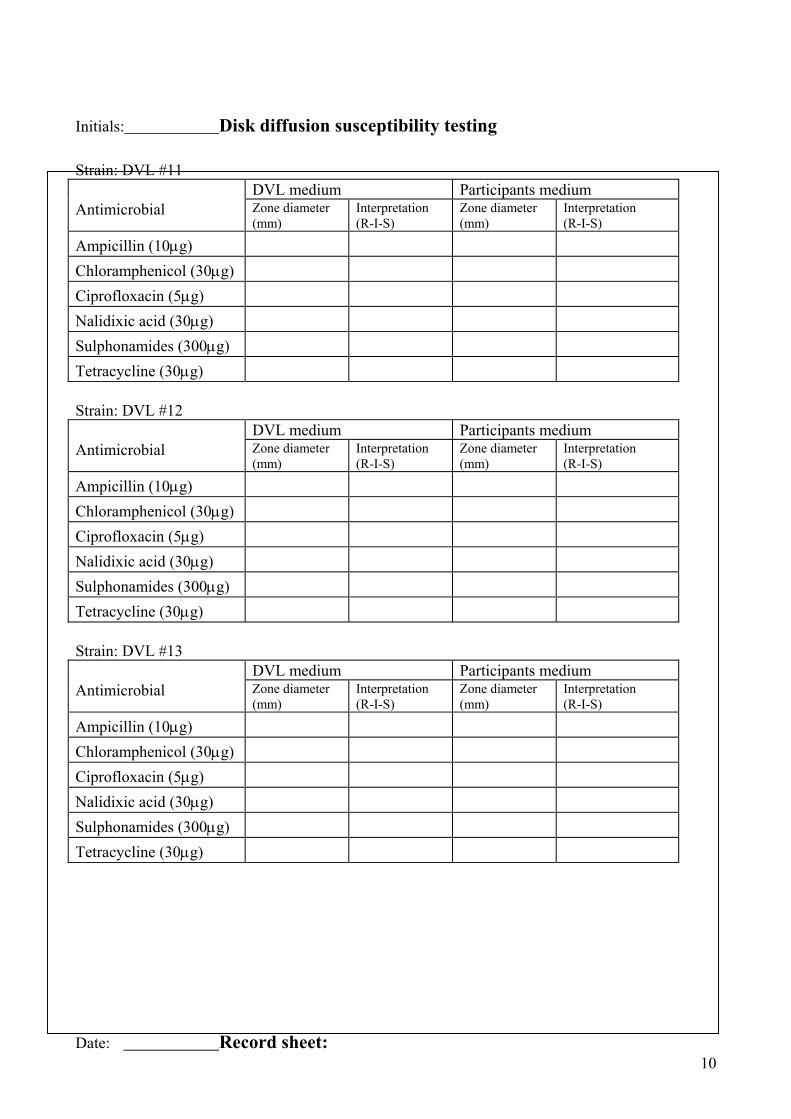
Initials: Disk diffusion susceptibility testing Strain: DVL #11 DVL medium Participants medium Antimicrobial Zone diameter
(mm) Interpretation (R-I-S)
Zone diameter (mm)
Interpretation (R-I-S)
Ampicillin (10µg)
Chloramphenicol (30µg)
Ciprofloxacin (5µg)
Nalidixic acid (30µg)
Sulphonamides (300µg)
Tetracycline (30µg) Strain: DVL #12 DVL medium Participants medium Antimicrobial Zone diameter
(mm) Interpretation (R-I-S)
Zone diameter (mm)
Interpretation (R-I-S)
Ampicillin (10µg)
Chloramphenicol (30µg)
Ciprofloxacin (5µg)
Nalidixic acid (30µg)
Sulphonamides (300µg)
Tetracycline (30µg) Strain: DVL #13 DVL medium Participants medium Antimicrobial Zone diameter
(mm) Interpretation (R-I-S)
Zone diameter (mm)
Interpretation (R-I-S)
Ampicillin (10µg)
Chloramphenicol (30µg)
Ciprofloxacin (5µg)
Nalidixic acid (30µg)
Sulphonamides (300µg)
Tetracycline (30µg) Date: Record sheet:
10

Initials: Disk diffusion susceptibility testing Strain: DVL #14 DVL medium Participants medium Antimicrobial Zone diameter
(mm) Interpretation (R-I-S)
Zone diameter (mm)
Interpretation (R-I-S)
Ampicillin (10µg)
Chloramphenicol (30µg)
Ciprofloxacin (5µg)
Nalidixic acid (30µg)
Sulphonamides (300µg)
Tetracycline (30µg) Strain: DVL #15 DVL medium Participants medium Antimicrobial Zone diameter
(mm) Interpretation (R-I-S)
Zone diameter (mm)
Interpretation (R-I-S)
Ampicillin (10µg)
Chloramphenicol (30µg)
Ciprofloxacin (5µg)
Nalidixic acid (30µg)
Sulphonamides (300µg)
Tetracycline (30µg) Strain: ATCC 25922 DVL medium Participants medium Antimicrobial Zone diameter
(mm) Within the QC Interval.
Zone diameter (mm)
Within the QC Interval.
Ampicillin (10µg)
Chloramphenicol (30µg)
Ciprofloxacin (5µg)
Nalidixic acid (30µg)
Sulphonamides (300µg)
Tetracycline (30µg) Date: Record sheet:
11

Initials: Disk diffusion susceptibility testing Strain: DVL #11 DVL medium Local organiser medium Antimicrobial Zone diameter
(mm) Interpretation (R-I-S)
Zone diameter (mm)
Interpretation (R-I-S)
Ampicillin (10µg)
Chloramphenicol (30µg)
Ciprofloxacin (5µg)
Nalidixic acid (30µg)
Sulphonamides (300µg)
Tetracycline (30µg) Strain: DVL #12 DVL medium Local organiser medium Antimicrobial Zone diameter
(mm) Interpretation (R-I-S)
Zone diameter (mm)
Interpretation (R-I-S)
Ampicillin (10µg)
Chloramphenicol (30µg)
Ciprofloxacin (5µg)
Nalidixic acid (30µg)
Sulphonamides (300µg)
Tetracycline (30µg) Strain: DVL #13 DVL medium Local organiser medium Antimicrobial Zone diameter
(mm) Interpretation (R-I-S)
Zone diameter (mm)
Interpretation (R-I-S)
Ampicillin (10µg)
Chloramphenicol (30µg)
Ciprofloxacin (5µg)
Nalidixic acid (30µg)
Sulphonamides (300µg)
Tetracycline (30µg)
12

Date: Record sheet: Initials: Disk Diffusion susceptibility testing Strain: DVL #14 DVL medium Local organiser medium Antimicrobial Zone diameter
(mm) Interpretation (R-I-S)
Zone diameter (mm)
Interpretation (R-I-S)
Ampicillin (10µg)
Chloramphenicol (30µg)
Ciprofloxacin (5µg)
Nalidixic acid (30µg)
Sulphonamides (300µg)
Tetracycline (30µg) Strain: DVL #15 DVL medium Local organiser medium Antimicrobial Zone diameter
(mm) Interpretation (R-I-S)
Zone diameter (mm)
Interpretation (R-I-S)
Ampicillin (10µg)
Chloramphenicol (30µg)
Ciprofloxacin (5µg)
Nalidixic acid (30µg)
Sulphonamides (300µg)
Tetracycline (30µg) Strain: ATCC 25922 DVL medium Local organiser medium Antimicrobial Zone diameter
(mm) Within the QC Interval.
Zone diameter (mm)
Within the QC Interval.
Ampicillin (10µg)
Chloramphenicol (30µg)
Ciprofloxacin (5µg)
Nalidixic acid (30µg)
Sulphonamides (300µg)
Tetracycline (30µg)
13