Embed Size (px)
Citation preview

LABORATORY TESTING IN
PRIMARY CARE OPTOMETRY
Tammy P. Than, MS, OD, FAAOCarl Vinson VAMC
Dublin, GA

Microbiology
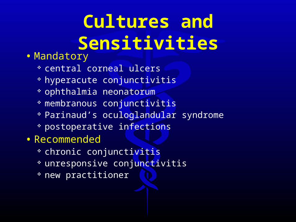
Cultures and Sensitivities
• Mandatory central corneal ulcers hyperacute conjunctivitis ophthalmia neonatorum membranous conjunctivitis Parinaud’s oculoglandular syndrome postoperative infections
• Recommended chronic conjunctivitis unresponsive conjunctivitis new practitioner

Cultures and Sensitivities
• specimen preparation is important• no anesthetic – if possible• sterile swab plate onto culture media• culturette• media:
Thioglycolate broth Blood agar Chocolate agar Saboraud’s agar

Transport Media
• Amies media without charcoal Higher yield than other media Comparable to plates
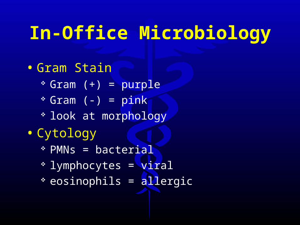
In-Office Microbiology
• Gram Stain Gram (+) = purple Gram (-) = pink look at morphology
• Cytology PMNs = bacterial lymphocytes = viral eosinophils = allergic

Diagnostic Imaging
• plain film X-Ray• CT scan• MRI
75 million in 2003
• Ultrasonography• Angiography• GDx, OCT, HRT• etc…

Resources
• Imaging of the Globe and Orbit: A Guide to Differential Diagnosis Hosten and Bornfield Publisher Thieme
• http://www.med.harvard.edu/AANLIB/home.html
• http://www.loni.ucla.edu/index.shtml

X-Ray: The Basics
• Incident X-Ray enters tissue• Beam is attenuated• Exit X-Ray leaves tissue exposes film
White areas = not exposed Dark areas = film exposed
• 3-D represented by 2-D• Black = air (no attenuation)• White = bone• Gray = soft tissue
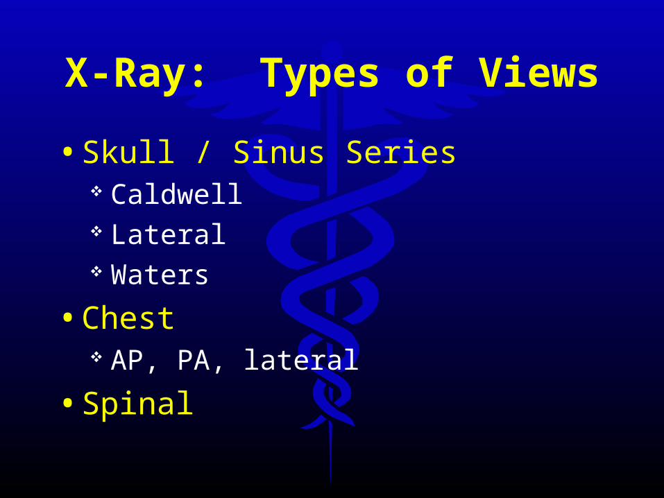
X-Ray: Types of Views
• Skull / Sinus Series Caldwell Lateral Waters
• Chest AP, PA, lateral
• Spinal

CALDWELL

CALDWELCALDWELLL

LATERAL

LATERALLATERAL
WATERS

WATERSWATERS

X-Ray Indications• Confirm the integrity of the orbit
Intraocular Foreign Body Intraorbital Foreign Body
• Trauma muscle entrapment?

X-Ray Indications
• Sinusitis R/O Orbital cellulitis
• Horner’s syndrome
• Uveitis
• Ankylosing spondylitis
• Reiter’s syndrome
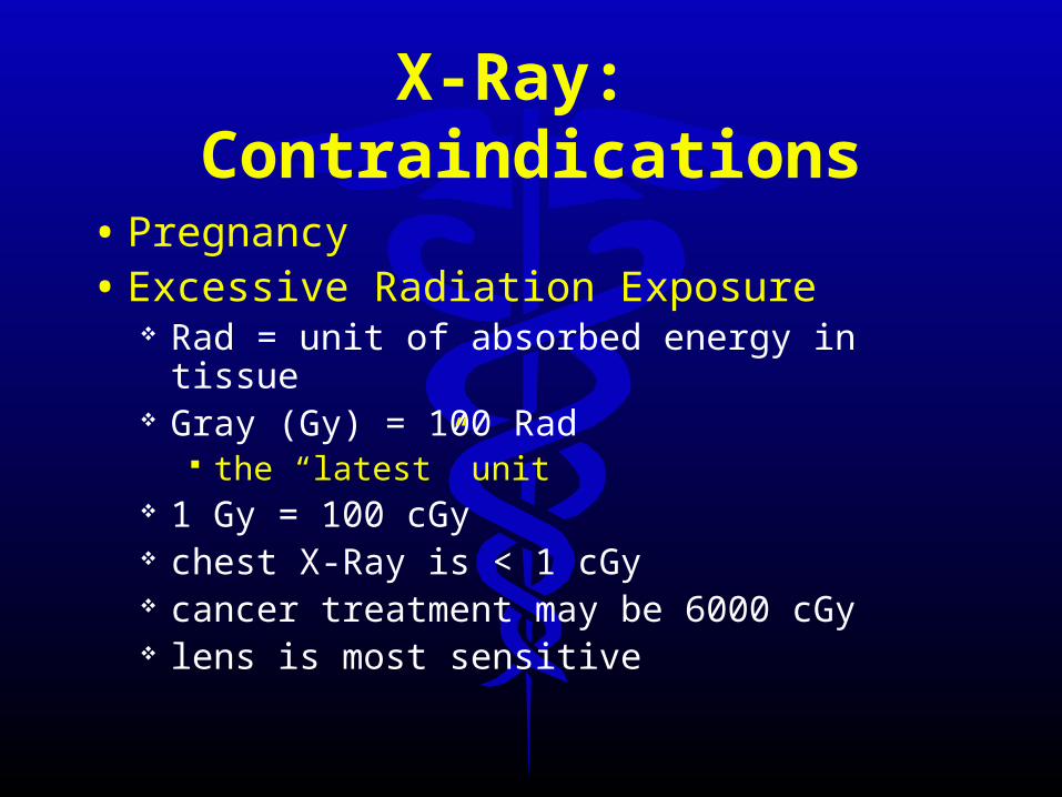
X-Ray: Contraindications
• Pregnancy• Excessive Radiation Exposure
Rad = unit of absorbed energy in tissue Gray (Gy) = 100 Rad
the “latest” unit 1 Gy = 100 cGy chest X-Ray is < 1 cGy cancer treatment may be 6000 cGy lens is most sensitive

X-Rays
• Pros Inexpensive Readily available Rapid results
• Cons Radiation exposure No information about soft tissue 2-D interpretation can be difficult

Case #1
• 17 YOM
• Hit in eye x 1 day
• + pain
• +diplopia

Work-Up
• EOMs
• Exophthalmometry
• Crepitus?
• Nerve sensation
• IOP
• Imaging

Management
• Nasal decongestants
• Oral antibiotics broad spectrum
e.g. Keflex 500 mg qid
• Don’t blow nose!
• +/- Sx in 1-2 weeks

Other Considerations…
• R/O Seidel’s sign
• Anterior Segment Pathology uveitis corneal abrasion subconjunctival hemorrhage
• Commotio Retinae

CT Scan: The Basics• Series of thin X-Ray sections
flat panel detectors may eliminate slices
• Emitted X-Rays
• Diode sensors
• Computer reconstructs views

CT Scan: The Basics
• CT Numbers density < water = negative CT# density > water = positive CT#
• “Windowing”
• Gray Scale White = bone Black = air Gray = brain

CT Scan• Views
coronal paranasal sinuses, orbital integrity
sagittal chiasmal pathology
axial orbital and visual pathways

CT – The Exam• Specific protocols
orbital chiasmal brain sinuses
• Slice thickness and slice increment• Cranial
~1 cm / no overlap
• Orbital and Chiasmal 3 mm with 2 mm between allows overlap
• Gantry• 10-20 minutes / scan
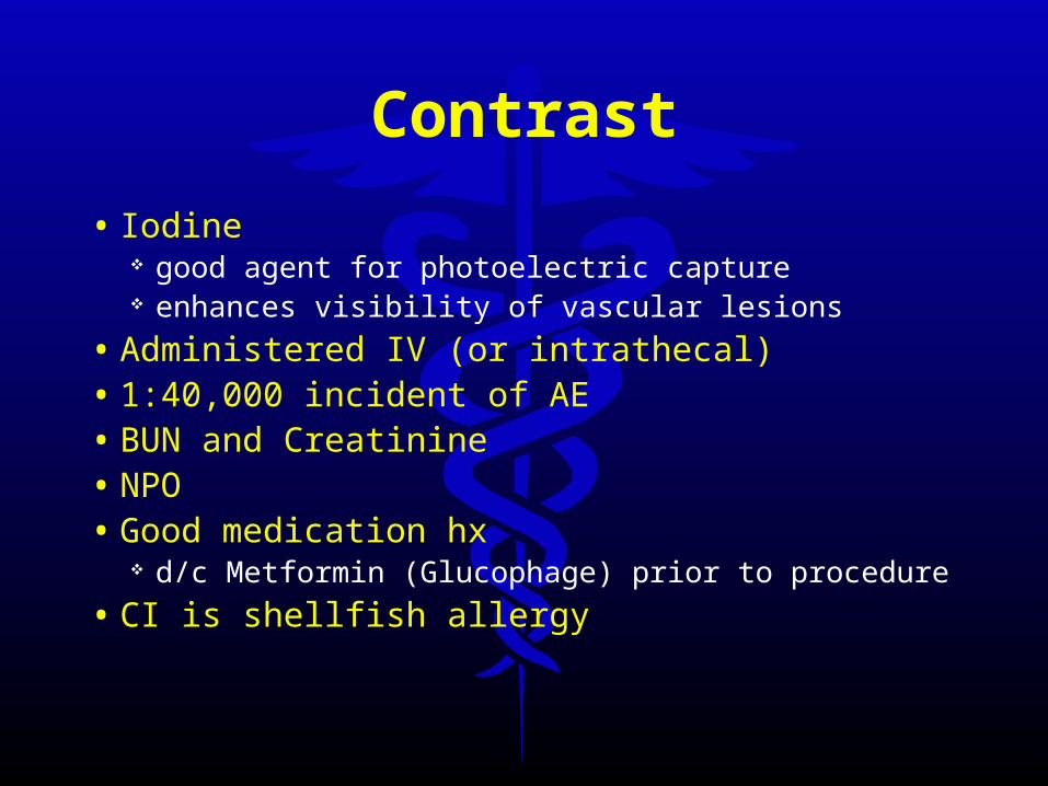
Contrast
• Iodine good agent for photoelectric capture enhances visibility of vascular lesions
• Administered IV (or intrathecal)• 1:40,000 incident of AE• BUN and Creatinine• NPO• Good medication hx
d/c Metformin (Glucophage) prior to procedure
• CI is shellfish allergy

BUN (Blood Urea Nitrogen)
• actually performed on serum or plasma 12% higher than blood
• nitrogen portion of urea• urea is formed in liver from protein breakdown• filtered through renal glomeruli
small amount reabsorbed in the tubules remainder excreted in urine
• azotemia – elevated BUN nonspecific prerenal, renal, or postrenal

BUN (Blood Urea Nitrogen)
• must be compared over time or evaluated with other tests renal function – also assess creatinine levels
• fasting not required• Adult 5-20 mg/dL• >60 8-21 mg/dL• increased BUN
many conditions and many drugs
• decreased BUN alcohol abuse, diet lacking protein, liver destruction, late
pregnancy

CREATININE
• product of anaerobic energy-producing creatine-phosphate metabolism in skeletal muscle
• excreted by kidneys increased levels indicative of decreased glomerular
filtration rate
• Avoid excessive exercise for 8 hours and avoid excessive red meat for 24 hours before testing
CREATININE
• Normal females 0.5 – 1.1 mg/dL males 0.6 –1.2 mg/dL elderly – may be lower
• Creatinine clearance, urine 24 hour collection
• Creatinine clearance, serum urine 6, 12, or 24 hour collection blood sample collected anytime during urine collection
period

CT Scan: Artifacts
• Motion
• Dental Fillings
• Partial volume phenomenon

CT Indications
• bone imaging
• calcification
• blood detection acute
• meningiomas
• when MRI contraindicated

CT Contraindications
• pregnancy
• excessive radiation exposure
• contrast contraindication iodine sensitivity shellfish allergy kidney disease

CT Scan
• Pros High diagnostic yield Good for bone Can reconstruct different views
• Cons Expensive Human risk Motion artifacts Hard to ddx tumors
MRIUnpaired protons (H) = tiny magnets
from water and fat body is 63% hydrogen atoms
• Disrupt with radio pulse
• Protons return to original state
• Release energy -> MRI

MRI
• Signal strength: proton density• Relaxation time: surrounding tissue• T1 weighted
Proton density tissue composition
• T2 weighted Tissue differences
• Intermediate• fat suppression

MRI
• White Matter and Fat T1 = bright T2 = dark
• Gray Matter and CSF T1 = dark T2 = bright
• Vitreous T1 = dark T2 = bright
• blood, air = black• EOMs and optic nerves = intermediate density

MRI: The Examination
• Gantry• Flux
0.5 – 1.5 Tesla
• Energy detected• Image reconstructed• 40 minutes• +/- gadolinium contrast
paramagnetic highlights images of similar density

MRI Indications
• tumors posterior visual pathway brain stem pituitary
• infarcts• posterior fossa• MS

MRI Indications• elevated optic nerve head(s)
• unilateral proptosis
• field loss hemianopia bitemporal
• cranial nerve palsies

MRI Contraindications
• pregnancy
• metallic FB
• pacemakers
• kidney disease (if using contrast)
• claustrophobia?

Latest…• Short bore
high field >1.5 Tesla advantages of tunnel and open MRIs

MRI
• Pros More accurate 3-D image Good structural detail No radiation
• Cons $$$ Time consuming Won’t show recent hemorrhage

Case #3.
• 46 YOWF
• CC: Time to change her glasses
• HabRx: OD –4.25 DS OS –5.00 DS
• BVA OD: -4.00 20/20 OS: -0.25 20/20

Other Imaging Tests…
Magnetic Resonance Angiography (MRA)
• Non-invasive method for investigation of blood vessels
• Surgical planning
• 3-D view
• Picks up rapid blood flow
• highly accurate for stenosis >50%

Positron Emission Tomography (PET)
• Biochemical and physiologic function – in vivo
• Radioactive “Tracer” compound Injected or inhaled C, N, O, F
18F labeled fluorodeoxyglucose Image of brain activity

Functional MRI (fMRI)
• Physiology of visual system
• With or without contrast
• Many advantages over PET
• The new “lie detector?”

So… You want to order a scan??

Before You Order Tests...
• Good case history any contraindications??
• Comprehensive exam• Narrow ddx• Is there anything YOU can do?• Avoid “fishing expedition” or “shot-gun”
approaches• Select most appropriate test
MRI not always needed

Communicating with the Lab
• Which test(s)? with contrast?
• Code?• Which insurance?• What do you want to view?• DDX?• Be available

Getting the Job Done
• Communicate with the Patient Explain why imaging is necessary Explain the test
• Insurance Issues Can you order the test?? Is the patient insured??

If You Order Tests...
• Written report of findings and copies of the films
• Communicate
• Treat
• Comanage / Refer

CLIA
• Clinical Laboratory Improvement Act
• regulates all lab tests performed on humans in US
• ensures quality laboratory testing
• Waived tests determined by FDA or CDC to be so simple that
there is little risk of error
• www.cms.hhs.gov/clia
Missouri Contact
• Missouri Dept of Health and Senior Services – CLIA Section PO Box 570 Jefferson City, MO 65102 573-751-6318 Contact: William Nugent

CLIA
• can file for “Waived Status” approximately 40 tests
random blood glucose ESR urine pregnancy tests
• must meet criteria: enroll in CLIA program pay fees biennially ($150 for waived) follow manufacturers’ test instructions

Glad you looked!
• 58 year old female
• CC: SpRx broken
• OHx: unremarkable
• MHx: unremarkable, no meds
• 20/20 OD; 20/20 OS

Random Blood Glucose
• note when patient ate last e.g. 220 mg/dL pp 3 hours pp = post-prandial
• diabetic if: 200 mg/dL with symptoms
• can do in-office
• encourage patients to do this!

Fasting Plasma Glucose
• no food or drink for 8-12 hours
• diabetes if 126 mg/dL must repeat if asymptomatic
• IFG = 100 – 125 mg/dL
• also increased with: steroids stress diuretics

What’s in a Name??
• home monitoring whole blood glucose
• laboratory methods plasma glucose
• plasma glucose usually 10-15% higher than whole blood
• SOME home monitors calibrate to plasma
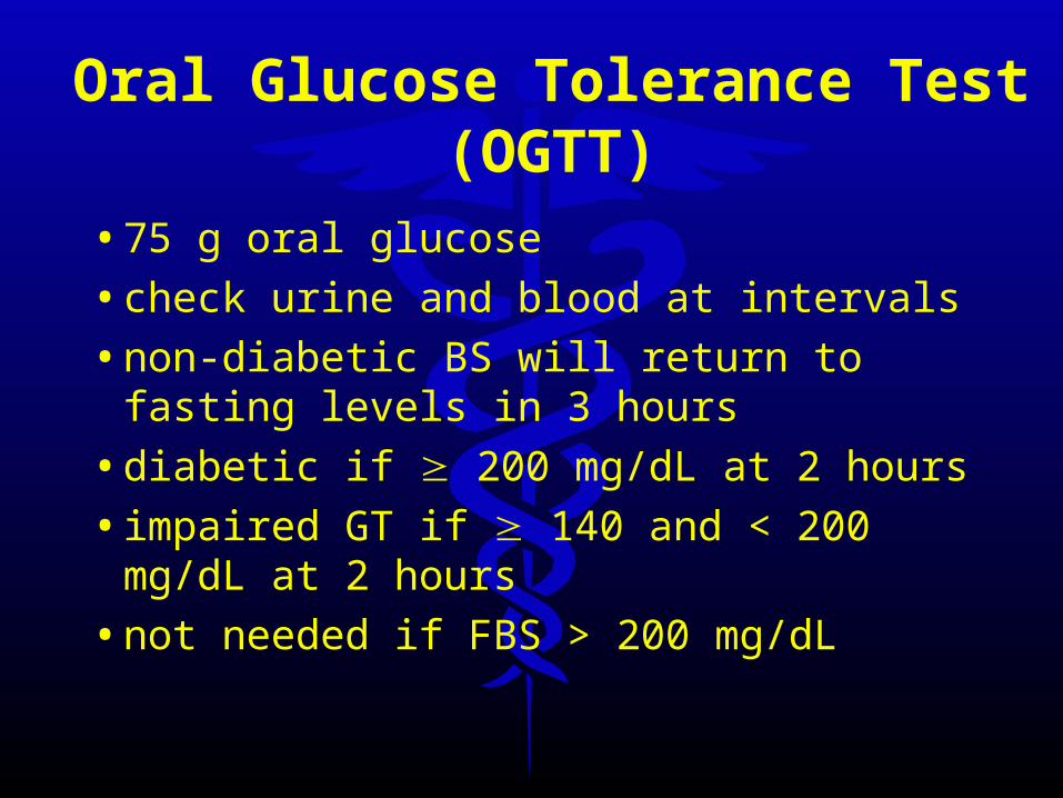
Oral Glucose Tolerance Test (OGTT)
• 75 g oral glucose
• check urine and blood at intervals
• non-diabetic BS will return to fasting levels in 3 hours
• diabetic if 200 mg/dL at 2 hours
• impaired GT if 140 and < 200 mg/dL at 2 hours
• not needed if FBS > 200 mg/dL
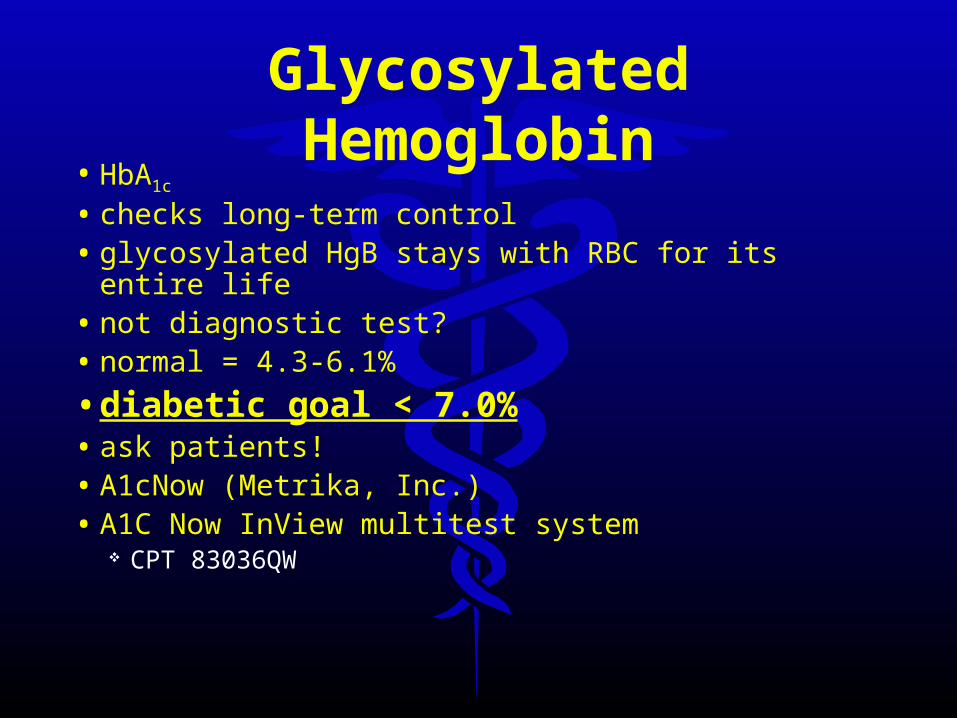
Glycosylated Hemoglobin
• HbA1c
• checks long-term control• glycosylated HgB stays with RBC for its entire life• not diagnostic test?• normal = 4.3-6.1%
• diabetic goal < 7.0%• ask patients!• A1cNow (Metrika, Inc.) • A1C Now InView multitest system
CPT 83036QW

Glycoslyated HemoglobinA1C Blood Glucose
Levels
12% 345 mg/dL
11 310
10 275
9 240
8 205
7 170
6 135
5 100
4 65
1% A1C = 30 mg/dL1% A1C = 30 mg/dL

What Can We Do?• Pre-diabetes – new term!• 61% of US adults overweight• Diabetes Prevention Program
pts with IGT (N=3234) lifestyle changes vs metformin vs placebo reduced risk
58% with lifestyle– 30 minutes daily activity; weight loss of 5-7% BW30 minutes daily activity; weight loss of 5-7% BW
31% with medication
• Educate patients honesty best policy…
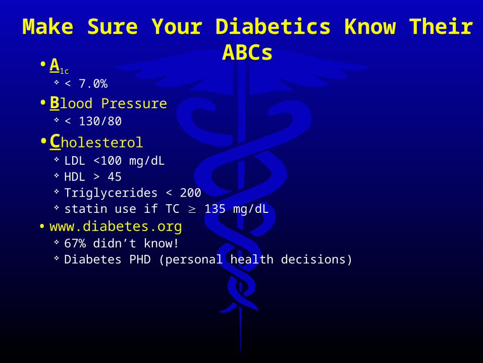
Make Sure Your Diabetics Know Their ABCs• A1c
< 7.0%
• Blood Pressure < 130/80
• Cholesterol LDL <100 mg/dL HDL > 45 Triglycerides < 200 statin use if TC 135 mg/dL
• www.diabetes.org 67% didn’t know! Diabetes PHD (personal health decisions)

Fasting Plasma Glucose
• fluctuating vision get stable reading before new SpRx
• retinopathy
• diplopia
• vascular occlusions
• optic neuropathy

HIV Testing
• Home-use HIV test kits NOT FDA approved Available on Internet
• FDA-approved Home sample collection kits
• Enzyme Immunoassay
• Western Blot
• Nucleic acid testing (viral load)

OraQuick Rapid HIV-1/2 Antibody Test
• approved in 2002 for testing with blood HIV-1 and HIV-2 CLIA waived status
• March 26, 2004 approved using oral fluid results in 20 minutes! only for HIV-1 not for screening blood donors not CLIA waived status yet
• 31% do NOT return for HIV testing results• Also Uni-Gold Recombigen HIV (7/2004)