Embed Size (px)
Citation preview
that the heparin on his surfaces is in equilibrium with endogenous heparin in the blood and that the Gott surfaces will remain heparinized for periods of more than 2 years.
It has been found (3) that unmod- ified plastics strongly adsorb blood pro- teins. Changes in 4-potential (with time) of treated and untreated polystyrene surfaces in contact with Ringer solutions containing fibrinogen, y-globulin, or albumin at one-thousandth the usual physiological concentrations have been recorded, and Fig. 1 is typical of the result. This change in a-potential is attributed to the adsorption of pro- teins on the plastic surfaces. As a result of this adsorption, the solution no long- er "sees" the plastic surface, but rather sees the adsorbed protein layer. Similar results were obtained with other treated and untreated plastic surfaces. In the case of albumin a relatively small differ- ence was found which may be due to the fact that albumin is more negatively charged than the other proteins.
The nonthrombogenicity of the hep-
An extensive survey of bovine tis- sues and fluids, using highly specific immunochemical methods, found the enzyme lactoperoxidase only in the salivary and mammary glands (1). Ex- tension of this investigation showed for the first time that lactoperoxidase is also present in the harderian and lacrimal glands, which are also exo- crine glands of ectodermal origin. A second protein, the nonheme, iron- containing "red protein," which had been isolated with lactoperoxidase from raw bovine milk (2, 3, 4), was also found there. The crude preparation of lactoperoxidase obtained by treating raw skim milk with ion-exchange resin (3, 4) also contains the red, nonheme, iron protein. Both the lactoperoxidase (3, 4) and the red protein (5) were further purified by ion-exchange chromatog- raphy on IRC-50.
Lacrimal and harderian glands of both cow and steer were excised at the slaughterhouse and packed in ice; the
1626
arinized surfaces as well as that of the Gott surfaces may be related to the reduced adsorption of blood proteins and perhaps other blood components on the heparinized surfaces. It has also been thought that thrombus formation may be initiated by the "sticking" of formed elements such as leucocytes and platelets to a surface (4). In some ex- periments formed elements of the blood adhered to chemically heparinized sur- faces to a significantly smaller degree than to the unmodified surfaces.
R. I. LEININGER, C. W. COOPER
R. D. FALB, G. A. GRODE Battelle Memorial Institute, Columbus, Ohio
References and Notes
1. I. Gore and B. Larkey, J. Lab. Clin. Med. 56, 839 (1960).
2. V. Gott, J. D. Whiffen, R. C. Dutton, Science 142, 129 (1963).
3. R. I. Leininger, in Biophysical Mechanisms in Vascular Homeostasis and Intravascular Thrombosis, P. N. Sawyer, Ed. (Appleton- Century-Crofts, New York, 1965), pp. 288-296.
4. E. Ponder, ibid., pp. 53-60. 5. Supported by NIH contract PH43-64-496; Dr.
F. W. Hastings, project monitor.
4 April 1966 I
tissue was freed of extraneous fat and connective tissue as soon as possible. Each gram was homogenized at 4?C with 1 ml of 0.1M phosphate buffer containing 1 percent cholate, pH 7.4; the homogenate was centrifuged for 30 minutes at 20,000g. The soluble ex- tracts thus obtained were examined di- rectly by immunodiffusion analysis.
Essentially the same procedure as for the salivary gland (1) was used to isolate the enzyme from the lacrimal gland. The gland was similarly ex- tracted, except that a second extraction was made of the insoluble residue and the combined extracts were dialyzed. To each 100 ml of the extract, 2.7 g of the sodium form of IRC-50 resin was added, and the pH of the solution was adjusted to 7.0; the suspension was stirred for 30 minutes. Then, after re- moval of the resin by filtration or cen- trifugation, the extract was treated with resin a second time. The resins were combined and washed free of
material absorbing light at 280 m,u with distilled water on a sintered-glass fun- nel, and then washed into a glass tube 2 cm in diameter. The proteins ad- sorbed to the resin were first eluted with a solution of 0.5M sodium acetate, the eluate being collected on an automatic fraction collector.
After a greenish-colored crude-en- zyme fraction was obtained, the elut- ing solution was changed to a solution that was 0.5M with respect to sodium acetate and 0.5M as to sodium chloride. Collection of the eluate on the auto- matic fraction collector continued until another fraction red in color was ob- tained. The contents of the tubes con- taining the green-colored crude enzyme were combined and dialyzed against distilled water. The crude lactoper- oxidase could be freed of contaminating hemoproteins (6) such as cytochrome c by passage down a Sephadex G-75 or G-100 column (3, 4). Alternatively, the crude dialyzed enzyme could be further purified by chromatography on IRC-50, 200 to 400 mesh (3, 4, 6).
The contents of the tubes containing the red-colored fraction were com- bined; their color resulted primarily from a nonheme iron protein that could be further purified by passage down a Sephadex G-100 column or by ion-exchange chromatography (5).
Rabbit antiserums were prepared against partially purified lactoperoxidase and crude red protein (4, 6, 7); the immunization procedure has been de- tailed (4, 6, 7). Antiserums obtained from two rabbits (R78 and R79) dur- ing the course of hyperimmunization with red protein were pooled and se- rially absorbed by small additions of purified lactoperoxidase, whole bovine serum, lactalbumin, and lactoglobulin to provide a reagent (R7879 abs.) specific for red protein. A pool of antiserum to lactoperoxidase was serial- ly absorbed by small additions of pur- ified red protein to provide a reagent (R7377) specific for lactoperoxidase. Absorbed antiserums gave only a single band of precipitation when examined by immunodiffusion or immunoelectro- phoresis employing crude antigen or skim milk.
The presence in each antiserum of an excess of the antigen used in absorp- tion was evident from immunodiffusion findings. As shown in Fig. 1 (lower
material absorbing light at 280 m,u with distilled water on a sintered-glass fun- nel, and then washed into a glass tube 2 cm in diameter. The proteins ad- sorbed to the resin were first eluted with a solution of 0.5M sodium acetate, the eluate being collected on an automatic fraction collector.
After a greenish-colored crude-en- zyme fraction was obtained, the elut- ing solution was changed to a solution that was 0.5M with respect to sodium acetate and 0.5M as to sodium chloride. Collection of the eluate on the auto- matic fraction collector continued until another fraction red in color was ob- tained. The contents of the tubes con- taining the green-colored crude enzyme were combined and dialyzed against distilled water. The crude lactoper- oxidase could be freed of contaminating hemoproteins (6) such as cytochrome c by passage down a Sephadex G-75 or G-100 column (3, 4). Alternatively, the crude dialyzed enzyme could be further purified by chromatography on IRC-50, 200 to 400 mesh (3, 4, 6).
The contents of the tubes containing the red-colored fraction were com- bined; their color resulted primarily from a nonheme iron protein that could be further purified by passage down a Sephadex G-100 column or by ion-exchange chromatography (5).
Rabbit antiserums were prepared against partially purified lactoperoxidase and crude red protein (4, 6, 7); the immunization procedure has been de- tailed (4, 6, 7). Antiserums obtained from two rabbits (R78 and R79) dur- ing the course of hyperimmunization with red protein were pooled and se- rially absorbed by small additions of purified lactoperoxidase, whole bovine serum, lactalbumin, and lactoglobulin to provide a reagent (R7879 abs.) specific for red protein. A pool of antiserum to lactoperoxidase was serial- ly absorbed by small additions of pur- ified red protein to provide a reagent (R7377) specific for lactoperoxidase. Absorbed antiserums gave only a single band of precipitation when examined by immunodiffusion or immunoelectro- phoresis employing crude antigen or skim milk.
The presence in each antiserum of an excess of the antigen used in absorp- tion was evident from immunodiffusion findings. As shown in Fig. 1 (lower left), diffusion of antiserum to red protein (well 1) against antiserum to lactoperoxidase (well 2) gave two bands of precipitation: one band showed complete fusion with the single
SCIENCE, VOL. 152
left), diffusion of antiserum to red protein (well 1) against antiserum to lactoperoxidase (well 2) gave two bands of precipitation: one band showed complete fusion with the single
SCIENCE, VOL. 152
Lactoperoxidase: Identification and Isolation
from Harderian and Lacrimal Glands
Abstract. Investigation of bovine lacrimal and harderian glands revealed the presence of the enzyme lactoperoxidase, which was isolated and purified. A nonheine, iron-containing protein was identified at the same time. Both proteins are present in milk, mammary glands, and salivary glands. Their roles are dis- cussed: The lactoperoxidase may be important in controlling bacterial flora.
band given by red protein (well B); the other, complete fusion with the
single band given by lactoperoxidase (well C).
Immunodiffusion analysis of extract of bovine lacrimal gland revealed the presence of components immunological- ly indistinguishable from red protein and lactoperoxidase isolated from cow's milk. As shown in Fig. 1 (top left and bottom right), lacrimal-gland ex- tract (wells A) gave a single band of
precipitation with antiserum to red pro- tein (wells 1), which fuses completely with the purified reference red protein placed in wells labeled B. Lactoper- oxidase (wells C) showed no reaction with the antiserum to red protein. Sim- ilarly, the antiserum to lactoperoxidase revealed the presence of an antigen in the lacrimal-gland extract, indistin- guishable from purified lactoperoxidase from milk (Fig. 1, top right). Ex- tracts of the harderian gland gave simi- lar results.
Presence of lactoperoxidase and the red protein in the lacrimal gland was further confirmed by their isolation. As we have said, when the gland extracts were treated with the ion-exchange res- in, both lactoperoxidase and red, non- heme, iron proteins were adsorbed to the resin; they were separated from each other almost quantitatively by the elution procedure. The eluted fractions were analyzed for enzyme activity by the guaiacol assay procedure (2, 3, 4). More than 95 percent of the peroxi- dase activity was contained in the frac- tion eluted with 0.5M sodium acetate; the lactoperoxidase was, however, con- taminated with other hemoproteins such as cytochrome c and hemoglobin. On a molar basis, almost twice as much lactoperoxidase as cytochrome c was extracted by the procedure employed.
Although the proteins and enzyme activity in tears and lacrimal glands have been variously studied (8, 9), this is the first report, to our knowledge, of the presence of a peroxidase. The occurrence of both lactoperoxidase and red protein in the salivary and lacrimal glands of steers as well as cows shows that these proteins are not only products of the mammary gland.
While human YA immunoglobulins of parotid secretions and colostrum resem- ble one another, they differ significantly in their immunological properties from YA found in human serum (9). It is noteworthy that lactoperoxidase ob- tained from lacrimal and salivary glands is immunologically indistinguishable from that obtained from the mammary 17 JUNE 1966
Fig. 1. Immunodiffusion analysis of bovine lacrimal-gland extract. Wells labeled 1 contain rabbit antiserum (R7879 abs.) prepared against red protein from cow's milk. Wells labeled 2 contain rabbit antiserum (R7377 abs) to lactoperoxidase from cow's milk. Bovine lacrimal-gland extract in wells A, purified red protein in wells B, and purified lactoperoxidase in wells C.
gland; similarly, the red proteins appear to be immunochemically identical.
The presence of lactoperoxidase in the four exocrine glands of ectodermal origin suggests a common function. The role of the enzyme is still a matter for speculation, although there is evidence that it may be protective (10); our studies have shown that it can inhibit certain bacteria under aerobic conditions, so that it may serve to con- trol bacterial flora of the oral, nasal, and ocular area. Interestingly the en-
zyme lysozyme also has been found in milk (11), saliva (12), and tears (13, 14), and is believed to be an anti- bacterial agent.
Immunoelectrophoretic studies (14) have shown that under mild trauma human tears contain the nonheme iron protein transferrin. Red nonheme, iron
proteins isolated from milk have been referred to as a transferrin or lacto- transferrin (15). This nomenclature is used primarily because the spectral properties of this iron protein resemble those of serum transferrin. However, the iron protein in the lacrimal gland is not identical with serum transferrin in electrophoretic, chromatographic, or im- munochemical properties; moreover, it
has not been detected in any tissue other than salivary, harderian, lacrimal, and mammary glands. The name lacto- transferrin may be misleading since it implies that the protein has a role as a milk-specific iron-transfer protein; eval- uation of its role may reveal another function. Recent work on nonheme iron proteins has shown their importance in biological oxidation systems.
MARTIN MORRISON
Department of Biochemistry, City of Hope Medical Center, Duarte, California
PETER Z. ALLEN
Department of Microbiology, University of Rochester Medical Center, Rochester, New York
References and Notes
1. M. Morrison, P. Allen, J. Bright, W. Jaya- singhe, Arch. Biochem. Biophys. 111, 126 (1965).
2. B. D. Polis and H. W. Shmukler, J. Biol. Chem. 201, 475 (1953).
3. M. Morrison and D. Hultquist, ibid. 238, 2847 (1963).
4. P. Allen and M. Morrison, Arch. Biochem. Biophys. 102, 106 (1963).
5. M. Morrison and P. Allen, unpublished. 6. P. Allen and M. Morrison, Fed. Proc. 22, 264
(1963). 7. -- , Arch. Biochem. Biophys., in press. 8. J. Smolens, I. H. Leopold, J. Parker, Amer.
J. Ophthalnol. 32, 153 (1949); R. Brunish, Arch. Ophthalmol. 57, 554 (1957); J. Francois
1627
and M. Rabaey, Amer. J. Ophthalmol. 50, 793 (1960); W. B. Chodirker and T. B. Tomasi, Science 142, 1080 (1963).
9. T. B. Tomasi, E. M. Tan, A. Solomon, R. A. Prendergast, J. Exp. Med. 121, 101 (1965).
10. G. R. Jago and M. Morrison, Proc. Soc. Exp. Biol. Med. 111, 585 (1962); S. J. Klebanoff and R. G. Luebke, ibid. 118, 483 (1965).
11. P. Jolles and J. Jolles, Nature 192, 1187 (1961); R. C. Chandan, K. M. Shahani, R. G. Holly, Nature 204, 76 (1964).
12. J. F. Petit and P. Jolles, ibid. 200, 168 (1963); H. H. Chauncey, J. Amer. Dental Assoc. 43, 361 (1961).
13. V. Krause, Acta Ophthalmzol. Supp. 53 1959); J. Allerhand et al., J. Pediat. 62, 85 (1963).
14. A. S. Josephson and D. W. Lockwood, J. Immunol. 93, 532 (1964).
15. M. L. Groves, J. Am1er. Chemi. Soc. 82, 3345 (1960); B. Johanson, Acta. Chem. Scand. 14, 510 (1960); J. Montreuil, J. Tonnelat, S. Mullet, Biochim. Biophys. Acta 45, 413 (1960); B. Blanc and H. Islike, Bull. Soc. Chitn. Biol. 43, 929 (1961); W. G. Gordon, J. Ziegler, J. J. Basch, Biochim. Biophys. Acta 60, 410 (1962).
16. Aided by PHS grant GM-08964. Tissues gen- erously supplied by E. B. Manning and Sons, Pico Rivera, Calif.
17. We thank J. Bright and 0. Finley for assist- ance.
21 February 1966
Electrophoretic Heterogeneity of the
Polypeptide Chains of Human G-Myeloma Proteins
Abstract. The light and heavy poly- peptide chains derived from human G- myeloma proteins are electrophoretical- ly heterogeneous as judged by disc electrophoresis of the polypeptide chains in urea-acrylamide gels. Individ- ual myeloma proteins contained as many as eight light-chain and nine heavy-chain components.
Normal human serum immunoglobu- lin G (IgG) consists of a group of molecules that are heterogeneous when judged by immunochemical and elec- trophoretic criteria (1). Human G-mye- loma proteins, by contrast, have been considered homogeneous (2). It has be- come increasingly apparent, however, that myeloma proteins are only rela- tively homogeneous by electrophoretic criteria.
Electrophoretic heterogeneity in the starch-gel patterns of intact G-myeloma proteins has been demonstrated by Askonas, Fahey, and others (3) using myeloma proteins from both man and mouse. The constituent light and heavy polypeptide chains of human myeloma proteins are also electrophoretically heterogeneous. Cohen and Porter (4) demonstrated two bands in alkaline urea starch-gel analyses of light chains
and M. Rabaey, Amer. J. Ophthalmol. 50, 793 (1960); W. B. Chodirker and T. B. Tomasi, Science 142, 1080 (1963).
9. T. B. Tomasi, E. M. Tan, A. Solomon, R. A. Prendergast, J. Exp. Med. 121, 101 (1965).
10. G. R. Jago and M. Morrison, Proc. Soc. Exp. Biol. Med. 111, 585 (1962); S. J. Klebanoff and R. G. Luebke, ibid. 118, 483 (1965).
11. P. Jolles and J. Jolles, Nature 192, 1187 (1961); R. C. Chandan, K. M. Shahani, R. G. Holly, Nature 204, 76 (1964).
12. J. F. Petit and P. Jolles, ibid. 200, 168 (1963); H. H. Chauncey, J. Amer. Dental Assoc. 43, 361 (1961).
13. V. Krause, Acta Ophthalmzol. Supp. 53 1959); J. Allerhand et al., J. Pediat. 62, 85 (1963).
14. A. S. Josephson and D. W. Lockwood, J. Immunol. 93, 532 (1964).
15. M. L. Groves, J. Am1er. Chemi. Soc. 82, 3345 (1960); B. Johanson, Acta. Chem. Scand. 14, 510 (1960); J. Montreuil, J. Tonnelat, S. Mullet, Biochim. Biophys. Acta 45, 413 (1960); B. Blanc and H. Islike, Bull. Soc. Chitn. Biol. 43, 929 (1961); W. G. Gordon, J. Ziegler, J. J. Basch, Biochim. Biophys. Acta 60, 410 (1962).
16. Aided by PHS grant GM-08964. Tissues gen- erously supplied by E. B. Manning and Sons, Pico Rivera, Calif.
17. We thank J. Bright and 0. Finley for assist- ance.
21 February 1966
Electrophoretic Heterogeneity of the
Polypeptide Chains of Human G-Myeloma Proteins
Abstract. The light and heavy poly- peptide chains derived from human G- myeloma proteins are electrophoretical- ly heterogeneous as judged by disc electrophoresis of the polypeptide chains in urea-acrylamide gels. Individ- ual myeloma proteins contained as many as eight light-chain and nine heavy-chain components.
Normal human serum immunoglobu- lin G (IgG) consists of a group of molecules that are heterogeneous when judged by immunochemical and elec- trophoretic criteria (1). Human G-mye- loma proteins, by contrast, have been considered homogeneous (2). It has be- come increasingly apparent, however, that myeloma proteins are only rela- tively homogeneous by electrophoretic criteria.
Electrophoretic heterogeneity in the starch-gel patterns of intact G-myeloma proteins has been demonstrated by Askonas, Fahey, and others (3) using myeloma proteins from both man and mouse. The constituent light and heavy polypeptide chains of human myeloma proteins are also electrophoretically heterogeneous. Cohen and Porter (4) demonstrated two bands in alkaline urea starch-gel analyses of light chains from most partially reduced and alky- lated G-myeloma proteins. Heavy poly- peptide chains (y-chains) yielded a dif- fuse electrophoretic zone that did not
1628
from most partially reduced and alky- lated G-myeloma proteins. Heavy poly- peptide chains (y-chains) yielded a dif- fuse electrophoretic zone that did not
1628
resolve into bands. Poulik (5) reported even greater heterogeneity; he found at least five components in some mye- loma light chains analyzed by starch- gel electrophoresis at alkaline pH. His studies in acid urea starch gel also sug- gested the presence of multiple com-
ponents in myeloma protein y-chains. Myeloma proteins are the most
homogeneous preparations of immuno- globulin molecules now available. For this reason they have served as models for structural analyses of immunoglobu- lin molecules. The high resolving pow- er of disc electrophoresis in acrylamide gels prompted us to reexamine the elec- trophoretic homogeneity of polypeptide chains isolated from myeloma proteins.
A G-myeloma protein molecule is constructed of two light polypeptide chains that are antigenically either of type K (K-chains) or type L (A-chains) (6) and two heavy polypeptide chains of one of the four antigenic subclasses
(y2a, 721,, y72e 72Y1) (7). Four human
G-myeloma proteins were selected to represent the four heavy chain sub- classes and both types of light chains. These proteins were isolated (8) and found by immunochemical means to be relatively uncontaminated (9).
In previous electrophoretic studies of myeloma polypeptide chains (4, 5) the chains were obtained by partial reduc- tion of the myeloma proteins, under conditions that resulted in the break- ing predominantly of interpolypeptide- chain disulfide bonds. To minimize the possibility that folding of polypeptide chains (caused by intrachain disulfide bridges) might contribute to electro- phoretic heterogeneity, the isolated pro- teins were extensively reduced in 7M guanidine hydrochloride (10) with O.1M dithioerythritol, and then alky- lated in 5.2M guanidine hydrochloride with 0.22M iodoacetamide at pH 8.2.
Light and heavy polypeptide chains were isolated by gel filtration on Sepha- dex G-200 in 5M guanidine hydro- chloride (10). Since it is not possible to perform electrophoresis in the pres- ence of high concentrations of charged molecules (such as guanidine hydro- chloride), the polypeptide chains were dialyzed against 8.5M urea and then analyzed by disc electrophoresis on 4 percent polyacrylamide gels at pH 9.4 in the presence of 8.5M deionized urea (11).
The acrylamide-gel patterns of heavy-chain preparations from myelo- ma proteins representing the four anti- genic subclasses of IgG all showed
The acrylamide-gel patterns of heavy-chain preparations from myelo- ma proteins representing the four anti- genic subclasses of IgG all showed
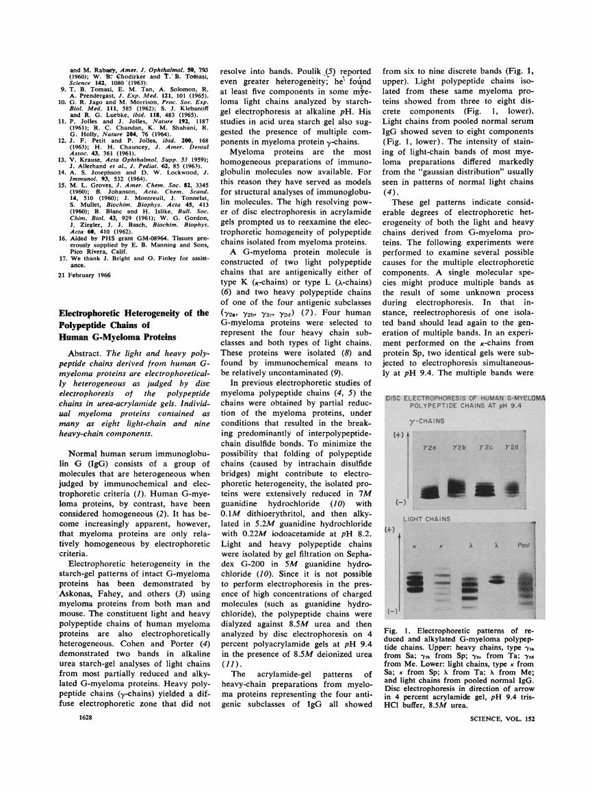
from six to nine discrete bands (Fig. 1, upper). Light polypeptide chains iso- lated from these same myeloma pro- teins showed from three to eight dis- crete components (Fig. 1, lower). Light chains from pooled normal serum IgG showed seven to eight components (Fig. 1, lower). The intensity of stain-
ing of light-chain bands of most mye- loma preparations differed markedly from the "gaussian distribution" usually seen in patterns of normal light chains
(4). These gel patterns indicate consid-
erable degrees of electrophoretic het-
erogeneity of both the light and heavy chains derived from G-myeloma pro- teins. The following experiments were
performed to examine several possible causes for the multiple electrophoretic components. A single molecular spe- cies might produce multiple bands as the result of some unknown process during electrophoresis. In that in-
stance, reelectrophoresis of one isola- ted band should lead again to the gen- eration of multiple bands. In an experi- ment performed on the K-chains from
protein Sp, two identical gels were sub-
jected to electrophoresis simultaneous-
ly at pH 9.4. The multiple bands were
DISC ELECTROPHORESIS OF HUMAN G-MYELOMA POLYPEPTIDE CHAINS AT pH 9.4
y-CHAINS
+)t +)t
(-)I (-)I
Fig. 1. Electrophoretic patterns of re- duced and alkylated G-myeloma polypep- tide chains. Upper: heavy chains, type 72a from Sa; 72b from Sp; Y2c from Ta; 7Y2
from Me. Lower: light chains, type K from Sa; K from Sp; X from Ta; X from Me; and light chains from pooled normal IgG. Disc electrophoresis in direction of arrow in 4 percent acrylamide gel, pH 9.4 tris- HC1 buffer, 8.5M urea.
SCIENCE, VOL. 152
- ) - )