Embed Size (px)
DESCRIPTION
This paper presents a microfluidic method for precisecontrol of the size and polydispersity of surfactant−DNA nanoparticles. Amixture of surfactant and DNA dispersed in 35% ethanol is focused betweentwo streams of pure water in a microfluidic channel. As a result, a rapidchange of solvent quality takes place in the central stream, and thesurfactant-bound DNA molecules undergo a fast coil−globule transition. Byadjusting the concentrations of DNA and surfactant, fine-tuning of thenanoparticle size, down to a hydrodynamic diameter of 70 nm with apolydispersity index below 0.2, can be achieved with a good reproducibility.
Citation preview
On-Chip Controlled Surfactant−DNA Coil−Globule Transition byRapid Solvent Exchange Using Hydrodynamic Flow FocusingCiprian Iliescu,*,† Catalin Marculescu,‡ Shrinivas Venkataraman,† Baptiste Languille,§ Hanry Yu,†,∥,⊥,#
and Guillaume Tresset*,§
†Institute of Bioengineering and Nanotechnology, 31 Biopolis Way, The Nanos #04-01, Singapore 138669, Singapore‡National Institute for Research and Development in Microtechnologies, IMT-Bucharest, Bucharest 077190, Romania§Laboratoire de Physique des Solides, Universite Paris-Sud, CNRS, 91400 Orsay, France∥Department of Physiology, Yong Loo Lin School of Medicine, National University of Singapore, Singapore 119077, Singapore⊥Singapore-MIT Alliance for Research and Technology, 65 Nanyang Dr, Singapore 637460, Singapore#Mechanobiology Institute, National University of Singapore, Singapore 119077, Singapore
ABSTRACT: This paper presents a microfluidic method for precisecontrol of the size and polydispersity of surfactant−DNA nanoparticles. Amixture of surfactant and DNA dispersed in 35% ethanol is focused betweentwo streams of pure water in a microfluidic channel. As a result, a rapidchange of solvent quality takes place in the central stream, and thesurfactant-bound DNA molecules undergo a fast coil−globule transition. Byadjusting the concentrations of DNA and surfactant, fine-tuning of thenanoparticle size, down to a hydrodynamic diameter of 70 nm with apolydispersity index below 0.2, can be achieved with a good reproducibility.
■ INTRODUCTIONRecent developments in molecular biology and genomicresearch have given a detailed characterization of geneticdiseases such as Alzheimer’s, Parkinson’s, and Huntington’s.Gene therapy, the transfection of therapeutic genes into cells,offers the opportunity to treat, prevent, and control suchdiseases. Despite the ethical debate regarding the use of genetherapy in medical practice,1 the main challenges in genetherapy are related to cell targeting, transfection efficiency, geneexpression regulation, and safety issues related to gene carriers.2
Current research on gene delivery vector development focuseson two major directions: viral and synthetic vectors. The use ofviral vectors have recorded some successful clinical trials,3,4 butside effects such as immunogenic response5 or vector-inducedcancer6 have limited so far the development of this method.Nonviral vectors such as lipid-based containers,7 inorganicnanoparticles,8 and polymer-based carriers9,10 confer advan-tages in terms of simplicity of use, structural and chemicalflexibility in exerting control over physical and chemicalproperties, feasibility of large-scale production, lack of specificimmune response, and a larger gene-loading capacity.1,9 The“synthetic solutions” must protect the DNA from intracellularand extracellular degradation,10,11 neutralize the negativecharges of DNA, avoid the interactions with cell surface,12,13
and have an appropriate size for transfection. Large vector sizesfavor in vitro tests due to their sedimentation to the bottom ofthe cell culture dish and at the same time exhibit a high
transfection efficiency; however, in vivo, their effect is irrelevantbecause either they are easily captured and degraded by theimmune system or they cannot diffuse efficiently throughtissues toward target cells.14 It was demonstrated for examplethat particles with hydrodynamic radii over 200 nm typicallyexhibit a more rapid rate of blood clearance than particles withradii under 200 nm and are mostly sequestrated in the spleenand liver.15 Likewise, a low value of the polydispersity index(PDI) of the nanocarriers can play an important role inachieving good transfection efficiencies because the entirety of amonodisperse population of nanocarriers can potentially reachtarget cells, whereas the largest species of a polydispersepopulation are blocked by the host body thus lowering theoverall transfection efficiency. In addition, the size distributionof polydisperse populations can be distorted from batch tobatch which leads to a poor repeatability of the results.Three main strategies are currently used in nonviral gene
delivery: encapsulation,16 adsorption,17 and condensation.18,19
The encapsulation method consists of trapping DNA moleculesinside biodegradable spherical micro- or nanoparticles. Themethod involves high shear stress or relatively high temperaturethat can degrade the DNA.8,20 The adsorption method relies onelectrostatic binding of DNA molecules on biodegradable orinorganic particles; in these conditions DNA is exposed to
Received: April 11, 2014
Article
pubs.acs.org/Langmuir
© XXXX American Chemical Society A dx.doi.org/10.1021/la5035382 | Langmuir XXXX, XXX, XXX−XXX
enzymatic degradation.8,21 The condensation of anionic DNAby a cationic surfactant leads to a partial collapse of the DNAchains.22 In general, the size of the resulting particle is poorlycontrolled because the assembly process is driven by kineticsand the system quickly becomes trapped into a metastable state.Microfluidic platforms can play an important role in the
development of gene therapy and nanomedicine.23,24 Theseplatforms can be used for both screening nanocarriers libraries(through in vitro systems mimicking in vivo conditions)25,26
and/or for the synthesis of nanocarriers with an excellentcontrol on their size distribution, composition and morphol-ogy.27 Recent works are related to the synthesis of multilayeredpolyelectrolyte nanocarriers as reported by Qi et al.28 orassembly of lipid nanoparticles by rapid mixing in serpentinemicrochannels.29 Kim et al. reported the processing of lipid−polymer hybrid nanoparticles (in the range 30-170 nm) using apattern-tunable microvortex platform.30 In a recent work, thesame group proposed a microfluidic “factory” for single stepsynthesis of high-density lipoprotein with opportunity forencapsulation of hydrophobic drugs as well as quantum dots forbioimaging applications.31 Other important achievements arerelated to liposome-hydrogel hybrid nanoparticles,32 formationof lipid vesicles,33,34 cross-linked alginate nanoparticles,35
chitosan-based nanoparticles,36,37 and diblock copolymersnanoparticles.38 The most simple microfluidic method forachieving a precise control of the nanoparticle synthesis usingdiffusion-mixing process of miscible liquids is hydrodynamicflow focusing.39,40 In our previous work,41 we used hydro-dynamic flow focusing in order to get a controlled mixingkinetics and a uniform distribution of charges at the mixinginterface between polyelectrolyte (sodium carboxymethylcellu-lose; carboxyMC) and surfactant (dodecyl trimethylammoniumbromide; DTAB) streams. The results showed a well-controlledand tunable nanoparticle size with a small polydispersity. Acondition for an effective control is that the mixing time τmix beshorter than the adsorption time τad over which the surfactant−polyelectrolyte association takes place. In other words,surfactant and polyelectrolyte must be dispersed homoge-neously in the solution before they bind to each other.Unfortunately, for the compaction of DNA by surfactant, largesizes of nanoparticles with broad distributions were achieved,similar to what is obtained by mixing in bulk. These poorresults may be due to an adsorption time τad of surfactants ontoDNA shorter than onto carboxyMC used previously, whereasτmix is limited by the diffusion of surfactants in both cases and isthereby unchanged. Indeed, the linear charge density of DNA ismore than twice as high (5.9 e‑/nm) as that of carboxyMC (2.5e‑/nm), which, in the same ionic conditions, lowers significantlyτad. Consequently, if the mixing time τmix required tohomogenize DNA and surfactants is not reduced below theiradsorption time τad, the association process gives rise to largenanoparticles with uncontrolled size distribution. The mecha-nism is reminiscent of the self-assembly of block copolymernanoparticles by rapid change of solvent quality: when τmix isbelow τagg, the nanoparticle size remains unchanged at aminimum value, whereas above τagg, the size increases as apower law of τmix.
42
Here, we report the use of microfluidic hydrodynamic flowfocusing to enable an accurate control over the size distributionof surfactant−DNA nanoparticles by a rapid change of solventquality. Indeed, rather than trying to further reduce the mixingtime between DNA and surfactant, which is quite challengingwith large molecules, we opted for a solvent exchange. It should
be noted here that we were primarily interested in long DNAchains, i.e. ∼2000 base pairs which is the typical size of a gene.The compaction of short oligonucleotides such as silencingRNA (∼20 base pairs) into small monodisperse nanoparticles iseasier and does not require a sophisticated mixing strategy. Thechosen surfactant was again DTAB because it is a chemicallysimple, well-established, and thoroughly documented cationicsurfactant model. Upon bulk mixing, DNA collapses in anoncontrolled manner with local excess concentrations yieldinglarge aggregates non suitable for efficient gene deliveryapplications. In 35% ethanol, however, DNA, and nucleicacids in general, as well as surfactant are soluble, and surfactantmolecules are weakly bound to DNA chains through electro-static interactions but do not form compact globules.43 Giventhe small sizes of solvent molecules and their subsequent largediffusion coefficients (∼10−9 m2 s−1), the solvent quality can bechanged rapidly in such a way that the surfactant-bound DNAchains collapse due to the poor solubility of the hydrocarbonchains of surfactants without having enough time to aggregatewith each other by diffusion; they thereby form nearlymonomolecular DNA-based nanoparticles. Compared toprevious studies reporting monomolecular DNA-based nano-particles,14 the microfluidics approach avoids resorting tochemical additives or harsh formulation processes. Besides, itoffers a greater versatility than bulk synthesis, and it will enableus to finely tune the sequential assembly of multicomponentsoft nanoparticles.
■ EXPERIMENTAL SECTIONReagents. Dodecyl trimethylammonium bromide (DTAB) and
cetyltrimethylammonium bromide (CTAB) were purchased fromSigma-Aldrich with a purity >99%. DTAB and CTAB were dissolvedin Millipore Milli-Q deionized water (conductivity <10−6 S m−1) at astock concentration of 20 mM. The critical micelle concentration forDTAB in pure water is 15 mM and therefore micelles could not beformed in any of our experiments involving DTAB. The critical micelleconcentration for CTAB in pure water is 1 mM. Calf thymus DNA andλ-DNA were purchased from Invitrogen. Unless otherwise stated inthe text, calf thymus DNA was always used for the experiments. Themixing of surfactant, DNA and 35% ethanol at the desiredconcentration was performed within 5-10 min before each experiment.Bulk mixing was performed under agitation with a magnetic stirrer.
Dynamic Light Scattering Measurements. The size andpolydispersity of surfactant−DNA nanoparticles were measured bydynamic light scattering (DLS) using a Zetasizer model ZS-90(Malvern Instrument, Ltd., UK). Data were collected at a back-scattering angle of 173° with temperature maintained at 25 oC.Because the final solutions of nanoparticles contained less than 5%ethanol, we set the refractive index to that of pure water at 25 °C. Sizeanalysis was carried out by the cumulants method and thepolydispersity index (PDI) was estimated accordingly. It should benoted that the size distributions returned by the Contin method alwaysexhibited a single peak, at least for the experiments with DTAB. Foreach sample, three measurements were performed, each measurementbeing an average of ten values. The standard deviation of thehydrodynamic diameters was calculated for these three values.
Transmission Electron Microscopy (TEM). The morphologiesof surfactant−DNA complexes were observed under a FEI Tecnai G2F20 electron microscope using an acceleration voltage of 200 keV. TheTEM samples were prepared by first placing a drop of surfactant−DNA complexes (4.0 μL) onto a formvar-coated 200 mesh copper grid(Ted Pella Inc., USA). After 1 min, the excess solution was wicked offby using filter paper. Then the staining agent, phosphotungstic acidsolution (2% w/v in deionized water; 4.0 μL), was placed on the grid,and after 1 min, the excess solution was wicked off and the grid was leftto dry under the ambient conditions.
Langmuir Article
dx.doi.org/10.1021/la5035382 | Langmuir XXXX, XXX, XXX−XXXB
Fabrication of the Microfluidic Device. The microfluidicchannel (40-μm-depth) was fabricated in silicon using classical DeepRIE Bosch process through a 5-μm-thick photoresist, while the etch-through holes were performed using same method through aphotoresist/SiO2 mask. A 150 nm-thick dry-oxide layer was grownin a furnace on the silicon surface in order to achieve a permanenthydrophilic surface of the microfluidic channel. The silicon wafer wasanodically bonded to a glass wafer for the sealing of the microfluidicchannels. Finally the bonded wafers were diced in individual chips andNanoport microfluidic connectors (Upchurch Scientific) weremounted for the inlets and outlet. Detailed considerations regardingfabrication process are presented in our previous work.41
Microfluidic Experimental Setup. A two-channel pumpingsystem MFCS-FLEX (Fluigent, France) was used for controlling theflow of reactants through the microfluidic chip. The system wasequipped with three pumping units and three flow-meters (0-50 μL/min), only two were used for the current experiment, and a softwarethat allowed a fine-tuning of the flow rates (by varying the appliedpressure) in the microfluidic device. The chip was mounted on a chipholder, and an optical microscope (Keydance) allowed the observationof the flow in the microfluidic device.Computer Simulations. The numerical three-dimensional (3D)
geometry and the structured orthogonal mesh were generated usingGAMBIT preprocessor (ANSYS), consisting of 1 188 000 hexahedralfinite volumes. The dimensions of the reconstructed microfluidicchannel were carefully selected to capture the diffusion process with anacceptable computational cost. Therefore, the length of the inletchannels was 300 μm, the length of the outlet channel was 3000 μm,and the cross sections were 60 × 40 μm in all cases. The 3D pressure-driven flow in the microchannel configuration was simulated using the
FLUENT CFD package (ANSYS). The code computed the isothermallaminar flows of the Newtonian working fluids, with double precisionand with a 10−10 convergence criterion. The FLUENT code solves theCauchy equation of motion in which the extra-stress tensor isexpressed as a generalized Newtonian model:
ρ ρ η γ∂∂
+ ∇ = − ∇ + ∇ ⎡⎣⎢
⎤⎦⎥t
pu
u u b E( ) 2 ( ( ) )(1)
where ρ is the fluid density, b the specific mass force, E the strain ratetensor, t the time, u the velocity vector, p the pressure, and η theviscosity function dependent on the shear rate γ. All the quantitiesentering eq 1 varied with the position r and the time t. Along with themomentum equation, the diffusion equation was also solved in orderto obtain an accurate solution
ρ ρ ρ∂∂
= ∇ ∇tt
D tr
r( , )
[ ( ) ( , )](2)
where D(ρ) is the collective diffusion coefficient depending on thelocal fluid density ρ.
The numerical simulations were performed using a Volume of Fluidmethod with 35% ethanol in aqueous solution (ρ = 930 kg/m3 and η =0.0011 Pa s) injected on the central stream and water (ρ = 998.2 kg/m3 and η = 0.001003 Pa s) on the focusing streams. The flow rate ratioused was 5/45, considering the central streamflow rate and the sum ofthe focusing streams flow rates, respectively. The Reynolds numbersassociated with each stream were ∼1.5 and ∼8.5 respectively. By usinga user defined scalar (UDS), specific to the FLUENT package, wewere able to activate given fluid properties such as diffusioncoefficients. The UDS allowed us to customize the transport equation
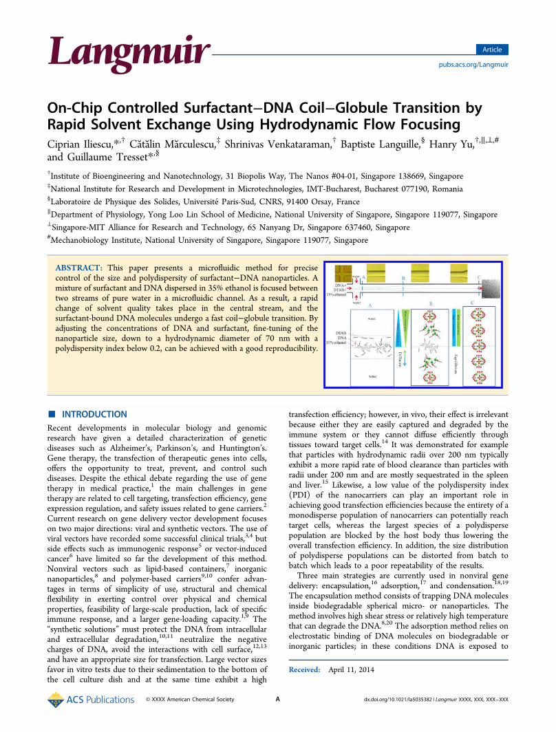
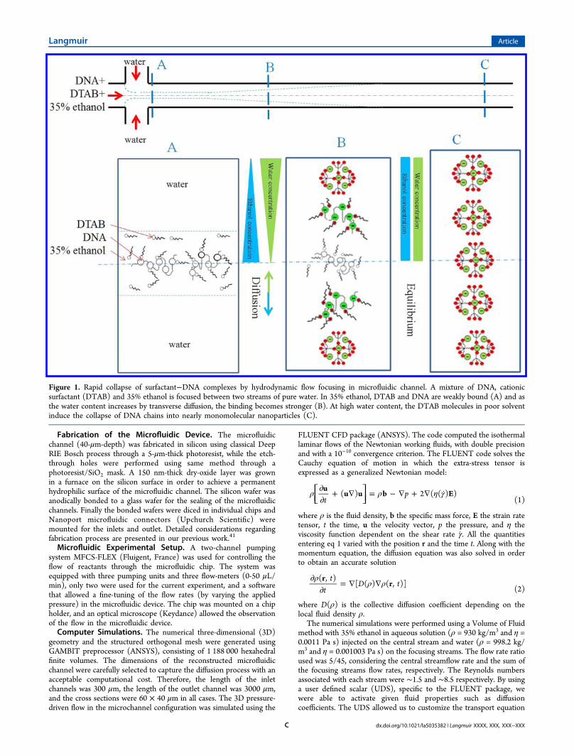
Figure 1. Rapid collapse of surfactant−DNA complexes by hydrodynamic flow focusing in microfluidic channel. A mixture of DNA, cationicsurfactant (DTAB) and 35% ethanol is focused between two streams of pure water. In 35% ethanol, DTAB and DNA are weakly bound (A) and asthe water content increases by transverse diffusion, the binding becomes stronger (B). At high water content, the DTAB molecules in poor solventinduce the collapse of DNA chains into nearly monomolecular nanoparticles (C).
Langmuir Article
dx.doi.org/10.1021/la5035382 | Langmuir XXXX, XXX, XXX−XXXC
by defining solution zones, the flux function, the unsteady function andother parameters so as to obtain an accurate simulation of the diffusionprocess. The value for the diffusion coefficient of ethanol in water was0.625 × 10−9 m2/s.44 No chemical reaction was taken into account andthe ethanol was therefore assumed to be a passive tracer in water.
■ RESULTS AND DISCUSIONWorking Principle. The compaction mechanism of DNA
by cationic surfactant in aqueous solutions is driven by twointeractions that take place almost simultaneously. Theelectrostatic interaction between the anionic backbone ofDNA and the cationic headgroup of surfactant is supplementedby a hydrophobic interaction between the alkyl chains of thesurfactant molecules.45 As a result, the surfactant-bound DNAchains collapse (a process referred to as coil−globuletransition) to form nano- or micrometer-size particles. Thesize of the particles is strongly dependent on the number of theDNA molecules trapped in a particle during the condensationprocess. Also, the presence of salt in solution favors theaggregation of particles during the course of their assembly byscreening the repulsive electrostatic interactions and it generallygives rise to large particle sizes. In the present work, we proposeto split the compaction process into two distinctive steps: onein bulk for the electrostatic association between DNA andsurfactant and the other one on chip for the collapse ofsurfactant-bound DNA chains driven by the hydrophobicity ofthe surfactant alkyl chains in a solvent with high water content.If the solution used for solubilizing DNA and surfactantcontains a high enough fraction of organic solvent such asethanol, the alkyl chains of surfactant molecules are in a good
solvent quality and remain dissociated so that the surfactant−DNA complexes are in a coil conformation.43 Note that thefraction of ethanol must be below 50% otherwise DNAprecipitates. At this stage, there is only electrostatic interactionbetween the cationic headgroup of surfactant and the anionicbackbone of DNA. This step can be easily achieved in bulk bymixing DNA and surfactant in a 35% ethanol solution. In thesecond step, a simple change of solvent quality through waterdilution enhances the hydrophobic interactions between thealkyl chains of the surfactant molecules leading to a compactionof the surfactant−DNA complexes. Classical bulk dilutionresults in a poorly controlled DNA compaction, mainly due to alack of homogeneity of molecules and charges throughout thesolution yielding large aggregates and/or high polydispersitynot suitable for efficient delivery applications. Forcing DNAand surfactant at high velocity into a turbulent vessel isprohibited because the high shear rates generated locally in thesolution will break apart the DNA chains. A gentle method forachieving a rapid change of solvent quality involves hydro-dynamic flow focusing in a microfluidic channel. When a centralstream of good quality solvent is flown through a microfluidicchannel and sandwiched between two side streams of purewater, its width is strongly reduced due to a focusing effect,which is possible only because of the low Reynolds number ofthe liquids and their subsequent laminar flow.46,47 The purewater diffuses transversely through the central stream (and viceversa) in a mixing time τmix all the shorter as its width isreduced and changes accordingly the solvent quality. Thissecond step of the process developed on-chip is illustrated in
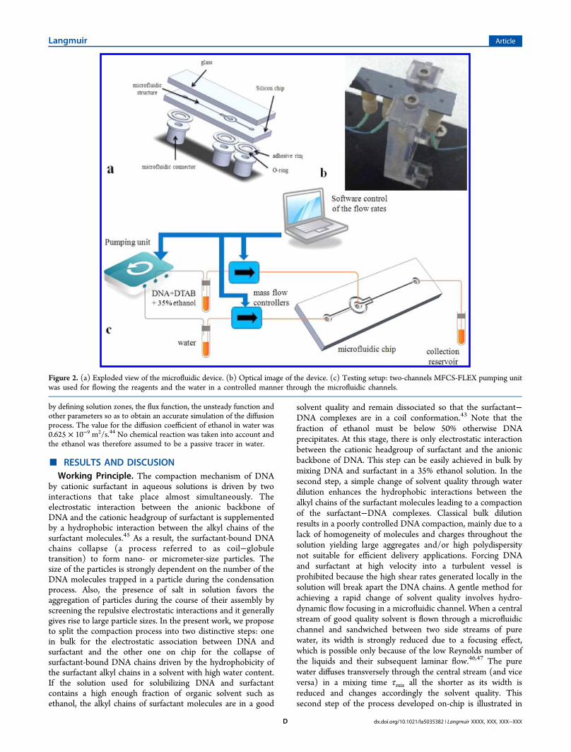
Figure 2. (a) Exploded view of the microfluidic device. (b) Optical image of the device. (c) Testing setup: two-channels MFCS-FLEX pumping unitwas used for flowing the reagents and the water in a controlled manner through the microfluidic channels.
Langmuir Article
dx.doi.org/10.1021/la5035382 | Langmuir XXXX, XXX, XXX−XXXD
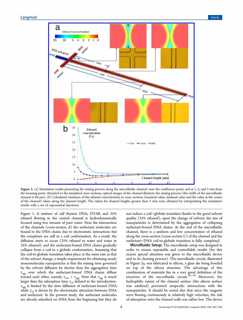
Figure 1. A mixture of calf thymus DNA, DTAB, and 35%ethanol flowing in the central channel is hydrodynamicallyfocused using two streams of pure water. Near the intersectionof the channels (cross-section A) the surfactant molecules arebound to the DNA chains due to electrostatic interactions butthe complexes are still in a coil conformation. As a result, thediffusion starts to occur (35% ethanol in water and water in35% ethanol), and the surfactant-bound DNA chains graduallycollapse from a coil to a globule conformation. Assuming thatthe coil-to-globule transition takes place at the same rate as thatof the solvent change, a simple requirement for obtaining nearlymonomolecular nanoparticles is that the mixing time governedby the solvent diffusion be shorter than the aggregation timeτagg over which the surfactant-bound DNA chains diffusetoward each other, namely τmix < τagg. Note that τagg is muchlarger than the adsorption time τad defined in the introduction:τagg is limited by the slow diffusion of surfactant-bound DNAwhile τad is driven by the electrostatic attraction between DNAand surfactant. In the present study, the surfactant moleculesare already adsorbed on DNA from the beginning but they do
not induce a coil−globule transition thanks to the good solventquality (35% ethanol); upon the change of solvent, the size ofnanoparticles is determined by the aggregation of collapsingsurfactant-bound DNA chains. At the end of the microfluidicchannel, there is a uniform and low concentration of ethanolalong the cross-section (cross-section C) of the channel and thesurfactant−DNA coil-to-globule transition is fully completed.
Microfluidic Setup. The microfluidic setup was designed inorder to ensure repeatable and controllable results (for thisreason special attention was given to the microfluidic deviceand to its cleaning process). The microfluidic circuit, illustratedin Figure 2a, was fabricated in silicon, a glass die being bondedon top of the silicon structure. The advantage of thiscombination of materials lies in a very good definition of thestructure of the microfluidic circuit.48−50 Moreover, thehydrophilic nature of the channel surface (the silicon surfacewas oxidized) prevented unspecific interactions with thenanoparticles. It should be noted also that since the reagentswere flowing continuously at relatively high velocities, the riskof adsorption onto the channel walls was rather low. The device
Figure 3. (a) Simulation results presenting the mixing process along the microfluidic channel: near the confluence point, and at 1, 2, and 3 mm fromthe focusing point. Attached to the simulated cross sections, optical images of the channel illustrate the mixing process (the width of the microfluidicchannel is 60 μm). (b) Calculated variations of the ethanol concentration in cross sections (maximal value, minimal value and the value at the centerof the channel) taken along the channel length. The values for channel lengths greater than 3 mm were obtained by extrapolating the simulationresults with a set of exponential functions.
Langmuir Article
dx.doi.org/10.1021/la5035382 | Langmuir XXXX, XXX, XXX−XXXE
could be easily cleaned after each experiment by flowing NMP(N-methylpyrrolidone) for 15 seconds through the centralinlet. This operation was performed without any modificationof the testing setup, just by simple replacement of the reservoirconnected to the main channel. A buffer solution (35%ethanol) was flown (for 1 min) after NMP in order to avoid anycontamination of the surfactant−DNA sample. This point iscrucial not only for the accuracy of the experiments (all theexperiments were performed on the same chip and in the sameflowing conditions) but also for the scalability of thenanoparticle synthesis: the device can be reused and anautomatic cleaning process can be setup after a certainprocessing time. The microfluidic chip and its fabricationprocess were completely described in our previous work,41 forthis reason we will underline here only the main characteristics.The device presented two inlets: the central one for theDTAB/DNA/35% ethanol solution, and the other one, thatassured the hydrodynamic focusing of the previouslymentioned reagents, containing pure water. The water channelwas divided into two symmetrical branches in order to ensure asymmetrical focusing and an identical pressure drop on thebranches. The depth of the microfluidic channel was 40 μm, thewidth was 60 μm, while its length was 1.5 cm.41 A photographof the microfluidic chip and its holder is presented in Figure 2b.The microfluidic setup is represented in Figure 2c, the pumpingunit and mass flow controllers were software-controlled andassured a fine-tuning of the flowing parameters in themicrofluidic device.Simulation Results. Numerical simulations illustrate with
accuracy the three-dimensional process of solvent homoge-nization while fluids are flowing through the channel. Theresults of the simulations as well as some optical images (topview) with the mixing process in microfluidic channel arepresented in Figure 3a. For the visualization of the mixingprocess, four cross sections through the microfluidic channelwere selected at fixed intervals. First point was selected veryclose to the focusing area (40 μm), and from the simulation avery strong gradient in ethanol concentration (0-35%) isobserved. Also the contrast on the optical image between thecentral stream (ethanol) and the lateral streams is evident.Once the diffusion process started the main stream became lessand less visible due to the progress in the mixing process. Themaximal calculated value of ethanol concentration alsodecreases (from 35%, the initial value, to 14% for a channellength of 1 mm, 7% for 2 mm, and 4.7% for 3 mm). Thegraphical variations of the maximal and minimal calculatedvalues of the ethanol concentration as well as the values in thecenter of the microfluidic channel are presented in Figure 3b.By extrapolating the numerical solution obtained over 3 mm ofchannel with a set of exponential functions, the curvesconverged to a uniform concentration (3.5% ethanol) at theend of a 15 mm-long microfluidic channel demonstrating thatthe mixing process was complete within the channel. Theextrapolated values of the ethanol concentration close to theoutlet were: maximal 3.7%, minimal 3.41%, center point 3.56%.Analytical Considerations. Let QA and QB be the flow
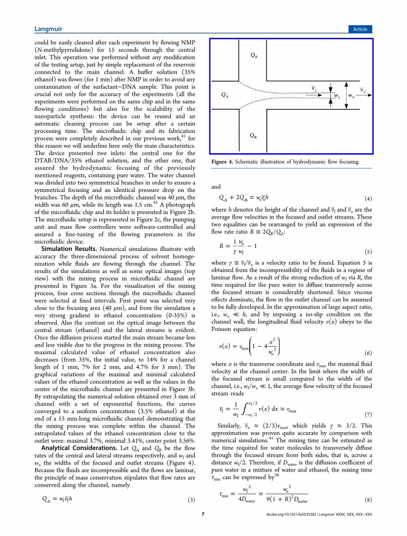
rates of the central and lateral streams respectively, and wf andwo the widths of the focused and outlet streams (Figure 4).Because the fluids are incompressible and the flows are laminar,the principle of mass conservation stipulates that flow rates areconserved along the channel, namely
= Q w v hA f f (3)
and
+ = Q Q w v h2A B o o (4)
where h denotes the height of the channel and vf and vo are theaverage flow velocities in the focused and outlet streams. Thesetwo equalities can be rearranged to yield an expression of theflow rate ratio R ≡ 2QB/QA:
γ= −R
ww
11o
f (5)
where γ ≡ vf/vo is a velocity ratio to be found. Equation 5 isobtained from the incompressibility of the fluids in a regime oflaminar flow. As a result of the strong reduction of wf via R, thetime required for the pure water to diffuse transversely acrossthe focused stream is considerably shortened. Since viscouseffects dominate, the flow in the outlet channel can be assumedto be fully developed. In the approximation of large aspect ratio,i.e., wo ≪ h, and by imposing a no-slip condition on thechannel wall, the longitudinal fluid velocity v(x) obeys to thePoisson equation:
= −⎛⎝⎜
⎞⎠⎟v x v
xw
( ) 1 4max
2
o2
(6)
where x is the transverse coordinate and vmax the maximal fluidvelocity at the channel center. In the limit where the width ofthe focused stream is small compared to the width of thechannel, i.e., wf/wo ≪ 1, the average flow velocity of the focusedstream reads
∫ = ≈−
vw
v x x v1
( ) dw
w
ff /2
/2
maxf
f
(7)
Similarly, vo ≈ (2/3)vmax, which yields γ ≈ 3/2. Thisapproximation was proven quite accurate by comparison withnumerical simulations.41 The mixing time can be estimated asthe time required for water molecules to transversely diffusethrough the focused stream from both sides, that is, across adistance wf/2. Therefore, if Dwater is the diffusion coefficient ofpure water in a mixture of water and ethanol, the mixing timeτmix can be expressed by38
τ = ≈+
wD
wR D4 9(1 )mix
f2
water
o2
2water (8)
Figure 4. Schematic illustration of hydrodynamic flow focusing.
Langmuir Article
dx.doi.org/10.1021/la5035382 | Langmuir XXXX, XXX, XXX−XXXF
With Dwater ≈ 10−9 m2 s−1, ωo = 60 μm, and R = 9, the mixingtime is 4 ms. This value must be compared to the aggregationtime τagg over which two surfactant-bound DNA chainsencounter each other by diffusion. As a rule of thumb, τaggcan be estimated from the diffusion-limited rate constantbetween two uncharged surfactant-bound DNA chains suchthat
τ πρ≈− DR16agg1
h (9)
with ρ the number density of chains which is of the order of1019 m−3 for a concentration around 20 mg/L, D their diffusioncoefficient, and Rh their hydrodynamic radius. Notice that theaggregation time given with this value of ρ is a lower boundbecause the chains also diffuse transversely as they travel alongthe channel and ρ thereby decreases by dilution. Following theStokes-Einstein relationship, DRh = kBT/6πη, with kB theBoltzmann constant, T the temperature, and η the viscosity ofthe solvent which is ∼2.3 mPa s for 35% ethanol at 25 °C. Thisyields τagg ≈ 21 ms and we are indeed in the regime where thesolvent exchange occurs faster than the aggregation ofsurfactant-bound DNA chains, i.e., τmix < τagg.Parameters Effect on Size and Polydispersity of
Surfactant−DNA Nanoparticles. Influence of Calf ThymusDNA Concentration. The influence of DNA concentration was
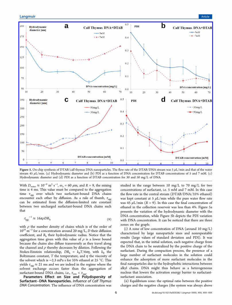
studied in the range between 10 mg/L to 70 mg/L for twoconcentrations of surfactant, i.e. 5 mM and 7 mM. In this casethe flow rate in the central stream (DTAB/DNA/35% ethanol)was kept constant at 5 μL/min while the pure water flow ratewas 45 μL/min (R = 9). In this case the final concentration ofethanol in the collection reservoir was less than 4%. Figure 5apresents the variation of the hydrodynamic diameter with theDNA concentration, while Figure 5b depicts the PDI variationwith DNA concentration. It can be noticed that there are threezones on the graph:(i) A zone of low concentration of DNA (around 10 mg/L)
characterized by large nanoparticle sizes and nonrepeatableresults (large values of standard deviation and PDI). It wasexpected that, in the initial solution, each negative charge fromthe DNA chain to be neutralized by the positive charge of thesurfactant. During the compaction process, the presence of alarge number of surfactant molecules in the solution couldenhance the adsorption of more surfactant molecules in thefinal nanoparticles due to the hydrophobic interactions betweenalkyl chains. DNA might thus behave as a heterogeneousnucleus that lowers the activation energy barrier to surfactant-surfactant association.(ii) Equilibrium zone: the optimal ratio between the positive
charges and the negative charges (the system was always above
Figure 5. On-chip synthesis of DTAB/calf thymus DNA nanoparticles. The flow rate of the DTAB/DNA stream was 5 μL/min and that of the waterstream 45 μL/min. (a) Hydrodynamic diameter and (b) PDI as a function of DNA concentration for DTAB concentrations of 5 and 7 mM. (c)Hydrodynamic diameter and (d) PDI as a function of DTAB concentration for 30 and 50 mg/L of DNA.
Langmuir Article
dx.doi.org/10.1021/la5035382 | Langmuir XXXX, XXX, XXX−XXXG
the isoelectric point because the surfactants carrying positivecharges were in excess. Note that for 5 mM DTAB, 1.65 g/L ofDNA were necessary to achieve neutralization). It is interestingto observe that the hydrodynamic diameter of the particles withDNA concentration did not vary much for DNA concentrationsbetween 20 mg/L up to 60 mg/L. For the concentration ofsurfactant of 5 mM, it could be noticed an optimum point inDNA concentration (50 mg/L) where the hydrodynamicdiameter of the nanoparticles was below 70 nm.(iii) High concentration of DNA: the size of the nano-
particles started to slowly increase. The increase of thehydrodynamic diameter in this zone can be explained by thepresence of multiple DNA chains inside a single nanoparticle.According to eq 9, τagg ∝ ρ−1; therefore, a higher DNAconcentration lowers the aggregation time. We then arrived atan aggregation time comparable to or smaller than the mixingtime (τmix ≈ τagg) and the surfactant-bound DNA chains couldaggregate during the solvent exchange. It is important to noticethat the PDI decreased with the increase of DNAconcentration. A value of DNA concentration between 30 to70 mg/L is then recommended for achieving uniform and well-controlled surfactant−DNA nanoparticles.Influence of Surfactant (DTAB) Concentration. The
variation of the hydrodynamic diameter of the nanoparticleswith the surfactant concentration was studied for DTABconcentrations between 3 mM and 10 mM, for two values ofDNA concentration: 30 and 50 mg/L. Figure 5c,d illustrates theinfluence of DNA concentration on the hydrodynamic diameterof the nanoparticles and PDI, respectively. The graph presentedin Figure 5c shows the same trend as the graph representing theinfluence of DNA concentration. Both curves present aminimum value. The low DNA concentration zone from thegraph presented in Figure 5a has a correspondence in Figure 5cin the area with high surfactant concentration, while the areawith high concentration of DNA from Figure 5a is similar tothe area with low surfactant concentration. This aspect wassomehow expected, the ratio between positive and negativecharges playing an important role in the compaction of DNA. Itmust be mentioned that 3 mM was a limit for the concentrationof the surfactant (the results presented a large value of PDI),also for large values of DTAB concentration (9 mM and 10mM), the size of the nanoparticles started to increase as well asPDI. We can conclude that values of DTAB concentration of 5mM up to 8 mM are the most recommended for tuning the sizeof nanoparticles while keeping a reasonable polydispersity (PDIbelow 0.2).Influence of Flow Rate Ratio. For the influence of the flow
rate ratio on the hydrodynamic diameter and polydispersity, wecompared the nanoparticle sizes obtained by on-chip and bulkmixing. To do so, we used 50 mg/L of calf thymus DNA and6.5 mM of DTAB in 35% ethanol. The flow rate of pure waterwas controlled between 40 and 45 μL/min while the flow rateof the DTAB/DNA/35% ethanol solution was varied between1 and 10 μL/min. The results are presented in Table 1.Strikingly, the nanoparticles were much larger and morepolydisperse when prepared by bulk mixing than by on-chipmixing for the same final concentrations of DNA and DTAB.The batch-to-batch reproducibility was also less good. Besides,it can be noticed that there was no significant influence of theflow rate ratio when R ≥ 7.5. The change of solvent qualitytook place over a time τmix smaller than the aggregation time,i.e., τmix < τagg whatever the flow rate ratio: τmix was estimated tobe 5.5 and 0.189 ms for R = 7.5 and 45, respectively, while τagg
was around 9 ms for the DNA concentration considered here.As such, the kinetics of nanoparticle formation was governedsolely by the collapse of surfactant-bound DNA chains.Therefore, the hydrodynamic diameter of the resultingnanoparticles was only a function of the concentrations ofDTAB and DNA and hardly depended on the flowingcondition. This finding is opposite to the results from ourprevious work41 where the flowing conditions played animportant role on the hydrodynamic diameter of thenanoparticles. Indeed, the mixing time was limited by thediffusion of reagents, surfactant and polyelectrolyte, whereas inthe present study, it relied on the diffusion coefficient of thesolvent which was one or two orders of magnitude higher thanthat of the reagents due to the small size of the solventmolecules. In our previous work, τmix was thus greater orcomparable to τad and as a consequence, the final nanoparticleswere dependent on the mixing kinetics, i.e., on the flow rateratio between the streams carrying both reagents. Such asituation was observed in the present study when R ≤ 5 (seeTable 1): for example, at R = 5, τmixwas estimated to be 11 ms,that is, slightly larger than an estimated τagg of 9 ms. As aconsequence, the nanoparticle size increased to 200 nm simplybecause the assembly started to depend on the mixing kinetics.
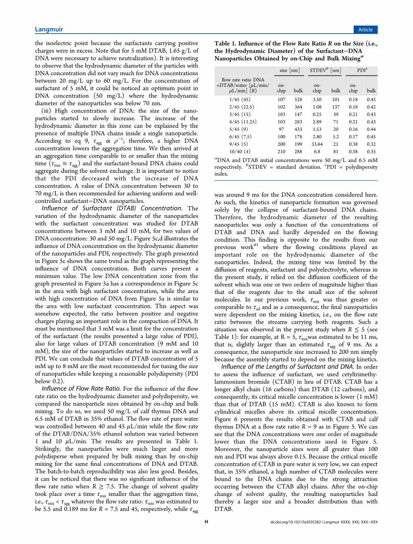
Influence of the Lengths of Surfactant and DNA. In orderto assess the influence of surfactant, we used cetyltrimethy-lammonium bromide (CTAB) in lieu of DTAB. CTAB has alonger alkyl chain (16 carbons) than DTAB (12 carbons), andconsequently, its critical micelle concentration is lower (1 mM)than that of DTAB (15 mM). CTAB is also known to formcylindrical micelles above its critical micelle concentration.Figure 6 presents the results obtained with CTAB and calfthymus DNA at a flow rate ratio R = 9 as in Figure 5. We cansee that the DNA concentrations were one order of magnitudelower than the DNA concentrations used in Figure 5.Moreover, the nanoparticle sizes were all greater than 100nm and PDI was always above 0.15. Because the critical micelleconcentration of CTAB in pure water is very low, we can expectthat, in 35% ethanol, a high number of CTAB molecules werebound to the DNA chains due to the strong attractionoccurring between the CTAB alkyl chains. After the on-chipchange of solvent quality, the resulting nanoparticles hadthereby a larger size and a broader distribution than withDTAB.
Table 1. Influence of the Flow Rate Ratio R on the Size (i.e.,the Hydrodynamic Diameter) of the Surfactant−DNANanoparticles Obtained by on-Chip and Bulk Mixinga
size [nm] STDEVb [nm] PDIc
flow rate ratio DNA+DTAB/water [μL/min/
μL/min] (R)on-chip bulk
on-chip bulk
on-chip bulk
1/45 (45) 107 528 3.50 101 0.18 0.452/45 (22.5) 102 364 1.08 137 0.18 0.423/45 (15) 103 147 0.25 39 0.21 0.434/45 (11.25) 103 283 2.89 71 0.21 0.435/45 (9) 97 433 1.53 20 0.16 0.446/45 (7.5) 100 178 2.80 5.2 0.17 0.459/45 (5) 200 199 33.64 21 0.38 0.3210/40 (4) 210 288 6.8 81 0.38 0.35
aDNA and DTAB initial concentrations were 50 mg/L and 6.5 mMrespectively. bSTDEV = standard deviation. cPDI = polydispersityindex.
Langmuir Article
dx.doi.org/10.1021/la5035382 | Langmuir XXXX, XXX, XXX−XXXH
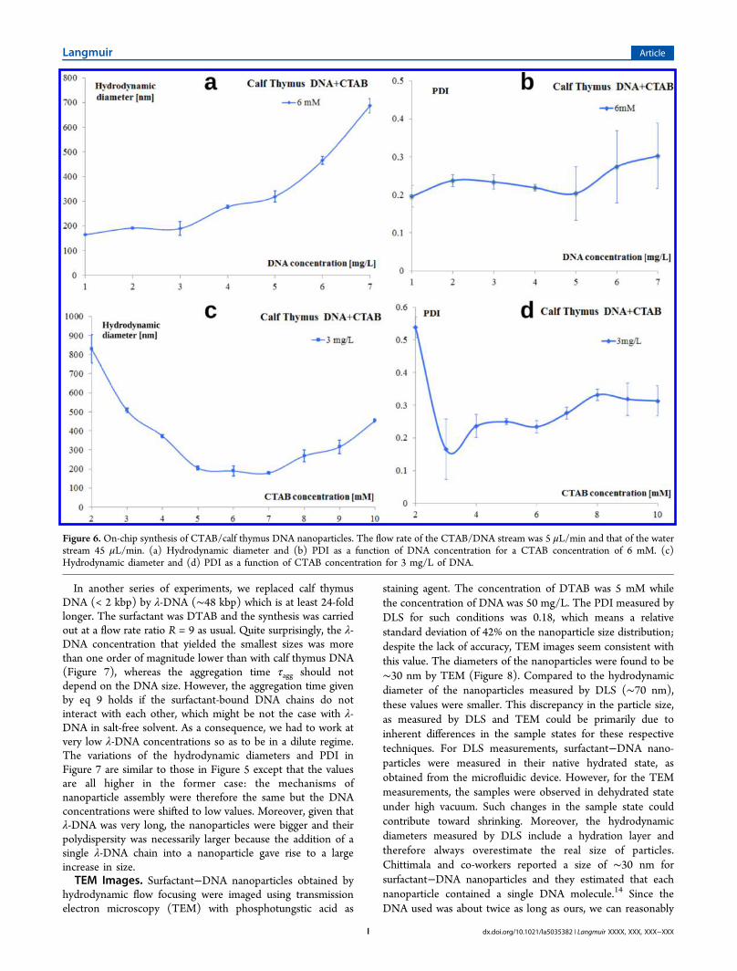
In another series of experiments, we replaced calf thymusDNA (< 2 kbp) by λ-DNA (∼48 kbp) which is at least 24-foldlonger. The surfactant was DTAB and the synthesis was carriedout at a flow rate ratio R = 9 as usual. Quite surprisingly, the λ-DNA concentration that yielded the smallest sizes was morethan one order of magnitude lower than with calf thymus DNA(Figure 7), whereas the aggregation time τagg should notdepend on the DNA size. However, the aggregation time givenby eq 9 holds if the surfactant-bound DNA chains do notinteract with each other, which might be not the case with λ-DNA in salt-free solvent. As a consequence, we had to work atvery low λ-DNA concentrations so as to be in a dilute regime.The variations of the hydrodynamic diameters and PDI inFigure 7 are similar to those in Figure 5 except that the valuesare all higher in the former case: the mechanisms ofnanoparticle assembly were therefore the same but the DNAconcentrations were shifted to low values. Moreover, given thatλ-DNA was very long, the nanoparticles were bigger and theirpolydispersity was necessarily larger because the addition of asingle λ-DNA chain into a nanoparticle gave rise to a largeincrease in size.TEM Images. Surfactant−DNA nanoparticles obtained by
hydrodynamic flow focusing were imaged using transmissionelectron microscopy (TEM) with phosphotungstic acid as
staining agent. The concentration of DTAB was 5 mM whilethe concentration of DNA was 50 mg/L. The PDI measured byDLS for such conditions was 0.18, which means a relativestandard deviation of 42% on the nanoparticle size distribution;despite the lack of accuracy, TEM images seem consistent withthis value. The diameters of the nanoparticles were found to be∼30 nm by TEM (Figure 8). Compared to the hydrodynamicdiameter of the nanoparticles measured by DLS (∼70 nm),these values were smaller. This discrepancy in the particle size,as measured by DLS and TEM could be primarily due toinherent differences in the sample states for these respectivetechniques. For DLS measurements, surfactant−DNA nano-particles were measured in their native hydrated state, asobtained from the microfluidic device. However, for the TEMmeasurements, the samples were observed in dehydrated stateunder high vacuum. Such changes in the sample state couldcontribute toward shrinking. Moreover, the hydrodynamicdiameters measured by DLS include a hydration layer andtherefore always overestimate the real size of particles.Chittimala and co-workers reported a size of ∼30 nm forsurfactant−DNA nanoparticles and they estimated that eachnanoparticle contained a single DNA molecule.14 Since theDNA used was about twice as long as ours, we can reasonably
Figure 6. On-chip synthesis of CTAB/calf thymus DNA nanoparticles. The flow rate of the CTAB/DNA stream was 5 μL/min and that of the waterstream 45 μL/min. (a) Hydrodynamic diameter and (b) PDI as a function of DNA concentration for a CTAB concentration of 6 mM. (c)Hydrodynamic diameter and (d) PDI as a function of CTAB concentration for 3 mg/L of DNA.
Langmuir Article
dx.doi.org/10.1021/la5035382 | Langmuir XXXX, XXX, XXX−XXXI
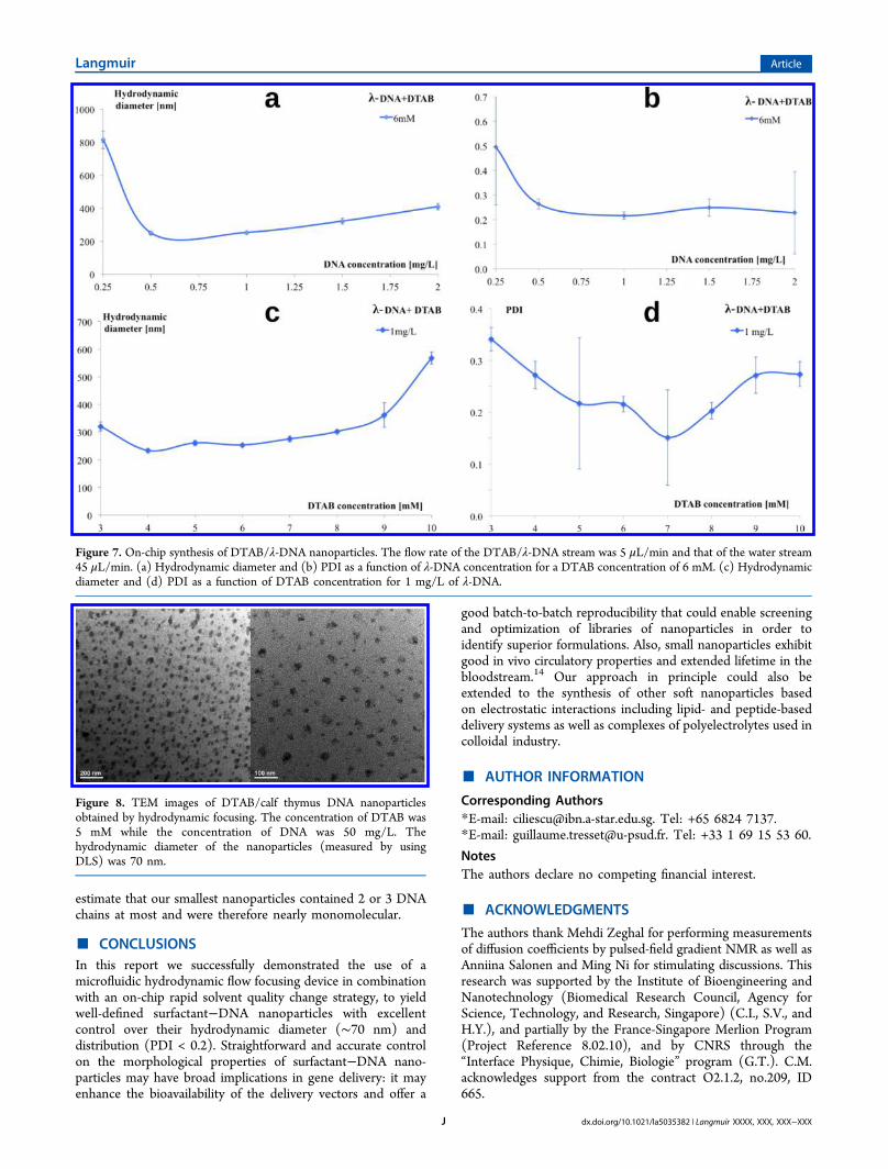
estimate that our smallest nanoparticles contained 2 or 3 DNAchains at most and were therefore nearly monomolecular.
■ CONCLUSIONSIn this report we successfully demonstrated the use of amicrofluidic hydrodynamic flow focusing device in combinationwith an on-chip rapid solvent quality change strategy, to yieldwell-defined surfactant−DNA nanoparticles with excellentcontrol over their hydrodynamic diameter (∼70 nm) anddistribution (PDI < 0.2). Straightforward and accurate controlon the morphological properties of surfactant−DNA nano-particles may have broad implications in gene delivery: it mayenhance the bioavailability of the delivery vectors and offer a
good batch-to-batch reproducibility that could enable screeningand optimization of libraries of nanoparticles in order toidentify superior formulations. Also, small nanoparticles exhibitgood in vivo circulatory properties and extended lifetime in thebloodstream.14 Our approach in principle could also beextended to the synthesis of other soft nanoparticles basedon electrostatic interactions including lipid- and peptide-baseddelivery systems as well as complexes of polyelectrolytes used incolloidal industry.
■ AUTHOR INFORMATION
Corresponding Authors*E-mail: [email protected]. Tel: +65 6824 7137.*E-mail: [email protected]. Tel: +33 1 69 15 53 60.
NotesThe authors declare no competing financial interest.
■ ACKNOWLEDGMENTS
The authors thank Mehdi Zeghal for performing measurementsof diffusion coefficients by pulsed-field gradient NMR as well asAnniina Salonen and Ming Ni for stimulating discussions. Thisresearch was supported by the Institute of Bioengineering andNanotechnology (Biomedical Research Council, Agency forScience, Technology, and Research, Singapore) (C.I., S.V., andH.Y.), and partially by the France-Singapore Merlion Program(Project Reference 8.02.10), and by CNRS through the“Interface Physique, Chimie, Biologie” program (G.T.). C.M.acknowledges support from the contract O2.1.2, no.209, ID665.
Figure 7. On-chip synthesis of DTAB/λ-DNA nanoparticles. The flow rate of the DTAB/λ-DNA stream was 5 μL/min and that of the water stream45 μL/min. (a) Hydrodynamic diameter and (b) PDI as a function of λ-DNA concentration for a DTAB concentration of 6 mM. (c) Hydrodynamicdiameter and (d) PDI as a function of DTAB concentration for 1 mg/L of λ-DNA.
Figure 8. TEM images of DTAB/calf thymus DNA nanoparticlesobtained by hydrodynamic focusing. The concentration of DTAB was5 mM while the concentration of DNA was 50 mg/L. Thehydrodynamic diameter of the nanoparticles (measured by usingDLS) was 70 nm.
Langmuir Article
dx.doi.org/10.1021/la5035382 | Langmuir XXXX, XXX, XXX−XXXJ
■ REFERENCES(1) Niidome, T.; Huang, L. Gene therapy progress and prospects:nonviral vectors. Gene Ther. 2002, 9, 1647−1652.(2) Lin, Y.; Zhang, Y.; Qiao, Y.; Huang, J.; Xu, B. Light and host−guest inclusion mediated salmon sperm DNA/surfactant interactions.J. Colloid Interface Sci. 2011, 362, 430−438.(3) Kay, M. A.; Manno, C. S.; Ragni, M. V.; Larson, P. J.; Couto, L.B.; McClelland, A.; Glader, B.; Chew, A. J. Evidence for gene transferand expression of factor IX in haemophilia B patients treated with anAAV vector. Nat. Genet. 2000, 24, 257−261.(4) Cavazzana-Calvo, M.; Hacein-Bey, S.; de Saint Basile, G.; Gross,F.; Yvon, E.; Nusbaum, P.; Selz, F.; Hue, C.; Certain, S.; Casanova, J.-L. Gene therapy of human severe combined immunodeficiency(SCID)-X1 disease. Science 2000, 288, 669−672.(5) Marshall, E. Gene therapy on trial. Science 2000, 288, 951−957.(6) Check, E. Gene therapy: a tragic setback. Nature 2002, 420, 116−118.(7) Morrissey, D. V.; Lockridge, J. A.; Shaw, L.; Blanchard, K.;Jensen, K.; Breen, W.; Hartsough, K.; Machemer, L.; Radka, S.; Jadhav,V. Potent and persistent in vivo anti-HBV activity of chemicallymodified siRNAs. Nat. Biotechnol. 2005, 23, 1002−1007.(8) Xu, Z. P.; Zeng, Q. H.; Lu, G. Q.; Yu, A. B. Inorganicnanoparticles as carriers for efficient cellular delivery. Chem. Eng. Sci.2006, 61, 1027−1040.(9) Wong, S. Y.; Pelet, J. M.; Putnam, D. Polymer systems for genedeliverypast, present, and future. Prog. Polym. Sci. 2007, 32, 799−837.(10) Maury, B.; Goncalves, C.; Tresset, G.; Zeghal, M.; Cheradame,H.; Guegan, P.; Pichon, C.; Midoux, P. Influence of pDNA availabilityon transfection efficiency of polyplexes in non-proliferative cells.Biomaterials 2014, 35, 5977−5985.(11) Abdelhady, H. G.; Allen, S.; Davies, M. C.; Roberts, C. J.;Tendler, S. J.; Williams, P. M. Direct real-time molecular scalevisualisation of the degradation of condensed DNA complexes exposedto DNase I. Nucleic Acids Res. 2003, 31, 4001−4005.(12) Lechardeur, D.; Sohn, K.; Haardt, M.; Joshi, P.; Monck, M.;Graham, R.; Beatty, B.; Squire, J.; O’brodovich, H.; Lukacs, G.Metabolic instability of plasmid DNA in the cytosol: a potential barrierto gene transfer. Gene Ther. 1999, 6, 482−497.(13) Schaffer, D. V.; Lauffenburger, D. A. Optimization of cell surfacebinding enhances efficiency and specificity of molecular conjugategene delivery. J. Biol. Chem. 1998, 273, 28004−28009.(14) Chittimalla, C.; Zammut-Italiano, L.; Zuber, G.; Behr, J.-P.Monomolecular DNA nanoparticles for intravenous delivery of genes.J. Am. Chem. Soc. 2005, 127, 11436−11441.(15) Owens, D. E.; Peppas, N. A. Opsonization, biodistribution, andpharmacokinetics of polymeric nanoparticles. Int. J. Pharm. 2006, 307,93−102.(16) Jang, J.-H.; Shea, L. D. Intramuscular delivery of DNA releasingmicrospheres: microsphere properties and transgene expression. J.Controlled Release 2006, 112, 120−128.(17) Kasturi, S. P.; Sachaphibulkij, K.; Roy, K. Covalent conjugationof polyethyleneimine on biodegradable microparticles for delivery ofplasmid DNA vaccines. Biomaterials 2005, 26, 6375−6385.(18) Bielinska, A. U.; Kukowska-Latallo, J. F.; Baker, J. R. Theinteraction of plasmid DNA with polyamidoamine dendrimers:mechanism of complex formation and analysis of alterations inducedin nuclease sensitivity and transcriptional activity of the complexedDNA. Biochim. Biophys. Acta - Gene Struct. Express. 1997, 1353, 180−190.(19) Tresset, G.; Cheong, W. C. D.; Lam, Y. M. J. Phys. Chem. B2007, 111, 14233−14238.(20) Ando, S.; Putnam, D.; Pack, D. W.; Langer, R. PLGAmicrospheres containing plasmid DNA: preservation of supercoiledDNA via cryopreparation and carbohydrate stabilization. J. Pharm. Sci.1999, 88, 126−130.(21) Oster, C.; Kim, N.; Grode, L.; Barbu-Tudoran, L.; Schaper, A.;Kaufmann, S.; Kissel, T. Cationic microparticles consisting of poly
(lactide-co-glycolide) and polyethylenimine as carriers systems forparental DNA vaccination. J. Controlled Release 2005, 104, 359−377.(22) Li, D.; Wagner, N. J. Universal binding behavior for ionic alkylsurfactants with oppositely charged polyelectrolytes. J. Am. Chem. Soc.2013, 135, 17547−17555.(23) Yeo, L. Y.; Chang, H. C.; Chan, P. P.; Friend, J. R. Microfluidicdevices for bioapplications. Small 2011, 7, 12−48.(24) Khan, I. U.; Serra, C. A.; Anton, N.; Vandamme, T.Microfluidics: A focus on improved cancer targeted drug deliverysystems. J. Controlled Release 2013, 172, 1065−1074.(25) Valencia, P. M.; Farokhzad, O. C.; Karnik, R.; Langer, R.Microfluidic technologies for accelerating the clinical translation ofnanoparticles. Nat. Nanotechnol. 2012, 7, 623−629.(26) Kim, C. S.; Duncan, B.; Creran, B.; Rotello, V. M. Triggerednanoparticles as therapeutics. Nano Today 2013, 8, 439−447.(27) Marre, S.; Jensen, K. F. Synthesis of micro and nanostructures inmicrofluidic systems. Chem. Soc. Rev. 2010, 39, 1183−1202.(28) Qi, A.; Chan, P.; Ho, J.; Rajapaksa, A.; Friend, J.; Yeo, L.Template-free synthesis and Encapsulation technique for layer-by-layerpolymer nanocarrier fabrication. ACS Nano 2011, 5, 9583−9591.(29) Belliveau, N. M.; Huft, J.; Lin, P. J.; Chen, S.; Leung, A. K.;Leaver, T. J.; Wild, A. W.; Lee, J. B.; Taylor, R. J.; Tam, Y. K.Microfluidic synthesis of highly potent limit-size lipid nanoparticles forin vivo delivery of siRNA. Mol. Ther. 2012, 1, e37.(30) Kim, Y.; Lee Chung, B.; Ma, M.; Mulder, W. J.; Fayad, Z. A.;Farokhzad, O. C.; Langer, R. Mass production and size control oflipid−polymer hybrid nanoparticles through controlled microvortices.Nano Lett. 2012, 12, 3587−3591.(31) Kim, Y.; Fay, F.; Cormode, D. P.; Sanchez-Gaytan, B. L.; Tang,J.; Hennessy, E. J.; Ma, M.; Moore, K.; Farokhzad, O. C.; Fisher, E. A.Single step reconstitution of multifunctional high-density lipoprotein-derived nanomaterials using microfluidics. ACS Nano 2013, 7, 9975−9983.(32) Hong, J. S.; Stavis, S. M.; DePaoli Lacerda, S. H.; Locascio, L. E.;Raghavan, S. R.; Gaitan, M. Microfluidic directed self-assembly ofliposome-hydrogel hybrid nanoparticles. Langmuir 2010, 26, 11581−11588.(33) Jahn, A.; Stavis, S. M.; Hong, J. S.; Vreeland, W. N.; DeVoe, D.L.; Gaitan, M. Microfluidic mixing and the formation of nanoscale lipidvesicles. ACS Nano 2010, 4, 2077−2087.(34) Tresset, G.; Iliescu, C. Electrical control of loaded biomimeticfemtoliter vesicles in microfluidic system. Appl. Phys. Lett. 2007, 90,173901.(35) Rondeau, E.; Cooper-White, J. J. Biopolymer microparticle andnanoparticle formation within a microfluidic device. Langmuir 2008,24, 6937−6945.(36) Dashtimoghadam, E.; Mirzadeh, H.; Taromi, F. A.; Nystrom, B.Microfluidic self-assembly of polymeric nanoparticles with tunablecompactness for controlled drug delivery. Polymer 2013, 54, 4972−4979.(37) Majedi, F. S.; Hasani-Sadrabadi, M. M.; Emami, S. H.;Taghipoor, M.; Dashtimoghadam, E.; Bertsch, A.; Moaddel, H.;Renaud, P. Microfluidic synthesis of chitosan-based nanoparticles forfuel cell applications. Chem. Com. 2012, 48, 7744−7746.(38) Karnik, R.; Gu, F.; Basto, P.; Cannizzaro, C.; Dean, L.; Kyei-Manu, W.; Langer, R.; Farokhzad, O. C. Microfluidic platform forcontrolled synthesis of polymeric nanoparticles. Nano Lett. 2008, 8,2906−2912.(39) Knight, J. B.; Vishwanath, A.; Brody, J. P.; Austin, R. H.Hydrodynamic focusing on a silicon chip: mixing nanoliters inmicroseconds. Phys. Rev. Lett. 1998, 80, 3863−3866.(40) Lim, J.-M.; Bertrand, N.; Valencia, P. M.; Rhee, M.; Langer, R.;Jon, S.; Farokhzad, O. C.; Karnik, R. Parallel microfluidic synthesis ofsize-tunable polymeric nanoparticles using 3D flow focusing towards invivo study. Nanomed.-Nanotechnol. Biol. Med. 2014, 10, 401−409.(41) Tresset, G.; Marculescu, C.; Salonen, A.; Ni, M.; Iliescu, C. Finecontrol over the size of surfactant−polyelectrolyte nanoparticles byhydrodynamic flow focusing. Anal. Chem. 2013, 85, 5850−5856.
Langmuir Article
dx.doi.org/10.1021/la5035382 | Langmuir XXXX, XXX, XXX−XXXK
(42) Johnson, B. K.; Prud’homme, R. K. Mechanism for rapid self-assembly of block copolymer nanoparticles. Phys. Rev. Lett. 2003, 91,118302.(43) Rudorf, S.; Radler, J. O. Self-Assembly of stable monomolecularnucleic acid lipid particles with a size of 30 nm. J. Am. Chem. Soc. 2012,134, 11652−11658.(44) Tyn, M. T.; Calus, W. F. Temperature and concentrationdependence of mutual diffusion coefficients of some binary liquidsystems. J. Chem. Eng. Data 1975, 20, 310−316.(45) Trabelsi, S.; Raspaud, E.; Langevin, D. Aggregate formation inaqueous solutions of carboxymethylcellulose and cationic surfactants.Langmuir 2007, 23, 10053−10062.(46) Weigl, B. H.; Yager, P. Microfluidic diffusion-based separationand detection. Science 1999, 283, 346−347.(47) Castillo-Leon, J.; Rodriguez-Trujillo, R.; Gauthier, S.; Jensen, A.C.; Svendsen, W. E. Micro-“factory” for self-assembled peptidenanostructures. Microelectron. Eng. 2011, 88, 1685−1688.(48) Iliescu, C.; Taylor, H.; Avram, M.; Miao, J.; Franssila, S. Apractical guide for the fabrication of microfluidic devices using glassand silicon. Biomicrofluidics 2012, 6, 016505.(49) Iliescu, C.; Tresset, G.; Xu, G. Continuous field-flow separationof particle populations in a dielectrophoretic chip with threedimensional electrodes. Appl. Phys. Lett. 2007, 90, 234104.(50) Iliescu, C.; Tresset, G.; Xu, G. Dielectrophoretic field-flowmethod for separating particle populations in a chip with asymmetricelectrodes. Biomicrofluidics 2009, 3, 044104.
Langmuir Article
dx.doi.org/10.1021/la5035382 | Langmuir XXXX, XXX, XXX−XXXL