Embed Size (px)
Citation preview

저 시-비 리- 경 지 2.0 한민
는 아래 조건 르는 경 에 한하여 게
l 저 물 복제, 포, 전송, 전시, 공연 송할 수 습니다.
다 과 같 조건 라야 합니다:
l 하는, 저 물 나 포 경 , 저 물에 적 된 허락조건 명확하게 나타내어야 합니다.
l 저 터 허가를 면 러한 조건들 적 되지 않습니다.
저 에 른 리는 내 에 하여 향 지 않습니다.
것 허락규약(Legal Code) 해하 쉽게 약한 것 니다.
Disclaimer
저 시. 하는 원저 를 시하여야 합니다.
비 리. 하는 저 물 리 목적 할 수 없습니다.
경 지. 하는 저 물 개 , 형 또는 가공할 수 없습니다.

의학 석사 학위논문
Laparoscopic double tract proximal gastrectomy for
proximal early gastric cancer
2013년 1월
서울대학교 대학원
의학과 외과학 전공
안 상 훈

Laparoscopic double tract proximal gastrectomy for
proximal early gastric cancer
지도교수 김 형 호
이 논문을 의학 석사 학위논문으로 제출함
2013년 1월
서울대학교 대학원
의학과 외과학 전공
안 상 훈
안상훈의 의학 석사 학위논문을 인준함
2013년 1월 10일
위 원 장 김 나 영 (인)
부위원장 김 형 호 (인)
위 원 강 성 범 (인)
Laparoscopic double tract proximal gastrectomy for
proximal early gastric cancer
by
Sang-Hoon Ahn
A Thesis Submitted to the Department of Surgery
in Partial Fulfillment of the Requirements
for the Degree of Master of Philosophy in Surgery
at the Seoul National University College of Medicine
January 2013
Approved by thesis committee:
Professor Nayoung Kim Chairman
Professor Hyung-Ho Kim Vice chairman
Professor Seong-Bum Kang

i
Abstract
Background: LAPG is not routinely performed because it is associated with increased
reflux symptoms and anastomotic strictures. The purpose of this study is to describe a
novel method of laparoscopy-assisted proximal gastrectomy (LAPG) with double tract
reconstruction (DTR) for proximal early gastric cancer (EGC), and to evaluate the
technical feasibility, safety, and short-term surgical outcomes, especially reflux
symptoms, after LAPG.
Methods: Retrospective review of the prospective cohort data of 43 patients who
presented to a single tertiary hospital from June 2009 through April 2012 and
underwent LAPG with DTR for proximal EGC. The data of this prospective cohort
were analyzed, and the reflux symptoms, clinicopathologic characteristics, surgical
outcomes, postoperative morbidities and mortalities, and follow-up findings were
analyzed.
Results: The mean surgical time was 180.7 minutes; mean estimated blood loss, 120.4
mL; mean length of the proximal resection margin, 4.13 cm; mean number of retrieved
lymph nodes, 41.2; and mean postoperative hospital stay, 7.1 days. Early complication
rate was 11.6% (n = 5); major complication (grade higher than Clavien-Dindo IIIa)
occurred in 1 patient (2.3%). Late complication rate was 11.6% (n = 5): 2 patients had
esophagojejunostomy stenosis, which was successfully treated with fluoroscopic
balloon dilatations; 1, chylous ascites; and 2 had Visick grade II reflux symptoms
(4.6%), managed by medication during the mean follow-up period of 21.6 months.
Conclusion: DTR after LAPG is a feasible, simple, and novel reconstruction method
with excellent postoperative outcomes in terms of preventing reflux symptoms. Its

ii
clinical applicability must be validated by prospective randomized trials.
Keywords : Gastric cancer, Laparoscopy, Proximal gastrectomy, Laparoscopy-
assisted proximal gastrectomy (LAPG), Double tract reconstruction (DTR),
Proximal EGC
Student Number : 2011-21847

iii
List of Tables
Table 1. Patient demographics and preoperative characteristics----------14
Table 2. Operative data and Short-term Surgical Outcomes------------16
Table 3. Early complication rate based on Clavien-Dindo Classifications------17
Table 4. Reflux symptoms based on Visick score------------------19
Table 5. Pathologic findings, Recurrence and Survival data----------21
Table 6. Nutritional parameter---------------------------26
Table 7. Comparison of DTR with EEG and SSEG which were analyzed in the
previous our study in our institution. ------------------------- 28

iv
List of Figures
Figure 1. Schematic illustration of double tract reconstruction-------------3
Figure 2. Port placement-------------------------------8
Figure 3. Combined retraction suture of left lateral lobe of liver and falciform
ligament-----------------------------------------8
Figure 4. Esophagus division by LapJack----------------------9
Figure 5. Transection of stomach---------------------------9
Figure 6. Insertion of anvil---------------------------------10
Figure 7. Preparation of jejunum--------------------------10
Figure 8. Intracorporeal esophagojejunostomy------------------11
Figure 9. Extrcorporeal gastrojejunostomy---------------------11
Figure 10. Common entry hole of gastrojejunostomy closure-------------12
Figure 11. Extracorporeal jejunostomy-----------------------12
Figure 12. Relative ration of intake food flow ---------------------23
Figure 13. Nutritional parameters -------------------------25

v
Contents
Abstract-----------------------------------------i
List of Tables---------------------------------------- iii
List of Figures----------------------------------------iv
Contents--------------------------------------------v
Introduction------------------------------------------1
Materials and Methods------------------------------------2
Results--------------------------------------------13
Discussion------------------------------------------29
References------------------------------------------35

1
Introduction
In Korea, over the last 2 decades, the incidence of early gastric cancer (EGC) and
proximal gastric cancer has gradually increased from 24.8% to approximately 50%
and from 5.3% to 14.0%, respectively. Proximal EGC comprises 30.3% of all
proximal gastric cancers, whereas distal EGC comprises 51.5% of all distal gastric
cancers. Consequently, the need for surgical treatment of proximal EGC, by total or
proximal gastrectomy has gradually been increasing. However, since proximal
gastrectomy often leads to reflux esophagitis and anastomotic strictures, it is not
routinely performed in Korea and other countries. In 2009, proximal gastrectomy
was performed in only 1% (139 cases) of all gastric operations in Korea, including
open cases [1-3]. Furthermore, although various reconstruction methods have
been reported thus far, the optimal reconstruction method after proximal
gastrectomy remains controversial [4, 5]. In general, total gastrectomy is
recommended due to the high morbidity rates associated with proximal
gastrectomy [6].
However, if the rate of reflux esophagitis and anastomotic stricture after proximal
gastrectomy can be lowered to that of total gastrectomy, proximal gastrectomy
may become a treatment of choice for proximal EGC. The purpose of this study
was to assess the feasibility, safety, and surgical outcomes of a novel technique
designed to prevent reflux symptoms—laparoscopy-assisted proximal gastrectomy
(LAPG) with double tract reconstruction (DTR). To our knowledge, thus far, this is
the first study to report a surgical procedure involving LAPG and DTR for proximal
EGC.

2
Materials and Methods
1. Patients
From June 2009 to April 2012, 43 patients underwent LAPG with DTR for proximal
EGC at Seoul National University Bundang Hospital, Korea. In this study, we
included patients with a preoperative diagnosis of a <5-cm wide T1N0 lesion in the
proximal stomach, in whom no lymph node (LN) enlargement was observed in LN
stations 5, 6, and 10, according to endoscopy, endoscopic ultrasonography, and
computed tomography (CT). Preoperative reflux esophagitis were evaluated by the
Visick score and endoscopic findings (Los Angeles classification). Double tract
reconstruction is a reconstruction method after proximal gastrectomy which
consists of 3 anastomosis: esophagojejunostomy(E-Jstomy),
gastrojejunostomy(G-JStomy) 15cm below E-Jstomy and jejunojejunostomy(J-
Jstomy) 20cm below G-Jstomy (Figure 1). Double tract means that food passage
after reconstruction flows simultaneously to the stomach and jejunum.

3
Figure 1. Schematic illustration of double tract reconstruction

4
2. Procedures
1) Laparoscopy-assisted proximal gastrectomy
The patient was placed in a reverse Trendelenburg position under general
anesthesia. The operator, a scopist, was positioned on the right side of the patient,
and the first assistant was positioned on the left side of the patient. Five working
ports were used during the surgical procedures (Figure 2). First, the falciform
ligament and left lobe of the liver were retracted by combined suture retraction of
the lesser omentum (Figure 3) [7]. Partial omentectomy was started about 4 cm
away from the gastroepiploic arcade. The left gastroepiploic vessels were ligated
distal to the omental branch to prevent omental infarction and then divided using
hemoclips. The omentum was dissected from the mesocolon around the transition
zone of LN stations 4d to 6, and the right gastroepiploic vessels were preserved.
The peritoneum along the superior edge of the pancreas was mobilized. The lesser
omentum was mobilized with careful preservation of the right gastric vessels and
the hepatic branch of the anterior vagus nerve. The hepatic and pyloric branches of
the vagus nerves were routinely preserved (this is an important step to prevent
delayed gastric emptying caused by pyloric dysfunction). Dissection proceeded
along the LN stations 7, 8a, and 9. The coronary vein (left gastric vein) and the left
gastric artery were then clipped and divided. Dissection was continued along with
the splenic artery up to the splenic hilum (LN stations 11p and 11d). The
esophagogastric junction was mobilized. Next, an intracorporeal purse-string
suture clamp “LapJack” (Eterne, Seoul, Korea) (Figure 4) was applied to the
esophagus, and endo-bulldog (B. Braun Melsungen AG, Melsungen, Germany) was
applied to its distal portion for the prevention of spillage from the stomach. After a

5
purse-string suture was made using a straight needle (Prolene 2-0), the
esophagus was transected. Dissection was carried out by the “ downstream
method” to dissect the LN stations 2 and 4sa. An approximately 3–4-cm long
transverse incision was made with extension of the left 12-mm trocar site. The
stomach was delivered through this mini-laparotomy, and the specimen was
transected by linear staplers after ensuring the distal resection margin and
trimming the gastroepiploic arcade (Figure 5).
2) Reconstructions
The anvil head of the circular stapler was placed in the abdominal cavity, and the
pneumoperitoneum was re-established using a wound retractor and glove. The
anvil head was intracorporeally inserted into the esophagus stump using a
laparoscopic anvil clamp, and the purse-string suture was tied laparoscopically
(Figure 7). After the purse-string suture was tied, an Endo-loop (Ethicon Endo-
Surgery, Somerville, NJ) was also added to the proximal portion of the first knot
for reinforcement. A Roux-en-Y E-Jstomy was performed by intracorporeal way
with a circular stapler (Figure 7 & 8), and the jejunal stump was closed with a
linear stapler. Next, side-to-side G-Jstomy, 15cm below the E-Jstomy, was
performed in an extracorporeal fashion using 2 linear staplers (Figure 9 & 10).
Finally, end-to-side jejunojejunostomy, 20cm below the G-Jstomy, was
performed by an extracorporeal hand-sewing suture (Figure 11). The abdominal
cavity was checked, 1 or 2 Jackson-Pratt (J-P) drainage tubes were placed
through the trocar wounds around the E-Jstomy, and the incisions were closed.

6
3. Postoperative care
During the postoperative period, the patients were managed according to our
hospital’s critical pathway protocols. Sips of water, a semifluid diet (SFD), and a
soft blended diet (SBD) were given to the patients on postoperative days 3, 4, and
5, respectively. After SBD intake, the J-P drainage tube was removed. Finally, the
patients were routinely discharged from the hospital on postoperative day 6 if they
exhibited no discomfort, abdominal pain, or abnormal laboratory test results.
4. Evaluation of the clinical parameters
The clinical features, surgical parameters (e.g., sex, age, tumor size, histological
type, length of resection margin, and number of retrieved and metastatic LNs),
early postoperative complications (0–30 days), and late postoperative
complications (>30 days) were analyzed based on the information obtained from
our prospectively maintained gastric cancer database and electric medical record.
Postoperative complications were classified according to the Clavien-Dindo
Classification, and grades of complications were recorded. Major complications
were defined as those with grades higher than Clavien-Dindo classification IIIa.
Patients were routinely followed at our outpatient clinic at 1, 3, 6, and 12 months
postoperatively and annually thereafter. Anastomotic stenosis and reflux symptoms
were diagnosed based on endoscopic findings and patient symptoms. The definition
of anastomotic stenosis was diagnosed when patients complained of dysphagia
during the postoperative follow-up and a 9mm diameter endoscope could not pass
the E-Jstomy. Reflux symptoms were diagnosed by modified Visick scores (Table
4). A gastric emptying scan was performed at 3 months after operation.

7
Radioactivity was measured in every 30 minutes up to 120minutes after the intake
of solid food mixed with 2mCi technetium-99m-DTPA(diethylene-triamine-
pentaacetate. The half life and the gastric emptying time were calculated by the
exponential function. The normal range of gastric emptying time in our institution is
70 to 150 minutes. Delayed gastric emptying and relative intake between the
stomach and the small bowel were analyzed. For evaluating the nutritional status,
body weight and serum levels of total protein and albumin were measured before
the operation, and at 3, 6, and 12 months after the operation. This study was
approved by the ethics committee of the hospital (No. B-1203/147-105).
5. Statistical analysis
Statistical analyses were performed with the SPSS statistical software, version
18.0, for Windows (SPSS, Chicago, IL, USA). All values are expressed as the mean
± standard deviation (SD) of the mean. Nutritional parameters were analyzed by
the paired samples t test. P < 0.05 was considered statistically significant.

8
Figure 2. Port placement
Figure 3. Combined retraction suture of left lateral lobe of liver and falciform
ligament

9
Figure 4. Esophagus division by LapJack.
Figure 5. Transection of stomach

10
Figure 6. Insertion of anvil
Figure 7. Preparation of jejunum

11
Figure 8. Intracorporeal esophagojejunostomy
Figure 9. Extracorporeal gastrojejunostomy

12
Figure 10. Common entry hole of gastrojejunostomy closure
Figure 11. Extracorporeal jejunostomy

13
Results
1) Patient demographics
Patient characteristics are described in Table 1. The study cohort included 35
men and 8 women, with a mean age of 59.9 years. Comorbidities existed in 17
patients (30.2%) and body mass index (BMI, kg/m2) was 23.7 (range 17.4–30.3).
Of the 43 patients, 10 patients had a history of previous abdominal operation. None
of the patients had gastroesophageal reflux disease according to the Los Angeles
Classification during the preoperative evaluation by endoscopy and Visick score.

14
Table 1. Patient demographics
LAPG (n=43) Range
Age (Years, Mean ± SD) 59.9 (± 11.9) 35-85
Gender (Male : Female) 35 : 8
Smoking 30.2% (n=13)
Body mass index (BMI, kg/m2) 23.7 (± 2.9) 17.4 ~ 30.3
Previous abdominal surgery 23.3% (n=10)
Comorbidity 39.5% (n=17)
Hypertension 11
Diabetes 4
Asthma 2
CAD 5
Liver cirrhosis 2
CVA 2
ASA score
1 17
2 22
3 3
Gastroesophageal reflux disease 0%
(Based on Visick score and LA
classification)
(CAD : coronary artery disease, CVA : cerebral vascular accident, ASA : American society of
anesthesiologists, LA : Los angeles)

15
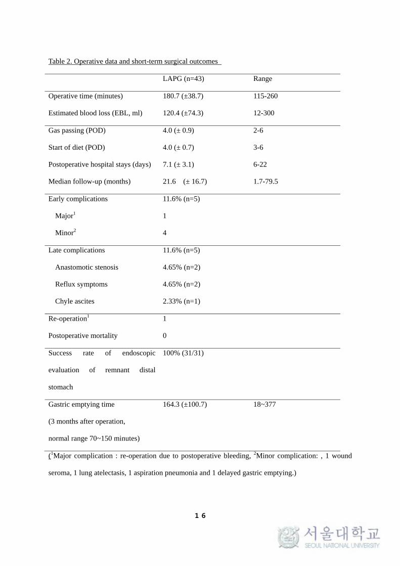
2) Surgical parameters and short-term surgical outcomes
All the surgeries, involving D1+beta lymphadenectomy without any open
conversion, were performed by a single surgeon. The surgical parameters of the
43 patients are shown in Table 2. The surgical time was calculated from the start
of the incision to the closure of the wound, and the mean surgical time was 180.7
minutes (range: 115–300 minutes). Figure 10 shows that the time taken for an
operation gradually decreased. The mean estimated blood loss was 120.4 mL
(range: 30–300 mL). No serious intraoperative events or complications were
observed. The median postoperative hospital stay was 7.1 days. The overall early
complication rate was 11.6% (n = 5); the early complications included 1 case each
of postoperative bleeding at the mesentery of the Roux limb, wound seroma, lung
atelectasis, aspiration pneumonia, and delayed gastric emptying. Wound and lung
complications were treated and improved by conservative management. In the case
of bleeding, the patient underwent immediate laparoscopic bleeding control on the
night of the operation. Delayed gastric emptying was improved by fasting for 5
days. A major complication, defined by a grade higher than Clavien-Dindo IIIa, was
observed in 1 patient (2.3%) (Table 3).
16
Table 2. Operative data and short-term surgical outcomes
LAPG (n=43) Range
Operative time (minutes) 180.7 (±38.7) 115-260
Estimated blood loss (EBL, ml) 120.4 (±74.3) 12-300
Gas passing (POD) 4.0 (± 0.9) 2-6
Start of diet (POD) 4.0 (± 0.7) 3-6
Postoperative hospital stays (days) 7.1 (± 3.1) 6-22
Median follow-up (months) 21.6 (± 16.7) 1.7-79.5
Early complications 11.6% (n=5)
Major1 1
Minor2 4
Late complications 11.6% (n=5)
Anastomotic stenosis 4.65% (n=2)
Reflux symptoms 4.65% (n=2)
Chyle ascites 2.33% (n=1)
Re-operation1 1
Postoperative mortality 0
Success rate of endoscopic
evaluation of remnant distal
stomach
100% (31/31)
Gastric emptying time
(3 months after operation,
normal range 70~150 minutes)
164.3 (±100.7) 18~377
(1Major complication : re-operation due to postoperative bleeding, 2Minor complication: , 1 wound
seroma, 1 lung atelectasis, 1 aspiration pneumonia and 1 delayed gastric emptying.)

17
Table 3. Early complication rate based on Clavien-Dindo classifications
Grade of complications, n(%) LAPG (n=43) %
I
Wound seroma 1 2.3%
Atelectasis 1 2.3%
II
Aspiration pneumonia 1 2.3%
Delayed gastric emptying 1 2.3%
IIIa
Bleeding 1 2.3%
Overall early complication rate 5 11.6%
18
3) Endoscopic evaluation of reflux esophagitis and a remnant distal stomach
At 3 months after the operation, we routinely performed endoscopy for evaluation
of reflux esophagitis and a remnant distal stomach. In the 31 patients who received
an endoscopy, no reflux esophagitis was found during the endoscopic evaluation
and no intubation failure was achieved during the examination for a remnant distal
stomach (Tables 2).
4) Late complications (reflux esophagitis and anastomotic stricture)
The overall rate of late complications was 11.6% (5 of 49 patients). These
complications included 2 cases of anastomotic stenosis, 2 cases of reflux
symptoms, and 1 case of chylous ascites. The 2 patients with stenosis were
successfully treated with fluoroscopic balloon dilatations. Reflux symptoms in the
other 2 patients were classified as Visick grade II, based on the Visick score, and
were easily controlled by medications (Table 4).

19
Table 4. Reflux symptoms based on Visick score
Preoperative reflux symptoms (n=43) Postoperative Reflux symptoms (n=43)
Total 0 4.65% (n=2)
I 0 2
II 0 0
III 0 0
IV 0 0

20
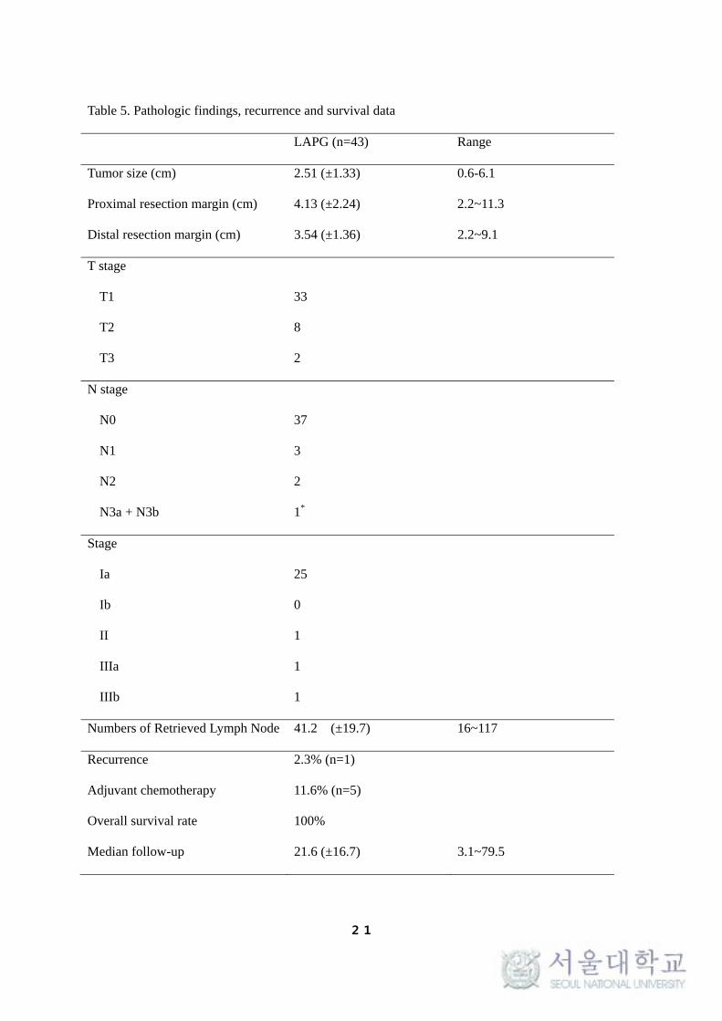
5) Pathologic findings, and recurrence and survival data
The pathologic findings of this study are shown in Table 5. All the patients were
diagnosed with proximal EGC during the preoperative examinations. All the LN
dissections were D1+beta, and the mean number of LNs retrieved was 41.2 (range:
16–117). The mean lengths of the proximal and distal resection margins were 4.13
and 3.54 cm, respectively. Adjuvant chemotherapy was recommended in 5 patients
with stage II, III, and IV disease, according to the AJCC/UICC sixth edition until
2010 and the AJCC/UICC seventh edition since 2010. The median follow-up period
was 21.6 (range 3.1–79.5) months. At the final follow-up, tumor recurrence was
found to occur in 1 patient, with a median follow-up. The disease stage in this
patient was stage IIIb. The recurrence pattern was peritoneal seeding. The overall
survival rate of our study group was 100%.
21
Table 5. Pathologic findings, recurrence and survival data
LAPG (n=43) Range
Tumor size (cm) 2.51 (±1.33) 0.6-6.1
Proximal resection margin (cm) 4.13 (±2.24) 2.2~11.3
Distal resection margin (cm) 3.54 (±1.36) 2.2~9.1
T stage
T1 33
T2 8
T3 2
N stage
N0 37
N1 3
N2 2
N3a + N3b 1*
Stage
Ia 25
Ib 0
II 1
IIIa 1
IIIb 1
Numbers of Retrieved Lymph Node 41.2 (±19.7) 16~117
Recurrence 2.3% (n=1)
Adjuvant chemotherapy 11.6% (n=5)
Overall survival rate 100%
Median follow-up 21.6 (±16.7) 3.1~79.5

22
6) Gastric emptying scan
We performed a routine gastric emptying scan at 3 months after the operation.
The mean gastric emptying time was 164.3 minutes (range: 18–377 minutes, n =
31); this finding shows that gastric emptying was delayed to some extent (Table
2). Approximately 60% of the food consumed remained in the stomach and 40% in
the jejunum. The relative ratio of food flow from the stomach to the small intestine
after DTR was approximately 3:2 (Figure 12).

23
Figure 12. Relative ratio of intake food flow
24
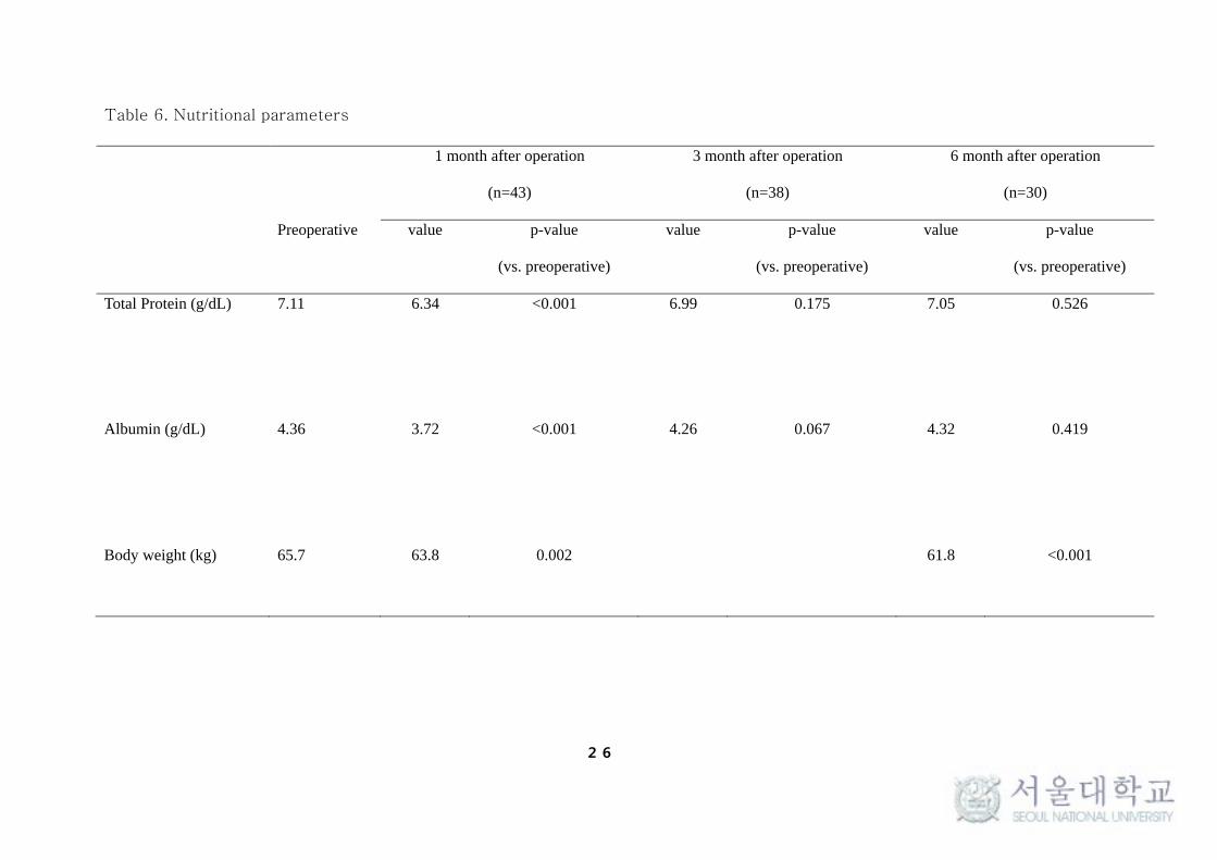
7) Nutritional parameters
To evaluate the postoperative nutritional status, the serum levels of total protein
and albumin at the first, third, and sixth postoperative months were measured and
compared with the preoperative data. The total protein and albumin levels were
both significantly decreased 1 month after the operation (7.11 versus 6.34, p <
0.0001; 4.36 versus 3.72, p < 0.0001, respectively). However, these levels
returned to normal 3 months after the operation (Figure 13). Additionally, the
mean weight loss 1, 6 months after the operation was 2.9 and 5.9%, respectively
(Table 6).

25
Figure 13. Nutritional parameters
26
Table 6. Nutritional parameters
1 month after operation
(n=43)
3 month after operation
(n=38)
6 month after operation
(n=30)
Preoperative value p-value
(vs. preoperative)
value p-value
(vs. preoperative)
value p-value
(vs. preoperative)
Total Protein (g/dL) 7.11 6.34 <0.001 6.99 0.175 7.05 0.526
Albumin (g/dL) 4.36 3.72 <0.001 4.26 0.067 4.32 0.419
Body weight (kg) 65.7 63.8 0.002 61.8 <0.001

27
8) Comparison with previous study.
When we compared this main results with our previous study[8], the mean
operation time and postoperative hospital stay were significantly shorter and the
early complication rates showed a trend toward being reduced in the DTR group.
The rate of reflux symptoms significantly decreased from the E-Gstomy group to
the DTR group. Furthermore, in the DTR group, there were no patients with
greater than Visick score of II (Table 7).

28
Table 7. Comparison of DTR with EEEG and SSEG which were analyzed in the previous our study in our institution.
EEEG (n=13) SSEG (n=37) DTR (n=43) p-value
Operation time (minutes) 190.0 225.6 180.7 0.009
Estimated blood loss ( ml) 103.3 121.5 120.4 0.727
Postoperative hospital stays (days) 12.7 11.0 7.1 0.017
Early complications 15.4% (n=2) 27.0% (n=10) 11.6% (n=5) 0.135
Anastomotic stenosis 46.2% (n=4) 0% (n=0) 4.65% (n=2) <0.001
Reflux symptoms 15.4% (n=2) 37.8% (n=14) 4.65% (n=2) 0.017
Visick II 1 7 2
Visick III 0 4 0
Visick IV 1 3 0
Body weight change (kg) -7.2 -5.4 -3.7
Median follow up (months) 34.3 30.5 21.6
(EEEG : end-to-end esophagogastrostomy, SSEG : side-to-side esophagogastrostomy, DTR : double tract reconstruction)

29
Discussion
In this study, we analyzed the surgical outcomes of LAPG with DTR in 43 patients
with proximal EGC. To our knowledge, this is first report to describe the
application of LAPG with DTR for proximal EGC, which shows excellent
postoperative outcomes, especially with respect to decreased reflux symptoms.
This novel procedure was found to have acceptable oncologic outcomes, surgical
time, and complications rates. Thus, we conclude that DTR after LAPG with
D1+beta LN dissection is a likely acceptable treatment for proximal EGC;
furthermore, it is a feasible, safe, and useful method for preventing reflux
esophagitis.
Proximal gastrectomy is not yet the standard treatment for patients with proximal
EGC. It is still classified as an investigational treatment by the Japanese gastric
cancer treatment guidelines (third edition) [9]. The application of proximal
gastrectomy to proximal EGC has been limited due to the following 3 main
concerns: oncologic safety, functional benefits, and late complications such as
reflux esophagitis and anastomotic stenosis. In a recent systematic and meta-
analysis comparing total gastrectomy with proximal gastrectomy, it was concluded
that total gastrectomy and proximal gastrectomy had similar overall survival
outcomes for proximal gastric cancer; however, proximal gastrectomy with
esophagogastrostomy showed a higher incidence of reflux esophagitis and
anastomotic stenosis. Total gastrectomy was therefore recommended for proximal
gastric cancer [10]. However, the number of cases of proximal EGC has been

30
increasing in Korea due to national screening programs and advances in endoscopic
diagnosis and devices [1-3]. Is it justified for all these patients with EGC, who are
capable of showing a good survival rate after surgery, to undergo open total
gastrectomy?
As a minimally invasive surgery, laparoscopic gastrectomy has several advantages
over open gastrectomy, especially with respect to early postoperative outcomes—
that is, it reduces postoperative pain, surgical stress, and estimated blood loss, it
accelerates recovery and return to normal bowel function and oral intake, and it
reduces the duration of hospital stay [11-14]. Because gastric cancer is mostly
located in the distal area in Eastern countries, laparoscopic distal gastrectomy has
been a more common procedure than laparoscopic total or proximal gastrectomy.
However, recently, positive outcomes of laparoscopic total or proximal
gastrectomy have been reported [8, 15, 16].
In this context, laparoscopic proximal gastrectomy is an attractive treatment
option for proximal EGC when considering the prognosis of EGC, the advantages of
a minimally invasive surgery and function preservation, including improved
nutrition, prevention of anemia, improved production of gut hormones, and a
reduction of postoperative complaints [17-20].
If the incidence of late complications such as reflux esophagitis and anastomotic
stenosis could be decreased to that of total gastrectomy, LAPG has the potential to
become the standard procedure for proximal gastrectomy. The most important
technical challenge of LAPG may be the reconstruction method, which needs to be
designed to prevent reflux symptoms and anastomotic strictures. Several
reconstruction methods have already been reported; however, an optimal

31
reconstruction after LAPG has not yet been established.
Several previous studies have applied direct esophagogastric anastomosis as the
reconstruction method, probably because it is simple and needs only 1 anastomosis.
Anti-reflux procedures such as a gastric tube formation, fundoplication,
esophagopexy with crural repair and pyloroplasty have been used for preventing
reflux esophagitis and anastomotic strictures. However, all these methods involved
esophagogastrostomy, and the results were disappointing since the rate of reflux
esophagitis and anastomotic stenosis were still high [4, 8, 21]. A good alternative
to esophagogastrostomy reconstruction after proximal gastrectomy is the Roux-
en-Y type E-Jstomy, which is the most powerful anti-reflux reconstruction.
There are 2 kinds of E-Jstomy that can be performed after proximal gastrectomy
—jejunal interposition and DTR. Jejunal interposition has been introduced as an
alternative method for preventing severe reflux and is widely performed in open
surgery; however, laparoscopic jejunal interposition has not yet gained acceptance
due to its technical complexities. These complexities include the formation of a
pedicled jejunal flap and the formation of 3 anastomoses. The mean surgical time
was also relatively long (233–614 minutes) [22, 23].
At our institution, LAPG with esophagogastrostomy was also performed since May
2003; however, the rate of reflux symptoms and anastomotic stenosis after
esophagogastrostomy was still high, even though we gradually began to perform a
few anti-reflux procedures as well (i.e. gastric tube formation, esophagopexy with
crural repair and fundoplication). Therefore, in April 2009, LAPG with DTR was
introduced at our institute.
The LAPG with DTR procedure showed a mortality rate of zero and a low rate of

32
early postoperative complications. The late complication rate was also low,
especially with respect to the rate of reflux symptoms and anastomotic stricture,
which was nearly equivalent to that of total gastrectomy and jejunal interposition
[19, 24].
This procedure has the following advantages. First, LAPG with DTR is easier to
perform, and it is a time-saving procedure in comparison to LATG with E-Jstomy.
This procedure involves the addition of just 1 more anastomosis, G-Jstomy by
stapling, which adds only 5–10 minutes to the conventional LATG anastomosis
procedure (E-Jstomy and J-Jstomy); moreover, we can save on surgical time
because we do not need to dissect LN stations 5, 6, 12a or divide the duodenum. It
is thought to be more natural than jejunal interposition because DTR does not need
mesentery division and maintains the continuity of the jejunum. Second, revision of
E-Jstomy does not involve re-operation of the gastric stump cancer, contrary to
esophagogastrostomy, and it is also easier than jejunal interposition because it is
easy to resect the efferent jejunal limb and to perform G-Jstomy and re-
anastomosis. Third, delayed gastric emptying is not a concern, because even if
delayed gastric emptying occurs, there exists an alternative passage route for food,
contrary to jejunal interposition. Thus, delayed gastric emptying after DTR is not a
serious problem. However, in order to perform DTR, surgeons should have
sufficient experience to independently perform secure laparoscopic E-Jstomy to
perfection.
Clinicians tend to consider body weight as a measure of nutritional status.
Difficulty in maintaining bodyweight is a defining characteristic of the
postgastrectomy syndrome. In this study, the mean weight loss 6 months after the

33
procedure was 5.9%, whereas an average weight loss of 16% after total
gastrectomy has been reported. Although various mechanism have been considered,
such as decrease of gastric acid level, reflux esophagitis, intestinal floral alteration,
and increased peristalsis and diarrhea, reduced food intake is the most conceivable
explanation for body weight loss after total gastrectomy [25, 26]. We speculate
that the difference in body weight loss is because of the limited reservoir function
in total gastrectomy. When we compared the functional outcomes between
esophago-gastrostomy and DTR in the view of historical comparison, DTR showed
the tendency of less body weight loss and rapid recovery of total protein and
albumin [8].
This study has several limitations. First, this was a retrospective study of a case
series. Second, we didn’t assess the quality of life of the patients because it was
not fully followed up by using a validated questionnaire, such as the Korean version
of GastroIntestinal Quality of Life Index (GIQLI) and the European Organization for
Research and Treatment of Cancer (EORTC) Quality of Life Questionnaire (QLQ)-
C30 and sto22. Third, the numbers of patients were relatively small. Fourth, we did
not investigate the overall functional outcomes using clinical assessments,
anthropometric tests, and laboratory tests. We only assessed the nutritional status
based on body weight changes and total protein and albumin levels.
However, to our knowledge, this is the first study to report the procedure for DTR
after LAPG. In this era of function preserving surgery and minimally invasive
surgery, this study provides an overview of the procedure for LAPG with DTR, the
surgical skills required, and other important surgery-related data. These

34
encouraging data lead us to plan phase III multicenter prospective randomized
clinical trial between LAPG versus LATG.
In conclusion, our initial case series demonstrated that DTR after LAPG is a
feasible, simple, and useful reconstruction method with excellent postoperative
outcomes in terms of preventing reflux symptoms. However, future prospective
randomized trials are warranted to validate its clinical usefulness.
Conflict of interest Dr. S.-H Ahn have no conflicts of interest or financial ties to
disclose.

35
References
1. 2004 Nationwide Gastric Cancer Survey in Korea. J Gastric Cancer 2005, 5(4):285-303.
2. Jeong O, Park Y-K: Clinicopathological Features and Surgical Treatment of Gastric
Cancer in South Korea: The Results of 2009 Nationwide Survey on Surgically Treated
Gastric Cancer Patients. J Gastric Cancer 2011, 11(2):69-77.
3. Ahn HS, Lee HJ, Yoo MW, Jeong SH, Park DJ, Kim HH, Kim WH, Lee KU, Yang HK:
Changes in clinicopathological features and survival after gastrectomy for gastric cancer
over a 20-year period. Br J Surg 2011, 98(2):255-260.
4. An JY, Youn HG, Choi MG, Noh JH, Sohn TS, Kim S: The difficult choice between total
and proximal gastrectomy in proximal early gastric cancer. Am J Surg 2008, 196(4):587-
591.
5. Nakano K, Fujita M, Shigeta H, Kurata N, Tanaka N, Sekimoto K: [A case of intractable
malabsorption after proximal subtotal gastrectomy (author's transl)]. Nihon Shokakibyo
Gakkai zasshi = The Japanese journal of gastro-enterology 1980, 77(5):784-788.
6. David W. Mercer EKR: Stomach. In: Sabiston Textbook of Surgery : The biological basis of
modern surgical practice. 18 edn. Edited by Townsend CM: Elsevier; 2008: 1265-1266.
7. Shabbir A, Lee JH, Lee MS, Park do J, Kim HH: Combined suture retraction of the
falciform ligament and the left lobe of the liver during laparoscopic total gastrectomy.
Surg Endosc 2010, 24(12):3237-3240.
8. Ahn SH, Lee JH, Park DJ, Kim HH: Comparative study of clinical outcomes between
laparoscopy-assisted proximal gastrectomy (LAPG) and laparoscopy-assisted total
gastrectomy (LATG) for proximal gastric cancer. Gastric Cancer 2012.
9. Japanese gastric cancer treatment guidelines 2010 (ver. 3). Gastric Cancer 2011,
14(2):113-123.
10. Wen L, Chen XZ, Wu B, Chen XL, Wang L, Yang K, Zhang B, Chen ZX, Chen JP, Zhou ZG
et al: Total vs. Proximal Gastrectomy for Proximal Gastric Cancer: A Systematic Review

36
and Meta-Analysis. Hepatogastroenterology 2012, 59(114):633-640.
11. Kim HH, Hyung WJ, Cho GS, Kim MC, Han SU, Kim W, Ryu SW, Lee HJ, Song KY:
Morbidity and mortality of laparoscopic gastrectomy versus open gastrectomy for
gastric cancer: an interim report--a phase III multicenter, prospective, randomized Trial
(KLASS Trial). Ann Surg 2010, 251(3):417-420.
12. Hwang SH, Park do J, Jee YS, Kim MC, Kim HH, Lee HJ, Yang HK, Lee KU: Actual 3-year
survival after laparoscopy-assisted gastrectomy for gastric cancer. Arch Surg 2009,
144(6):559-564; discussion 565.
13. Kim W, Song KY, Lee HJ, Han SU, Hyung WJ, Cho GS: The impact of comorbidity on
surgical outcomes in laparoscopy-assisted distal gastrectomy: a retrospective analysis of
multicenter results. Ann Surg 2008, 248(5):793-799.
14. Kim MC, Jung GJ, Kim HH: Morbidity and mortality of laparoscopy-assisted
gastrectomy with extraperigastric lymph node dissection for gastric cancer. Dig Dis Sci
2007, 52(2):543-548.
15. Eom BW, Kim YW, Lee SE, Ryu KW, Lee JH, Yoon HM, Cho SJ, Kook MC, Kim SJ:
Survival and surgical outcomes after laparoscopy-assisted total gastrectomy for gastric
cancer: case-control study. Surg Endosc 2012.
16. Lee JH, Ahn SH, Park DJ, Kim HH, Lee HJ, Yang HK: Laparoscopic Total Gastrectomy
with D2 Lymphadenectomy for Advanced Gastric Cancer. World J Surg 2012.
17. Adachi Y, Inoue T, Hagino Y, Shiraishi N, Shimoda K, Kitano S: Surgical results of
proximal gastrectomy for early-stage gastric cancer: jejunal interposition and gastric
tube reconstruction. Gastric Cancer 1999, 2(1):40-45.
18. Takeshita K, Saito N, Saeki I, Honda T, Tani M, Kando F, Endo M: Proximal gastrectomy
and jejunal pouch interposition for the treatment of early cancer in the upper third of
the stomach: surgical techniques and evaluation of postoperative function. Surgery 1997,
121(3):278-286.
19. Furukawa H, Hiratsuka M, Imaoka S, Ishikawa O, Kabuto T, Sasaki Y, Kameyama M,

37
Ohigashi H, Nakano H, Yasuda T: Limited surgery for early gastric cancer in cardia. Ann
Surg Oncol 1998, 5(4):338-341.
20. Kameyama J, Ishida H, Yasaku Y, Suzuki A, Kuzu H, Tsukamoto M: Proximal gastrectomy
reconstructed by interposition of a jejunal pouch. Surgical technique. Eur J Surg 1993,
159(9):491-493.
21. Yoo CH, Sohn BH, Han WK, Pae WK: Long-term results of proximal and total
gastrectomy for adenocarcinoma of the upper third of the stomach. Cancer Res Treat
2004, 36(1):50-55.
22. Uyama I, Sugioka A, Fujita J, Komori Y, Matsui H, Hasumi A: Completely laparoscopic
proximal gastrectomy with jejunal interposition and lymphadenectomy. J Am Coll Surg
2000, 191(1):114-119.
23. Kinoshita T, Gotohda N, Kato Y, Takahashi S, Konishi M: Laparoscopic proximal
gastrectomy with jejunal interposition for gastric cancer in the proximal third of the
stomach: a retrospective comparison with open surgery. Surg Endosc 2012.
24. Katai H, Sano T, Fukagawa T, Shinohara H, Sasako M: Prospective study of proximal
gastrectomy for early gastric cancer in the upper third of the stomach. Br J Surg 2003,
90(7):850-853.
25. Braga M, Zuliani W, Foppa L, Di Carlo V, Cristallo M: Food intake and nutritional status
after total gastrectomy: results of a nutritional follow-up. Br J Surg 1988, 75(5):477-480.
26. Bergh C, Sjostedt S, Hellers G, Zandian M, Sodersten P: Meal size, satiety and
cholecystokinin in gastrectomized humans. Physiology & behavior 2003, 78(1):143-147.