Embed Size (px)
Citation preview

JSLS
Laparoscopic Pyelolithotomy in a Pelvic Kidney:A Case Report and Review of the Literature
David M. Hoenig, MD,1 Arieh L. Shalhav, MD,1 Abdelhamid M. Elbahnasy, MD,1
Elspeth M. McDougall, MD,1 Ralph V. Clayman, MD1,2
ABSTRACT
Background and Objectives: Laparoscopic pyelolithoto-my was performed in a pelvic kidney with a large renalpelvis calculus.
Methods and Results: Laparoscopic pyelolithotomy wassuccessfully performed in a pelvic kidney with an operativetime of 310 minutes. The use of intraoperative fluoroscopyand a semi-automatic suturing device greatly facilitated theprocedure. The patient's operative pain was managed with3 doses of ketorolac; she resumed a regular diet the dayafter surgery, and was discharged on the first postoperativeday.
Conclusions: For patients with a large stone in the renalpelvis of an ectopic kidney, laparoscopic pyelolithotomyprovides an effective approach.
Key words: Laparoscopy, Pyelolithotomy, Ectopic kidney,Pelvic kidney, Shock wave lithotripsy, Percutaneousnephrolithotomy.
1Washington University Division of Urology, St. Louis, MO, USA.2Washington University Department of Radiology, St. Louis, MO, USA.
Address reprint request to: Ralph V. Clayman, MD, Suite 5101 Queeny Tower, OneBarnes Hospital Plaza, St. Louis, MO 63108, USA.
INTRODUCTION
Pelvic kidneys are typically incidental findings, but may pre-sent due to underlying obstructive or calculous disease.The anatomical characteristics of the ectopic kidney canpose a significant challenge to the treatment of calculousdisease. Treatment of larger renal calculi in these kidneyshad been uniformly by open surgery until laparoscopicallyguided percutaneous nephrostolithotomy (PCNL) wasdescribed, initially, by Esghi, et al.,1 and later by Toth, et al.2
Recently, 2 cases of laparoscopic pyelolithotomy have beenreported in a pelvic kidney.3,4 In one, the pyelolithotomywas completed but the pyelotomy was not closed.3 Due tourine leakage, an indwelling stent and urethral catheterwere maintained for 8 days. Hospitalization lasted 6 days.3
In the second case report, intraoperative fluoroscopy andlaparoscopic suturing were used to perform the procedure,but the pyelotomy closure was not tested for watertightnessand the patient experienced peritoneal leakage of urineafter the Foley catheter was removed.4 This necessitatedreplacement of the Foley catheter for an unspecified lengthof time and the length of hospitalization was not reported.4
Herein we report the third case of a laparoscopic pyelolitho-tomy in a pelvic kidney; in this case, use of the Carter-Thomason Needle-Point Suture Passer (Inlet Medical Inc.,Eden Prairie, MN) and laparoscopic suturing of the pyeloto-my with an Endostitch device (Auto Suture, Norwalk, CT)resulted in a waterproof closure.
CASE REPORT
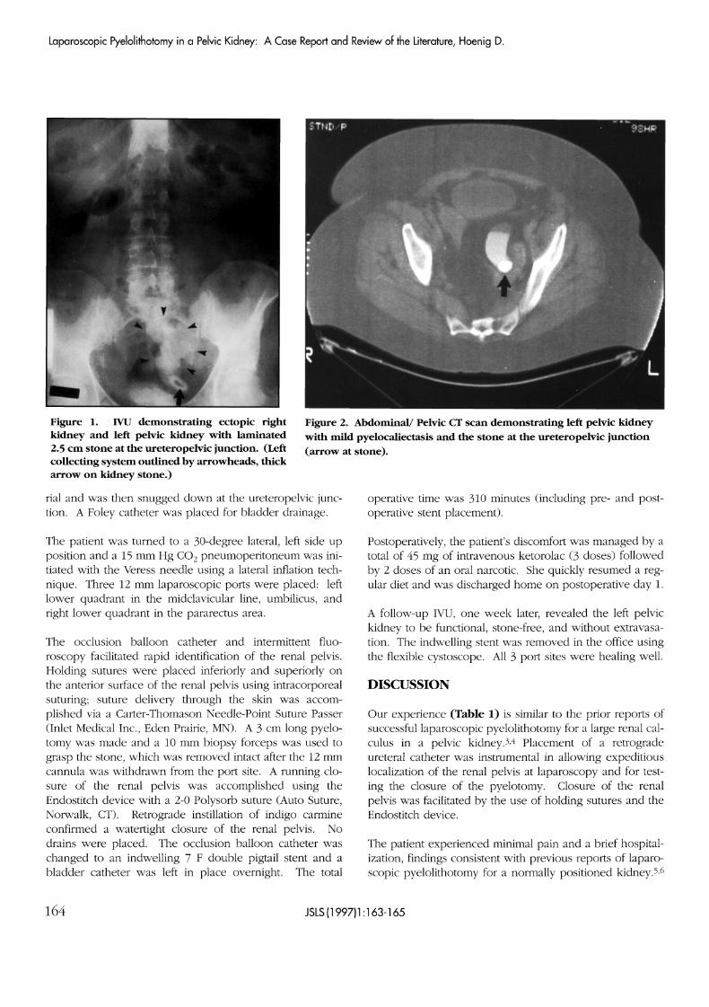
A 63-year old woman was referred for endourological treat-ment of a symptomatic 2.5-cm-x-1.5 cm renal pelvis stone inher left pelvic kidney. Intravenous urography (IVU) andabdominal/pelvic computed tomography defined the pelvickidney's location relative to other structures; there was mildpyelocaliectasis noted (Figures 1 & 2).
After initiating general anesthesia, the patient was main-tained in a supine position. Using flexible cystoscopy, a0.038 in. guidewire was advanced to the renal pelvis. A 7F/ 11.5 mm balloon occlusion catheter was then passedover the guidewire into the renal pelvis of the left pelvickidney. The balloon was inflated with 1 cc of contrast mate-
JSLS (1997)1:163-165 163
Laparoscopic Pyelolithotomy in a Pelvic Kidney: A Case Report and Review of the Literature, Hoenig D.
Figure 1. IVU demonstrating ectopic rightkidney and left pelvic kidney with laminated2.5 cm stone at the ureteropelvic junction. (Leftcollecting system outlined by arrowheads, thickarrow on kidney stone.)
rial and was then snugged down at the ureteropelvic junc-tion. A Foley catheter was placed for bladder drainage.
The patient was turned to a 30-degree lateral, left side upposition and a 15 mm Hg CO2 pneumoperitoneum was ini-tiated with the Veress needle using a lateral inflation tech-nique. Three 12 mm laparoscopic ports were placed: leftlower quadrant in the midclavicular line, umbilicus, andright lower quadrant in the pararectus area.
The occlusion balloon catheter and intermittent fluo-roscopy facilitated rapid identification of the renal pelvis.Holding sutures were placed inferiorly and superiorly onthe anterior surface of the renal pelvis using intracorporealsuturing; suture delivery through the skin was accom-plished via a Carter-Thomason Needle-Point Suture Passer(Inlet Medical Inc., Eden Prairie, MN). A 3 cm long pyelo-tomy was made and a 10 mm biopsy forceps was used tograsp the stone, which was removed intact after the 12 mmcannula was withdrawn from the port site. A running clo-sure of the renal pelvis was accomplished using theEndostitch device with a 2-0 Polysorb suture (Auto Suture,Norwalk, CT). Retrograde instillation of indigo carmineconfirmed a watertight closure of the renal pelvis. Nodrains were placed. The occlusion balloon catheter waschanged to an indwelling 7 F double pigtail stent and abladder catheter was left in place overnight. The total
Figure 2. Abdominal/ Pelvic CT scan demonstrating left pelvic kidneywith mild pyelocaliectasis and the stone at the ureteropelvic junction(arrow at stone).
operative time was 310 minutes (including pre- and post-operative stent placement).
Postoperatively, the patient's discomfort was managed by atotal of 45 mg of intravenous ketorolac (3 doses) followedby 2 doses of an oral narcotic. She quickly resumed a reg-ular diet and was discharged home on postoperative day 1.
A follow-up IVU, one week later, revealed the left pelvickidney to be functional, stone-free, and without extravasa-tion. The indwelling stent was removed in the office usingthe flexible cystoscope. All 3 port sites were healing well.
DISCUSSION
Our experience (Table 1) is similar to the prior reports ofsuccessful laparoscopic pyelolithotomy for a large renal cal-culus in a pelvic kidney.3,4 Placement of a retrogradeureteral catheter was instrumental in allowing expeditiouslocalization of the renal pelvis at laparoscopy and for test-ing the closure of the pyelotomy. Closure of the renalpelvis was facilitated by the use of holding sutures and theEndostitch device.
The patient experienced minimal pain and a brief hospital-ization, findings consistent with previous reports of laparo-scopic pyelolithotomy for a normally positioned kidney.5,6
164 JSLS (1997)1:163-165

Table 1.Comparison of laparoscopic pyelolithotomy and PCNL for renal pelvis stones > 1.5-2 cm.
Procedure:
Anesthesia:No. of access ports:Approach:Surgical time:Drains:Hospital stay:Stone free rate:# add. procedures (%):Blood Transfusion:# of patients:
Laparoscopicpyelolithotomy 3,4,5,6
General3-4Trans- or retroperitoneal2 to 5 hoursStent (1 week)1-2 days100%27% convert to open0%11 (9 eutopic)
PCNL 7,8,9
General1Retroperitoneal1 to 3 hoursPCNU (2 to 7 days)3 to 5 days95%1-2 (12-90%)2-10%494
SWL10, 11, 12
MAC/GeneralnoneExtracorporeal1 to 3 hoursstent (1 week)0-2 days52-78 %1-2 (50-60%)<1 %>2700
MAC= monitored anesthesia care, PCNU= percutaneous nephroureteral stent
In these reports, the operative time has averaged 2-5 hoursusing a 3- or 4-port approach. The renal pelvis was suturedclosed in only one case and all patients had a surgical drainplaced prior to fascial closure. While a stone-free rate of100% was recorded among 9 patients, it is of note that 3 ofthose 9 patients required conversion to an open proce-dure.6 While these reports concluded that laparoscopicpyelolithotomy in a eutopic kidney was of value in a situ-ation where shock wave lithotripsy (SWL) or PCNL hadfailed or could not be done, we believe this represents arare situation. Indeed, despite a vast experience atWashington University with laparoscopic renal surgery anda large surgical stone population (e.g. an approximateannual urolithiasis case load of 500 to 600 SWL, 100 to 150ureteroscopies, and 50 to 100 PCNL) this is the singular caseof a laparoscopic pyelolithotomy at our institution.
With regard to stone treatment in an ectopic or pelvic kid-ney, we believe the treatment strategy should be similar tothat used for stones in a eutopic kidney. Hence, SWL orureteroscopy remain first line therapy for smaller calculi(less than 2 cm), while laparoscopic-assisted percutaneous,pure laparoscopic, or open procedures are reserved forlarger calculi.
From this standpoint, it is of note that 2 cases of laparo-scopically guided PCNL have been reported in pelvic kid-neys.1,2 The operative time was not reported for thesecases and the length of hospital stay was 6 days in onereport.3 We believe that given advances in laparoscopicequipment it may be just as simple to handle the stoneremoval entirely laparoscopically. The benefits of thisapproach would potentially include a shorter hospital stay
and less postoperative morbidity as occurred in the presentcase. For urologists interested in laparoscopic pyelolitho-tomy in this situation, the use of intraoperative fluoroscopyto identify the renal pelvis, the placement of holdingsutures, the use of the Carter-Thomason Needle-PointSuture Passer, and intracorporeal suturing with theEndostitch device are recommended.
References:
1. Esghi AM, Roth JS, Smith AD. Percutaneous transperitonealapproach to a pelvic kidney for endourological removal of astaghorn calculus. J Urol. 1985;134:525.
2. Toth C, Holman E, Pasztor I, Khan AM. Laparoscopically con-trolled and assisted percutaneous transperitoneal nephrolithotomyin a pelvic dystopic kidney. J Endourol. 1993;7:303.
3. Harmon WJ, Kleer E, Segura JW. Laparoscopic pyelolithotomyfor calculus removal in a pelvic kidney. J Urol. 1996;155:2019.
4. Chang TD, Dretler SP. Laparoscopic pyelolithotomy in anectopic kidney. J Urol. 1996;156:1753.
5. Gaur DD, Agarwal DK, Purohit KC. Retroperitoneal laparo-scopic Gil-Vernet pyelolithotomy: an initial report. Min Inv Ther.
1994;3:55.
6. Gaur DD, Agarwal DK, Purohit KC, Darshane AS.Retroperitoneal laparoscopic pyelolithotomy. J Urol.1994;151:927.
JSLS (1997)1:163-165 165