Embed Size (px)
Citation preview


Laryngeal Masses
Tumour-Like Tumours
OCongential OAcquired OBenign Malignant Precancerous
Saccular cyst
Laryngeocele
Traumatic
Nodule
Polyp
INTUBATION
GRANULOMA
·INFLAMMATORY
REFLUX LARYNGITIS
·RIENKE’S OEDEMA
·VENTRICULAR
PROLAPSE
·REACTIVE
HYPERKERATOSIS
LEUKOPLAKIA
ERYTHOPLAKIA
Epithelial
Papilloma
Multiple papilloma
:CT
Hamangioma
Chondroma
PRIMARY
·SCC
·VERRUCOUS
·ADENOID CYS
·LYMPHOEPI
Sarcoma
Lymphoma
·SECONDARY
·HYPOPHARYN
·THYROID 2
·METASTATIC
Leukoplakia
Erythroplakia
Single papilloma
Larygeal keratosis

Benign Tumors of the Larynx
•Benign tumors of the larynx are relatively uncommon.
•Types:
– Epithelial– Epithelial
• Squamous epithelium
•Papilloma,Recurrent respiratory papillomatosis
•Keratinised papilloma
• Glandular
•Pleomorphic adenoma
•Oncocytic tumour
– Non-epithelial , Mesenchymal
•Vascular
•Haemangioma
Infantile: subglottic. Adult: supraglottic.
•Lymphangioma
•Cartilage and bone
Chondroma Osteoma Giant cell tumour
•Muscle
Leiomyoma Rhabdomyoma Angiomyoma Epithelioidleiomyoma
•Adipose: Lipoma
•Neural
Neurilemmoma Neurofibroma SchwannomaParaganglioma (Chemodectoma) Granular cell
• Pseudotumours : Inflammatory -fibroblastic Amyloid Laryngeal cysts

Single Papilloma of the Larynx
• Incidence
Adults (30-50 years) , males
True neoplasm
No recurrence if completely removed
Turn malignant 5%Turn malignant 5%
•Pathology:
• Site: The free edge of the of 2/3 of vocal cord is the commonest site.
• Solitary, Sessile or pedunculated mass move up and down with expiration and inspiration.
• Pink to deep red in colour
• With warty papilliferous surface and variable size

•Microscopically:
• A cord of vascular C.T., covered by hyperplastic stratified squamous epithelium.
• Cell are well differentiated , orderly arranged with some mitotic figure
• Intact BM
• Keratosis frequently seen especially in recurrent cases
• Loss of polarity in basal cell layer indicate early malignant • Loss of polarity in basal cell layer indicate early malignant changes
•Clinical picture
Dysphonia, stridor if large
Unilateral single whitish warty like mamillated
Management
MLS: remove the whole lesion
Biopsy


Multiple Papilloma of the Larynx
• Pathology
Recurrent respiratory papillomatosis; juvenile
multiple papillomatosis; viral papillomatosis
Not true neoplasm; viral infection by HPV
Children 5-15 years
Similar to squamous papilloma, sessile, smaller, Similar to squamous papilloma, sessile, smaller, multiple
Recurrent in same or different areas, regress after puberty
Does NOT turn malignant
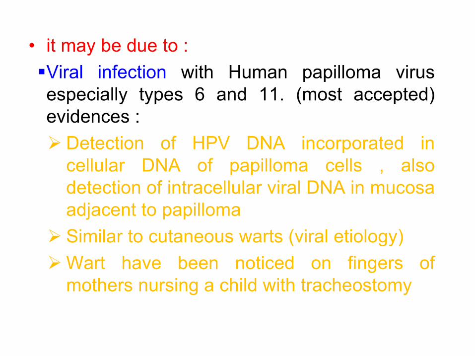
• it may be due to :
Viral infection with Human papilloma virus especially types 6 and 11. (most accepted) evidences :
Detection of HPV DNA incorporated in cellular DNA of papilloma cells , also detection of intracellular viral DNA in mucosa detection of intracellular viral DNA in mucosa adjacent to papilloma
Similar to cutaneous warts (viral etiology)
Wart have been noticed on fingers of mothers nursing a child with tracheostomy

In some patient disease transmitted at delivary from mother with vaginal warts
Tendency for sponatenous regression at any age and relapse may occur
Increased incidence of tracheobronchialinvolvement and even external tracheostomywound in patient with :wound in patient with :
Trachostomy
Repeated endoscopic procedures
Long duration of the disease
• Autoimmune disturbance or hormonal imbalance. Papillomas usually regress during puberty.

• Clinical picture
Stridor, dyspnea
Dysphonia
NPL-scope: multiple, sessile, warty like, pinkish granulations on vocal cords, supraglottic, subglottic, trachea, bronchi
Management Management
Removal by MLS, crosurgery, laser
Medical: estrogen, interferon, autogenous vaccines
Severe stridor: tracheostomy, Risk of implantation at tracheal site


Adult solitary papilioma Juvenile multiple papillomata
Age: Adults → 30 -50 years. Children→5 -15 years.
Aetiology: True benign tumour. Human papilioma virus.
Site: Commonly => anterior 2/3 of the vocal fold. Anywhere in the larynx.
Symptoms: Hoarseness of voice. 1- Stridor
2-Hoarseness of voice
Signs: Unilateral, single, pinkish, sessile, finely
tabulated, wart-like mass.
Bilateral, multiple, pinkish, sessile, finely
lobulated, wart-like masses.
Treatment: Micro-laryngoscopic excision → by surgical
instruments or laser surgery.
1- Repeated micro-la ryngoscopic excision → by
surgical instruments or better laser surgery.
2- Tracheostomy → when necessary.
3- Anti-viral medications →as interferon and
acyclovir are tried.
Prognosis: * Recurrence → uncommon.
* Malignant transformation → may occur.
* Recurrence → common but spontaneous
regression usually occurs at puberty.
* Malignant transformation → does not occur.

Carcinogensis
• Carcinogenesis in the upper aerodigestive tract is described as a multistep process. The exogenous agents cited above cause epithelial injury that evokes an epithelial response consisting of (hyper) regeneration (hyperplasia) and/or hyperkeratosis.(hyperplasia) and/or hyperkeratosis.
• With continued exposure to the noxious agents, there is a growing likelihood that foci of epithelial dysplasia will develop, spread, and eventually progress to carcinoma in situ.


Laryngeal cancer•Cancer = 2nd most common cause of death
•HNC = 6th cancer world wide
•HNC = of lowest 5-year survival
•HNC = 90% SCC
•Lx CA = 2% of body tumors•Lx CA = 2% of body tumors
•Lx CA = 40% of HN tumors
• Age:
occurs in old age (>40 , 60-70 years). However, it may affect younger age groups, particularly smokers.
• Sex:
more common in males >females of a ratio of 8:1

• Predisposing factors: Chronic mucosal irritation:
Smoking , Ex-smoker (up to 20years):90% of patients with larynx cancer have a history of heavy tobacco (take 10-20yr for a cell to turn into carcinoma insitu)
Burning cigarette produce 600 byproducts , 60 are carcinogenic , 26 affect reparatory tract
Alcohol:
The combination of smoking and alcohol use has synergistic carcinogenic effect on the larynx.synergistic carcinogenic effect on the larynx.
Chronic laryngitis:
considered a predisposing factor based on the presence of the same risk factors.
Neck irradiation
Exposure to asbestos, nickel , more common in wood workers.
GERD ????

• Precancerous conditions:
•Leukoplakia: 50%
•Laryngeal keratosis with severe dysplasia.
•Single papilloma in adults.


Severe dysplasia
CIS


Classification: Regional
•Supraglottic larynx : arise from buccopharyngealanlaoge (hypobranchial eminence) and arches III and IV
•The glottis and subglottis derivd from pulmonary anlage (laryngotracheal groove) and arch V
•The multiple arch derivation arise into horizontal •The multiple arch derivation arise into horizontal segmentation of laryngeal structures in supraglottis, glottis and subglottis

• Significance:
• The lymphatic drainage , vascular and nerve supply follow these anatomical boundaries:
•Supraglottic : drain in superior and middle cervical LN
•Glottis and subglottis: drain to juglo-omohyoid and inferior deep cervical LN
• Surface lymphatics freely connect between the sides • Surface lymphatics freely connect between the sides
• Deep submucosal lymphatic remain in compartment according vertical and horizontal segmentation
• Spread of cancer larynx respect this anatomic compartmentation

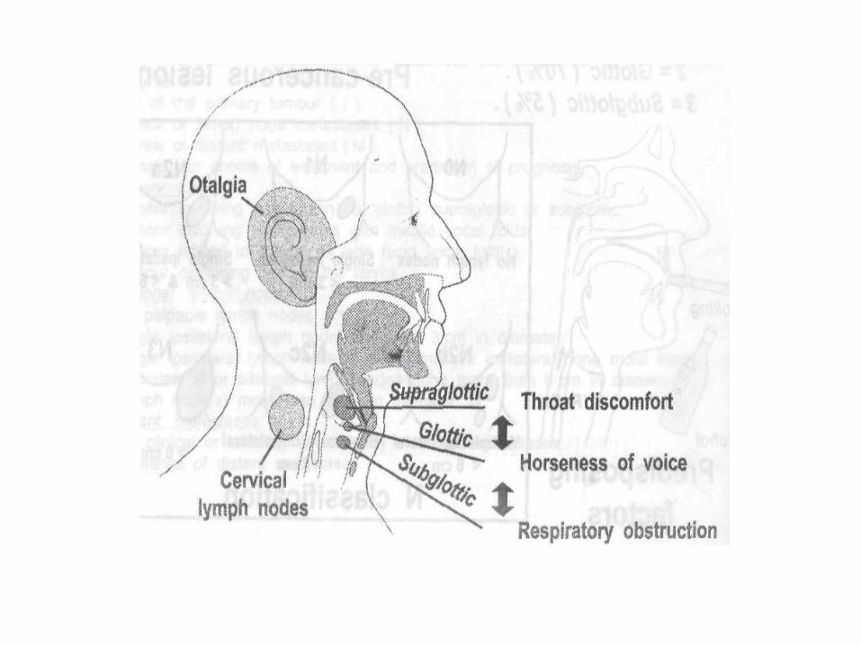
• Glottic: 60%
Vocal cords
Early presentation
No LN
• Supraglottic: 35%
Subdivided into :
Marginal zone: laryngeal surface of suprahyoidepiglottis , AE fold ,Arytenoidsepiglottis , AE fold ,Arytenoids
Supraglottic excluding marginal zone : infrahyoidepiglottis , Ventricle, VB,
Late presentation : silent area
Rich lymphatic: 30-50% LN

• Subglottic: 5%
Starts glottic
Dyspnea, stridor rapidly
LN 10-20%
• Transglottic carcinoma
•These represent tumours that crossed the laryngeal ventricle in a vertical plane, involving the supraglottis and ventricle in a vertical plane, involving the supraglottis and the glottis, with possible involvement of the subglottis.

Pathology
• Macroscopic
Cauliflower mass (supraglottic).
Malignant ulcer(commonly glottic and subglottic)
• Microscopic
90-95% SCC
• Border's calssification :
•Grade I: 75-100% well differentiated.
•Grade II: 50-75% well differentiated.
•Grade III: 25-50% well differentiated.
•Grade IV: 0-25% well differentiated.


• Undifferentiated carcinomas, well-differentiated verrucous carcinomas, and other rare entities.
• Other malignant tumours may :
• Metastasize to the larynx (secondary)—malignant melanoma, hypernephroma—and general
• or systemic malignancies may manifest themselves in the larynx—non-Hodgkin’s lymphoma
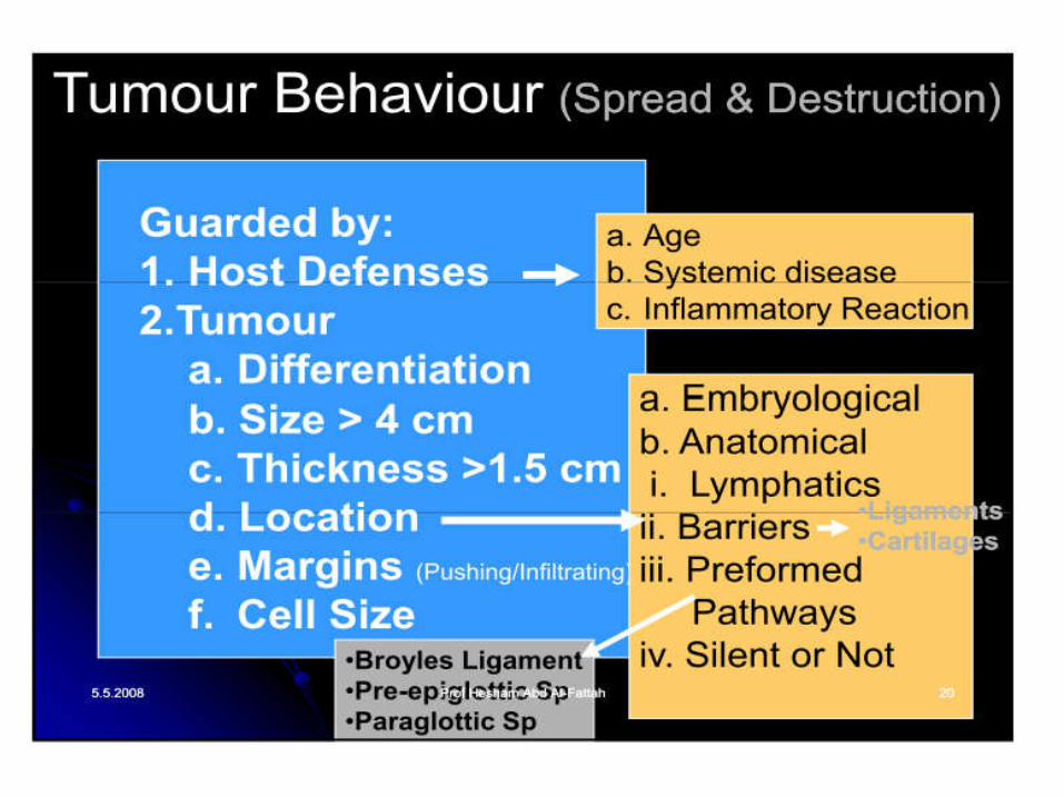
Spread
•Direct:
•From one region to another
•Liagmentous and cartilagenous structures of the larynx offer great resistance to invasion by carcinoma. once invaded , survival rate diminished dramatically
• Quadrangular membrane : separate supraglottic from paraglottic spaceparaglottic space
• Conus elasticus : separate glottis and subglottic space from paraglottic space and intrinsic laryngeal muscle → fixation
• Thyrohyoid membrane :
From anterior boundaries of pre-epiglottic space
If the space involved , tumour may spread through membrane to the neck or into the deep muscles of the tongue base

•Cricothyroid membrane :Subglottic tumour may spread through this membrane into neck
•Anterior commissure : Spread along lead to cartilage invasion
•Thyroid and cricoids cartilage ;
Cartilage may produce substance that inhibits tumourangiogensis
Cartilage invasion usually occur in ossified portions of cartilage (may related to vascularity association with cartilage (may related to vascularity association with ossification)

Connective tissue barriers within the larynx
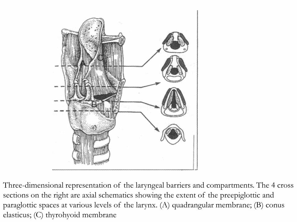
Three-dimensional representation of the laryngeal barriers and compartments. The 4 cross sections on the right are axial schematics showing the extent of the preepiglottic and paraglottic spaces at various levels of the larynx. (A) quadrangular membrane; (B) conus elasticus; (C) thyrohyoid membrane
• Laryngeal carcinoma is associated with a high incidence to develop a second primary 6-12 % , in the areodigestive tract especially bronchogeniccarcinoma.
• It occurs either synchronously (at the same time) in 0.5-1%, in 0.5-1%,
• or metasynchronously in 5-10% of cases. Therefore, proper assessment and prolonged follow up of laryngeal carcinoma is essential.

•Lymphatic• By permeation , Embolism , perilymphatic
• Most important factor in prognosis
• +ve LN = drop 50% in prognosis
• Supraglottic to upper deep cervical LNs (30-50 %)
• Glottic has no lymphatic drainage (early glotticcancer doesn’t spread to LNs) except after cancer doesn’t spread to LNs) except after spread to the adjacent areas.
• Subglottic to the lower cervical LNs then paratracheal LN and superior mediastinal LN.
•Blood: distant
•1% lung, bones, liver

There are several factors that determine cervical lymph node metastasis:
1. Site of the tumour
2. Size of the tumour: The incidence of nodal metastasis increases, with increased size of the tumour.
3. Stage: nodal metastasis is more in advanced (T3 & T4) lesions, than in early (Tl & T2) lesions.
4. The histologic characteristics of the tumor, such as 4. The histologic characteristics of the tumor, such as extracapsular spread in nodal metastases, angiolymphatic invasion, perineural spread, and a high histologic grade
5. Tumour differentiation: It is more with poorly, than with well differentiated tumours (according to Broders classification).
6. The incidence of nodal metastasis increases with cartilage invasion, extralaryngeal spread, and neural infiltration.

• Clinical picture:
Symptoms
1.Hoarseness of voice which is progressive and unremitting.
• It is the earliest symptom in glottic tumours, and may presist as the only symptom for several months.
• Early in glottis carcinoma, late in supraglottic and subglotticcarcinoma
• Supraglottic tumours produce a muffled voice rather than • Supraglottic tumours produce a muffled voice rather than hoarseness.
• An important consideration is the cancer which develops in one who sufferes from chronic laryngitis, as they are at risk from delay in diagnosis, because they already have hoarseness of voice.
• Any patient presenting with hoarseness of voice for more than two weeks should be examined and investigated properly.

•May be due to ;
•The slightest change in contour, thickness, or vibratory characteristics of the vocal cord will result in perceived changes in the voice.
•Interference with mucosal vibration.
•Invasion of vocalis muscle
•Invasion of cricoarytenoid joint•Invasion of cricoarytenoid joint
•Neural invasion

2.Dyspnoea and stridor due to airway obstruction.
• Most common in subglottic and may be the only symptoms. It occurs early in supraglottictumours.
• Late symptoms as sequale of neglected dysphonia ,indicates advanced T
• It is due to enchroachement upon the airway by:
Large tumour Large tumour
Fixation of the arytenoid or vocal cord.
Accumulated secretion and debris
Secondary odema
•Acute airway obstruction may be precipitated by :
•Infection
•Instrumentation

3. Pain ( odynophagia & otalgia )
• Late symptoms
• More typical in supraglottic lesions
• Referred ear pain is particularly sinister and should always promote a high suspicious to cancer.
• If accentuated by swallowing may indicate invasion of deep structures e.g base of the tongue and/ or hypopharynx or invasion of laryngeal skeleton.
Sensation of discomfort in the throat on swallowing, Sensation of discomfort in the throat on swallowing, complain of a foreign-body feeling in the throat may be an early symptom of supraglottic tumours.
Referred otalgia to the ipsilateral ear along the Arnold's branch of vagus.
Localized pain is rare, it may occur with supraglottictumour, or may be due to perichondritis.
4.Cough and irritation in the throat :
•May be the early non descriptive symptoms.
•Usually deveiops with supraglottic carcinoma due to involvement of the superior laryngeal nerve (especially in marginal zone)
5.Swelling (lump) in the neck or larynx mainly due to:
• Lymph node metastasis in the upper or middle • Lymph node metastasis in the upper or middle cervical groups.
• It may be due to direct penetration of the tumouroutside larynx invading thyroid cartilage and extralaryngeal spread.
• Perichondritis and abscess formation may give rise to painful, tender, oedematous swelling.

6.late symptoms:
•Dysphagia due to invasion of the hypopharynx.
•Haemoptysis :
Rare
Usually with large supraglotticUsually with large supraglotticcarcinoma especially in lesion of margins of epiglottis.
• Anorexia and cachexia.
•Foetid breath.

Examination:
•General: Assess general condition
• To assess the patient general condition
• Chest examination
• To detect possible distant metastasis
•Local :• Local :
– The nose, oral cavity and the oropharynx must be examined to detect :• Possible second primary cancer.
• Granulomatous disease.
• Dental status for sepsis and oral hygiene must also be assess
•Neck:
Broadending of thyroid cartilage
Tenderness = perichondritis
Movement of lx: T extension
Click: post cricoid extension
Neck: Lymph nodesNeck: Lymph nodes
•Laryngeal Examination:
• Shows site, extension of the lesion in the larynx
•Mobility of vocal cord
•Shows extension to pharynx

Investigations
•General:
Assess general condition: ESR, CBC, ECG, DM, Liver
DD: serology syphilis, TB sputum
Chest x ray, pulmonary function test
Metastasis work-up
Chest X ray
Abdominal US
Bone scan

• Imaging:
Plain x ray lateral neck
Barium swallow: pharynx
CT: best study:
Tumor extension
Cartilage involvement LN
Carotid artery, jagular vein
•Endoscopy: MLS
Assess tumor extension
Biopsy: confirm diagnosis, type of cancer, differentiation


Diagnosis difficulities:
• Negative biopsy
• Keratosis
• Previous radiation• A low-grade perichondritis may prevent a larynx from
returning to normal.
• CT scan , another deep biopsy and close follow up are • CT scan , another deep biopsy and close follow up are indicated PET is diagnostic
• Hidden areas :• Subglottic region, laryngeal surface of epiglottis , laryngeal
ventricles
• Miscellaneous conditions• such as chronic laryngitis, tuberculosis, syphilis and benign tumours
may give rise to diagnostic confusion or difficulty so biosy is recommended

Classification: TNM
•Universal method for tumor classification
•Standardization
•Decision making
•Predict prognosis
•T: tumor grade: T1,2,3,4
•N: nodes: N1,2,3
•M: metastasis: M0,1
•Stage: combination of TNM, I, II, III, IV

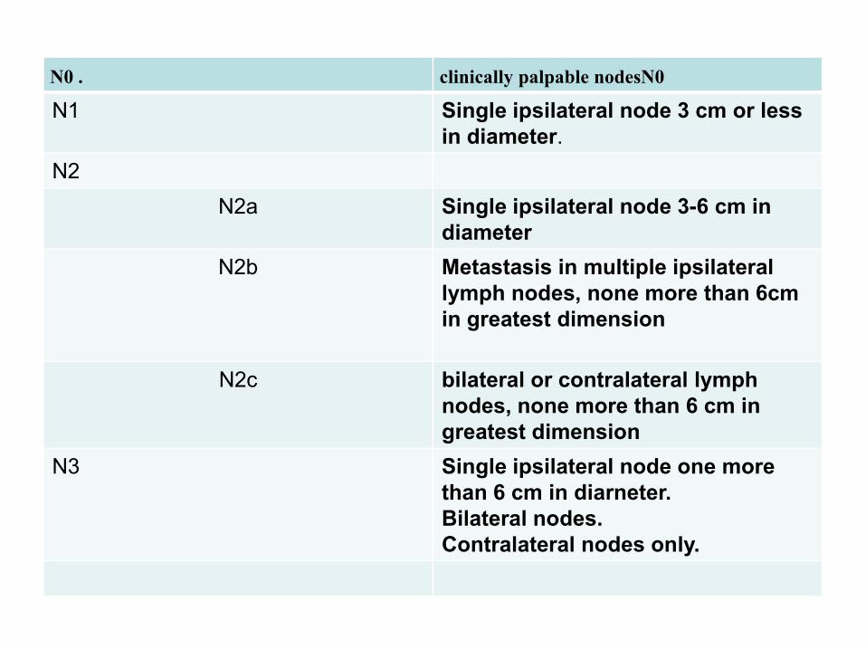
clinically palpable nodesN0N0 .
Single ipsilateral node 3 cm or less in diameter.
N1
N2
Single ipsilateral node 3-6 cm in diameter
N2a
Metastasis in multiple ipsilateral lymph nodes, none more than 6cm in greatest dimension
N2b
in greatest dimension
bilateral or contralateral lymph nodes, none more than 6 cm in greatest dimension
N2c
Single ipsilateral node one more than 6 cm in diarneter. Bilateral nodes. Contralateral nodes only.
N3

M
No evidence of distant metastasis.
M 0
Tumour with distant metastasis.
M 1.


• Treatment
• CA larynx potentially curable disease
Early diagnosis
Proper assessment
• Aim:
•Locoregional control
•Distant control
•Rehabilitation
• Treatment modalities:• Treatment modalities:
Curative:
Surgical/laser
Radiotherapy
Combination
Rehabilitation:
Palliative: inoperable disease
Distant metastasis
Advanced local disease
Poor general condition

Surgical Treatment
Early glottic T:
•Cordectomy, partial vertical laryngectomy
T1 lesion, one vocal cord, mobile, not reaching anterior commissurel or arytenoid
•Laser
•Radiotherapy: better voice•Radiotherapy: better voice
Early supraglottic T:
•Supraglottic: horizontal partial laryngectomy:
•Localized supraglottic lesions, normal cord mobility

•Advantages of partial resections:
•Preservation of the larynx: voice
•No permanent tracheostomy
•Better QOL
•Disadvantages of partial resections:
•Prolonged hospitalization•Prolonged hospitalization
•High rate of recurrence
•Strict follow up is mandatory
•Pulmonary complication: aspiration: Horizontal
•Difficult technically: need training

•Late CA LX: Total laryngectomy
•Indications:
• Cord fixation
• Large tumors + extralaryngeal
• Cartilage involved
• Recurrent after RTH
•Advantages of total resections:•Advantages of total resections:
• Radical surgery gives better cure?????
• Disadvantages of total resections:
• Loss of voice
• Permanent tracheostomy
• QOL: Limited activities: swimming, inability to increase intra-thoracic pressure
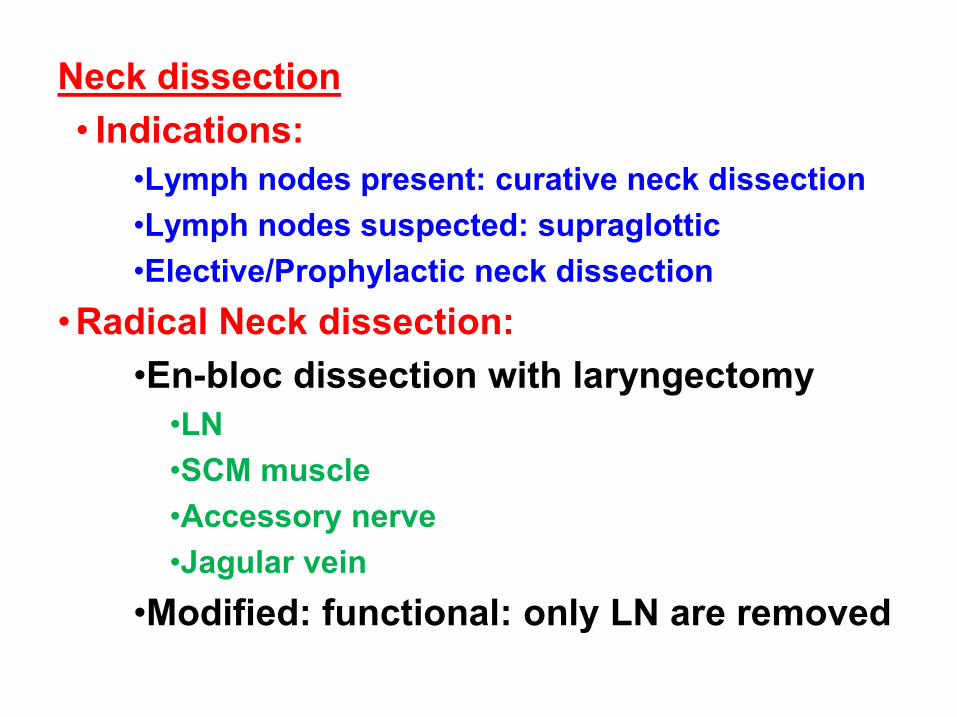
Neck dissection
• Indications:
•Lymph nodes present: curative neck dissection
•Lymph nodes suspected: supraglottic
•Elective/Prophylactic neck dissection
•Radical Neck dissection:
•En-bloc dissection with laryngectomy•En-bloc dissection with laryngectomy
•LN
•SCM muscle
•Accessory nerve
•Jagular vein
•Modified: functional: only LN are removed


TYPES OF LARYNGECTOMIES
1.Function-preservation laryngectomy:
•In conservation laryngeal surgery:
a. part of larynx remains
b. no permanent tracheostomyb. no permanent tracheostomy
c. patient can phonate
• Types of conservation laryngeal surgery:
1- For supraglottic cancer:
supraglottic (horizontal) laryngectomy
2-For glottic cancer:
a. cordectomy (microlarynx or laser )
b. hemilaryngectomy ( vertical )


2.Radical surgery (Total laryngectomy):
• In total laryngectomy:
a. All larynx is removed
b. There is a permanent tracheostomy
c. Patient can not phonate unless voice restoration is done.restoration is done.

Radiotherapy
•Dose: Cobalt 60 or linear acceleration/ 5500-7000 rad/ fractionated
on 5 session per week. Each session is 300-500 rad
• Indications:Early tumors with mobile cord
When surgery is contraindicated
Recurrence after surgeryRecurrence after surgery
Non-resectable LN
• Contraindications:Large tumors with fixed cord
Cervical LN
Cartilage involved

•Complications:
Skin coloration
Laryngeal edema
Xerostomia
Anorexia, malaise
Radio-perichondritisRadio-perichondritis
•Disadvantages:
Prolonged treatment
Follow up for residual or recurrent tumor is
difficult because of edema
Radiotherapy can not be repeated

Rehabilitation
• Partial laryngectomy:
• Voice therapy
• Total laryngectomy:
• Physiological methods: Esophageal • Physiological methods: Esophageal speech
• Surgical methods: Tracheo-esophageal valve
• Artificial methods: electrolarynx.
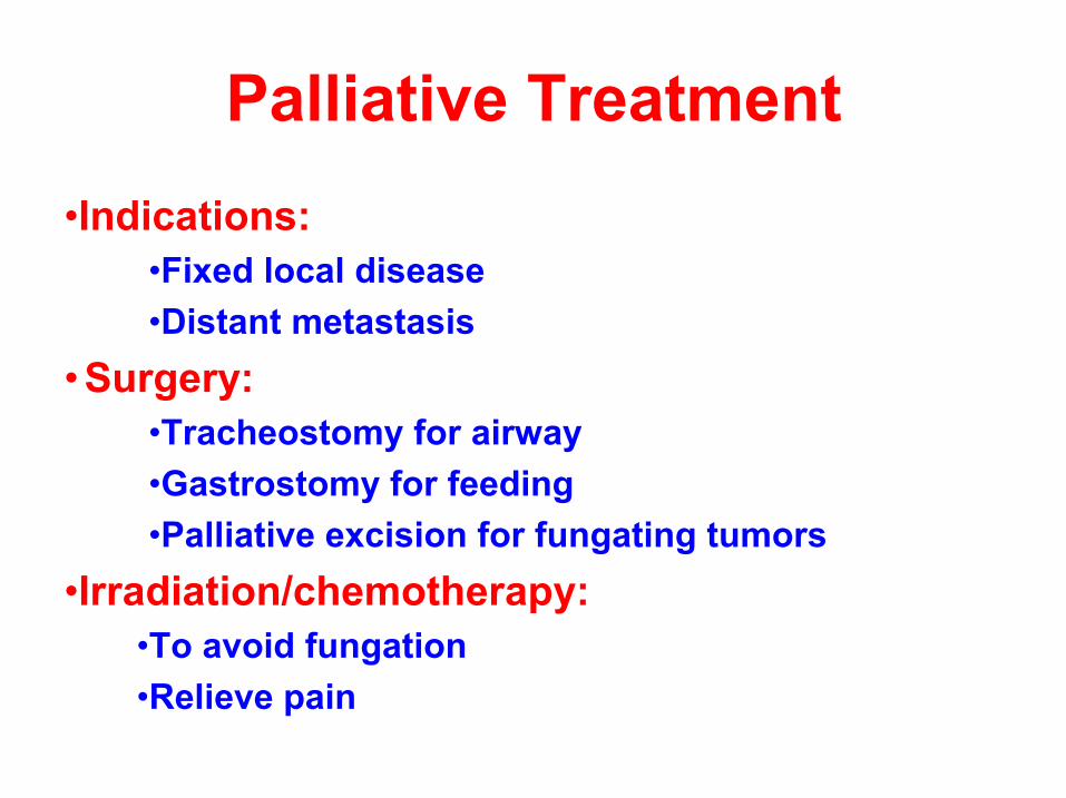
Palliative Treatment
•Indications:
•Fixed local disease
•Distant metastasis
•Surgery:•Surgery:
•Tracheostomy for airway
•Gastrostomy for feeding
•Palliative excision for fungating tumors
•Irradiation/chemotherapy:
•To avoid fungation
•Relieve pain

•Medical:
•Antibiotics for infection
•Pain control

Prognosis
•Glottic good prognosis
Early symptoms
No lymphatic
5 year survival 85-90% 5 year survival 85-90%
•Late cases: worse prognosis
T4: 30% 5 year survival