Embed Size (px)
Citation preview
Laryngopharyngeal RefluxGuest Editors: Wolfgang Issing, Petros D. Karkos, Oliver Reichel, and Marcus Hess
International Journal of Otolaryngology

Laryngopharyngeal Reflux
International Journal of Otolaryngology
Laryngopharyngeal Reflux
Guest Editors: Wolfgang Issing, Petros D. Karkos,Oliver Reichel, and Marcus Hess

Copyright © 2012 Hindawi Publishing Corporation. All rights reserved.
This is a special issue published in “International Journal of Otolaryngology.” All articles are open access articles distributed under theCreative Commons Attribution License, which permits unrestricted use, distribution, and reproduction in any medium, provided theoriginal work is properly cited.

Editorial Board
Rolf-Dieter Battmer, GermanyRobert Cowan, AustraliaP. H. Dejonckere, The NetherlandsJoseph E. Dohar, USAPaul J. Donald, USAR. L. Doty, USADavid W. Eisele, USAAlfio Ferlito, Italy
Ludger Klimek, GermanyLuiz Paulo Kowalski, BrazilRoland Laszig, GermanyCharles Monroe Myer, USAJan I. Olofsson, NorwayRobert H. Ossoff, USAJeffrey P. Pearson, UKPeter S. Roland, USA
Leonard P. Rybak, USAShakeel Riaz Saeed, UKMichael D. Seidman, USAMario A. Svirsky, USATed Tewfik, CanadaPaul H. Van de Heyning, BelgiumBlake S. Wilson, USAB. J. Yates, USA

Contents
Laryngopharyngeal Reflux, Petros D. Karkos, Wolfgang Issing, Oliver Reichel, and Marcus HessVolume 2012, Article ID 926806, 2 pages
Chronic Cough, Reflux, Postnasal Drip Syndrome, and the Otolaryngologist, Deborah C. Sylvester,Petros D. Karkos, Casey Vaughan, James Johnston, Raghav C. Dwivedi, Helen Atkinson, and Shah KortequeeVolume 2012, Article ID 564852, 5 pages
Impact of Laparoscopic Fundoplication for the Treatment of Laryngopharyngeal Reflux: Review of theLiterature, Guilherme da Silva Mazzini and Richard Ricachenevsky GurskiVolume 2012, Article ID 291472, 4 pages
Eosinophilic Esophagitis for the Otolaryngologist, Petros D. Karkos, R. Srivastava, S. Kaptanis,and C. VaughanVolume 2012, Article ID 181402, 5 pages
Endoscopic Laryngeal Findings in Japanese Patients with Laryngopharyngeal Reflux Symptoms,Nobuhiko Oridate, Ryoji Tokashiki, Yusuke Watanabe, Aki Taguchi, Osamu Kawamura,and Kazuma FujimotoVolume 2012, Article ID 908154, 4 pages
Reflux Revisited: Advancing the Role of Pepsin, Karna Dev Bardhan, Vicki Strugala, and Peter W. DettmarVolume 2012, Article ID 646901, 13 pages
Hindawi Publishing CorporationInternational Journal of OtolaryngologyVolume 2012, Article ID 926806, 2 pagesdoi:10.1155/2012/926806
Editorial
Laryngopharyngeal Reflux
Petros D. Karkos,1, 2 Wolfgang Issing,3 Oliver Reichel,4 and Marcus Hess5
1 Department of Otolaryngology-Head Neck Surgery, Queen Alexandra Hospital, Portsmouth PO63LY, UK2 Department of Otolaryngology, Aristotle university of Thessaloniki, Thessaloniki, Greece3 Department of Otolaryngology-Head Neck Surgery, The Freeman Hospital, Newcastle Upon Tyne, UK4 Department of Otolaryngology-Head Neck Surgery, Schwarzwald-Baar Hospital, Villingen-Schwenningen, Germany5 Department of Otolaryngology-Head Neck Surgery, University Medical Center Hamburg-Eppendorf, Hamburg, Germany
Correspondence should be addressed to Petros D. Karkos, [email protected]
Received 18 July 2012; Accepted 18 July 2012
Copyright © 2012 Petros D. Karkos et al. This is an open access article distributed under the Creative Commons AttributionLicense, which permits unrestricted use, distribution, and reproduction in any medium, provided the original work is properlycited.
Laryngopharyngeal reflux (LPR) is here to stay. Despiteadvances in the role of pepsin, better drugs, and less invasivetests, diagnosis is still difficult and is still in many places—based on empiric treatment rather than robust evidence.
In this special issue on LPR, we attempt to reviewsome hot topics regarding this entity which still puzzlesotolaryngologists all over the world.
The issue of chronic cough, postnasal drip, and refluxis quite a controversial topic and remains a therapeuticchallenge. Chronic cough can be associated with manydiseases that often overlap more than one medical specialty.A detailed assessment of the patient with chronic coughrelies on a multidisciplinary approach and close cooperationbetween pulmonary medicine, gastroenterology, and oto-laryngology. Gastroesophageal reflux (GERD) and postnasaldrip syndrome account for a significant number of casesof chronic nonproductive cough seen in otolaryngologypractice. Each may, alone or in combination, contribute tocough even when clinically silent and failure to recognisetheir contribution may lead to unsuccessful treatment.Many of these patients are notoriously difficult to diagnoseand treat but the literature suggests that a systematic andthorough approach in a multidisciplinary setting can leadto successful diagnosis and treatment in the majority ofpatients.
The role of surgery in management of LPR is also amatter of debate in the literature. Results of laparoscopicfundoplication for the treatment of classic GERD are wellestablished. In theory, surgery for persistent LPR after
failure of medical treatment should be equally as effective,if we agree on the notion that LPR and GERD sharecommon pathophysiology. Many studies have demonstratedsymptomatic improvement after surgical fundoplication.However, current knowledge does not allow this conclusion.Large multicenter, randomized control trials are needed,focusing on diagnostic tools to improve selection criteria,presenting standard endpoints and long-term follow-up.
Eosinophilic esophagitis (EE) is a great reflux mimic andoften presents with dysphagia, recurrent food bolus obstruc-tion and GERD-like symptoms. Despite higher awareness,the literature suggests that EE remains a commonly misdi-agnosed condition especially in the otolaryngology commu-nity. The treatment though differs than the LPR treatmentand EE should be part of the differential diagnosis whenfaced with “difficult to treat LPR or GERD”. The introductionof Transnasal Esophagoscopy in the ENT office over thelast decade has meant that increasingly more laryngologistsbecome accustomed in recognising esophageal pathologyincluding EE.
The presence of specific endoscopic laryngeal findingsin patients with suspected LPR has been well documentedin the literature. Because the first-line therapy for LPR isconsidered to be proton pump inhibitors, there are manystudies comparing the endoscopic laryngeal findings beforeand after acid suppression therapy.
Finally, the role of pepsin and the fact that LPR ismuch more dependent on pepsin-mediated damage in thelaryngeal and airway mucosa than on acid have been well

2 International Journal of Otolaryngology
described over the last few years. The crucial role of pepsin inLPR may, in turn, stimulate the development of drugs whichspecifically target this molecule. This may radically enhanceour knowledge and management of this condition.
Petros D. KarkosWolfgang Issing
Oliver ReichelMarcus Hess
Hindawi Publishing CorporationInternational Journal of OtolaryngologyVolume 2012, Article ID 564852, 5 pagesdoi:10.1155/2012/564852
Review Article
Chronic Cough, Reflux, Postnasal Drip Syndrome, andthe Otolaryngologist
Deborah C. Sylvester,1 Petros D. Karkos,1, 2 Casey Vaughan,1 James Johnston,2
Raghav C. Dwivedi,2 Helen Atkinson,1 and Shah Kortequee1
1 Department of Otolaryngology Head Neck Surgery, Bradford Royal Infirmary, Bradford BD96RJ, UK2 Department of Otolaryngology Head Neck Surgery, Queen Alexandra Hospital, Portsmouth PO63LY, UK
Correspondence should be addressed to Petros D. Karkos, [email protected]
Received 4 January 2012; Accepted 7 February 2012
Academic Editor: Wolfgang Issing
Copyright © 2012 Deborah C. Sylvester et al. This is an open access article distributed under the Creative Commons AttributionLicense, which permits unrestricted use, distribution, and reproduction in any medium, provided the original work is properlycited.
Objectives. Chronic cough is a multifactorial symptom that requires multidisciplinary approach. Over the last years, generalpractitioners refer increasingly more chronic cough patients directly to the otolaryngologist. The aim of this paper is to highlightthe issues in diagnosis and management of chronic cough patients from the otolaryngologist perspective. Design. Literature review.Results. Gastroesophageal reflux and postnasal drip syndrome remain one of the most common causes of chronic cough. Betterdiagnostic modalities, noninvasive tests, and high technology radiological and endoscopic innovations have made diagnosis ofthese difficult-to-treat patients relatively easier. Multidisciplinary assessment has also meant that at least some of these cases can bedealt with confidently in one stop clinics. Conclusions. As the number of referrals of chronic cough patients to an Ear Nose ThroatClinic increases, the otolaryngologist plays a pivotal role in managing these difficult cases.
1. Introduction
Chronic cough is a persistent and frustrating symptom formany adults and children and a frequent reason for primaryor secondary care visits or referrals. This condition generatessignificant healthcare and economic cost and is associatedwith a spectrum of disorders across multiple medical special-ties and can provide significant challenges for the involvedphysician or surgeon. Chronic cough is associated with dete-rioration in the quality of patients’ lives. Associated symp-toms and negative outcomes with this condition include lossof sleep, exhaustion, irritability, urinary incontinence, coughsyncope, social disability, and inability to perform daily activ-ities. Many patients experience chronic cough secondary toanother medical condition, such as COPD, asthma, rhinos-inusitis, Gastroesophageal reflux syndrome (GERD), postn-asal drip syndrome (PNDS), or unknown etiology. GERD isthought to be the most common cause of chronic cough ina nonsmoker nonasthmatic individual.
Thorough assessment of a patient with a chronic coughrelies on a multidisciplinary approach. The otolaryngologist
should be familiar with the diagnostic algorithm of chroniccough patients and should work closely with the gastroen-terologist and the pulmonologist, ideally in “cough clinics,”to confidently diagnose and treat these patients.
1.1. Reflux and Chronic Cough. Chronic nonspecific cough,defined as a nonproductive cough in the absence of identi-fiable respiratory disease or known cause [1] persisting formore than three to eight weeks [2], poses a significant burdento healthcare costs and considerably impairs quality of life.Gastroesophageal reflux disease (GERD) represents one ofthe three main causes of chronic cough (along with asthmaand upper airways cough/postnasal drip syndrome), impli-cated in up to 41% of chronic cough patients [3]. The clinicalfeatures of GERD-related cough include heartburn, regurgi-tation, and/or worsening of cough after foods or medicationsknown to decrease lower esophageal sphincter-pressure,with extraesophageal manifestations such as hoarseness,wheezing, sore throat, chest pain, and globus also described.
Whilst classic GERD symptoms are present in 6–10% ofchronic cough patients, GERD is clinically silent in up to
2 International Journal of Otolaryngology
75% of patients with GERD-related cough [4]. Diagnosis ofGERD is frequently based on the clinical responses of coughto antireflux therapy rather than on objective assessments ofGERD per se. Furthermore, an increased understanding ofthe pathophysiology of GERD and in particular the specificphenomenon of laryngopharyngeal reflux (LPR), has high-lighted the complexity of this condition, with the need for in-dividual patients assessment and tailoring of therapy becom-ing apparent.
Coughing may be provoked by reflux via a number ofmechanisms. The regurgitation of gastric contents into thelaryngopharynx can cause mechanical or pH-sensitive stim-ulation, with chronic inflammation leading to the sensitisa-tion of peripheral nerves mediating cough [5]. This may havean acid or nonacid (namely, bile and pepsin) basis. Adha-mi et al. [6] demonstrated that bile can injure the laryngealepithelium but only in an acidic environment, and further-more, Sasaki et al. [7] were able to demonstrate histologicallaryngeal injury in a rat model following bile exposurein neutral environments. Pepsin, the principal proteolyticenzyme of the stomach, is predominantly active in acidic pHand has been shown to cause laryngeal injury in this state [6];however proteolytic activity is still present up to pH 7 andcan be reactivated [8]. Johnston et al. [8] found the presenceof pepsin in the larynx of patients with the clinical diagnosisof LPR but not in controls and in these same patients, pepsinwas absent in their esophageal epithelium [9]. It has beensuggested that coughing can also be induced by “micro” or“silent” aspiration, caused by the direct activation of trach-eo-bronchial receptors by reflux entering the airway. Distalesophageal reflux may also induce coughing through vagalstimulation known as the oesophago-bronchial reflex [10],Ing et al. [11], demonstrating that infusing acid into theoesophagus of chronic cough patients increases coughing.Additionally whilst infusion of acid (compared to saline)into the oesophagus of those GERD patients without chroniccough had no effect, a sensitised cough reflex to capsaicin wasseen in those GERD patients with chronic cough [12].
An alternative pathophysiology is that coughing can infact be the causation in reflux: increased intra-abdominalpressure during strenuous coughing episodes negativelyimpacting the lower esophageal sphincter, possibly by way ofa positive feedback loop [13].
As discussed above, reflux associated cough can be alaryngopharyngeal or distal esophageal phenomena. LPR hasdistinct features, as first identified by Koufman and collea-gues. In a combined reported series of 899 patients, throatclearing was a complaint of 87% of LPR patients versus 3%of those with GERD, while only 20% of LPR patients com-plained of heartburn versus 83% in the GERD group [5]. Dif-ferences in body mass index (BMI) between GERD and LPRpatients have also been highlighted; in a retrospective studyof 500 patients attending for pH probe studies, the mean BMIof isolated LPR patients was 25.9 compared to 28.3 for thosewith GERD [14].
Identifying GERD as the cause of a chronic cough canbe challenging. Esophageal pH testing can demonstrate anincreased number of reflux events, prolonged exposure ofthe esophageal mucosa to reflux, or more convincingly a
significant temporal association between reflux events andcough. Although esophageal pH testing has a sensitivity ofapproximately 90% for the evaluation of chronic cough,specificity ranges from 66% to 100% [15, 16]. Addi-tional diagnostic tests include inhaled tussigenic challenges,endoscopy, examining bronchoalveolar lavage fluid, and/orsputum for lipid laden macrophages, barium swallow, Bern-stein test, radioisotope scintiscan, and radionucleotide emp-tying studies with solids [17]. Examination of the larynxmay reveal evidence of LPR: key examination findings beingvocal cord oedema and erythema as well as medial vocalcord erythema [18, 19]. Findings should, however, be takenin context: Hicks and colleagues finding that almost 80% ofstudy participants had a least one reflux-attributable findingon laryngoscopy when 100 healthy volunteers were examined[20].
1.2. Treatment of GERD-Associated Cough. Patient counsell-ing is essential in reducing GERD and related LPR. Dietaryadvice includes the avoidance of a high-fat diet and losingweight if obese, avoiding eating two hours before bedtimeand refraining from caffeine, carbonated drinks, alcohol, andcitrus products [21]. Patients should also be asked to refrainfrom smoking and elevate the head of the bed by 15 cm. Somemedications are associated with increase GERD, namely,anticholinergics, beta-agonists, bisphosphonates, calcium-channel blocker, corticosteroids, benzodiazepines, oestro-gens, opiates, progesterone, prostaglandins, and theophylline[17]. Other recommendations include nasal continuouspositive airway pressure if obstructive sleep apnoea is present[22] and avoiding exercise that may increase intra-abdominalpressure [23].
Proton pump inhibitors (PPIs) have commonly beenthe mainstay empirical treatment for GERD-related cough.Given the difficulty in clearly diagnosing this condition,Irwin [21] has described the clinical profile of such patientsin whom empirical therapy should be considered; those notexposed to environmental irritants, not a present smoker, noton an ACE inhibitor, with a normal/stable chest radiograph,and in whom symptomatic asthma, upper airways cough sy-ndrome, and nonasthmatic eosinophilic bronchitis has beenruled out. The use of empirical therapy has, however, beenquestioned. In a meta-analysis of 5 randomised controlledtrials on GERD treatment for cough in adults and childrenwithout primary lung disease, Chang et al. [24] found thatthere was no difference in cough resolution for patients whoreceived a placebo versus a PPI (OR 0.24 (95% CI 0.04 to1.27). There was, however, a significant difference in second-ary outcomes of mean cough score (mean difference of−0.51(−1.02 to 0.01)) and change in cough score (−0.29 (−0.62to 0.04)) at the end of the trial. This led the authors toconclude that the use of PPI had “some effect in some adults.”More recently, a Cochrane Database Systematic review byChang and colleagues [2] including 9 randomised controlledtrials of PPIs for adults with chronic cough found that usingintention-to-treat, pooled data from studies resulted in nosignificant difference between treatment and placebo in totalresolution of cough (OR 0.46; 95% CI 0.19 to 1.15 no overallsignificant improvement in cough outcomes (end of trial or
International Journal of Otolaryngology 3
change in cough scores). There was, however, a significantimprovement in cough scores at end of intervention (twoto three months) in those receiving PPI (standardised meandifference −0.41; 95% CI −0.75 to −0.07) using genericinverse variance analysis on cross-over trials. The authorswere unable to conclude definitely that GERD treatmentwith PPIs is universally beneficial for cough associated withGERD. Despite the current lack of evidence for definite treat-ment of empiric treatment, published guidelines from theACCP [21] and BTS [25] suggest that PPI therapy should becommenced, for example, omeprazole 20–40 mg twice dailyor equivalent taken before meals for at least 8 weeks [25].
Of particular interest in nonacidic refux, medicationssuch as Gaviscon or Gaviscon Advance, which act by forminga raft or physical barrier to reflux present a supplementary oreven alternative treatment option. McGlashan et al. [26] con-ducted a randomised controlled trial of Gaviscon Advancein 49 patients with a diagnosis LPR (based on the refluxsymptom index (RSI) and the reflux findings score (RFS)).Patients were assessed pretreatment and at 2, 4, and 6 monthsafter treatment. Significant differences in the mean (SD)between treatment and control were observed for RSI at the2-month (11.2 (7.0) versus 16.8 (6.4), P = 0.005) and 6-month (11.2 (8.1) versus 18.3 (9.4), P = 0.008) assessmentsand for RFS at the 6-month (7.1 (2.8) versus 9.5 (3.4), P =0.005) assessment. The details of the cough component of theRSI were not, however, detailed further in the report.
Gastroesophageal dysmotility has been implicated in thepathophysiology of GERD via abnormalities of delayed gas-tric emptying and reduced pressure or inappropriate transi-ent relaxation of the lower esophageal sphincter [27]. Severalprokinetic agents (e.g., bethanechol, metoclopramide, dom-peridone, cisapride, and macrolides such as erythromycin)can stimulate gastrointestinal motility and have, therefore,been proposed as useful adjuncts to antireflux medication.The evidence base for this lies only in unblinded, uncon-trolled studies, where, when in combination PPIs for treatingGERD-associated cough, cough or hoarseness improved by70% to 100% [10, 28–31]. In the recent Cochrane review,Chang et al. [2] found insufficient data to evaluate theevidence for the use of prokinetic agents in chronic cough.However, interestingly in 56 patients diagnosed with GERD-related cough, 24 responded to a PPI alone; however, 18 ofthe remaining patients improved with the addition of metoc-lopramide or cisapride [32]. These drugs, however, may havesignificant side-effects: erythromycin, for example, oftencauses nausea and abdominal pain and cisapride was with-drawn from the US market due to safety concerns.
Although surgery is more traditionally used to treat themore typical reflux symptoms, it may be of some value inthe management of reflux-related cough. Studies relating tothe outcomes of surgical treatment of GERD are, however, ofquestionable value as they suffer from lack of controls andblinding, use differing postoperative evaluation criteria, andare typically based on a highly selective group of patients.Kaufman and colleagues [33] reported their long-term(mean 53 months) outcomes of 128 patients treated with lap-aroscopic antireflux surgery. Cough and hoarseness was im-proved in 65% to 75% of cases compared to heartburn and
regurgitation in over 90% of subjects. In their review oftreatment options for GERD-related cough, Chandra andHarding [17] summarised the finding of 9 prospective stud-ies of surgical management, reporting that 586 of 689 sur-gically treated patients had a “significant cough response.”
GERD remains one of the leading causes of chroniccough; however, the difficulty in diagnosing this condition,especially as “classic” reflux symptoms are often absentmeans that it can be overlooked. The mainstay of treatment,for now, remains as lifestyle modification, dietary advice,and medical therapy. The role of traditional empirical treat-ment with PPIs is questionable, with evidence from ran-domised trials implying that there is some benefit in the rightpatient group. Identifying this patient group is, therefore,imperative: careful clinical history taking and laryngeal eval-uation along with objective reflux assessment being key. Theuse of alginate preparations seems to be quite popular in thelast few years, although evidence is currently lacking. Sur-gical management, although not as useful for cough symp-tomatology as for classic symptoms also has a role in patientsresistant to medical therapy.
2. Postnasal Drip Syndrome(or Upper Airway Cough Syndrome)
Postnasal drip (PND) or catarrh is the drainage of secretionsfrom the nose or paranasal sinuses into the pharynx. Clini-cally, the diagnosis of PND syndrome (PNDS) is very vague,made on history and examination and relies on the reportingof the patient of this sensation of something “dripping downthe throat,” rhinorrhoea and constant throat clearing [34].Nasendoscopy revealing rhinitis and mucopurulent secre-tions is suggestive, although not diagnostic. The issues whenattempting to diagnose PNDS, is that there are no objectivesensitive or specific tests and no way to quantify the amountof catarrh or to prove that it is directly responsible for causingcough. PNDS is associated with very nonspecific symptomsand a definitive diagnosis of PND-induced cough cannot bemade from the history and examination findings alone.
The differential diagnosis of PNDS-induced coughincludes all other causes of rhinitis including, allergic rhini-tis, perennial nonallergic rhinitis, bacterial sinusitis, allergicfungal sinusitis, rhinitis due to anatomic sinonasal abnor-malities, rhinitis due to physical or chemical irritants, occup-ational rhinitis, rhinitis medicamentosa, and rhinitis of pre-gnancy.
Another issue when attempting to diagnose PNDS-induced cough is that GERD is often associated with a highprevalence of upper respiratory symptoms and therefore caneither coexist or mimic PNDS [35]. The introduction of the-more widely accepted in Americas-term of “Upper AirwayCough Syndrome” (UACS) was made based on the need toanswer the question whether “the conditions listed aboveactually produce cough through a final common pathway ofPND or whether, in fact, in some circumstances they causeirritation or inflammation of upper airway structures thatdirectly stimulate cough receptors and produce cough indep-endently of or in addition to any associated PND” [35].
4 International Journal of Otolaryngology
It is obvious that because there are no objective tests fordiagnosing PND, treatment is often based on the specificdisease that is present. For example, avoidance of specific al-lergens after allergy testing has been done, nasal steroid treat-ment and antihistamines, treatment of concomitant infec-tion, and correction of any associated sinonasal anatomicalabnormalities can have an indirect effect on the managementof PND-induced cough.
The American College of Chest Physicians recommendsan empiric trial of therapy for UACS because improvementor resolution of cough in response to specific treatment is thepivotal factor in confirming the diagnosis of UACS as a causeof cough. That should especially be the case if no specificcause can be elicited from the history and examination [36].
A usual empiric therapy involves a first generation anti-histamine/decongestant. If a patient has resolution or partialresolution of cough, then UACS is considered to have beena cause of cough and antihistamine therapy is continued.Marked improvement or resolution of cough may take seve-ral weeks and occasionally as long as a few months [37].
If there is no response with a first-generation antihis-tamine, then the patient should undergo sinus imaging. Ch-ronic sinusitis causes a productive cough or can be clinicallysilent, in that the cough can be nonproductive, and none ofthe typical findings associated with acute sinusitis may bepresent [36, 38].
Allergy skin testing, measurement of serum Ig levels tosee whether (acquired) hypogammaglobulinemia is present,evaluation of the patient’s home and workplace if there isa potential environmental cause for persistent upper air-way symptom are all reasonable diagnostic strategies espe-cially if there is lack of response to sinusitis treatment. If nase-ndoscopy reveals nasal polyps, in the absence of any contra-indication, the patient should undergo a standard aspirinchallenge. If the results of the challenge are positive, thepatient should undergo desensitization, followed by theconsideration of chronic aspirin therapy unless it is con-traindicated.
3. Conclusions
Chronic cough can be associated with many diseases thatoften overlap more than one medical specialty. A detailedassessment of the patient with chronic cough relies on amultidisciplinary approach and close cooperation betweenpulmonary medicine, gastroenterology, and otolaryngology.Gastrooesophageal reflux and postnasal drip syndromeaccount for a significant number of cases of chronic non-productive cough seen in otolaryngology practice. Each may,alone or in combinationcontribute to cough even whenclinically silent, and failure to recognise their contributionmay lead to unsuccessful treatment. Many of these patientsare notoriously difficult to diagnose and treat but the liter-ature suggests that a systematic and thorough approach in amultidisciplinary setting can lead to successful diagnosis andtreatment in the majority of patients.
Conflict of Interests
The authors declare no conflict of interests.
References
[1] A. Chang and M. Asher, “A review of cough in children,” Jour-nal of Asthma, vol. 38, no. 4, pp. 299–309, 2001.
[2] A. B. Chang, T. J. Lasserson, J. Gaffney, F. L. Connor, and L. A.Garske, “Gastro-oesophageal reflux treatment for prolongednon-specific cough in children and adults,” Cochrane Databaseof Systematic Reviews, vol. 1, Article ID CD004823, 2011.
[3] A. H. Morice, “Epidemiology of cough,” Pulmonary Pharma-cology and Therapeutics, vol. 15, no. 3, pp. 253–259, 2002.
[4] R. S. Irwin, C. L. French, F. J. Curley, J. K. Zawacki, and F.M. Bennett, “Chronic cough due to gastroesophageal reflux:clinical, diagnostic, and pathogenetic aspects,” Chest, vol. 104,no. 5, pp. 1511–1517, 1993.
[5] J. A. Koufman, “The otolaryngologic manifestations of gas-troesophageal reflux disease (GERD): a clinical investigationof 225 patients using ambulatory 24-hour pH monitoring andan experimental investigation of the role of acid and pepsin inthe development of laryngeal injury,” Laryngoscope, vol. 101,supplement 53, no. 4, pp. 1–78, 1991.
[6] T. Adhami, J. R. Goldblum, J. E. Richter, and M. F. Vaezi,“The role of gastric and duodenal agents in laryngeal injury:an experimental canine model,” American Journal of Gastroen-terology, vol. 99, no. 11, pp. 2098–2106, 2004.
[7] C. T. Sasaki, J. Marotta, J. Chow, J. Hundal, and R. N. Eisen,“Bile-induced laryngitis: is there a basis in evidence?” Annalsof Otology, Rhinology and Laryngology, vol. 114, no. 3, pp. 192–197, 2005.
[8] N. Johnston, P. W. Dettmar, B. Bishwokarma, M. O. Lively, andJ. A. Koufman, “Activity/stability of human pepsin: implica-tions for reflux attributed laryngeal disease,” Laryngoscope, vol.117, no. 6, pp. 1036–1039, 2007.
[9] G. A. Gill, N. Johnston, A. Buda et al., “Laryngeal epithelialdefenses against laryngopharyngeal reflux: investigations ofE-cadherin, carbonic anhydrase isoenzyme III, and pepsin,”Annals of Otology, Rhinology and Laryngology I, vol. 114, no.12, pp. 913–921, 2005.
[10] R. S. Irwin, J. K. Zawacki, F. J. Curley, C. L. French, and P. J.Hoffman, “Chronic cough as the sole presenting manifestationof gastroesophageal reflux,” American Review of RespiratoryDisease, vol. 140, no. 5, pp. 1294–1300, 1989.
[11] A. J. Ing, M. C. Ngu, and A. B. X. Breslin, “Pathogenesisof chronic persistent cough associated with gastroesophagealreflux,” American Journal of Respiratory and Critical Care Med-icine, vol. 149, no. 1, pp. 160–167, 1994.
[12] N. Javorkova, S. Varechova, R. Pecova et al., “Acidification ofthe oesophagus acutely increases the cough sensitivity in pa-tients with gastro-oesophageal reflux and chronic cough,” Ne-urogastroenterology and Motility, vol. 20, no. 2, pp. 119–124,2008.
[13] S. M. Altschuler, J. T. Boyle, T. E. Nixon, A. I. Pack, and S.Cohen, “Simultaneous reflex inhibition of lower esophagealsphincter and crural diaphragm in cats,” The American Journalof Physiology, vol. 249, no. 5, pp. G586–G591, 1985.
[14] S. L. Halum, G. N. Postma, C. Johnston, P. C. Belafsky, and J.A. Koufman, “Patients with isolated laryngopharyngeal refluxare not obese,” Laryngoscope, vol. 115, no. 6, pp. 1042–1045,2005.
[15] R. S. Irwin and J. M. Madison, “Diagnosis and treatment ofchronic cough due to gastro-esophageal reflux disease andpostnasal drip syndrome,” Pulmonary Pharmacology and The-rapeutics, vol. 15, no. 3, pp. 261–266, 2002.
[16] A. W. Wunderlich and J. A. Murray, “Temporal correlationbetween chronic cough and gastroesophageal reflux disease,”

International Journal of Otolaryngology 5
Digestive Diseases and Sciences, vol. 48, no. 6, pp. 1050–1056,2003.
[17] K. M. Chandra and S. M. Harding, “Therapy insight: treat-ment of gastroesophageal reflux in adults with chronic cough,”Nature Clinical Practice Gastroenterology and Hepatology, vol.4, no. 11, pp. 604–613, 2007.
[18] D. G. Hanson, J. Jiang, and W. Chi, “Quantitative color analy-sis of laryngeal erythema in chronic posterior laryngitis,” Jour-nal of Voice, vol. 12, no. 1, pp. 78–83, 1998.
[19] W. Park, D. M. Hicks, F. Khandwala et al., “Laryngopharyngealreflux: prospective cohort study evaluating optimal dose ofproton-pump inhibitor therapy and pretherapy predictors ofresponse,” Laryngoscope, vol. 115, no. 7, pp. 1230–1238, 2005.
[20] D. M. Hicks, T. M. Ours, T. I. Abelson, M. F. Vaezi, and J. E.Richter, “The prevalence of hypopharynx findings associatedwith gastroesophageal reflux in normal volunteers,” Journal ofVoice, vol. 16, no. 4, pp. 564–579, 2002.
[21] R. S. Irwin, “Chronic cough due to gastroesophageal refluxdisease: ACCP evidence-based clinical practice guidelines,”Chest, vol. 129, supplement 1, 2006.
[22] B. T. Green, W. A. Broughton, and J. B. O’Connor, “Markedimprovement in nocturnal gastroesophageal reflux in a largecohort of patients with obstructive sleep apnea treated withcontinuous positive airway pressure,” Archives of InternalMedicine, vol. 163, no. 1, pp. 41–45, 2003.
[23] C. S. Clark, B. B. Kraus, J. Sinclair, and D. O. Castell, “Gastroe-sophageal reflux induced by exercise in healthy volunteers,”Journal of the American Medical Association, vol. 261, no. 24,pp. 3599–3601, 1989.
[24] A. B. Chang, T. J. Lasserson, T. O. Kiljander, F. L. Connor, J. T.Gaffney, and L. A. Garske, “Systematic review and meta-ana-lysis of randomised controlled trials of gastro-oesophageal ref-lux interventions for chronic cough associated with gastro-oe-sophageal reflux,” British Medical Journal, vol. 332, no. 7532,pp. 11–17, 2006.
[25] A. H. Morice, L. McGarvey, and I. Pavord, “Recommendationsfor the management of cough in adults,” Thorax, vol. 61,supplement 1, pp. i1–i24, 2006.
[26] J. A. McGlashan, L. M. Johnstone, J. Sykes, V. Strugala, and P.W. Dettmar, “The value of a liquid alginate suspension (Gavis-con Advance) in the management of laryngopharyngeal refl-ux,” European Archives of Oto-Rhino-Laryngology, vol. 266, no.2, pp. 243–251, 2009.
[27] D. Ang, K. Blondeau, D. Sifrim, and J. Tack, “The spectrum ofmotor function abnormalities in gastroesophageal reflux dis-ease and barrett’s esophagus,” Digestion, vol. 79, no. 3, pp. 158–168, 2009.
[28] R. S. Irwin, F. J. Curley, and C. L. French, “Chronic cough.The spectrum and frequency of causes, key components of thediagnostic evaluation, and outcome of specific therapy,” Amer-ican Review of Respiratory Disease I, vol. 141, no. 3, pp. 640–647, 1990.
[29] J. M. Fitzgerald, C. J. Allen, M. A. Craven, and M. T. New-house, “Chronic cough and gastroesophageal reflux,” Cana-dian Medical Association Journal, vol. 140, no. 5, pp. 520–524,1989.
[30] M. T. Dordal, M. A. Baltazar, I. Roca, L. Marques, M. T. Server,and J. Botoy, “Nocturnal spasmodic cough in the infant. Ev-olution after antireflux treatment,” Allergy and Immunology,vol. 26, no. 2, pp. 53–58, 1994.
[31] C. Dupont, P. Molkhou, N. Petrovic, and B. Fraitag, “Treat-ment using Motilium of gastro-esophageal reflux associatedwith respiratory manifestations in children,” Annals of Pedi-atric Cardiology, vol. 36, no. 2, pp. 148–150, 1989.
[32] R. H. Poe and M. C. Kallay, “Chronic cough and gastroe-sophageal reflux disease: experience with specific therapy fordiagnosis and treatment,” Chest, vol. 123, no. 3, pp. 679–684,2003.
[33] J. A. Kaufman, J. E. Houghland, E. Quiroga, M. Cahill, C.A. Pellegrini, and B. K. Oelschlager, “Long-term outcomesof laparoscopic antireflux surgery for gastroesophageal refluxdisease (GERD)-related airway disorder,” Surgical Endoscopyand Other Interventional Techniques, vol. 20, no. 12, pp. 1824–1830, 2006.
[34] R. A. Cathcart and J. A. Wilson, “Should chronic catarrh pa-tients seen in primary care be referred for further investiga-tions?” International Journal of Clinical Practice, vol. 49, no. 4,pp. 387–391, 2011.
[35] M. R. Pratter, “Chronic upper airway cough syndrome sec-ondary to rhinosinus diseases (previously referred to as post-nasal drip syndrome): ACCP evidence-based clinical practiceguidelines,” Chest, vol. 129, supplement 1, pp. 63S–71S, 2006.
[36] M. R. Pratter, C. E. Brightling, L. P. Boulet, and R. S. Irwin, “Anempiric integrative approach to the management of cough:ACCP evidence-based clinical practice guidelines,” Chest, vol.129, no. 1, 2006.
[37] R. S. Irwin and J. E. Richter, “Gastroesophageal reflux andchronic cough,” American Journal of Gastroenterology, vol. 95,supplement 1, pp. S9–S14, 2000.
[38] R. S. Natt, J. E. Earis, and A. C. Swift, “Chronic cough: a multi-disciplinary approach,” The Journal of Laryngology and Otol-ogy. In press.
Hindawi Publishing CorporationInternational Journal of OtolaryngologyVolume 2012, Article ID 291472, 4 pagesdoi:10.1155/2012/291472
Review Article
Impact of Laparoscopic Fundoplication forthe Treatment of Laryngopharyngeal Reflux:Review of the Literature
Guilherme da Silva Mazzini1, 2 and Richard Ricachenevsky Gurski1, 3
1 Gastrointestinal Surgery Department, Hospital de Clınicas de Porto Alegre, Ramiro Barcelos Street 2350,90035-903 Porto Alegre, RS, Brazil
2 Biochemstry Department, ICBS, Universidade Federal do Rio Grande do Sul, Ramiro Barcelos Street 2600 anexo,90035-000 Porto Alegre, RS, Brazil
3 Surgery Department, Medicine School, Universidade Federal do Rio Grande do Sul, Ramiro Barcelos Street 2400,90035-003 Porto Alegre, RS, Brazil
Correspondence should be addressed to Guilherme da Silva Mazzini, [email protected]
Received 1 September 2011; Accepted 9 November 2011
Academic Editor: Petros D. Karkos
Copyright © 2012 G. D. S. Mazzini and R. R. Gurski. This is an open access article distributed under the Creative CommonsAttribution License, which permits unrestricted use, distribution, and reproduction in any medium, provided the original work isproperly cited.
Laryngopharyngeal reflux (LPR) is part of the so-called extraesophageal manifestations of gastroesophageal reflux disease (GERD).It is presented by unspecific symptoms and signs and is believed to be caused by the reflux of gastric content to the proximalesophagus and larynx. However, evidence considering the role of the antireflux surgery for LPR has failed to demonstrate resultscomparable to those for GERD. The aim of this paper is to review the current literature regarding the impact of laparoscopicfundoplication for the treatment of LPR.
1. Introduction
Gastroesophageal reflux disease (GERD) is defined as acondition that develops when the reflux of stomach contentsinto the esophagus causes troublesome symptoms and/orcomplications and represents the failure of the antirefluxbarrier [1]. It has long been recognized as a significant publichealth concern, since heartburn afflicts nearly two-thirds ofUS adults at some point in their lives and accounts for agreat number of physician office visits every year [2]. Whilemost GERD patients suffer from typical esophageal manifes-tations, such as heartburn and regurgitation, there is a subsetof patients who experience the so-called laryngeal symptoms,which may be caused by laryngopharyngeal reflux (LPR)[3]. Table 1 displays the most frequent symptoms associatedwith LPR, which are unspecific and can be found in otherotolaryngologic disorders [4].
Despite the advances in medical management for GERD,the surgical treatment has been studied more methodicallysince the introduction of laparoscopic Nissen fundoplication
[5], and increasing numbers of patients have opted forthe surgical treatment since the advent of this minimallyinvasive technique. However, most of the studies regardingthe surgical treatment for LPR symptoms have failed toshow results as good as those for GERD symptoms [6].Interestingly, the efficacy of medical treatment for LPR is alsonot comparable to that for GERD [7].
Although LPR and GERD have different clinical pre-sentation and response to management, accumulating evi-dence shows the correlation between the pathophysiologyof both entities, which is the reflux of the gastric contentto esophagus or proximally, to the larynx [8]. Perry etal. 2008 [9] showed that, in the upright position, LPRpatients have the same degree of gastric cardia dilationthat is found in patients with typical GERD symptoms andthose with a mixed presentation, suggesting that the samepathophysiologic disturbance that predisposes typical GERDpatients to reflux is present in patients with symptoms ofLPR. Furthermore, acid reflux is thought to lead to not only
2 International Journal of Otolaryngology
Table 1: Laryngopharyngeal reflux symptoms.
Dysphonia
Swallowing difficulty (pseudodysphagia)
Globus
Throat clearing
Cough
Choking
Post nasal drip
Laryngospasm
Sore throat
laryngeal alterations associated with LPR, but also the directcontact of the laryngeal epithelium with gastric refluxatecontaining pepsin, bile acids, and other components, whichare not prevented by the proton pump inhibitors (PPIs) [8].Taking together, this evidence suggests that the antirefluxsurgery should have good results in LPR patients as well,since it provides an effective barrier to gastroesophagealreflux and eliminates both acid and nonacid reflux.
The aim of this paper is to present the current evidenceregarding the impact of laparoscopic fundoplication (LF) forthe treatment of LPR.
2. Materials and Methods
A Medline, PubMed, and Cochrane database search wasdone to find articles in the English language on surgeryfor LPR in adults. The following keywords were used:“surgery/fundoplication and extraesophageal manifestationsof gastroesophageal reflux,” “surgery/fundoplication andlaryngopharyngeal reflux,” and “surgery/fundoplication andatypical symptoms of gastroesophageal reflux.” Relatedarticles and links were searched. Additional articles wereidentified by a manual search of the references from the keyarticles. only articles regarding antireflux surgery specificallyfor the treatment of LPR and not including lower respiratoryor other extraesophageal symptoms of GERD were selected.
3. Results
A total of five studies were selected. There were no ran-domized clinical trials and only one study had a controlgroup. The inclusion criteria, preoperative evaluation, andendpoints are not standardized, making the data too hetero-geneous.
Westcott et al. 2004 [10] studied 41 patients submitted toLF due to LPR symptoms. After a mean follow-up time of 14months, he observed 84% improvement in reflux symptomindex (RSI). From the preoperative evaluation, the factorssignificantly associated with poor outcome after surgerywere structural changes seen in laryngoscopy (i.e., vocal-cordscarring, paresis, granuloma, and carcinoma or subglotticstenosis) and no improvement with PPIs treatment.
Swoger et al. 2006 [11] conducted a controlled studywith 25 patients presenting LPR symptoms that did notrespond to aggressive PPI treatment (omeprazole 40 mg
twice daily or lansoprazole 60 mg twice daily for 4 months).From this group, patients who decided to submit to LF(n = 10) were compared to the ones who decided tokeep on medical management (control group n = 15).No significant improvement in symptom scale was observedin both groups, despite a significant improvement in pHand laryngoscopy scores after surgery. Some patients havedemonstrated improvement in symptoms by treating addi-tional pathologies such as allergy or asthma.
Catania et al. 2007 [12] studied 58 patients submitted toLF for laryngopharyngeal reflux, for a mean followup of 15.4months. Since the first month after surgery, he observed 97%improvement in symptoms (decrease >5 points in RIS) and65% of total response (patients experiencing no symptoms).These results were maintained on late follow-up evaluation.Also, there was a significant increase in quality-of- life indexused measured by laryngopharyngeal reflux-health-relatedquality-of-life index.
Sala et al. 2008 [13] evaluated vocal and laryngealsymptoms in 22 patients submitted to LF for LPR, after a3 months course of medical treatment. Vocal and laryngealsymptoms significantly improved after 3 months of medicaltreatment and kept improving after surgery, showing a sta-tistically significant difference between pre-and postsurgicaltreatment too. However, voice quality and laryngeal findingsonly showed a significant improvement after surgery.
Wassenaar et al. 2011 [14] introduced a new approachto preoperative evaluation. While most of the studiesuse classical evaluation with dual-probe 24 h-esophagealpHmetry, upper endoscopy, laryngoscopy, in this study,patients were additionally evaluated with laryngeal pepsinmeasured by western blotting in sputum and posteriorlaryngeal biopsies. All but one patient with LPR symptomswere positive for pepsin in laryngeal biopsy before surgery.Also, sputum was collected preoperatively in 5 patientsand 4 of these were positive for pepsin, in correlationwith correspondent biopsy. Seven patients were submittedto LF, and 2 were submitted to endoscopic fundoplication(EsophyX, EndoGastric Solutions, Redmond, Wash). Fromthese 2 patients, 1 had to be submitted to LF for failureof the endoscopic treatment. Eight patients had symptomimprovement (6 good improvement and 2 mild), and 1had no improvement. From the 8 patients who experiencedimprovement, 7 were negative for pepsin in postoperativesputum analyses, and 1 had a consistent decrease in pepsin(from +++ preoperatively to + after surgery). The only onepatient who did not experience improvement was negativefor pepsin in preoperative biopsies and sputum.
4. Discussion
Literature review demonstrated additional studies regardingsurgical treatment for LPR, but including patients withother extraesophageal symptoms, such as lower respiratorysymptoms [15, 16]. We decided to exclude those studiesfrom this paper in order to achieve a more specific analysis,since even the studies directed to LPR present criticallyheterogeneous evidence. Whereas most studies had shownsome degree of symptomatic improvement after surgical
International Journal of Otolaryngology 3
fundoplication, further conclusions are challenging, due tothe weak evidence grade of the studies.
A possible explanation to the difficulty of the studies inproving the efficacy of LF for LPR is that, in most of the stud-ies, the diagnosis of LPR is focused on traditional measures ofgastroesophageal reflux (esophagoscopy or pH monitoring),identification of injury by laryngoscopy, pharyngeal pHmonitoring, or empiric treatment of symptoms by PPIs,which have demonstrated not to be reliable diagnostic tools[6]. LPR presents with a spectrum of symptoms and signsthat are very unspecific and most of the times not associatedto classical GERD symptoms [17]. Therefore, any advance inpreoperative evaluation that could lead to a more specificcharacterization of the LPR and the correlation of thesymptoms with the gastric reflux will help to study the effectof the restoration of the antireflux barrier in those patients.
Recent studies have focused on more specific methodsfor diagnosis and prediction of response to treatment inLPR patients [18]. Although not controlled and with a smallnumber of patients, the study by Wassenaar et al. 2011 [14]probably brought a more specific marker for LPR, witha good correlation between symptoms and reflux, whichmarked even the efficacy of LF. Pepsin in sputum and/or inlaryngeal biopsies must now be studied in large randomizedcontrolled trials.
Another important factor to be taken into accountis the body mass index (BMI) of subjects submitted tosurgery. From the studies presented above, only one hasprovided the BMI of the patients [14]. Several studies haveproposed a causative role for obesity on GERD [19, 20],but the correlation between obesity and LPR is still poorlyunderstood [21, 22]. Additionally, long-term control ofGERD by LF in obese patients seems to be worse than innormal weight subjects [23]. Therefore, patients BMI mustbe well characterized in any study regarding the efficacy of LF.
5. Conclusion
Results of LF for the treatment of GERD are well established[24], and, in theory, both GERD and LPR share a simi-lar pathophysiology. Consequently, well-indicated antirefluxsurgery should be as effective for LPR as for GERD. Manystudies have demonstrated symptomatic improvement aftersurgical fundoplication. However, the current knowledgepresented by the literature does not allow this conclusion.Large multicenter, randomized control trials are needed,focusing on diagnostic tools to improve selection criteria,presenting standard end-points and long-term followup.
Acknowledgment
The authors would like to thank the financial support fromFIPE/HCPA.
References
[1] P. J. Kahrilas, N. J. Shaheen, M. F. Vaezi et al., “AmericanGastroen- terological Association Medical Position Statement
on the management of gastroesophageal reflux diseas,” Gas-troenterology, vol. 135, no. 4, pp. 1383–1391, 2008, 1391:e1381–1385.
[2] J. E. Everhart, Ed., “The burden of digestive diseases in theUnited States,” US Department of Health and Human Ser-vices, Public Health Service, National Institutes of Health,National Institute of Diabetes and Digestive and Kidney Dis-eases, US Government Printing Office, Washington, DC, USA,NIH Publication No. 09-6443, 2008.
[3] J. A. Koufman, “he otolaryngologic manifestations of gastroe-sophageal reflux disease,” Laryngoscope, vol. 101, pp. 1–78,1991.
[4] C. N. Ford, “Evaluation and management of laryngopharyn-geal reflux,” Journal of the American Medical Association, vol.294, no. 12, pp. 1534–1540, 2005.
[5] B. Dallemagne, J. M. Weerts, C. Jehaes, S. Markiewicz, and R.Lombard, “Laparoscopic Nissen fundoplication: preliminaryreport,” Surgical Laparoscopy & Endoscopy, vol. 1, no. 3, pp.138–143, 1991.
[6] D. W. Barry and M. F. Vaezi, “Laryngopharyngeal reflux: morequestions than answers,” Cleveland Clinic Journal of Medicine,vol. 77, no. 5, pp. 327–334, 2010.
[7] P. D. Karkos and J. A. Wilson, “Empiric treatment of laryn-gopharyngeal reflux with proton pump inhibitors: a system-atic review,” Laryngoscope, vol. 116, no. 1, pp. 144–148, 2006.
[8] T. Adhami, J. R. Goldblum, J. E. Richter, and M. F. Vaezi,“The role of gastric and duodenal agents in laryngeal injury:an experimental canine model,” American Journal of Gastroen-terology, vol. 99, no. 11, pp. 2098–2106, 2004.
[9] K. A. Perry, C. K. Enestvedt, C. S. F. Lorenzo et al., “Theintegrity of esophagogastric junction anatomy in patients withisolated laryngopharyngeal reflux symptoms,” Journal of Gas-trointestinal Surgery, vol. 12, no. 11, pp. 1880–1887, 2008.
[10] C. J. Westcott, M. B. Hopkins, K. Bach, G. N. Postma, P. C.Belafsky, and J. A. Koufman, “Fundoplication for laryngo-pharyngeal reflux disease,” Journal of the American College ofSurgeons, vol. 199, no. 1, pp. 23–30, 2004.
[11] J. Swoger, J. Ponsky, D. M. Hicks et al., “Surgical fundopli-cation in laryngopharyngeal reflux unresponsive to aggressiveacid suppression: a controlled study,” Clinical Gastroenterologyand Hepatology, vol. 4, no. 4, pp. 433–441, 2006.
[12] R. A. Catania, S. M. Kavic, J. S. Roth et al., “Laparoscopicnissen fundoplication effectively relieves symptoms in patientswith laryngopharyngeal reflux,” Journal of GastrointestinalSurgery, vol. 11, no. 12, pp. 1579–1588, 2007.
[13] E. Sala, P. Salminen, S. Simberg, J. Koskenvuo, and J. Ovaska,“Laryngopharyngeal reflux disease treated with laparoscopicfundoplication,” Digestive Diseases and Sciences, vol. 53, no. 9,pp. 2397–2404, 2008.
[14] E. Wassenaar, N. Johnston, and A. Merati, “Pepsin detection inpatients with laryngopharyngeal reflux before and after fun-doplication,” Surgical Endoscopy, vol. 25, no. 12, pp. 3870–3876, 2011.
[15] D. R. Lindstrom, J. Wallace, T. A. Loehrl, A. L. Merati, and R.J. Toohill, “Nissen fundoplication surgery for extraesophagealmanifestations of gastroesophageal reflux (EER),” Laryngo-scope, vol. 112, no. 10, pp. 1762–1765, 2002.
[16] J. B. Y. So, S. M. Zeitels, and D. W. Rattner, “Outcomes ofatypical symptoms attributed to gastroesophageal reflux treat-ed by laparoscopic fundoplication,” Surgery, vol. 124, no. 1, pp.28–32, 1998.
[17] S. Tauber, M. Gross, and W. J. Issing, “Association of laryn-gopharyngeal symptoms with gastroesophageal reflux dis-ease,” Laryngoscope, vol. 112, no. 5, pp. 879–886, 2002.
4 International Journal of Otolaryngology
[18] J. Knight, M. O. Lively, N. Johnston, P. W. Dettmar, and J. A.Koufman, “Sensitive pepsin immunoassay for detection of lar-yngopharyngeal reflux,” Laryngoscope, vol. 115, no. 8, pp.1473–1478, 2005.
[19] R. Fass, “The pathophysiological mechanisms of GERD in theobese patient,” Digestive Diseases and Sciences, vol. 53, no. 9,pp. 2300–2306, 2008.
[20] H. Hampel, N. S. Abraham, and H. B. El-Serag, “Meta-analysis: obesity and the risk for gastroesophageal refluxdisease and its complications,” Annals of Internal Medicine, vol.143, no. 3, pp. 199–211, 2005.
[21] R. J. Hancox, R. Poulton, D. R. Taylor et al., “Associationsbetween respiratory symptoms, lung function and gastro-oe-sophageal reflux symptoms in a population-based birthcohort,” Respiratory Research, vol. 7, article 142, 2006.
[22] S. L. Halum, G. N. Postma, C. Johnston, P. C. Belafsky, and J.A. Koufman, “Patients with isolated laryngopharyngeal refluxare not obese,” Laryngoscope, vol. 115, no. 6, pp. 1042–1045,2005.
[23] K. Tekin, T. Toydemir, and M. A. Yerdel, “Is laparoscopicantireflux surgery safe and effective in obese patients?,” Sur-gical Endoscopy. In press.
[24] M. Fein, M. Bueter, A. Thalheimer et al., “Ten-year outcomeof laparoscopic antireflux surgery,” Journal of GastrointestinalSurgery, vol. 12, no. 11, pp. 1893–1899, 2008.
Hindawi Publishing CorporationInternational Journal of OtolaryngologyVolume 2012, Article ID 181402, 5 pagesdoi:10.1155/2012/181402
Review Article
Eosinophilic Esophagitis for the Otolaryngologist
Petros D. Karkos, R. Srivastava, S. Kaptanis, and C. Vaughan
Department of Otolaryngology, Bradford Royal Infirmary, Bradford BD96RJ, UK
Correspondence should be addressed to Petros D. Karkos, [email protected]
Received 2 November 2011; Accepted 13 November 2011
Academic Editor: Oliver Reichel
Copyright © 2012 Petros D. Karkos et al. This is an open access article distributed under the Creative Commons AttributionLicense, which permits unrestricted use, distribution, and reproduction in any medium, provided the original work is properlycited.
Objectives. This paper focuses on current diagnostic and treatment options for Eosinophilic Esophagitis (EE). Study Design. litera-ture review. Results. EE can be suspected on history and endoscopy although definitive diagnosis is strictly based on histopathology.It is a relatively new entity and is often misdiagnosed as gastroesophageal reflux (GERD). Eosinophilic infiltration of the esophagealmucosa is responsible for esophageal symptoms which can range from mild to debilitating dysphagia and food impaction, whenuntreated. In fact recurrent foreign body and food impaction can often be blamed for undiagnosed EE. There seems to be a strongfamilial component and association with allergy. The introduction of transnasal esophagoscopy in adult laryngology has enabledotolaryngologists to readily diagnose EE and promoted awareness of this often difficult to recognize entity. Conclusions. Despitehigher awareness, the literature suggests that EE remains a commonly misdiagnosed condition especially in the otolaryngologycommunity. Genetic studies are required to unfold the true familial and genetic component of this fascinating entity.
1. Introduction
Eosinophilic esophagitis (EE) previously known as idiopath-ic eosinophilic oesophagitis, atopic oesophagitis, and allergicoesophagitis is a clinicopathological entity that is being diag-nosed with increasing frequency. According to the latest con-sensus EE represents a chronic immune/antigen-mediatedesophageal disease characterized clinically by symptoms re-lated to esophageal dysfunction and histologically by eosin-ophil-predominant inflammation [1]. This disease is isolatedto the esophagus and has to be distinguished of any moderateeosinophilic infiltration associated to a generalized eosino-philic infiltration of a gut mucosa (gastroenteritis and colitis)[1]. The diagnostic criteria of EE include esophageal and/orupper gastrointestinal tract symptoms accompanied by ≥15intraepithelial eosinophils/high power field (HPF) in 1 ormore biopsy specimens without pathologic gastroesophagealreflux disease (GERD), as shown by normal pH monitoringof the distal esophagus or the lack of response to high-doseproton pump inhibitor (PPI) medication [2].
2. Epidemiology
EE was thought to be a rare condition; however a sharp risein its prevalence is recognized in most countries. This could
be due to a combination of a true escalation in its incidence,combined with an increasing recognition, awareness, andtesting amongst gastroenterologists, otolaryngologists, andpathologists. This notion is supported by the fact that thereare numerous reports of patients with multiple oesophagealrings with intraepithelial eosinophils that had been ascribedto acid reflux, but who did not respond to standard acidsuppression therapy. In retrospect, these patients may havehad EE [3–5].
The literature reveals an increase in frequency in bothpediatric [6] and adult [7] populations. One particular northAmerican study showed that the incidence of EE has in-creased dramatically from 0.35 per 100 000 between 1991 and1995 to 9.45 per 100000 between 2001 and 2005 making theprevalence of EE 55.0 per 100,000 people [8].
EE affects both sexes and all age groups with the typicalpatient being an atopic male presenting in childhood or the3rd or 4th decades of life [1]. The age at diagnosis can varythough. The disease affects 8 children and one adult, andmost pediatric cases appear in the first three years [1, 3].Moreover, children with eosinophilic esophagitis have ahigher frequency of atopic symptoms and peripheral eosin-ophilia than do adults [7]. The male-to-female prevalenceratio has been reported as 3 : 1 with cases extensively reported
2 International Journal of Otolaryngology
in patients of different ethnic origins [3]. Familial trends havebeen reported [9–12] with the majority of cases to date re-ported from North America and Europe and to a lesser extentAsia, Australia, and South America. No cases have been re-ported from Africa [10].
3. Etiology
There are a number of factors that are believed to play a rolein the origin of EE. These are genetics, allergy, seasonal vari-ation, and GERD.
There is more literature to support a genetic basis forEE. Studies have validated the expression of a unique EEtranscriptome and validated that it differentiates EE fromGERD, with eotaxin-3 being abundantly overexpressed in pa-tients with EE [1, 13]. IL-13 has been found to be specificallyupregulated in the esophagi of patients with EE and mightfunction as a master regulator of the EE transcriptome [14].Rothenberg et al. have identified the first genome-widesusceptibility locus at 5q22 [15]. Sherrill et al. have reportedthat polymorphisms in the thymic stromal lymphopoietin(TSLP) gene are risk factors for EE independent of under-lying allergy phenotypes [16]. They state there is a gender-specific association between single-nucleotide polymorph-isms (SNPs) in TSLP as well as a nonsynonymous SNP inthe TSLP receptor which suggests a mechanism for the malepredilection of the EoE [16]. Another SNP in the promoterof the TGF-β1 gene has been linked to reduced esophagealremodeling following topical steroid treatment. Familiarcases have also been reported [17].
More studies are supporting the concept that EE is anantigen-driven allergic condition, with a varying percentageof pediatric and adult patients having at least one more“allergic” disease. It is reported that 50%–60% of patientswith EE have a prior history of atopy [1, 8, 18]. The majorityof patients have evidence of or a familiar history of allergicrhinitis, asthma, eczema, or hypersensitivity to foods or ae-roallergens. The latter two are based on skin prick testingand IgE test results. Moawad et al. demonstrated a seasonalvariation in the diagnosis of EE which correlated with higherpollen count [19]. However, EE was still present duringperiods of lower atmospheric pollen concentrations and inpatients without a history of atopic disease, pointing towardsa possible multifactorial pathogenesis. In the pediatric litera-ture, food allergies have been implicated in the pathogenesisof EE [19, 20].
Allergens induce T-helper-2 (Th2) cells to produce in-terleukin (IL)-13, which can cause esophageal cells to over-express eotaxin-3 and fibroblasts. Activated Th2 cells alsoproduce IL-5, which regulates eosinophil numbers and theirresponse to eotaxin-3. In addition to eosinophils, mastcells and lymphocytes (including B cells) accumulate in theesophagus to contribute to the local inflammatory responsesobserved in patients with EE. The resulting injury leads toesophageal remodelling with wall thickening and fibrosis [9].The cytotoxic role of eosinophils in EE is directly relatedto the observed histopathological changes with destructionof the most superficial epithelial layers and the regenerativeresponse from the basal layers of the epithelium [21, 22].
The exact role of GERD in the development of EE is un-clear; however the latest updated consensus recommenda-tions states esophageal pH monitoring is useful to establishwhether GERD is present in EE or not [1].
4. Clinical Presentation
Clinical manifestations in children are less specific, whereasin adults they are more predictable. Feeding difficulties arethe manifestation in infants and toddlers, whereas vomitingand/or pain may be present in school-aged children. Dys-phagia is the main symptom in adolescents [1, 5]. Othersymptoms include a failure to thrive, heartburn, and isolatednausea [9]. To date, no pathognomonic features have beenidentified. Atopy is a common association in pediatric andadult patients with evidence of another allergic disease (al-lergic rhinitis, asthma, eczema, or hypersensitivity to foodsor aeroallergens) in more than half the cases. Family historyof atopy is frequent, with one study reporting a rate of 74%[9, 23].
The most common presenting symptom in adults is solidfood dysphagia. Others include food impaction (which mayor may not require endoscopic intervention), chest and up-per abdominal discomfort/pain, and resistant reflux symp-toms (despite a trial of acid suppression). One study showedthat more than half of patients with esophageal food impac-tion, based on clinicopathologic features are likely to have EE[24].
There is a subset of patients who have symptoms of EE,have had GERD diagnostically excluded but still demon-strate a clinicopathologic response to PPIs. Terms used todescribe these patients include PPI-responsive esophagealeosinophilia. The definition and diagnostic guidelines of EEinclude the term immune/antigen driven; however, studiesand clinical experience have identified a potential anti-inflammatory or “barrier healing” role for proton pump in-hibition in patients with esophageal eosinophilia [1].
5. Diagnosis
Esophagoscopy with biopsy is the ideal investigation forthe diagnosis of EE. There are various endoscopic findings,however none pathognomonic for EE. These include mu-cosal fragility (59% of cases), esophageal “trachealization”(multiple concentric rings resembling the trachea) in 49%,strictures in 40% of cases, furrows, white plaques, or papulesin 16% (aggregates of eosinophilic microabscesses), irregularmucosa, reddish changes in esophageal mucosal pattern,esophageal tears, and a narrow caliber in 5% [10]. Many ofthese features, including longitudinal furrows, are subtle andcan be missed. Between 9% and 32% of patients with symp-toms suggesting eosinophilic esophagitis have normal endo-scopic findings, and studies have shown patients can havehistologically proven EE yet normal macroscopic appearanceon endoscopy [25].
Radiological investigations are not recommended exceptin selected cases in order to elicit anatomical abnormali-ties/variations.

International Journal of Otolaryngology 3
Histology is essential in making the diagnosis of EE. Thelatest consensus recommends that 2 to 4 mucosal biopsyspecimens of the proximal and distal esophagus should beobtained, as various studies have shown the eosinophilicinfiltration is similar in these sections of the esophagus. Inchildren and, when indicated in adults, biopsy specimens ofthe gastric antrum and duodenum should be obtained onceto exclude other potential causes of esophageal eosinophilia[1]. No prospective studies have determined a thresholdnumber of esophageal eosinophils that can establish a di-agnosis of EE with high specificity and sensitivity and con-sistently allow differentiation of EE from other causes ofesophageal eosinophilia. It is recommended that, until morestudies are performed, all histologic features including eos-inophil microabscess formation, superficial layering of eos-inophils, extracellular eosinophil granules, basal cell hyper-plasia, dilated intercellular spaces, rete peg elongation, sub-epithelial lamina propria fibrosis, and increases in other celltypes be noted in pathology reports [1].
One-third to one-half of patients have peripheral eos-inophilia, and up to 55% have increased serum levels of im-munoglobulin E (IgE), therefore the search for specific IgEs isstrongly advised [20]. Although peripheral eosinophilia cancorrelate with tissue eosinophilia in some patients with EE,changes in the former need to be considered with caution [1].Because of the association with allergic diseases, a completeevaluation of aeroallergen and immediate type food allergy iswarranted.
6. Treatment
Management of patients EE remains controversial. EE is achronic disease and its activity may fluctuate independentlyof any therapeutic intervention, and, although it affects qual-ity of life, it does not seem to limit life expectancy or beassociated with malignant or premalignant conditions. Sev-eral treatment modalities have been tested. We focus onmedical, dietary, and surgical interventions according to the2011 recommendation [1], which have recently replaced the2007 recommendations.
Surgical intervention is usually reserved for the compli-cations of EE, namely, stenosis of the esophagus. Esophagealdilation with either Savary dilators or unsedated transnasalballoon techniques is associated with an 83% symptom re-sponse rate and a low complication rate of 5% [26]. Othersmaller studies report excellent symptomatic relief, for bothadults and children [27, 28]. Although dilation does notimprove the underlying inflammatory process and will prob-ably need to be repeated, perhaps it is an adequate strategyfor the healthy, young to middle-aged men commonly af-fected by the disease, who might prefer it to regular medica-tions or diet [29]. Although complications are more frequentthan those associated with dilation for other benign condi-tions [30], the risk of perforation appears to have been exag-gerated—a systematic review of 18 studies [31] identified 1perforation in 671 dilations (0.1% risk).
Medical treatment for EE needs to be both effective andsafe, considering the fact that EE is a chronic disease requir-ing prolonged courses of therapy for remission. Medications
used include corticosteroids, cromolyn sodium, proton-pump inhibitors (PPIs), leukotriene receptor antagonists,immunosuppressive agents, and monoclonal antibodies.
PPIs are used both for diagnostic and therapeutic pur-poses. Acid suppression is important to rule out secondaryesophageal eosinophilia due to GERD, although some au-thors feel EE’s contribution to refractory GERD is not sig-nificant [32]. A high PPI dose of 20–40 mg twice daily isrecommended for 8 to 12 weeks in adults (1 mg/kg twicedaily in children with the adult dose as maximum). PPItherapy alone is not effective for patients with EE; howeverit might alleviate symptoms related to secondary GERD[1]. It has been suggested that PPI-responsive esophagealeosinophilia is a different clinical entity. A small randomizedcontrolled trial comparing PPIs versus topical corticosteroidsfailed to show a difference between the groups [33].
Topical corticosteroids are effective in children andadults, inducing remission in most cases. A number of ran-domized controlled trials have evaluated this in recent years:a 15-day course of treatment with budesonide (viscous sus-pension, 1 mg twice daily for adults, 0.5 mg twice daily forchildren under 10 years old) is well tolerated and highlyeffective in inducing a histologic and clinical remission [1].Fluticasone (440–880 mg twice daily for adults, 88–440 mgtwice to 4 times daily for children, puffed and swallowedthrough a metered-dose inhaler) has also been used in adultsand children and was favoured before 2007 [33]. There isno significant evidence that either treatment is superior tothe other [1]. Systemic corticoids are not recommended dueto their adverse effects (up to 40%) as other treatments arealmost equally effective [33]. For severe urgent cases requir-ing hospitalization, Prednisone 1-2 mg/kg should be consid-ered [1].
Other treatment modalities [1, 33] include
(a) cromolyn sodium, which was shown to be ineffectiveand is not recommended,
(b) leukotriene receptor antagonists, of which mon-telukast might have a role in maintaining remissionin children but was proven inefficient in adults and isnot recommended,
(c) immune suppressants, such as azathioprine or 6-mercaptopurine, which were shown to be ineffectiveand is not recommended
(d) monoclonal antibodies, such as Mepolizumab, a hu-manized monoclonal antibody against interleukin-5(IL-5) which is not effective in adults and although itpromotes a histologic response it is not clinically ef-fective in children, Omalizumab (a monoclonal anti-IgE antibody) which is not effective [34], and Inflix-imab (a chimeric monoclonal anti tumor necrosisfactor-α antibody) which is not effective.
Diet modification (elimination of specific foods guidedby skin prick and atopy patch testing) is effective in over75% of patients and should be attempted in children. Inadults results are mixed, possibly because of poor compliance[1]. Elemental diet is not a real option in adults and elim-ination diets (directed, or empirical of milk, egg, soy, peanut,

4 International Journal of Otolaryngology
tree nut, wheat, shellfish, and fish) do not achieve consistentresults.
7. Conclusions
Despite higher awareness, the literature suggests that EEremains a commonly misdiagnosed condition especially inthe otolaryngology community. The introduction of Tran-snasal Esophagoscopy in the ENT office over the last decadehas meant that increasingly more and more laryngologistsbecome accustomed in recognising esophageal pathology in-cluding EE.
Genetic studies are required to unfold the true familialand genetic component of this fascinating entity.
References
[1] C. A. Liacouras, G. T. Furuta, I. Hirano et al., “Eosinophilicesophagitis: updated consensus recommendations for childrenand adults,” Journal of Allergy and Clinical Immunology, vol.128, no. 1, pp. 3.e6–20.e6, 2011.
[2] G. T. Furuta, C. A. Liacouras, M. H. Collins et al., “Eosinophil-ic esophagitis in children and adults: a systematic review andconsensus recommendations for diagnosis and treatment,”Gastroenterology, vol. 133, no. 4, pp. 1342–1363, 2007.
[3] P. Cant and R. Penagini, “Eosinophilic oesophagitis: the essen-tials for daily practice,” Scandinavian Journal of Gastroenterol-ogy, vol. 45, no. 5, pp. 528–532, 2010.
[4] F. Chang and S. Anderson, “Clinical and pathological featuresof eosinophilic oesophagitis: a review,” Pathology, vol. 40, no.1, pp. 3–8, 2008.
[5] S. S. Aceves, R. O. Newbury, R. Dohil, J. Schwimmer, and J. F.Bastian, “Distinguishing eosinophilic esophagitis in pediatricpatients: clinical, endoscopic, and histologic features of anemerging disorder,” Journal of Clinical Gastroenterology, vol.41, no. 3, pp. 252–256, 2007.
[6] S. Cherian, N. M. Smith, and D. A. Forbes, “Rapidly increasingprevalence of eosinophilic oesophagitis in Western Australia,”Archives of Disease in Childhood, vol. 91, no. 12, pp. 1000–1004,2006.
[7] A. Straumann and H. U. Simon, “Eosinophilic esophagitis:escalating epidemiology?” Journal of Allergy and ClinicalImmunology, vol. 115, no. 2, pp. 418–419, 2005.
[8] G. A. Prasad, J. A. Alexander, C. D. Schleck et al., “Epidemiol-ogy of eosinophilic esophagitis over three decades in OlmstedCounty, Minnesota,” Clinical Gastroenterology and Hepatology,vol. 7, no. 10, pp. 1055–1061, 2009.
[9] M. Remedios, D. Jones, and P. Kerlin, “Eosinophilic oesoph-agitis: epidemiology, pathogenesis and management,” Drugs,vol. 71, no. 5, pp. 527–540, 2011.
[10] A. J. Lucendo, S. Gonzalez-Castillo, D. Guagnozzi, J. L. Yague-Compadre, and A. Arias, “Eosinophilic esophagitis: currentaspects of a recently recognized disease,” GastroenterologyResearch, vol. 3, no. 2, pp. 52–64, 2010.
[11] R. J. Noel, P. E. Putnam, and M. E. Rothenberg, “Eosinophilicesophagitis,” The New England Journal of Medicine, vol. 351,no. 9, pp. 940–941, 2004.
[12] S. M. Patel and K. R. Falchuk, “Three brothers with dys-phagia caused by eosinophilic esophagitis,” GastrointestinalEndoscopy, vol. 61, no. 1, pp. 165–167, 2005.
[13] B. Bhattacharya, J. Carlsten, E. Sabo et al., “Increased expres-sion of eotaxin-3 distinguishes between eosinophilic esophagi-tis and gastroesophageal reflux disease,” Human Pathology, vol.38, no. 12, pp. 1744–1753, 2007.
[14] C. Blanchard, M. K. Mingler, M. Vicario et al., “IL-13 in-volvement in eosinophilic esophagitis: transcriptome analysisand reversibility with glucocorticoids,” Journal of Allergy andClinical Immunology, vol. 120, no. 6, pp. 1292–1300, 2007.
[15] M. E. Rothenberg, J. M. Spergel, J. D. Sherrill et al., “Com-mon variants at 5q22 associate with pediatric eosinophilicesophagitis,” Nature Genetics, vol. 42, no. 4, pp. 289–291, 2010.
[16] J. D. Sherrill, P. S. Gao, E. M. Stucke et al., “Variants of thy-mic stromal lymphopoietin and its receptor associate with eos-inophilic esophagitis,” Journal of Allergy and Clinical Immunol-ogy, vol. 126, no. 1, pp. 160.e3–165.e3, 2010.
[17] A. J. Lucendo and B. Lucendo, “An update on the immu-nopathogenesis of eosinophilic esophagitis,” Expert Review ofGastroenterology and Hepatology, vol. 4, no. 2, pp. 141–148,2010.
[18] C. Almansa, M. Krishna, A. M. Buchner et al., “Seasonal dis-tribution in newly diagnosed cases of eosinophilic esophagitisin adults,” American Journal of Gastroenterology, vol. 104, no.4, pp. 828–833, 2009.
[19] F. J. Moawad, G. R. Veerappan, J. M. Lake et al., “Correlationbetween eosinophilic oesophagitis and aeroallergens,” Alimen-tary Pharmacology and Therapeutics, vol. 31, no. 4, pp. 509–515, 2010.
[20] J. M. Spergel, J. L. Beausoleil, M. Mascarenhas, and C. A.Liacouras, “The use of skin prick tests and patch tests toidentify causative foods in eosinophilic esophagitis,” Journal ofAllergy and Clinical Immunology, vol. 109, no. 2, pp. 363–368,2002.
[21] B. B. Dahms, “Reflux esophagitis: sequelae and differential di-agnosis in infants and children including eosinophilic esopha-gitis,” Pediatric and Developmental Pathology, vol. 7, no. 1, pp.5–16, 2004.
[22] A. J. Lucendo, G. Carrion, S. M. Navarro et al., “Eosinophilicesophagitis in adults, an emerging cause of dysphagia. De-scription of 9 cases,” Revista Espanola de Enfermedades Diges-tivas, vol. 97, no. 4, pp. 229–239, 2005.
[23] C. A. Liacouras, J. M. Spergel, E. Ruchelli et al., “Eosinophilicesophagitis: a 10-year experience in 381 children,” ClinicalGastroenterology and Hepatology, vol. 3, no. 12, pp. 1198–1206,2005.
[24] T. K. Desai, V. Stecevic, C. H. Chang, N. S. Goldstein, K.Badizadegan, and G. T. Furuta, “Association of eosinophilicinflammation with esophageal food impaction in adults,” Gas-trointestinal Endoscopy, vol. 61, no. 7, pp. 795–801, 2005.
[25] A. Dahshan and R. Rabah, “Correlation of endoscopy and his-tology in the gastroesophageal mucosa in children: are routinebiopsies justified?” Journal of Clinical Gastroenterology, vol. 31,no. 3, pp. 213–216, 2000.
[26] E. S. Dellon, W. B. Gibbs, T. C. Rubinas et al., “Esophageal dila-tion in eosinophilic esophagitis: safety and predictors of clin-ical response and complications,” Gastrointestinal Endoscopy,vol. 71, no. 4, pp. 706–712, 2010.
[27] M. Bohm, J. E. Richter, S. Kelsen, and R. Thomas, “Esophagealdilation: simple and effective treatment for adults with eos-inophilic esophagitis and esophageal rings and narrowing,”Diseases of the Esophagus, vol. 23, no. 5, pp. 377–385, 2010.
[28] C. Robles-Medranda, F. Villard, C. le Gall et al., “Severedysphagia in children with eosinophilic esophagitis and eso-phageal stricture: an indication for balloon dilation?” Journalof Pediatric Gastroenterology and Nutrition, vol. 50, no. 5, pp.516–520, 2010.
[29] I. Hirano, “Dilation in eosinophilic esophagitis: to do or notto do?” Gastrointestinal Endoscopy, vol. 71, no. 4, pp. 713–714,2010.

International Journal of Otolaryngology 5
[30] K. W. Jung, N. Gundersen, J. Kopacova et al., “Occurrence ofand risk factors for complications after endoscopic dilation ineosinophilic esophagitis,” Gastrointestinal Endoscopy, vol. 73,no. 1, pp. 15–21, 2011.
[31] J. W. Jacobs Jr. and S. J. Spechler, “A systematic review of therisk of perforation during esophageal dilation for patients witheosinophilic esophagitis,” Digestive Diseases and Sciences, vol.55, no. 6, pp. 1512–1515, 2010.
[32] C. C. de Sa, H. S. Kishi, A. L. Silva-Werneck et al., “Eosin-ophilic esophagitis in patients with typical gastroesophagealreflux disease symptoms refractory to proton pump inhibitor,”Clinics, vol. 66, no. 4, pp. 557–561, 2011.
[33] E. J. Elliott, D. Thomas, and J. E. Markowitz, “Non-surgicalinterventions for eosinophilic esophagitis,” Cochrane Databaseof Systematic Reviews, vol. 3, Article ID CD004065, 2010.
[34] R. Rocha, A. B. Vitor, E. Trindade et al., “Omalizumab inthe treatment of eosinophilic esophagitis and food allergy,”European Journal of Pediatrics, vol. 170, no. 11, pp. 1471–1474,2011.
Hindawi Publishing CorporationInternational Journal of OtolaryngologyVolume 2012, Article ID 908154, 4 pagesdoi:10.1155/2012/908154
Research Article
Endoscopic Laryngeal Findings in Japanese Patients withLaryngopharyngeal Reflux Symptoms
Nobuhiko Oridate,1 Ryoji Tokashiki,2 Yusuke Watanabe,3 Aki Taguchi,4
Osamu Kawamura,5 and Kazuma Fujimoto6
1 Department of Otolaryngology-Head and Neck Surgery, Hokkaido University Graduate School of Medicine, Kita 15, Nishi 7,Kita-ku, Sapporo 060-8638, Japan
2 Department of Otolaryngology, Tokyo Medical University, Tokyo 160-0023, Japan3 Department of Otorhinolaryngology, International University of Health and Welfare Mita Hospital, Tokyo 108-8329, Japan4 Department of Otolaryngology, Ehime University Graduate School of Medicine, Ehime 791-0295, Japan5 Department of Gastroenterology, Gunma University Hospital, Maebashi, Gunma 371-8511, Japan6 Department of Internal Medicine, Saga Medical School, Saga 849-8501, Japan
Correspondence should be addressed to Nobuhiko Oridate, [email protected]
Received 29 July 2011; Accepted 16 September 2011
Academic Editor: Wolfgang Issing
Copyright © 2012 Nobuhiko Oridate et al. This is an open access article distributed under the Creative Commons AttributionLicense, which permits unrestricted use, distribution, and reproduction in any medium, provided the original work is properlycited.
Objective. To know the characteristics of endoscopic laryngeal and pharyngeal abnormalities in Japanese patients with laryngophar-yngeal reflux symptoms (LPRS). Methods. A total of 146 endoscopic images of the larynx and pharynx (60 pairs for the rabeprazolegroup and 13 pairs for the control group) were presented to 15 otolaryngologists blinded to patient information and were scoredaccording to several variables potentially associated with laryngopharyngeal reflux. The median value of the 15 scores for each itemfrom each image was obtained. The mean pretreatment scores of each item and total score were assessed in both rabeprazole andcontrol groups. In the rabeprazole group, the endoscopic findings before and after the 4-week treatment with rabeprazole werecompared. Changes between corresponding duration in the control group were also evaluated. Results. The median and mean pre-treatment total score was 3 and 3.02, respectively, from the 73 patients with LPRS. No significant differences were observed beforeand after treatment in either the rabeprazole or control groups for any item or total score. In 24 patients with a high pretreatmentscore (total score ≥ 4) from the rabeprazole group, significant decreases in scores for “thick endolaryngeal mucous” (0.54 to 0.17,P = 0.017) and total (4.77 to 3.58, P = 0.0003) were observed after the 4-week treatment.
1. Introduction
Gastroesophageal reflux is a recognized cause of ENT symp-toms [1]. Laryngopharyngeal symptoms and signs were re-ferred to as laryngopharyngeal reflux (LPR) [2]. The laryngo-pharyngeal findings attributed to gastroesophageal refluxhave been reported in the posterior pharyngeal wall, truevocal folds, and arytenoid medial wall [3]. However, accurateassessment of signs in the larynx and pharynx is likely tobe difficult because these signs observed during a laryngos-copic examination cannot be reliably determined from clini-cian to clinician [4]. The sensitivity and specificity of laryn-gopharyngeal findings, therefore, remain uncertain, chal-lenging the diagnostic accuracy of LPR. In this study, we con-
ducted a multicenter clinical trial to explore the presence ofendoscopic laryngeal findings in Japanese patients with lar-yngopharyngeal reflux symptoms (LPRSs). Because the first-line therapy for LPRS is considered to be proton pump inhi-bitor (PPI) [2], we also compared the endoscopic laryngealfindings before and after a 4-week acid suppression therapy.
2. Methods
Subjects consisted of outpatients visiting the otolaryngologydepartments of participating institutions between October2007 and May 2008 who had at least one LPRS such as lumpin the throat, throat pain, irritation in the throat, chronic
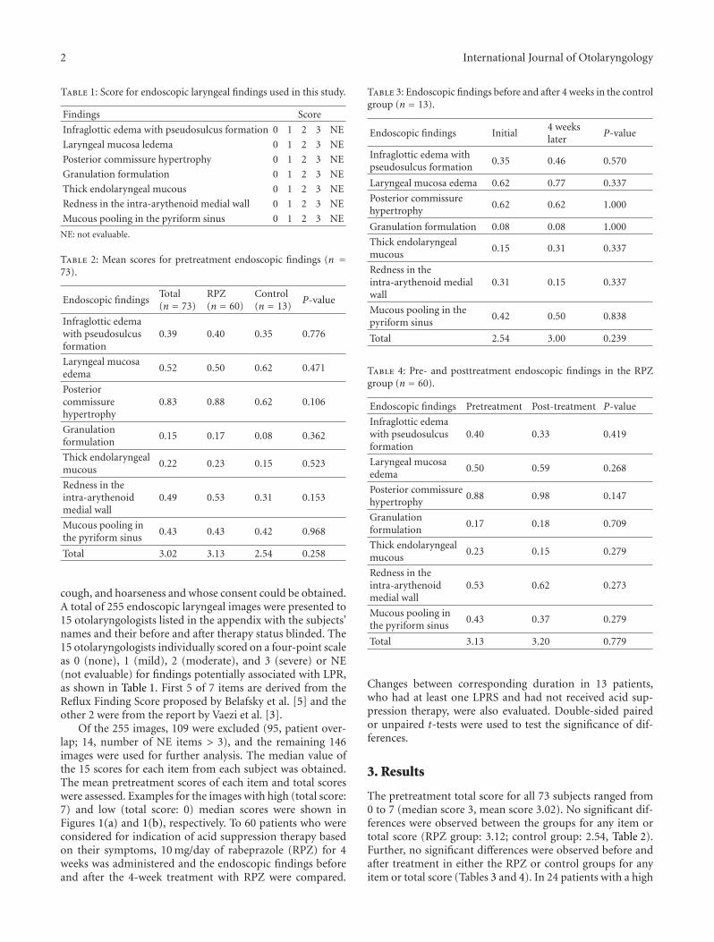
2 International Journal of Otolaryngology
Table 1: Score for endoscopic laryngeal findings used in this study.
Findings Score
Infraglottic edema with pseudosulcus formation 0 1 2 3 NE
Laryngeal mucosa ledema 0 1 2 3 NE
Posterior commissure hypertrophy 0 1 2 3 NE
Granulation formulation 0 1 2 3 NE
Thick endolaryngeal mucous 0 1 2 3 NE
Redness in the intra-arythenoid medial wall 0 1 2 3 NE
Mucous pooling in the pyriform sinus 0 1 2 3 NE
NE: not evaluable.
Table 2: Mean scores for pretreatment endoscopic findings (n =73).
Endoscopic findingsTotal(n = 73)
RPZ(n = 60)
Control(n = 13)
P-value
Infraglottic edemawith pseudosulcusformation
0.39 0.40 0.35 0.776
Laryngeal mucosaedema
0.52 0.50 0.62 0.471
Posteriorcommissurehypertrophy
0.83 0.88 0.62 0.106
Granulationformulation
0.15 0.17 0.08 0.362
Thick endolaryngealmucous
0.22 0.23 0.15 0.523
Redness in theintra-arythenoidmedial wall
0.49 0.53 0.31 0.153
Mucous pooling inthe pyriform sinus
0.43 0.43 0.42 0.968
Total 3.02 3.13 2.54 0.258
cough, and hoarseness and whose consent could be obtained.A total of 255 endoscopic laryngeal images were presented to15 otolaryngologists listed in the appendix with the subjects’names and their before and after therapy status blinded. The15 otolaryngologists individually scored on a four-point scaleas 0 (none), 1 (mild), 2 (moderate), and 3 (severe) or NE(not evaluable) for findings potentially associated with LPR,as shown in Table 1. First 5 of 7 items are derived from theReflux Finding Score proposed by Belafsky et al. [5] and theother 2 were from the report by Vaezi et al. [3].
Of the 255 images, 109 were excluded (95, patient over-lap; 14, number of NE items > 3), and the remaining 146images were used for further analysis. The median value ofthe 15 scores for each item from each subject was obtained.The mean pretreatment scores of each item and total scoreswere assessed. Examples for the images with high (total score:7) and low (total score: 0) median scores were shown inFigures 1(a) and 1(b), respectively. To 60 patients who wereconsidered for indication of acid suppression therapy basedon their symptoms, 10 mg/day of rabeprazole (RPZ) for 4weeks was administered and the endoscopic findings beforeand after the 4-week treatment with RPZ were compared.
Table 3: Endoscopic findings before and after 4 weeks in the controlgroup (n = 13).
Endoscopic findings Initial4 weekslater
P-value
Infraglottic edema withpseudosulcus formation
0.35 0.46 0.570
Laryngeal mucosa edema 0.62 0.77 0.337
Posterior commissurehypertrophy
0.62 0.62 1.000
Granulation formulation 0.08 0.08 1.000
Thick endolaryngealmucous
0.15 0.31 0.337
Redness in theintra-arythenoid medialwall
0.31 0.15 0.337
Mucous pooling in thepyriform sinus
0.42 0.50 0.838
Total 2.54 3.00 0.239
Table 4: Pre- and posttreatment endoscopic findings in the RPZgroup (n = 60).
Endoscopic findings Pretreatment Post-treatment P-value
Infraglottic edemawith pseudosulcusformation
0.40 0.33 0.419
Laryngeal mucosaedema
0.50 0.59 0.268
Posterior commissurehypertrophy
0.88 0.98 0.147
Granulationformulation
0.17 0.18 0.709
Thick endolaryngealmucous
0.23 0.15 0.279
Redness in theintra-arythenoidmedial wall
0.53 0.62 0.273
Mucous pooling inthe pyriform sinus
0.43 0.37 0.279
Total 3.13 3.20 0.779
Changes between corresponding duration in 13 patients,who had at least one LPRS and had not received acid sup-pression therapy, were also evaluated. Double-sided pairedor unpaired t-tests were used to test the significance of dif-ferences.
3. Results
The pretreatment total score for all 73 subjects ranged from0 to 7 (median score 3, mean score 3.02). No significant dif-ferences were observed between the groups for any item ortotal score (RPZ group: 3.12; control group: 2.54, Table 2).Further, no significant differences were observed before andafter treatment in either the RPZ or control groups for anyitem or total score (Tables 3 and 4). In 24 patients with a high
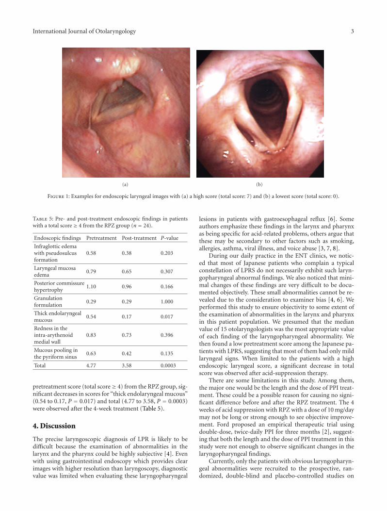
International Journal of Otolaryngology 3
(a) (b)
Figure 1: Examples for endoscopic laryngeal images with (a) a high score (total score: 7) and (b) a lowest score (total score: 0).
Table 5: Pre- and post-treatment endoscopic findings in patientswith a total score ≥ 4 from the RPZ group (n = 24).
Endoscopic findings Pretreatment Post-treatment P-value
Infraglottic edemawith pseudosulcusformation
0.58 0.38 0.203
Laryngeal mucosaedema
0.79 0.65 0.307
Posterior commissurehypertrophy
1.10 0.96 0.166
Granulationformulation
0.29 0.29 1.000
Thick endolaryngealmucous
0.54 0.17 0.017
Redness in theintra-arythenoidmedial wall
0.83 0.73 0.396
Mucous pooling inthe pyriform sinus
0.63 0.42 0.135
Total 4.77 3.58 0.0003
pretreatment score (total score≥ 4) from the RPZ group, sig-nificant decreases in scores for “thick endolaryngeal mucous”(0.54 to 0.17, P = 0.017) and total (4.77 to 3.58, P = 0.0003)were observed after the 4-week treatment (Table 5).
4. Discussion
The precise laryngoscopic diagnosis of LPR is likely to bedifficult because the examination of abnormalities in thelarynx and the pharynx could be highly subjective [4]. Evenwith using gastrointestinal endoscopy which provides clearimages with higher resolution than laryngoscopy, diagnosticvalue was limited when evaluating these laryngopharyngeal
lesions in patients with gastroesophageal reflux [6]. Someauthors emphasize these findings in the larynx and pharynxas being specific for acid-related problems, others argue thatthese may be secondary to other factors such as smoking,allergies, asthma, viral illness, and voice abuse [3, 7, 8].
During our daily practice in the ENT clinics, we notic-ed that most of Japanese patients who complain a typicalconstellation of LPRS do not necessarily exhibit such laryn-gopharyngeal abnormal findings. We also noticed that mini-mal changes of these findings are very difficult to be docu-mented objectively. These small abnormalities cannot be re-vealed due to the consideration to examiner bias [4, 6]. Weperformed this study to ensure objectivity to some extent ofthe examination of abnormalities in the larynx and pharynxin this patient population. We presumed that the medianvalue of 15 otolaryngologists was the most appropriate valueof each finding of the laryngopharyngeal abnormality. Wethen found a low pretreatment score among the Japanese pa-tients with LPRS, suggesting that most of them had only mildlaryngeal signs. When limited to the patients with a highendoscopic laryngeal score, a significant decrease in totalscore was observed after acid-suppression therapy.
There are some limitations in this study. Among them,the major one would be the length and the dose of PPI treat-ment. These could be a possible reason for causing no signi-ficant difference before and after the RPZ treatment. The 4weeks of acid suppression with RPZ with a dose of 10 mg/daymay not be long or strong enough to see objective improve-ment. Ford proposed an empirical therapeutic trial usingdouble-dose, twice-daily PPI for three months [2], suggest-ing that both the length and the dose of PPI treatment in thisstudy were not enough to observe significant changes in thelaryngopharyngeal findings.
Currently, only the patients with obvious laryngopharyn-geal abnormalities were recruited to the prospective, ran-domized, double-blind and placebo-controlled studies on
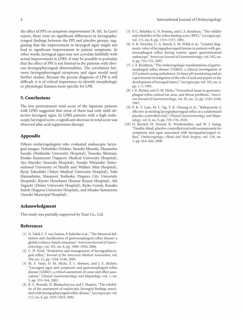
4 International Journal of Otolaryngology
the effect of PPI on symptom improvement [9, 10]. In Lam’sreport, there were no significant differences in laryngopha-ryngeal findings between the PPI and placebo groups, sug-gesting that the improvement in laryngeal signs might notlead to significant improvement in patient symptoms. Inother words, laryngeal signs may not correlate faithfully withactual improvement in LPRS. It may be possible to postulatethat the effect of PPI is not limited to the patients with obvi-ous laryngopharyngeal abnormalities. The correlation bet-ween laryngopharyngeal symptoms and signs would needfurther studies. Because the precise diagnosis of LPR is stilldifficult, it is of critical importance to identify morphologicor physiologic features more specific for LPR.
5. Conclusions
The low pretreatment total score of the Japanese patientswith LPRS suggested that most of them had only mild ob-jective laryngeal signs. In LPRS patients with a high endo-scopic laryngeal score, a significant decrease in total score wasobserved after acid suppression therapy.
Appendix
Fifteen otolaryngologists who evaluated endoscopic laryn-geal images: Nobuhiko Oridate, Yasushi Mesuda, MasanobuSuzuki (Hokkaido University Hospital), Tomoko Shintani,Etsuko Kanaizumi (Sapporo Medical University Hospital),Aya Maruko (Jusendo Hospital), Yusuke Watanabe (Inter-national University of Health and Welfare Mita Hospital),Ryoji Tokashiki (Tokyo Medical University Hospital), YukiHamashima, Masanori Yoshioka (Nagoya City UniversityHospital), Kiyoto Hosokawa (Kansai Rousai Hospital), AkiTaguchi (Ehime University Hospital), Rieko Gotoh, KanakoIndoh (Kagawa University Hospital), and Misako Yamamoto(Sanuki Municipal Hospital).
Acknowledgment
This study was partially supported by Eisai Co., Ltd.
References
[1] N. Vakil, S. V. van Zanten, P. Kahrilas et al., “The Montreal def-inition and classification of gastroesophageal reflux disease: aglobal evidence-based consensus,” American Journal of Gastro-enterology, vol. 101, no. 8, pp. 1900–1920, 2006.
[2] C. N. Ford, “Evaluation and management of laryngopharyn-geal reflux,” Journal of the American Medical Association, vol.294, no. 12, pp. 1534–1540, 2005.
[3] M. F. Vaezi, D. M. Hicks, T. I. Abelson, and J. E. Richter,“Laryngeal signs and symptoms and gastroesophageal refluxdisease (GERD): a critical assessment of cause and effect asso-ciation,” Clinical Gastroenterology and Hepatology, vol. 1, no.5, pp. 333–344, 2003.
[4] R. C. Branski, N. Bhattacharyya, and J. Shapiro, “The reliabil-ity of the assessment of endoscopic laryngeal findings associ-ated with laryngopharyngeal reflux disease,” Laryngoscope, vol.112, no. 6, pp. 1019–1024, 2002.
[5] P. C. Belafsky, G. N. Postma, and J. A. Koufman, “The validityand reliability of the reflux finding score (RFS),” Laryngoscope,vol. 111, no. 8, pp. 1313–1317, 2001.
[6] S. R. Vavricka, C. A. Storck, S. M. Wildi et al., “Limited diag-nostic value of laryngopharyngeal lesions in patients with gas-troesophageal reflux during routine upper gastrointestinalendoscopy,” American Journal of Gastroenterology, vol. 102, no.4, pp. 716–722, 2007.
[7] J. A. Koufman, “The otolaryngologic manifestations of gastro-esophageal reflux disease (GERD): a clinical investigation of225 patients using ambulatory 24-hour pH monitoring and anexperimental investigation of the role of acid and pepsin in thedevelopment of laryngeal injury,” Laryngoscope, vol. 101, no. 4,pp. 1–7, 1991.
[8] J. E. Richter and D. M. Hicks, “Unresolved issues in gastroeso-phageal reflux-related ear, nose, and throat problems,” Ameri-can Journal of Gastroenterology, vol. 92, no. 12, pp. 2143–2144,1997.
[9] P. K. Y. Lam, M. L. Ng, T. K. Cheung et al., “Rabeprazole iseffective in treating laryngopharyngeal reflux in a randomizedplacebo-controlled trial,” Clinical Gastroenterology and Hepa-tology, vol. 8, no. 9, pp. 770–776, 2010.
[10] O. Reichel, H. Dressel, K. Wiederanders, and W. J. Issing,“Double-blind, placebo-controlled trial with esomeprazole forsymptoms and signs associated with laryngopharyngeal re-flux,” Otolaryngology—Head and Neck Surgery, vol. 139, no.3, pp. 414–420, 2008.
Hindawi Publishing CorporationInternational Journal of OtolaryngologyVolume 2012, Article ID 646901, 13 pagesdoi:10.1155/2012/646901
Review Article
Reflux Revisited: Advancing the Role of Pepsin
Karna Dev Bardhan,1 Vicki Strugala,2 and Peter W. Dettmar2
1 Rotherham General Hospital, Moorgate Road, Rotherham, South Yorkshire S60 2UD, UK2 Technostics Ltd., The Deep Business Centre, Kingston Upon Hull, East Yorkshire HU1 4BG, UK
Correspondence should be addressed to Karna Dev Bardhan, [email protected]
Received 28 July 2011; Accepted 9 September 2011
Academic Editor: Petros D. Karkos
Copyright © 2012 Karna Dev Bardhan et al. This is an open access article distributed under the Creative Commons AttributionLicense, which permits unrestricted use, distribution, and reproduction in any medium, provided the original work is properlycited.
Gastroesophageal reflux disease is mediated principally by acid. Today, we recognise reflux reaches beyond the esophagus, wherepepsin, not acid, causes damage. Extraesophageal reflux occurs both as liquid and probably aerosol, the latter with a further reach.Pepsin is stable up to pH 7 and regains activity after reacidification. The enzyme adheres to laryngeal cells, depletes its defences,and causes further damage internally after its endocytosis. Extraesophageal reflux can today be detected by recognising pharyngealacidification using a miniaturised pH probe and by the identification of pepsin in saliva and in exhaled breath condensate by arapid, sensitive, and specific immunoassay. Proton pump inhibitors do not help the majority with extraesophageal reflux butspecifically formulated alginates, which sieve pepsin, give benefit. These new insights may lead to the development of novel drugsthat dramatically reduce pepsinogen secretion, block the effects of adherent pepsin, and give corresponding clinical benefit.
“For now we see through a glass, darkly.”—First epistle, Chapter 13, Corinthians
1. Introduction
This quotation from the Bible, often used in drama andthrillers, symbolises “that the clarity of a situation is oftenobscured”. It is in our view an apt description of thesyndrome we today recognise as extraesophageal reflux(EER). The inspired insight in the 1990s that the symptomsand findings might constitute a distinct entity [1] wasfollowed by growing awareness in the last decade thatthe underlying cause was gastroesophageal reflux. Hence,the Montreal classification included several extraesophagealfeatures within the spectrum of gastroesophageal refluxdisease (GERD), the association considered “established” forlaryngeal symptoms, cough, and asthma (and “proposed”for recurrent otitis media, idiopathic pulmonary fibrosis,pharyngitis, and sinusitis) [2]. This was a remarkableforesight, for at the time, the data on which we today regardfor EER was still emerging. Today, however, there is strongevidence that laryngeal damage from EER is mediated bypepsin. This decade may see these discoveries lead to clearer
understanding of the disease process and consequently leadto the development of effective therapy.
The earlier confusion surrounding the entity of EER andthe story now unfolding is reminiscent of the early 1980swhen an unusual organism came to be identified in thestomach, particularly in those with peptic ulcer. Lookingback, it may seem self-evident that Helicobacter pylori (H.pylori) was closely related to the development of duodenalulcer (DU)—but it certainly did not seem so at the time.
1.1. Acid—and Beyond. GERD has, with good reason, beenwidely regarded as the consequence of excessive reflux fromthe stomach into the lower esophagus, the acid componentof the refluxate damaging the esophageal mucosa. This linkis made indelible in the clinician’s mind by the rapid andsometimes dramatic relief proton pump inhibitors (PPIs)give, through profound acid suppression. Today, we areincreasingly aware reflux can reach much further, extendingbeyond the upper esophagus into the pharynx, larynx,
2 International Journal of Otolaryngology
airways, and middle ear, and may damage these structures[3, 4].
The benefits of the PPIs are striking in patients withtypical GERD, that is, those with lower esophageal symp-toms, principally retrosternal burning (“heartburn”), andregurgitation (the two together commonly referred to asthe “classical symptoms” of reflux) with or without erosiveesophageal changes. This is reflected by numerous clinicaltrials and reinforced by worldwide experience [5, 6]. Incontrast, PPIs generally give little benefit when symptomsarise from refluxate-damaged organs further away, indicatingthat unlike in the esophagus, acid may not be the damagingagent. In EER, the damaging agent we suggest is pepsin (andperhaps bile acids).
Pepsin is produced only in the stomach; hence evidenceof its presence in these organs signifies refluxate has reachedthem. The enzyme has recently been identified within thelaryngeal epithelium, in the saliva of patients with suspectedreflux laryngitis, and in the exhaled breath of those withairways and lung disease, where reflux is thought to haveplayed a part. Pepsin has also been found in the middle earin otitis media effusions (where bile acids, too, have recentlybeen identified).
“Host factors” too are presumably involved which influ-ence who develops disease and how severe it becomes.These factors may also have a bearing whether the disordermanifests with typical reflux symptoms arising from theesophagus or as EER. Many with EER have little or noretrosternal burning or regurgitation; this is surprising, for toreach the extraesophageal areas, the refluxate would first haveto travel through the esophagus. Refluxate is liquid, but thereis growing awareness that it may also be an aerosol. Firmevidence is, however, yet to emerge. Nevertheless, an aerosolremains an “attractive” possibility for it would account forobservations as yet unexplained. For example, intuitively, itwould seem that the concentration of hydrogen ions wouldbe less in an aerosol (compared to that in liquid refluxate),perhaps below a threshold to trigger esophageal symptoms.
The concept of pepsin and bile acids playing a rolein reflux disease developed many years ago but has beenoften overlooked in recent times, as the very success of PPIsincreasingly focused attention on acid (to the exclusion ofother factors) but also in part because the relevant literatureis not usually referred to by gastroenterologists.
2. Aim
The aim of this paper is, therefore, to draw the key evidencetogether and to raise awareness of EER amongst gastroen-terologists, who today are increasingly invited by ENT andrespiratory specialists to help investigate patients in whomthis condition is suspected. Bile acids are mentioned but thefocus is on pepsin; GERD is often referred to but mainly tocompare and contrast with EER, the main thrust.
We have broadly adhered to the philosophy of theMontreal classification of GERD, [2] departing only whenit does not sufficiently accommodate more recent findingsand evolving concepts of pathophysiology. Thus, a degree ofreflux into the lower esophagus and without any symptoms
is regarded as physiological gastroesophageal reflux (GER).Gastroesophageal reflux disease (GERD) is the term appliedwhen reflux is accompanied by typical symptoms, with orwithout erosive mucosal damage. At this stage, the totalduration of esophageal acid exposure is considerably longerthan in physiological reflux. Whilst extraesophageal refluxdisease is increasingly recognised, relatively little is known if“physiological” extraesophageal reflux (EER) occurs.
3. History
3.1. GERD: A New Concept Emerges. In 1934, Asher Winkel-stein first raised the possibility that the symptoms in fiveof his patients might have arisen from peptic esophagitis, acondition resulting “from the irritant action on the mucosaof hydrochloric acid and pepsin” [7–9]. From the late 1950s,elegant experimental studies have demonstrated the complexinterrelationship between bile acids, pepsin and hydrochloricacid (HCl) interacting and leading to esophageal damage.
3.2. Shifting Perceptions. The role of these nonacid factors,however, appeared to diminish in the clinician’s percep-tion when the histamine H2 receptor antagonists (H2RA)emerged in 1976. These were the first drugs to powerfullyreduce acid secretion and proved highly effective in con-trolling peptic ulcer, thus demonstrating the central role ofacid in the disease process. By extension, it seemed likely toalso be of use in GERD, but the clinical benefits proved tobe only modest. Acid inhibition, powerful with the H2RA,was profound with the new class of drugs, the proton pumpinhibitors (PPIs), which became available in 1989. Thesedrugs proved markedly superior to H2RA in the treatment ofGERD, which reinforced the growing perception that it wasthe acid component of refluxed gastric contents that was thecause of esophageal damage.
3.3. Pepsin and Bile Acids: At the Beginning
3.3.1. Pepsin. The elegant experimental studies of Goldberget al. [10] clearly demonstrated pepsin can damage theesophagus. Cat esophagi were infused for an hour with HCl,the pH ranging from 1 to 2.3. Acid at pH 1 proved verydamaging, whereas at pH 2.3 was without effect; addingpepsin to each of these infusates caused no further damage.In the intermediate acidity range of pH 1.6 and 2.0, however,the damage was proportional to the amount of pepsin added(25 and 50 µg/mL). Blocking the enzyme effect by firstpremixing with amylopectin sulphate (a synthetic pepsininhibitor) protected the esophagus, thereby confirming thatpepsin can, in the appropriate circumstances, cause damage.
HCl at pH 1 is probably not encountered in the gastriclumen (other than in exceptional circumstances), for thesecreted acid is rapidly diluted. A pH of 1.6–3 is common,however, and it is in this range when gastric refluxate con-taining acid and pepsin is most damaging to the esophagealmucosa. Pepsinogen arises from the gastric peptic cells (alsocalled the chief cells) which share space in the same glandsas the acid-secreting parietal cells: the two secretions areindependently controlled but almost always occur together.
International Journal of Otolaryngology 3
Reflux, therefore, irrespective of its pH, always containspepsin (see below).
3.3.2. Bile Acids. Experimental studies in the 1980s demon-strated the role of bile acids in damaging the esophagealmucosa. In a series of studies by Harmon and colleagues[11] and by Schweitzer et al. [12], varying concentrations oftaurine-conjugated and unconjugated bile salts were infusedinto rabbit esophagi at pH 2, 5 and 7. Significant disruptionof the esophageal mucosal barrier occurred at bile acidconcentrations similar to those in the stomach of patientswith esophagitis [13]. The evidence, however, suggested thatsuch disruption was probably not the direct effect of bileacids solubilising the cell membrane phospholipids. Instead,bile acids enter the epithelial cells and disrupt cellularmachinery from within, hence interfere with the cell barrierfunction. Such entry is determined by the physicochemicalproperties of the bile acids. Taurine-conjugates have a pKa of∼2 that is, half the molecules are in solution at pH 2 and,being charged, cannot penetrate the lipid bilayer of the cellmembrane. As the pH progressively rises, more bile acidscome out of solution and by pH 7 are insoluble, lose theircharge and as a result can now enter the epithelial cells.
These experimental studies have particular clinical rele-vance, for bile is present in esophageal refluxate [14–16] andis most frequently noted in patients with severe esophagitisor complicated Barrett’s esophagus [17, 18] in whom it ispresent at high concentrations [19].
3.4. Conceptualizing the Mechanism of Damage. Putting theseobservations together, one can conceptualise circumstanceswhere pepsin in the refluxate disrupts the esophagealmucosal barrier by acting on the epithelial cell surface, whilstbile acids achieve the same effect by diffusing into the cell anddamaging from within.
These seemingly complex mechanisms contrast sharplyfrom the “corrosive” action of acid, an effect “simpler” topicture. Gastric acid consists of H+ and Cl− ions in water.When in high concentrations, intuitively, the fluid is more“corrosive”; hence, the longer the time in contact with theesophagus, the greater the likelihood it will damage themucosa. Conversely, lower concentrations are less damaging.Importantly, the basic constituents of acid are unchanged,only its concentration.
4. Pepsin: Nature, Activation, Acidity, andEnzyme Activity: Clinical Significance
4.1. Nature and Activation. Pepsin is an ancient moleculeand present in all vertebrates studied, such as fishes andmammals. The stomach is largely devoid of live organisms(with the exception of H. pylori), a state widely believedto result from the presence of gastric acid, which acts as a“bulk steriliser”. A second important action of acid is theactivation of pepsinogen. This releases pepsin which initiatesdigestion through proteolysis, an action which also probablyhelps keep the stomach free of most bacteria [20].
The peptic chief cells produce and store pepsinogen,the precursor of the active enzyme. Pepsin, an aspartic
proteinase, is a large bilobed molecule and concave on onesurface, the concavity occupied by the detachable pro-part.When in contact with acid, the pro-part detaches exposingthe concavity, the active site for enzyme action. The enzymeattaches to its substrate at this point and cleaves it. Acid (pH< 6) is required to convert inert pepsinogen to active pepsinbut once converted, the pepsin continues the autocatalyticprocess sustaining the cascade in the absence of acid [3].
4.2. Pepsin: Isoenzymes. Pepsin has traditionally been stud-ied by gel electrophoresis of gastric juice and tissuehomogenates, which typically shows eight zones of lysis towhich various names and designations have been appliedbased on their electrophoretic mobility [21, 26]. The patternreflects the fact that pepsin is not a single molecule butencompasses a family of isoenzymes which structurally aresimilar. Today, the pepsin isoenzymes in gastric juice can beseparated by high-performance anion exchange chromatog-raphy (HPAEC) using chloride counter ion gradient elution(Figure 1). Each pepsin isoenzyme has its own “optimalpH level” when its action is at a maximum, thus ensuringdigestion across a wide range of gastric pH (see Table 1).
4.3. Pepsin: pH and Stability. The activity and stability of theenzyme is closely related to the prevailing pH of its environ-ment, a relationship investigated over the last 40 years usingdifferent sources of pepsin, various substrates, and changinganalytical methods. The results were broadly similar, anddifferences were attributed to the experimental conditions.Recent studies reexamining the pH-pepsin relationship inconditions of low acidity have given important new insightsinto enzyme stability and activity which have major clinicalsignificance (and are discussed in detail further on).
Individual pepsin isoenzymes were noted to be stablefor 24 hours even at body temperature but were ultimatelydegraded by autocatalysis if stored at its pH optima. Incontrast, a mixture of isoenzymes, as would be found ingastric secretion, proved to be more stable [22]. For example,purified porcine pepsin was irreversibly denatured at pH 7.1,whereas peptic activity of human gastric juice persisted untilexposed to pH 7.8. [27]. A “dormant” phase was observedbetween pH 6 and 8 when the enzyme was inactive but intact,hence it could be activated on return to pH <6 [28].
4.4. Pepsin: pH and Enzyme Activity: The Traditional View. Ithas long been known that pepsin is at its most active at pH2 to 3, and activity is declining as acidity diminishes [29].Emerging evidence has shown that refluxate reaching theextraesophageal areas is characterised by low acidity or noneat all in those on high-dose PPI treatment. Such conditionshave been widely regarded to destroy pepsin or to render itinactive, hence the scepticism that pepsin played a significantrole in damaging extra-esophageal tissues. Recently, however,important new information challenges such beliefs.
4.5. Pepsin: pH and Enzyme Activity: The Emerging Evidence.This interrelation has recently been reassessed, now usingpepsin isoenzyme 3B purified from human gastric juice[30]. It is the largest fraction of pepsin and accounts for70% of the total enzyme effect (Table 1), for which it serves
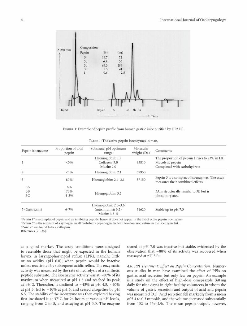
4 International Journal of Otolaryngology
Inject Pepsin 5 3c 3b 3a
Time
1
Composition
Pepsin
53c3b3c1
16.76.9
66.39.50.6
(µg)
7230
28641
2.5
(%)A 280 mm
Figure 1: Example of pepsin profile from human gastric juice purified by HPAEC.
Table 1: The active pepsin isoenzymes in man.
Pepsin isoenzymeProportion of total
pepsinSubstrate: pH optimum
rangeMolecular
weight (Da)Comments
1 <5%Haemoglobin: 1.9
Collagen: 3.0Mucin: 2.0
43810The proportion of pepsin 1 rises to 23% in DUMucolytic pepsinComplexed with carbohydrate
2 <1% Haemoglobin: 2.1 39950
3 80% Haemoglobin: 2.4–3.1 37150Pepsin 3 is a complex of isoenzymes. The assaymeasures their combined effects.
3A3B3C
6%70%4-5%
Haemoglobin: 3.23A is structurally similar to 3B but isphosphorylated
5 (Gastricsin) 6-7%Haemoglobin: 2.0–3.6
(maximum at 3.2)Mucin: 3.5–5
31620 Stable up to pH 7.3
“Pepsin 4” is a complex of pepsin and an inhibiting peptide, hence, it does not appear in the list of active pepsin isoenzymes.“Pepsin 6” is the remnant of a zymogen, in all probability pepsinogen, hence it too does not feature in the isoenzyme list.“Zone 7” was found to be a cathepsin.References [21–25].
as a good marker. The assay conditions were designedto resemble those that might be expected in the humanlarynx in laryngopharyngeal reflux (LPR), namely, littleor no acidity (pH 6.8), when pepsin would be inactiveunless reactivated by subsequent acidic reflux. The enzymaticactivity was measured by the rate of hydrolysis of a syntheticpeptide substrate. The isoenzyme activity was at ∼80% of itsmaximum when measured at pH 1.5 and reached its peakat pH 2. Thereafter, it declined to ∼45% at pH 4.5, ∼40%at pH 5, fell to ∼10% at pH 6, and ceased altogether by pH6.5. The stability of the isoenzyme was then explored havingfirst incubated it at 37◦C for 24 hours at various pH levels,ranging from 2 to 8, and assaying at pH 3.0. The enzyme
stored at pH 7.0 was inactive but stable, evidenced by theobservation that ∼80% of its activity was recovered whenreassayed at pH 3.0.
4.6. PPI Treatment: Effect on Pepsin Concentration. Numer-ous studies in man have examined the effect of PPIs ongastric acid secretion but only few on pepsin. An exampleis a study on the effect of high-dose omeprazole (60 mgdaily for nine days) in eight healthy volunteers in whom thevolume of gastric secretion and output of acid and pepsinwas measured [31]. Acid secretion fell markedly from a meanof 5.4 to 0.3 mmol/h, and the volume decreased substantiallyfrom 132 to 36 mL/h. The mean pepsin output, however,

International Journal of Otolaryngology 5
fell only modestly, from 126 to 101 mg/h, but because of thereduced volume, its concentration rose from 90 mg to 290 mgper 100 mL.
This study, like most others on gastric secretion, relied onthe measurement of stomach contents aspirated through anasogastric tube. A novel noninvasive approach was recentlyused to measure gastric volume by magnetic resonance imag-ing [32]. Unlike the study cited earlier [31], the reductionin the volume of gastric secretion on PPI was only 12%,hence the concentration of pepsin would have increased onlyslightly. The clinical significance of these contrasting findingsis discussed further on.
4.7. PPI Treatment: pH, Pepsin Activity, and H. Pylori.The PPIs commonly used today (e.g., omeprazole) onconventional dosing (single 20 mg dose in the morning) canelevate gastric pH to ≥6 but only for short periods; for muchof the time, the pH is around 4 to 5 [6] and falls at nightwhen acid secretion breaks through. Thus, for the majority of24 hours, pepsin in gastric juice is still active or dormant butstable, hence capable of reactivation when acidity returns.
High-dose PPI treatment (e.g., omeprazole 40 mg twicedaily) has a greater effect and is longer lasting, and the newerPPIs (e.g., tenatoprazole) [33] may enhance this. Theseconditions may keep pepsin inactive, but it seems unlikelythat the pH will be elevated to levels which will result in anysubstantial degradation of the enzyme.
The presence of H. pylori increases the effect of PPI, afeature sometimes overlooked yet likely to have a bearingon the efficacy of PPI therapy in the uninfected or in thosein whom the organism has been eradicated. Several studiesconfirm this, an example being the seminal investigationcarried out in DU patients [34]. Here, the median 24-hourintragastric pH when PPIs were not used was similar beforeand after H. pylori eradication, 1.0 and 1.1, respectively. Onomeprazole 20 mg, however, there was a major differencein pH, 5.5 before eradication but only 3.0 after it. Thesignificance is that whilst the majority of DU patients areinfected with H. pylori, its prevalence is much lower in GERDpatients (and similar to that in the general population),hence theoretically, PPIs might have a lesser effect.
4.8. PPIs, Pepsin, and Reflux: Clinical Significance. PPI ther-apy suppresses acid profoundly, has a variable effect on thevolume of secretion (as indicated earlier) which is difficult toexplain, but does not reduce the frequency of reflux episodes[35]. When volume is reduced only slightly [32], much fluidremains in the stomach and is available to reflux, carryingpepsin to the extraesophageal areas. When volume is reducedsubstantially, the concentration of pepsin rises [31] but refluxcontinues [35], although less is available to reflux, whatreaches the extraesophageal areas is rich in pepsin, hence isdamaging.
4.9. Pepsin, pH, and Cell Damage. Pepsin is refluxed to theextraesophageal areas where it adheres to the epithelium[36]. If activated by acid in the refluxate, it damages thecells but even in the absence of acid the enzyme has thecapacity to damage, for, though dormant, it is stable. Two
mechanisms operate. The first is by its reactivation whenexposed to acid in subsequent reflux episodes. The secondmechanism is independent of such reflux reacidification: it istaken up within epithelial cells by endocytosis and activatedfrom within [37]. This remarkable observation, based onlaryngeal cell studies, is a recent discovery with far reachingconsequences (and is discussed further on). The significanceis that refluxate always contains pepsin; even if devoid of acid(as might happen on high dose PPI treatment), the enzymewill still be damaging if reflux reaches the extraesophagealareas.
4.10. PPIs and Pepsin: Potential Clinical Relevance. Basedon older studies, pepsin is commonly assumed to becomeinactive at pH ≥4 and to be denatured at pH ≥5.5, hencethe widespread perception that PPI treatment renders theenzyme inactive by elevating the gastric pH. This view, wesuggest, now needs to be readjusted taking into considerationthe new evidence which clearly shows the enzyme retainsmuch of its activity at pH≥4, is still intact up to nearly pH 8,can be reactivated when exposed to acid once again but candamage cells even in the absence of acid.
In clinical practice, PPIs will continue to be used inEER, frequently in high dose, for they help some who inaddition to EER symptoms also have features of classicalGERD as well as the few who do not [38]. From theevidence above, however, it seems unlikely that profoundacid suppression with PPIs as the sole treatment strategywill give results comparable to those with typical esophagealsymptoms (heartburn and regurgitation) with or withouterosive esophagitis.
5. The Effect of Pepsin on Epithelial Cells
In laboratory studies, pepsin swiftly breaks down protein,the basis of its chemical assay. Its effect on extraesophagealtissues is in contrast subtle and perhaps sustained, depletesthe cells of its defences and threatens its viability. Thesechanges have been demonstrated in ongoing clinical andlaboratory studies by Johnston and colleagues who exploredthe effects of human pepsin 3B (purified from gastric juice)on laryngeal epithelium using ex vivo systems and cell culturestudies [36, 37, 39, 40].
5.1. Pepsin: Entry into Epithelial Cells. Based on esophagealand laryngeal biopsies from LPR patients and from controlsubjects, and employing a variety of analytical methods,they made three major observations: pepsin adhered toepithelial cells, was endocytosed, and caused internal cellderangements.
Pepsin was found adherent to the surface of laryngealepithelial cells obtained from LPR patients but not to thosefrom control subjects [36]; the absence in the latter groupis to be expected, for significant reflux had already beenexcluded by esophageal physiology studies. The enzyme wasnot found adherent to the esophageal epithelium (in LPRpatients); this is surprising bearing in mind that to reach thelarynx the refluxate has first to travel along the length of theesophagus. When active, the adherent enzyme damages the

6 International Journal of Otolaryngology
intercellular junctions and depletes proteins within the cellinvolved in its defence (and is discussed further on).
Inactive pepsin is taken up within the cells by endocytosisthrough a competitive receptor-mediated mechanism and isfound in vesicles located in the region of the Golgi system[37]. Such endocytosis, the second observation, is surprising,for it seems unlikely that receptors specific for pepsin existin laryngeal tissues. Presumably, such receptors serve someother purpose but when exposed to pepsin, they “shuttle” theenzyme into the cells.
When cells were exposed to human pepsin 3B at pH 7.4, alevel at which the enzyme is inactive, several major changes,nonetheless, occurred affecting the inner cell structure andfunction [39], the third major observation. The Golgi systemhas a pH of ∼5.5, together with its associated endosomesthese process large molecules such as proteins and receptorsthrough its slightly acidic environment. The inference is thatthe changes observed (see below) result from reactivation ofthe dormant enzyme within the cell.
The cells swelled and structural damage to the mito-chondria and to the Golgi system became visible on electronmicroscopy within an hour and increased by 12 hours. Theearly damage was accompanied by increased expression ofseven genes involved in cell stress and toxicity includingcertain heat shock proteins (as a family, the production ofheat shock proteins is activated when the cell is stressed andits survival threatened) and the late changes by the decreasedexpression of another 18 such stress genes. The investigatorsalso used in parallel a cell toxicity assay which measuresmitochondrial activity in living cells. There was a significantincrease in toxicity after pepsin exposure at pH 7.4 whichcorrelated well with the mitochondrial changes noted onelectron microscopy.
The evidence strongly argues for the following chain ofevents: inactive pepsin is endocytosed, is activated withinthe cells, and causes cell damage; this induces oxidativestress and the accumulation of free oxygen radicals which,in turn, damage mitochondria and may lead to cell death.In the experimental system used, the cells were exposed onlyonce to pepsin, thus mimicking what might happen with anisolated episode of LPR. Though damaged, the cells were stillviable at 12 hours but with repeated exposure, as would beexpected in chronic LPR, the damaged cells may not survive[39].
5.2. Pepsin: Depletion of Cell Defences. The effect of pepsinwas explored using a pig laryngeal epithelial cell model.Human pepsin 3B markedly depleted cell defences only whenthe enzyme was made active by the presence of acid (pH4). In contrast, acid on its own had no effect, nor did theenzyme when rendered inactive by raising pH to 7.4 or whenits activity was blocked with its inhibitor, pepstatin [30].
In a series of studies, the specific cell defence changesnoted were depletion of the carbonic anhydrase isoenzymeCA3 and the stress protein Sep 70, reduction of E-cadherin,and the alteration of the subtype profile of protective mucinproduced [30, 36, 40, 41].
The isoenzyme CA3 is widely expressed in tissues,including the basal layers of both esophageal and laryngeal
epithelium. It mediates the rapid two-way conversion ofCO2 and water to carbonic acid, bicarbonate, and H+, henceplays a key role in the regulation of cell pH. When theesophageal epithelium is exposed to acid, the isoenzyme isalso expressed in the more superficial cells, thus offeringgreater protection to the epithelium nearest the refluxed acid.In contrast, its production remains limited to the basal layersin the laryngeal epithelium.
Sep 70, like most other stress proteins, is a molecularchaperone which regulates the correct folding and unfoldingof intracellular proteins during their passage through thecell. E-cadherin is crucial for maintaining adhesion betweencells, and thereby mucosal integrity and its barrier function.There are several subtypes of mucin, some more prominentin specific tissues than others: collectively, they afford pro-tection. In chronic LPR, MUC-2, -3, and -5AC are amongstthe defensive mucins depleted, and in vitro studies confirmpepsin interferes with their production [42].
5.3. Tissue Damage in GER and EER: A Comparison. Theintensity of damage of the esophageal mucosa by acid reflux(pH < 4) is proportional to the duration of contact. Adegree of reflux occurs in health, particularly after meals, butperistalsis rapidly returns the refluxate to the stomach; anyresidue is neutralised by bicarbonate secreted in saliva andby the esophageal mucus glands.
In striking contrast, the larynx and extraesophagealstructures have no mechanism for bulk removal of damagingagents, hence they must rely on intracellular defences; but asindicated earlier, such cell defences are much depleted afterexposure to reflux. Hence, even slight exposure to reflux cancause disproportionate damage.
5.4. The Significance of Dilated Intercellular Spaces in theSquamous Epithelium. Acid injury to the esophageal squa-mous epithelium results in the dilatation of the intercellularspaces, which almost double in width; these changes arevisible only by electron microscopy. The phenomenon is nowwell established in patients with both erosive and nonerosivereflux disease [43, 44]. It was recently also observed inhealthy volunteers in whom the lower esophagus was infusedwith only weak levels of acid (pH 5.5) that the changesare no greater when strong acid (pH 2) with added pepsin(±bile acid) was infused. The changes were widespread andoccurred not only at the site of infusion, but also well awayfrom it [45].
Such dilatation has been reproduced experimentally inrabbit epithelium exposed to acid at pH 1.1 or at pH 2 butwith added pepsin [44]. These tissues had reduced electricalresistance mainly due to “leakiness” of the paracellular path-ways, the “leak” in proportion to the size of dextran particleswhich could enter the damaged tissue. The significance is thatthe “leak” is physical not virtual.
These studies clearly show the sensitivity of theesophageal epithelium to even low concentrations of acid.Its possible relevance to EER comes from the observationthat dilatation of intercellular spaces has also been notedin the laryngeal epithelium in patients with LPR [36, 46],which, unlike that in the esophagus, has not attracted

International Journal of Otolaryngology 7
attention. Whilst there is little acid in refluxate reaching theextraesophageal areas, it does contain pepsin, which maypotentially gain entry into the dilated spaces in the laryngealepithelium.
The nerve endings in the esophageal epithelium (inmonkeys) are located in the intercellular spaces and aresuperficial, appearing at a depth of only three cell layers,a major finding [43]. These sensory nerves are thoughtto be chemosensitive and respond to even low levels ofacid, pH 5.2 to 6.9, chronic irritation leading to secondaryhypersensitisation which perpetuates symptoms. The larynx,too, is richly innervated and is exquisitely sensitive. A similartrain of events may, theoretically, occur in the larynx, whichprolongs the symptoms even when the stimulus is muchreduced.
5.5. Summary. In summary, esophageal mucosal damage ismediated principally by acid, and laryngeal epithelial (andpossibly other extraesophageal epithelium) damage by pepsin(and presumably other agents). The environmental pH in thelarynx and hypopharynx is ∼5.5 to 6, a level at which pepsinis only slightly active or dormant. Even when inactive, pepsindamages cells after it is endocytosed and reacidified withinthem. The enzyme adheres to the laryngeal epithelium andwhen inactive is stable and can be reactivated by acidic refluxeven when the episodes are infrequent. These advances in ourunderstanding perhaps explain the apparent paradox notedin classical observations comparing EER and GER. As littleas three EER episodes per week can damage the larynx, itwas noted, whereas up to 50 GER episodes of acidic reflux(pH < 4) per day can be seen in the asymptomatic individual[1, 47].
6. The Reach and the Nature of Reflux
6.1. Introduction. Reflux into the esophagus is usually imag-ined as fluid welling up from the stomach and/or spurting injets which sometimes reach high, a plausible interpretationof pHmetry and impedance study results, the mental imagereinforced by artistic impressions in advertisements. Forgastroesophageal reflux to cause disease beyond the esoph-agus, however, the refluxate must self-evidently reach theseareas.
Reflux into the extraesophageal areas is not a new con-cept. Almost two decades ago, Koufman [1] speculated on itspossibility as an explanation for chronic laryngeal symptomsseen in several patients but supporting evidence was toemerge only several years later as technology advanced. Thepresence of refluxed material in the extraesophageal areassuggests it may play a role in causing symptoms from thelarynx and airways, but the growing evidence that pepsin cancause damage makes the case more compelling.
These insights spurred the development of new tech-nology to detect reflux in the extraesophageal areas. In thecourse of these investigations, two findings emerged almostas a “byproduct” (hence seemed to attract little notice)which are likely to change the concept of EER disease. First,the refluxate loses much of its acidity as it travels upwards(presumably by neutralisation with bicarbonate in saliva
and from the esophageal mucosal glands) [48, 49]. Second,refluxate can be both liquid and aerosol [49–51].
6.2. How Far Up the Esophagus Does Reflux Reach? StandardpHmetry readily detects acid (defined as pH < 4) refluxedinto the lower esophagus by its single sensor stationed 5 cmabove the lower esophageal sphincter (LES). Dual pHmetry,in which the second sensor is stationed in the vicinity ofthe upper esophageal sphincter (UES), confirmed proximalreflux does occur [52]. By this time, there was also growinginterest in whether refluxate containing lesser concentrationsof acid was potentially damaging. The newly developedmethod of combined impedance pHmetry provided themeans to detect liquid reflux independent of its pH and todetermine how far proximally such refluxate reaches. Theprinciple is that fluid is a better electrical conductor thanair, hence impedance (resistance) falls, the higher the acidconcentration, the greater the fall. The catheter has twopH electrodes, as with dual channel pHmetry (see below),and six pairs of impedance (resistance) sensors, each paircomprising two sensors separated by a tiny gap. Just a film offluid is sufficient to bridge this gap and complete the circuit.In the absence of any fluid, the sensors are exposed to air andrecord high impedance.
The pH electrodes are stationed 5 cm and 20 cm abovethe LES, whilst the impedance sensors extend from aboveto below the pH electrodes. The resistance sensor detectingchange furthermost from the LES indicates the proximalreach of reflux. These studies have clearly demonstratedrefluxate frequently rises to the proximal esophagus, thesignificance being it is in a position to reach extraesophagealareas [53–56]. Investigations in untreated GERD patientsshowed 63% of reflux episodes were acidic (and 72% ofsymptom episodes were associated with acid reflux). Incontrast, 80% of reflux episodes on PPI were weakly acid orweakly alkaline (and most symptoms were associated withrefluxate of this nature) [57].
6.3. Acid Reflux into the Pharynx. A recent advance is thedevelopment of a nasopharyngeal probe bearing a speciallyconstructed pH sensor at its end (available as the Dx-pH probe http://www.restech-corp.com/). The sensor isstationed in the mid-pharynx (i.e., away from the upperesophageal sphincter), where it is kept moistened with eachexhaled breath. The environmental pH detected with thismethod is ∼5.5; set against this relatively high pH, refluxcontaining even only little acid is readily detected [50].
6.4. Reflux into the Oropharynx and Airways: The Presenceof Pepsin. Pepsin has recently been detected in the salivaof patients with suspected EER using a highly sensitiveimmunoassay which utilises two unique monoclonal anti-bodies against human pepsin 3 [58]. The same immunoassayhas detected pepsin in the exhaled breath in patients withchronic cough thought to be due to EER. The breath sampleis captured, kept cold, and the immunoassay carried out onthe condensate which forms [51].
8 International Journal of Otolaryngology
6.5. The Acidity of Refluxate: Its Relevance to EER Disease.Refluxate detected in impedance studies is arbitrarily dividedas acid (pH < 4), weakly acid (pH 4–7), and nonacidic(pH > 7) [59]. Reflux at pH <4 is widely regarded as beingdamaging to the esophagus because of its high acidity, hencepH 4–7 is less injurious, and pH >7 probably without effect.The real significance of refluxate containing low or no acid,however, is that it always contains pepsin, which potentiallycan be carried to the extraesophageal areas, where it damagesthe epithelium.
As indicated earlier, esophageal damage in typical GERDis dominated by acid; in contrast, EER disease is mediatedprincipally by pepsin. This perhaps explains why PPIs fail torapidly improve EER disease, unlike their effect in classicalGERD.
6.6. The Physical Form of Refluxate: Liquid and Aerosol . Theconcept that reflux may also be an aerosol has only recentlyemerged, at this stage more plausible (albeit persuasive) thanproven. It arises from detecting acid in the pharynx andpepsin in the saliva but particularly in the exhaled breath(see below). Their presence, so far away from the stomach,is more plausibly explained if they were airborne, that is,carried in an aerosol, rather than in a column of liquid risingfrom the stomach.
Refluxate as an aerosol has several implications. First, asindicated above, it more plausibly explains the presence ofrefluxate deep in the lungs [60, 61] and in the middle earin otitis media effusions [62] (where recently bile acids toohave been identified) [63]. Second, liquid refluxate probablyhas higher concentrations of acid and pepsin, but an aerosolis more likely to carry these damaging agents further intothe extraesophageal areas. Third, their presence confirmsthat refluxate has reached. When in excess, and in theappropriate clinical circumstances, the findings are arguablypotentially diagnostic of EER. Finally, it draws attention toa major unmet therapeutic need, namely, the developmentof new approaches to more effectively decrease pepsin inrefluxate.
6.7. GER and EER and the Role of the Sphincters: Speculation.The esophageal lumen is occupied by mucosal folds and air,both swallowed and refluxed. Impaired LES function resultsin excessive reflux, and presumably liquid refluxate rises inthe lumen along the mucosal folds. We suggest that the airin the esophagus provides the medium through which anaerosol ascends.
The LES in health allows air from the stomach tobe vented whilst minimising the escape of liquids andsemisolids; such separation is less efficient in GERD, wherethe sphincter function is impaired. The role of the UES inreflux is less well understood, but we speculate that like theLES, it too in health can distinguish liquid from gas, holdingback the former and allowing the latter to be vented. Wethink, however, it would be difficult to distinguish betweentypes of gas, air that contains aerosol refluxate from air thatdoes not. Hence, both are vented and refluxate reaches theextraesophageal areas.
7. Detecting Extraesophageal Reflux
7.1. Diagnosis: A Note. Whilst it is beyond the scope of thispaper to consider the issues surrounding the diagnosis ofEER in any detail, we would like to highlight the following.
7.2. Clinical Manifestations of EER. Today, EER is increas-ingly considered as a potential cause of symptoms in adultswith chronic problems arising from the larynx, throat, andairways. Changes are often seen in the larynx, but there is nofeature characteristic of damage by EER. Furthermore, EERwhen present may be one of several contributory factors, forexample, smoking.
7.3. Technology Currently Available. Impedance pHmetryclearly identifies liquid reflux and indicates how far proxi-mally it reaches. Here, it is assumed that liquid is poised topenetrate into the extraesophageal areas hence EER can beinferred. Conventionally, one looks to correlation betweenproximal reflux episodes and symptoms as evidence of acausative link; but bearing in mind that even infrequentepisodes of EER can be damaging, the absence of such cor-relation does not necessarily exclude reflux-related damage.
Impedance pHmetry (MIIpH) has given us remarkableinsights into the pathophysiology of gastroesophageal reflux;it readily detects liquid refluxate irrespective of its pH (hence,it is particularly useful when investigating patients alreadyon PPI) and can determine how often and how far up theesophagus the refluxate reaches—but not beyond it (as yet),nor can it, as currently configured, identify aerosol reflux. Todetect EER, therefore, we suggest its role is more supportivethan diagnostic.
7.4. Emerging Technologies: Acid and Pepsin in the Pharynx,Saliva and Breath. Aerosol acid reflux into the pharynxcan today be detected by the Dx-pH nasopharyngeal probe(ResTech). Diagnostic criteria have been developed, and a pHlevel <5.5 is regarded as abnormal [50].
Pepsin immunoassay now makes it possible to detectminute amounts of the enzyme in saliva and in exhaledbreath. It has recently been adapted as a lateral flow-basedtest (PeptestTM, RD BioMed Ltd., UK), easy to use, with adetection lower limit of 16 ng/mL, and with results availablewithin minutes. The sensitivity is only slightly lower thanwith conventional ELISA, which is laborious and moretimen consuming. The test is noninvasive and will provemore acceptable to patients, particularly if serial assays areclinically required. Determining its clinical usefulness indiagnosis and in monitoring treatment will, however, requiremore extensive studies.
Venting air is normal and with it a tiny amount ofaerosolised gastric content is likely to escape, hence theasymptomatic may have pepsin detectable in the saliva. Howshould such a finding be interpreted? Lessons learnt twodecades ago of the diagnosis of GER by pHmetry give guid-ance. Some reflux of acid into the esophagus is physiologicaland not associated with any symptoms; that is, it is the norm.Symptoms (with or without erosive esophageal damage)develop only when reflux is excessive. Thus, the differencebetween health and disease is a quantitative one, that is,
International Journal of Otolaryngology 9
the degree of acid reflux. The same model may apply todetecting pepsin in the saliva. The test is quantitative; we may,therefore, find that as with GER it is the quantity of pepsinin extraesophageal areas that correlates best with disease (asopposed to the presence of a tiny amount).
8. Medical Therapy
8.1. The Place of PPIs. PPIs, dramatically effective in typicalreflux, are rather less so in EER. Several clinical trials andmeta-analyses have failed to show clear benefit in LPR [64]other than one single study that observed a benefit of twicedaily PPI for LPR symptoms and signs [65]. Patients withasthma when considered as a group showed no discerniblebenefit with PPIs; the subset with GER symptoms, however,were helped [66] but, interestingly, not those with pHmetry-proven reflux alone (in the absence of GER symptoms) [67].Patients with chronic cough in whom EER was suspected alsodid not benefit [68, 69].
Tenatoprazole, a new PPI with a much longer half-lifethan those currently available, correspondingly suppressesacid for a longer period in the 24 hours [33]. Other PPIsand potassium-competitive acid blockers are in development[5, 6]. These may prove helpful in patients with typical refluxwhen currently available PPIs give insufficient response; butfor the reasons stated earlier, they are unlikely to make muchdifference in EER.
Nevertheless, the occasional patient does show improve-ment, generally partial, with PPIs. In the absence of reallyeffective treatment, such anecdotes encourage continuedwidespread use of profound acid inhibition.
8.2. Antipepsins. Drugs with antipepsin activity have beenused in several clinical studies in patients with peptic ulcerbut were found not to be effective. Examples are amylopectinsulphate [70–72] and pepstatin [73].
8.3. Alginates: “Sieving Pepsin” from Gastric Secretion. Algi-nates, widely available for almost 40 years, have recentlybeen shown to have a powerful effect on pepsin and bilein refluxate in vitro [74] and potentially offer an effectivetreatment [75].
Recent in vitro studies confirm that Gaviscon Advance(GA), a specific alginate formulation, removes ∼90% ofpepsin and bile in the first “reflux episode”, declining toabout 50% by the tenth. Their rate of depletion was similar,suggesting a common mechanism, most probably selectivebinding [74].
8.4. Alginates: Effectiveness in EER? Only a few studies have asyet been done, mainly in LPR, and give encouraging results.
8.4.1. LPR. A UK study assessed the benefit of addingGA to standard vocal hygiene advice (control group) [75]and a USA investigation on the outcome of adding GA topreexisting PPI therapy [76]. In both, a dose of 10 mL × 4daily was used; this is the recommended dose when the drugis used for dyspepsia and is generally used only for shortperiods (4 to 8 weeks) but for the trials treatment was givenfor six months.
Allocation to treatment in the UK study was randomisedand blinded. The outcome was assessed by those unawareof the patients’ treatment group. Two scoring instrumentswere used, both validated and semiquantitative. The refluxsymptom index (RSI) measures the symptom burden and thereflux finding score (RFS) the degree of change observed onendoscopic examination.
The baseline scores were similar in both groups. RSIimproved significantly in the group compared to baselineboth at 2 and 6 months, but the improvement was muchgreater for those on GA. Treatment with GA gave a significantimprovement in RSI compared to the control group at 2 and6 months. The RFS did not change in the control group butimproved significantly in the GA-treated group but only atsix months; this suggests that endoscopic improvement lagsbehind symptom relief [75].
The USA study [76] used both RSI and RSF and, inaddition, a voice-related quality of life index (QLI). Bothtreatment groups had similar RSI symptom scores at the start(comparable to that in the UK study) and similar degree ofimprovement at two months. Thereafter, however, there wasno further symptom improvement in those on PPI alone,whereas the group on PPI + GA continued to make gains, thedifferences being significant at 4 and 6 months. This changein symptom intensity was also reflected in the RFS and QLI.
8.4.2. Chronic Cough. EER is often suspected in patients withchronic cough. It is, therefore, surprising that no major studyhas explored the value of using GA in this group.
8.5. Alginates: Reengineering the Polymer? Alginates are hy-drocolloids of vegetable origin and are a structural compo-nent of marine brown algae to which it gives strength andflexibility. These hydrocolloids are polymers and have theproperty of forming gels, films, thickeners, and stabilisers.The polymer is composed of two monomers, mannuronateand guluronate, and differences in the properties of alginatesare determined by their ratio. Added calcium binds tospecific sites and stiffens the overall structure.
The remarkable properties of these natural polymers can,we suggest, be enhanced by modern polymer chemistry,making it possible to develop derivatives with more powerfuland specific actions.
8.6. Focusing on Pepsin at Source and at Target. The treatmentof EER today is as we were circa 1970 for the treatmentof peptic ulcer: the need to reduce acid secretion power-fully was increasingly recognized, but the only drugs thenavailable were antacids (the use of anticholinergics, whichalso reduced acid secretion, restricted by their side effects).Today, we increasingly recognise the importance of pepsin;alginates help—but effective treatment will probably requiresubstantial if not profound suppression of pepsin secretion(as was achieved for acid with the H2RAs initially and thenwith PPIs).
Pepsinogen is secreted by the gastric peptic cells. Agreat deal is known about the intricate physiology of acidsecretion from the parietal cells but not nearly as muchon the regulation of pepsinogen secretion. To powerfully
10 International Journal of Otolaryngology
reduce the secretion of this proenzyme at the cellular levelrequires deeper understanding of mechanisms and probablythe development of specific inhibitors: this, we suggest, is anavenue for the future.
Minimising or halting the damage caused when theenzyme is adherent to the extraesophageal epithelium andpositioned to be endocytosed requires a different strategy[37, 39]. Hence, the development of an irreversible pepsininhibitor has recently been mooted.
We speculate that these two strategies, if and whendeveloped, are likely to be used together in troublesomeEER disease, the irreversible inhibitor against pepsin alreadyadherent, and a secretion inhibitor to markedly reducepepsinogen secretion and hence pepsin at source, therebypreventing further damage.
8.7. Surgery. Antireflux surgery for GERD gives very goodresults in the majority of patients who are carefully selectedfor this operation, and extensive clinical and trial experiencehas defined its role in patient management. Because gastroe-sophageal reflux is at the root of both GERD and EER, itis tempting to presume antireflux operation will also givesimilar benefit in extraesophageal reflux. There are indeedanecdotal instances where operation has helped individualpatients but as yet this cannot be generalized, for there aresubstantial differences between the two conditions.
There are as yet no specific selection criteria with whichto identify those with EER who are likely to benefit fromsurgery. The development of such criteria for GERD and theoptimisation of antireflux surgery developed over the last twodecades of the 20th century. It is likely to take several years fora similar position to be reached for the surgical treatment ofEER.
9. Conclusion
Knowledge of GERD emerged in the last third of the 20thcentury, as growth accelerating after the development ofPPIs, which triggered many studies on the pathophysiologyand dominated treatment. Knowledge of EER, a part ofthe reflux spectrum but with distinct characteristics, is stillemerging and, like GERD, may prove to be a worldwideproblem.
The presence of excessive acid in the esophagus is crucialfor the development of GERD symptoms and mucosaldamage, hence the benefit of PPIs which selectively andprofoundly inhibit it. When extended to EER, however,the results are poor. Emerging knowledge now provides apersuasive explanation: EER is much more dependent onpepsin-mediated damage in the laryngeal and airway mucosathan with acid.
It was the recognition that acid might play a major role inpeptic ulcer which led to the development of the H2RA andsubsequently to PPIs, the real power of which was to be foundin GERD, where profound acid reduction is important. Webelieve the recognition now of the crucial role of pepsin inEER may, in turn, stimulate the development of drugs whichspecifically target this molecule. This may radically enhanceour knowledge and management of this condition.
Abbreviations
DU: Duodenal ulcerEER: Extraesophageal reflux
GA: Gaviscon AdvanceGERD: Gastroesophageal reflux diseaseH2RA: Histamine H2 receptor antagonistsHCl: Hydrochloric acidH. pylori: Helicobacter pyloriLES: Lower esophageal sphincterLPR: Laryngopharyngeal refluxPPI: Proton pump inhibitorQLI: Quality of life indexRFS: Reflux finding scoreRSI: Reflux symptom indexUES: Upper esophageal sphincter.
Conflict of Interest
K. D. Bardhan has received a small unrestricted educa-tional grant from Reckitt Benckiser towards his researchprogramme. V. Strugala and P. W. Dettmar act as consultantsfor Reckitt Benckiser. The authors confirm that this paperwas developed independently and they have maintained fulleditorial ownership.
Acknowledgments
We are very grateful to our friend and colleague Dr. R. C.Heading for carefully scrutinising the paper and for his veryhelpful suggestions. The authors are indebted to BeverleyMason, K. D. Bardhan’s secretary, who has taken this paperthrough its many stages of development.
References
[1] J. A. Koufman, “The otolaryngologic manifestations of gas-troesophageal reflux disease (GERD): a clinical investigationof 225 patients using ambulatory 24-hour pH monitoring andan experimental investigation of the role of acid and pepsin inthe development of laryngeal injury,” Laryngoscope, vol. 101,no. 4, pp. 1–78, 1991.
[2] N. Vakil, S. V. van Zanten, P. Kahrilas et al., “The Montrealdefinition and classification of gastroesophageal reflux disease:a global evidence-based consensus,” American Journal ofGastroenterology, vol. 101, no. 8, pp. 1900–1920, 2006.
[3] N. Johnson and R. J. Toohill,, Effects, Diagnosis and Man-agement of Extra-Esophageal Reflux, Nova Science Publishers,New York, NY, USA, 2010.
[4] P. W. Dettmar, D. O. Castell, and R. C. Heading, “Reflux and itsconsequences,” Alimentary Pharmacology & Therapeutics, vol.33, supplement 1, pp. 1–71, 2011.
[5] V. Savarino, F. di Mario, and C. Scarpignato, “Proton pumpinhibitors in GORD. An overview of their pharmacology,efficacy and safety,” Pharmacological Research, vol. 59, no. 3,pp. 135–153, 2009.
[6] C. Scarpignato and R. H. Hunt, “Proton pump inhibitors: thebeginning of the end or the end of the beginning?” CurrentOpinion in Pharmacology, vol. 8, no. 6, pp. 677–684, 2008.
International Journal of Otolaryngology 11
[7] W. J. Dodds, W. J. Hogan, J. F. Helm, and J. Dent, “Pathogen-esis of reflux esophagitis,” Gastroenterology, vol. 81, no. 2, pp.376–394, 1981.
[8] A. Winkelstein, “Peptic esophagitis,” The Journal of theAmerican Medical Association, vol. 104, pp. 906–909, 1935.
[9] E. L. Wolf, “The esophagus,” Mount Sinai Journal of Medicine,vol. 67, no. 1, pp. 25–31, 2000.
[10] H. I. Goldberg, W. J. Dodds, S. Gee, C. Montgomery, and F.F. Zboralske, “Role of acid and pepsin in acute experimentalesophagitis,” Gastroenterology, vol. 56, no. 2, pp. 223–230,1969.
[11] J. W. Harmon, L. F. Johnson, and C. L. Maydonovitch, “Effectsof acid and bile salts on the rabbit esophageal mucosa,”Digestive Diseases and Sciences, vol. 26, no. 1, pp. 65–72, 1981.
[12] E. J. Schweitzer, J. W. Harmon, B. L. Bass, and S. Batzri, “Bileacid efflux precedes mucosal barrier disruption in the rabbitesophagus,” The American Journal of Physiology, vol. 247, no.5, pp. G480–G485, 1984.
[13] L. F. Johnson and J. W. Harmon, “Experimental esophagitisin a rabbit model. Clinical relevance,” Journal of clinicalgastroenterology, vol. 8, pp. 26–44, 1986.
[14] D. C. Gotley, A. P. Morgan, and M. J. Cooper, “Bileacid concentrations in the refluxate of patients with refluxoesophagitis,” British Journal of Surgery, vol. 75, no. 6, pp. 587–590, 1988.
[15] S. Y. Iffikhar, S. Ledingham, R. J. Steele et al., “Bile refluxin columnar-lined Barrett’s oesophagus,” Annals of the RoyalCollege of Surgeons of England, vol. 75, no. 6, pp. 411–416,1993.
[16] F. Johnsson, B. Joelsson, C. H. Floren, and A. Nilsson,“Bile salts in the esophagus of patients with esophagitis,”Scandinavian Journal of Gastroenterology, vol. 23, no. 6, pp.712–716, 1988.
[17] M. F. Vaezi and J. E. Richter, “Role of acid and duodenogas-troesophageal reflux in gastroesophageal reflux disease,” Gas-troenterology, vol. 111, no. 5, pp. 1192–1199, 1996.
[18] W. K. Kauer, J. H. Peters, T. R. DeMeester et al., “Compositionand concentration of bile acid reflux into the esophagus ofpatients with gastroesophageal reflux disease,” Surgery, vol.122, no. 5, pp. 874–881, 1997.
[19] D. Nehra, P. Howell, C. P. Williams, J. K. Pye, and J.Beynon, “Toxic bile acids in gastro-oesophageal reflux disease:influence of gastric acidity,” Gut, vol. 44, no. 5, pp. 598–602,1999.
[20] H. Zhu, C. A. Hart, D. Sales, and N. B. Roberts, “Bacterialkilling in gastric juice—effect of pH and pepsin on Escherichiacoli and Helicobacter pylori,” Journal of Medical Microbiology,vol. 55, no. 9, pp. 1265–1270, 2006.
[21] N. B. Roberts, “Human pepsin multiplicity and function: rolein reflux disease,” Alimentary Pharmacology and Therapeutics,vol. 24, no. 2, pp. 2–9, 2006.
[22] V. Walker and W. H. Taylor, “Pepsin 5 in gastric juice:determination and relationship to the alkali-stable pepticactivity,” Gut, vol. 20, no. 11, pp. 977–982, 1979.
[23] J. P. Pearson, R. Ward, and A. Allen, “Mucus degradation bypepsin: comparison of mucolytic activity of human pepsin 1and pepsin 3: implications in peptic ulceration,” Gut, vol. 27,no. 3, pp. 243–248, 1986.
[24] A. Allen, D. A. Hutton, A. J. Leonard, J. P. Pearson, and L. A.Sellars, “Pepsins,” in Endogenous Mediators of GastrointestinalDisease, L. W. John, Ed., pp. 53–69, CRC Press, New York, NY,USA, 1990.
[25] N. B. Roberts and W. H. Taylor, “Comparative pepstatin inhi-bition studies on individual human pepsins and pepsinogens
1,3 and 5(gastricsin) and pig pepsin A,” Journal of EnzymeInhibition and Medicinal Chemistry, vol. 18, no. 3, pp. 209–217, 2003.
[26] D. J. Etherington and W. H. Taylor, “The pepsins of normalhuman gastric juice,” Biochemical Journal, vol. 113, no. 4, pp.663–668, 1969.
[27] A. L. Tasker, Otitis media with effusion: key factors, Ph.D. thesis,University of Newcastle Upon Tyne, 2003.
[28] L. A. Campos and J. Sancho, “The active site of pepsin isformed in the intermediate conformation dominant at mildlyacidic pH,” FEBS Letters, vol. 538, no. 1–3, pp. 89–95, 2003.
[29] D. W. Piper and B. H. Fenton, “pH stability and activity curvesof pepsin with special reference to their clinical importance,”Gut, vol. 6, no. 5, pp. 506–508, 1965.
[30] N. Johnston, P. W. Dettmar, B. Bishwokarma, M. O. Lively, andJ. A. Koufman, “Activity/stability of human pepsin: implica-tions for reflux attributed laryngeal disease,” Laryngoscope, vol.117, no. 6, pp. 1036–1039, 2007.
[31] R. W. Ten Kate, H. A. Tuynman, H. P. Festen, G. Pals, and S. G.Meuwissen, “Effect of high dose omeprazole on gastric pepsinsecretion and serum pepsinogen levels in man,” EuropeanJournal of Clinical Pharmacology, vol. 35, no. 2, pp. 173–176,1988.
[32] A. Babaei, V. Bhargava, S. Aalam, M. Scadeng, and R.Mittal, “Effect of proton pump inhibition on the gastricvolume: assessed by magnetic resonance imaging,” AlimentaryPharmacology and Therapeutics, vol. 29, no. 8, pp. 863–870,2009.
[33] C. Scarpignato and I. Pelosini, “Review article: the opportu-nities and benefits of extended acid suppression,” AlimentaryPharmacology and Therapeutics, vol. 23, no. 2, pp. 23–34, 2006.
[34] J. Labenz, B. Tillenburg, U. Peitz et al., “Helicobacter pyloriaugments the pH-increasing effect of omeprazole in patientswith duodenal ulcer,” Gastroenterology, vol. 110, no. 3, pp.725–732, 1996.
[35] A. P. Tamhankar, J. H. Peters, G. Portale et al., “Omeprazoledoes not reduce gastroesophageal reflux: new insights usingmultichannel intraluminal impedance technology,” Journal ofGastrointestinal Surgery, vol. 8, no. 7, pp. 888–896, 2004.
[36] N. Johnston, J. Knight, P. W. Dettmar, M. O. Lively, andJ. Koufman, “Pepsin and carbonic anhydrase isoenzyme IIIas diagnostic markers for laryngopharyngeal reflux disease,”Laryngoscope, vol. 114, no. 12, pp. 2129–2134, 2004.
[37] N. Johnston, C. W. Wells, J. H. Blumin, R. J. Toohill, and A.L. Merati, “Receptor-mediated uptake of pepsin by laryngealepithelial cells,” Annals of Otology, Rhinology and Laryngology,vol. 116, no. 12, pp. 934–938, 2007.
[38] R. A. Franco Jr., D. G. Deschler, and P. Eamranond, “Laryn-gopharyngeal reflux,” UpToDate (http://www.uptodate.com/),vol. Version 17.3, 2009.
[39] N. Johnston, C. W. Wells, T. L. Samuels, and J. H. Blumin,“Pepsin in nonacidic refluxate can damage hypopharyngealepithelial cells,” Annals of Otology, Rhinology and Laryngology,vol. 118, no. 9, pp. 677–685, 2009.
[40] N. Johnston, P. W. Dettmar, M. O. Lively et al., “Effect ofpepsin on laryngeal stress protein (Sep70, Sep53, and Hsp70)response: role in laryngopharyngeal reflux disease,” Annals ofOtology, Rhinology and Laryngology, vol. 115, no. 1, pp. 47–58,2006.
[41] G. A. Gill, N. Johnston, A. Buda et al., “Laryngeal epithelialdefenses against laryngopharyngeal reflux: investigations ofE-cadherin, carbonic anhydrase isoenzyme III, and pepsin,”Annals of Otology, Rhinology and Laryngology, vol. 114, no. 12I, pp. 913–921, 2005.
12 International Journal of Otolaryngology
[42] T. L. Samuels, E. Handler, M. L. Syring et al., “Mucin geneexpression in human laryngeal epithelia: effect of laryngopha-ryngeal reflux,” Annals of Otology, Rhinology and Laryngology,vol. 117, no. 9, pp. 688–695, 2008.
[43] W. J. Barlow and R. C. Orlando, “The pathogenesis ofheartburn in nonerosive reflux disease: a unifying hypothesis,”Gastroenterology, vol. 128, no. 3, pp. 771–778, 2005.
[44] N. A. Tobey, S. S. Hosseini, C. M. Argote, A. M. Dobrucali,M. S. Awayda, and R. C. Orlando, “Dilated intercellularspaces and shunt permeability in nonerosive acid-damagedesophageal epithelium,” American Journal of Gastroenterology,vol. 99, no. 1, pp. 13–22, 2004.
[45] R. Farre, F. Fornari, K. Blondeau et al., “Acid and weaklyacidic solutions impair mucosal integrity of distal exposed andproximal non-exposed human oesophagus,” Gut, vol. 59, no.2, pp. 164–169, 2010.
[46] S. E. Axford, N. Sharp, P. Dettmar et al., “Cell biology oflaryngeal epithelial defenses in health and disease: preliminarystudies,” Annals of Otology, Rhinology and Laryngology, vol.110, no. 12, pp. 1099–1108, 2001.
[47] L. F. Johnson and T. R. Demeester, “Twenty four hour pHmonitoring of the distal esophagus. A quantitative measureof gastroesophageal reflux,” American Journal of Gastroenterol-ogy, vol. 62, no. 4, pp. 325–332, 1974.
[48] B. K. Oelschlager, E. Quiroga, J. A. Isch, and F. Cuenca-Abente, “Gastroesophageal and pharyngeal reflux detectionusing impedance and 24-hour pH monitoring in asymp-tomatic subjects: defining the normal environment,” Journalof Gastrointestinal Surgery, vol. 10, no. 1, pp. 54–62, 2006.
[49] G. J. Wiener, R. Tsukashima, C. Kelly et al., “OropharyngealpH monitoring for the detection of liquid and aerosolizedsupraesophageal gastric reflux,” Journal of Voice, vol. 23, no.4, pp. 498–504, 2009.
[50] S. Ayazi, J. C. Lipham, J. A. Hagen et al., “A new technique formeasurement of pharyngeal pH: normal values and discrimi-nating pH threshold,” Journal of Gastrointestinal Surgery, vol.13, no. 8, pp. 1422–1429, 2009.
[51] V. Strugala, P. W. Dettmar, and A. H. Morice, “Detectionof pepsin in sputum and exhaled breath condensate: couldit be a useful marker for reflux-related respiratory disease?”Gastroenterology, vol. 136, p. S1895, 2009.
[52] M. F. Vaezi, “Therapy Insight: gastroesophageal reflux diseaseand laryngopharyngeal reflux,” Nature Clinical Practice Gas-troenterology and Hepatology, vol. 2, no. 12, pp. 595–603, 2005.
[53] S. Shay, R. Tutuian, D. Sifrim et al., “Twenty-four hourambulatory simultaneous impedance and pH monitoring:a multicenter report of normal values from 60 healthyvolunteers,” American Journal of Gastroenterology, vol. 99, no.6, pp. 1037–1043, 2004.
[54] Y. L. Xiao, J. K. Lin, T. K. Cheung et al., “Normal valuesof 24-hour combined esophageal multichannel intraluminalimpedance and pH monitoring in the Chinese population,”Digestion, vol. 79, no. 2, pp. 109–114, 2009.
[55] P. Zentilin, E. Iiritano, P. Dulbecco et al., “Normal values of24-h ambulatory intraluminal impedance combined with pH-metry in subjects eating a Mediterranean diet,” Digestive andLiver Disease, vol. 38, no. 4, pp. 226–232, 2006.
[56] F. Zerbib, S. B. des Varannes, S. Roman et al., “Normal valuesand day-to-day variability of 24-h ambulatory oesophagealimpedance-pH monitoring in a Belgian-French cohort ofhealthy subjects,” Alimentary Pharmacology and Therapeutics,vol. 22, no. 10, pp. 1011–1021, 2005.
[57] G. E. Boeckxstaens and A. Smout, “Systematic review: roleof acid, weakly acidic and weakly alkaline reflux in gastro-oesophageal reflux disease,” Alimentary Pharmacology andTherapeutics, vol. 32, no. 3, pp. 334–343, 2010.
[58] V. Strugala, J. A. McGlashan, M. G. Watson et al., “Evaluationof a non-invasive pepsin dipstick test for the diagnosis ofextra-oesophageal reflux—results of a pilot study,” Gastroen-terology, vol. 132, pp. A99–A100, 2007.
[59] A. J. Bredenoord, R. Tutuian, A. J. Smout, and D. O.Castell, “Technology review: esophageal impedance monitor-ing,” American Journal of Gastroenterology, vol. 102, no. 1, pp.187–194, 2007.
[60] F. D’Ovidio, M. Mura, M. Tsang et al., “Bile acid aspiration andthe development of bronchiolitis obliterans after lung trans-plantation,” Journal of Thoracic and Cardiovascular Surgery,vol. 129, no. 5, pp. 1144–1152, 2005.
[61] C. Ward, I. A. Forrest, I. A. Brownlee et al., “Pepsin likeactivity in bronchoalveolar lavage fluid is suggestive of gastricaspiration in lung allografts,” Thorax, vol. 60, no. 10, pp. 872–874, 2005.
[62] A. Tasker, P. W. Dettmar, M. Panetti, J. A. Koufman, J. P.Birchall, and J. P. Pearson, “Reflux of gastric juice and glue earin children,” Lancet, vol. 359, no. 9305, p. 493, 2002.
[63] J. J. Klokkenburg, H. L. Hoeve, J. Francke, M. H. Wieringa,J. Borgstein, and L. Feenstra, “Bile acids identified in middleear effusions of children with otitis media with effusion,”Laryngoscope, vol. 119, no. 2, pp. 396–400, 2009.
[64] L. Gatta, D. Vaira, G. Sorrenti, S. Zucchini, C. Sama, and N.Vakil, “Meta-analysis: the efficacy of proton pump inhibitorsfor laryngeal symptoms attributed to gastro-oesophagealreflux disease,” Alimentary Pharmacology and Therapeutics,vol. 25, no. 4, pp. 385–392, 2007.
[65] O. Reichel, H. Dressel, K. Wiederanders, and W. J. Issing,“Double-blind, placebo-controlled trial with esomeprazolefor symptoms and signs associated with laryngopharyngealreflux,” Otolaryngology: Head and Neck Surgery, vol. 139, no.3, pp. 414–420, 2008.
[66] B. D. Havemann, C. A. Henderson, and H. B. El-Serag, “Theassociation between gastro-oesophageal reflux disease andasthma: a systematic review,” Gut, vol. 56, no. 12, pp. 1654–1664, 2007.
[67] J. G. Mastronarde, N. R. Anthonisen, M. Castro et al., “Efficacyof esomeprazole for treatment of poorly controlled asthma,”New England Journal of Medicine, vol. 360, no. 15, pp. 1487–1499, 2009.
[68] A. B. Chang, T. J. Lasserson, T. O. Kiljander, F. L. Connor, J.T. Gaffney, and L. A. Garske, “Systematic review and meta-analysis of randomised controlled trials of gastro-oesophagealreflux interventions for chronic cough associated with gastro-oesophageal reflux,” British Medical Journal, vol. 332, no. 7532,pp. 11–17, 2006.
[69] T. M. Ours, M. S. Kavuru, R. J. Schilz, and J. E. Richter, “Aprospective evaluation of esophageal testing and a double-blind, randomized study of omeprazole in a diagnostic andtherapeutic algorithm for chronic cough,” American Journal ofGastroenterology, vol. 94, no. 11, pp. 3131–3138, 1999.
[70] J. H. Baron, R. J. Gribble, D. J. Holdstock, and J. J.Misiewicz, “Double-blind controlled trial of amylopectin sul-phate (Depepsen) in the symptomatic treatment of duodenalulcer,” Gut, vol. 18, no. 9, pp. 723–724, 1977.
[71] J. B. Cocking, “A trial of amylopectin sulfate (SN-263) andpropantheline bromide in the long term treatment of chronic
International Journal of Otolaryngology 13
duodenal ulcer,” Gastroenterology, vol. 62, no. 1, pp. 6–10,1972.
[72] K. D. Landecker, E. M. McCallum, D. I. Fevre et al., “Effect ofsodium amylosulfate (Depepsen) on the healing of duodenalulcer,” Gastroenterology, vol. 71, no. 5, pp. 723–725, 1976.
[73] O. Bonnevie, L. B. Svendsen, J. Holst-Christensen et al.,“Double-blind randomised clinical trial of a pepsin-inhibitorypentapeptide (pepstatin) in the treatment of duodenal ulcer,”Gut, vol. 20, no. 7, pp. 624–628, 1979.
[74] V. Strugala, J. Avis, I. G. Jolliffe, L. M. Johnstone, and P. W.Dettmar, “The role of an alginate suspension on pepsin andbile acids—key aggressors in the gastric refluxate. Does thishave implications for the treatment of GORD?” Journal ofPharmacy and Pharmacology, vol. 61, no. 8, pp. 1021–1028,2009.
[75] J. A. McGlashan, L. M. Johnstone, V. Strugala, and P. W.Dettmar, “The value of a liquid alginate suspension (GavisconAdvance) in the management of laryngopharyngeal reflux,”European Archives of Oto-Rhino-Laryngology, vol. 266, no. 2,pp. 243–251, 2009.
[76] V. Strugala and P. W. Dettmar, “Alginate in the treatment ofextra-oesophageal reflux,” in Effects, Diagnosis and Manage-ment of Extra-Esophageal Reflux, N. Johnson and R. J. Toohill,Eds., pp. 145–168, Nova Science Publishers, New York, NY,USA, 2010.


![Role of Laryngopharyngeal Reflux in the Pathogenesis of ... · laryngopharyngeal reflux (LPR) [1]. One potential cause of OME is the reflux of gastric contents into the region of](https://img.pdfslide.net/doc/110x75/5ce753d788c993082d8c9080/role-of-laryngopharyngeal-reflux-in-the-pathogenesis-of-laryngopharyngeal.jpg)