Embed Size (px)
Citation preview

Laser Treatment in Patients with BilateralLarge DrusenThe Complications of Age-Related Macular DegenerationPrevention Trial
Complications of Age-Related Macular Degeneration Prevention Trial Research Group*
Objective: To evaluate the efficacy and safety of low-intensity laser treatment in the prevention of visualacuity (VA) loss among participants with bilateral large drusen.
Design: Multicenter randomized clinical trial. One eye of each participant was assigned to treatment, and thecontralateral eye was assigned to observation.
Participants: A total of 1052 participants who had �10 large (�125 �m) drusen and VA�20/40 in each eyeenrolled through 22 clinical centers.
Intervention: The initial laser treatment protocol specified 60 barely visible burns applied in a grid patternwithin an annulus between 1500 and 2500 �m from the foveal center. At 12 months, eyes assigned to treatmentthat had sufficient drusen remaining were retreated with 30 burns by targeting drusen within an annulus between1000 and 2000 �m from the foveal center.
Main Outcome Measure: Proportion of eyes at 5 years with loss of �3 lines of VA from baseline.Secondary outcome measures included the development of choroidal neovascularization or geographicatrophy (GA), change in contrast threshold, change in critical print size, and incidence of ocular adverseevents.
Results: At 5 years, 188 (20.5%) treated eyes and 188 (20.5%) observed eyes had VA scores � 3 lines worsethan at the initial visit (P � 1.00). Cumulative 5-year incidence rates for treated and observed eyes were 13.3%and 13.3% (P � 0.95) for choroidal neovascularization and 7.4% and 7.8% (P � 0.64) for GA, respectively. Thecontrast threshold doubled in 23.9% of treated eyes and in 20.5% of observed eyes (P � 0.40). The critical printsize doubled in 29.6% of treated eyes and in 28.4% of observed eyes (P � 0.70). Seven treated eyes and 14observed eyes had an adverse event of a �6-line loss in VA in the absence of late age-related maculardegeneration or cataract.
Conclusion: As applied in the Complications of Age-Related Macular Degeneration Prevention Trial, low-intensity laser treatment did not demonstrate a clinically significant benefit for vision in eyes of people withbilateral large drusen. Ophthalmology 2006;113:1974–1986 © 2006 by the American Academy of Ophthalmol-ogy.
People with early age-related macular degeneration (AMD)face a substantial risk of progressing to the late stages of AMDand severe loss of visual acuity (VA). The presence of multiplelarge drusen is associated with an increased risk of developinglate AMD and loss of vision.1–4 Laser treatment of theretina has been shown to reduce the extent of drusen.5–14
Results from small studies have indicated that laser treat-
ment may have a beneficial effect on vision,13,15–18 butunanimity on this point is lacking.19 Moreover, lasertreatment has been associated with an accelerated inci-dence of choroidal neovascularization when applied to thefellow eye of patients with unilateral choroidal neovascu-larization.12,20–22 The Complications of Age-Related Mac-ular Degeneration Prevention Trial (CAPT) is a multicenterrandomized clinical trial sponsored by the National EyeInstitute to evaluate low-intensity laser treatment for theprevention of vision loss from AMD in patients with bilat-eral large drusen.
Participants and Methods
Details of the design and methods and a description of the baselinecharacteristics of the participants have been reported.23,24 Only the
Originally received: August 9, 2006.Accepted: August 11, 2006. Manuscript no. 2006-891.
Supported by the National Eye Institute, Bethesda, Maryland (grant nos.:EY012211, EY012261, EY012279).
The Manuscript Writing Team has no conflict of interest with regard to thematerial presented in the article.
Reprint requests to CAPT Coordinating Center, University of Pennsylva-nia, 3535 Market Street, Suite 700, Philadelphia, PA 19104-3309.
*For Research Group membership, see “Appendix.”
1974 © 2006 by the American Academy of Ophthalmology ISSN 0161-6420/06/$–see front matterPublished by Elsevier Inc. doi:10.1016/j.ophtha.2006.08.015

major features of the CAPT relevant to the interpretation of theoutcome data are described below.
Enrollment and Evaluation of Participants
Participants enrolled through 22 clinical centers between May1999 and March 2001. The institutional review board associatedwith each center approved the study, and written informedconsent was obtained from each participant. Both eyes of theparticipants were enrolled into the CAPT; one eye of eachparticipant was selected randomly for laser treatment, and thecontralateral eye was assigned to observation. The CAPT eli-gibility criteria specified that each eye have �10 drusen at least125 �m in diameter and VA�20/40. Neither eye was to haveevidence of choroidal neovascularization, serous pigment epi-thelial detachment, geographic atrophy (GA) within 500 �m ofthe foveal center or �1 Macular Photocoagulation Study discareas in size, or other ocular conditions likely to compromiseVA or contraindicate application of laser treatment. Participantshad to be �50 years old and free of conditions that would likelypreclude 5 years of follow-up.
Local CAPT-certified ophthalmologists examined the partici-pants and determined whether they qualified for the clinical trial.A member of the CAPT Coordinating Center reviewed an eligi-bility checklist with the local ophthalmologist and clinic coordi-nator during a teleconference before disclosing which of the twoeyes was assigned to laser treatment. Treatment assignments weregenerated using a randomly permuted block method, stratified byclinical center and using a randomly chosen block size.
During the initial visit, participants provided information ondemographic characteristics, history of diabetes mellitus, his-tory of cigarette smoking, current use of aspirin, and current useof vitamins and dietary supplements. Blood pressure (BP) wasmeasured one time while the participant was sitting. Visualfunction examiners certified by the CAPT performed subjectiverefraction and then measured monocular VA, contrast sensitiv-ity, and critical print size. Refraction and measurement of VAwere performed using procedures developed for the EarlyTreatment Diabetic Retinopathy Study as adapted for the Age-Related Eye Disease Study (AREDS).25,26 Modified EarlyTreatment Diabetic Retinopathy Study charts 1 and 2 were usedat an initial distance of 3.2 m. Pelli–Robson charts were used at
a distance of 1.0 m for testing contrast threshold.27 MN Readcharts were used to determine the critical print size as a measureof reading function. The Snellen VA equivalent of the print sizecorresponding to a decrease in reading speed was determined byan algorithm by Mansfield et al.28 Photographers certified bythe CAPT obtained stereoscopic color fundus photographs anda fluorescein angiogram of the macula of each eye.
Laser Treatment
The CAPT protocol specified that laser treatment be performedon the same day as randomization and again at 12 months ifthere had not been sufficient resolution of drusen. Stereoscopiccolor photographs of the treated eye were taken within 48 hoursof treatment, typically on the same day as treatment. Initialtreatment consisted of 60 burns in a grid pattern using a 100-�mspot size with a 0.1-second duration. Treatment was appliedwithin an annulus between 1500 and 2500 �m from the fovealcenter (Fig 1). The desired intensity was a barely visible lesion.Fifteen burns were applied per quadrant without regard todrusen (i.e., no effort was made to hit or avoid drusen) butavoiding retinal blood vessels. Topical anesthesia was admin-istered before treatment. Argon green (514 nm) was the pre-ferred wavelength; however, other wavelengths could be used ifan argon green laser was not available.
Additional treatment was performed at 12 months if �10drusen with a �125-�m diameter (or an equivalent area) re-mained within 1500 �m of the foveal center in the treated eye(Fig 1). During this treatment session, 30 burns were adminis-tered in the annulus between 1000 and 2000 �m from the fovealcenter (Fig 1). Drusen were targeted for direct application oflaser burns. If all drusen within the annulus could be treatedwith fewer than 30 burns, the remainder of the burns wereapplied evenly within the treatment annulus, avoiding retinalvessels and the lesions from the initial treatment. Additionaltreatment was not performed if neovascularization or any othercomplication of AMD had developed in either eye. Decisions tore-treat were made by the local ophthalmologist. If no treatmentwas performed at the 12-month visit and the CAPT PhotographReading Center (Philadelphia, Pennsylvania) later determinedthat the patient was eligible for additional treatment, the oph-
Figure 1. Location of burns in laser treatments in the Complications of Age-Related Macular Degeneration Prevention Trial. Left, Diagram of locationof treatment burns at the initial visit. Right, Diagram of location of treatment burns at 1 year. Reproduced by permission of Sage Publications, ThousandOaks, London and New Delhi, from Complications of Age-Related Macular Degeneration Prevention Trial Research Group, The Complications ofAge-Related Macular Degeneration Prevention Trial (CAPT): rationale, design and methodology. © The Society for Clinical Trials, 2004.
CAPT Research Group � Laser Treatment in Patients with Bilateral Drusen
1975
thalmologist was encouraged to recall the patient for treatmentwithin 18 months of randomization.
Participant Follow-upParticipants originally were scheduled to be observed for 5years. Annual visits consisted of recording interim ocular his-tory, refraction, VA testing, contrast threshold testing, ophthal-mologic examination, color stereoscopic photography, and flu-orescein angiography. An additional visit conducted at 6months consisted of the same procedures except for fluoresceinangiography. In October 2001, the CAPT clinical staff sentparticipants a letter providing a summary of the results from theAREDS and advising them to discuss use of dietary supple-ments with their CAPT ophthalmologist if they had questions.Beginning in April 2002, participants were questioned at eachannual visit about their intake of dietary supplements contain-ing vitamins A, C, or E; zinc; and copper. The reading test wasadministered at only the 3- and 5-year visits. Brief safety visits,consisting of an interim history, VA screening (i.e., VA testingdid not need to conform to the standardized protocol and did notcontribute to the data analysis of VA scores), and an ophthal-mologic examination, were conducted 3 months after each lasertreatment. Staff performing evaluation procedures followed thesame standardized procedures used during the initial visit un-less a patient could not read at least 15 letters; then, the testingdistance was reduced from 3.2 m to 1.0 m. Visual functionexaminers were masked to which eye of the patient had beentreated. Clinic coordinators telephoned participants at 18, 30,42, and 54 months to ask about changes in vision and to remindthem of their next annual visit. Participants who enrolled beforeApril 1, 2000 were asked to consent to an additional year offollow-up, through 6 years. Participants with ocular symptomscould be examined by a CAPT ophthalmologist at any time.
Evaluation of PhotographsGraders at the Photograph Reading Center interpreted color pho-tographs and angiograms. Photographs taken at the initial visit
before treatment were evaluated to assess compliance with eligi-bility criteria and to describe the characteristics of drusen, pigmen-tary abnormalities, and atrophy of the retinal pigment epithelium(RPE). Graders assessing eligibility criteria were masked to treat-ment assignment. Fundus features were described using a gradingsystem that incorporated methods from the Wisconsin Age-Related Maculopathy Grading System29 and International Classi-fication and Grading System for Age-Related Maculopathy andAge-Related Macular Degeneration.30 The entire retinal areawithin 3000 �m of the foveal center was considered when gradingthe percent of area covered by drusen, predominant size of drusen,largest drusen, confluent drusen, and diameter of the circle thatcould accommodate all areas of focal hyperpigmentation. Fundusfeatures also were graded considering only the central area within500 �m of the foveal center, annulus from 500 to 1500 �m, andannulus from 1500 to 3000 �m of the foveal center.
Color photographs taken after laser treatment were assessedfor compliance with the treatment protocol. The number andintensity (degree of retinal whitening as compared to standardphotographs; Fig 2) of visible burns and their relationship to thefoveal center were recorded. A second assessment of treatmentintensity was made by recording the number of burns visible onthe 1-year fluorescein angiogram.
Photographs taken at follow-up visits were evaluated todescribe a subset of fundus features, to describe changes infundus features from the initial visit, and to detect the devel-opment of choroidal neovascularization, serous RPE detach-ment, and GA. Color photographs were used to grade 2 drusenfeatures: (1) percentage change in the area of the specific drusen(�63 �m) present at initial visit and (2) increase or decreasefrom the initial visit in the area of drusen in each subfield andthe entire retinal area within 3000 �m of the foveal center.These determinations were based on the judgment of the grad-ers after viewing photographs from the initial and follow-upvisit in a side-by-side manner. Fluorescein angiograms wereused to identify choroidal neovascularization, defined as expan-sion or persistent staining of an area of hyperfluorescence as thetime from injection increased, and serous detachment of the
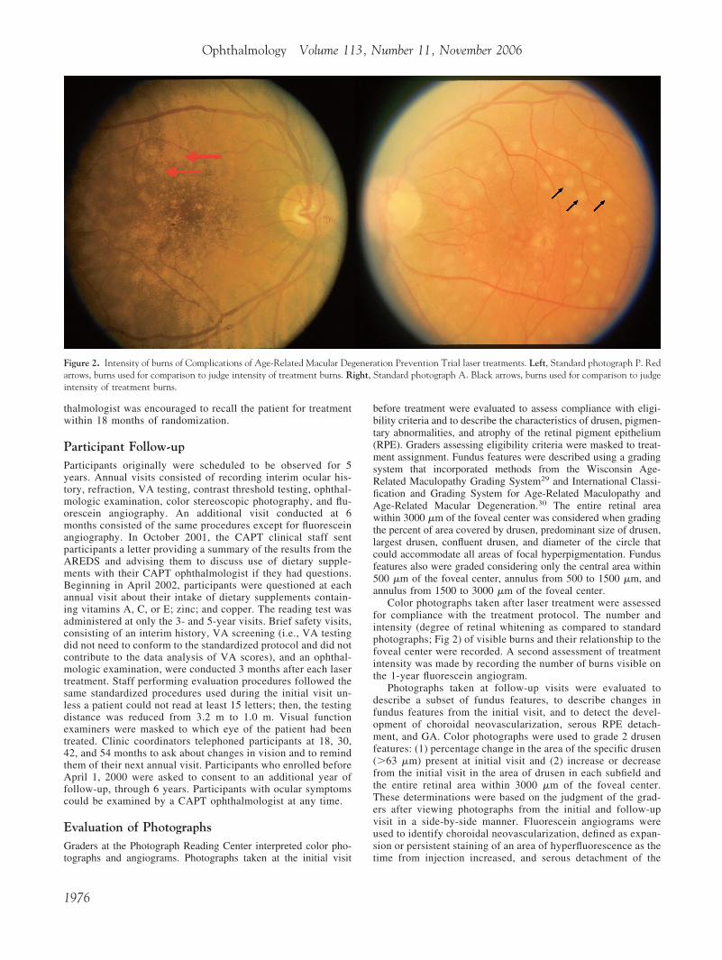
Figure 2. Intensity of burns of Complications of Age-Related Macular Degeneration Prevention Trial laser treatments. Left, Standard photograph P. Redarrows, burns used for comparison to judge intensity of treatment burns. Right, Standard photograph A. Black arrows, burns used for comparison to judgeintensity of treatment burns.
Ophthalmology Volume 113, Number 11, November 2006
1976

pigment epithelium, defined as a smooth dome-shaped elevationof the RPE with uniform fluorescein dye pooling and well-defined borders. Geographic atrophy was considered presentwhen the color photographs showed an area of atrophy at least250 �m in diameter accompanied by 2 of the following 3features: visible choroidal vessels, sharp edges, and approxi-mately circular shape. End point GA was defined as a total of�1 Macular Photocoagulation Study disc areas of atrophy whenall areas of GA were combined.
Outcome Measures
The primary outcome measure was a loss of �15 letters (3 lines)of VA between the initial visit and 5 years. Change in VA, changein contrast threshold, change in critical print size, and incidence oflate AMD (choroidal neovascularization, serous pigment epithe-lium detachment, or end point GA) were secondary outcomemeasures.
Serious adverse events warranting a report to the local institu-tional review board were defined as (1) treatment of the center ofthe foveal avascular zone, (2) a break in Bruch’s membrane at thetime of treatment as evidenced by blood or pigment and reportedby the treating ophthalmologist, (3) hemorrhage reported at thetime of CAPT laser treatment by the treating ophthalmologist orobserved by the graders in the Photograph Reading Center onposttreatment color photographs, or (4) a loss of �6 lines (30letters) of VA from the initial visit without the development ofchoroidal neovascularization, serous pigment epithelial detach-ment, GA, or cataract.
Sample Size and Power
Sample size calculations yielded a goal of 2000 eyes of 1000participants. Calculations involved several assumptions andestimates. The estimated rate of choroidal neovascularization(4%/year) through 5 years was based on a report in the litera-
ture31 and from a confidential report from the AREDS onparticipants assigned to a placebo who had bilateral drusen. Theproportion of eyes with choroidal neovascularization losing �3lines of VA by the completion of 5-year follow-up was esti-mated to be 75%, based on the experience of fellow eyes ofparticipants who enrolled in the Macular PhotocoagulationStudy. Thus, the estimated 5-year rate of �3 lines of VA lossdue to development of choroidal neovascularization was 15%(5*4%*75%). A 30% relative reduction, 15% to 10.5%, wasassumed to be the smallest effect of laser treatment that wouldbe of clinical importance. Additional assumptions included that4.4% of participants would develop loss of VA in both eyes; an� error of 0.05; statistical power of 0.90; and that 16% ofparticipants would be lost to follow-up at 5 years due to death,illness, and other reasons.
Data AnalysisData from CAPT clinical centers and the Photograph ReadingCenter that were entered into the database at the CAPT Coor-dinating Center by June 30, 2006 are the basis for this report.All comparisons of the 2 treatment groups were made on anintention-to-treat basis.
Definite hypertension was defined as systolic BP � 160 mmHg,diastolic BP � 95 mmHg, or current use of antihypertensivemedications. Suspect hypertension was defined as either systolicBP � 140 but � 160 mmHg or diastolic BP � 90 but � 95 mmHgin participants not taking antihypertensive medications. Intake ofantioxidant vitamins (A, C, and E) and zinc as reported by partic-ipants was summarized with respect to the doses used in theAREDS.
Analyses were conducted using statistical methods forpaired data because of the correlation between eyes of the sameperson.24 Differences between treatment groups in proportionswere assessed with the McNemar test. Differences betweentreatment groups in continuous data were assessed with eitherthe paired t test or the Wilcoxon signed rank test. Differencesbetween treatment groups in the proportion with VA loss wereassessed further using repeated-measures logistic regressionwith robust variance estimation data.32 The time to events suchas diagnosis of late AMD was described using Kaplan–Meierestimates of the cumulative proportion with the event. Differ-ences were assessed with proportional hazards modeling ac-commodating correlated data.33 The P values associated withcomparisons of secondary outcome measures after specific in-tervals of follow-up were not adjusted for multiple compari-sons. All analyses were conducted using SAS (version 9.1, SASInstitute, Inc., Cary, NC).
A data and safety monitoring committee reviewed treatmentsafety and study performance data twice a year and reviewedanalyses of treatment efficacy once a year. Although the pri-mary outcome measure was for loss of VA at 5 years afterenrollment, the committee specified guidelines following theO’Brien and Fleming approach, as expanded by Lan andDeMets, as a basis for discussion of early release of the databecause of treatment efficacy as assessed by repeated-measureslogistic regression.34,35
Results
Characteristics of Participants and EyesA total of 1052 participants enrolled. Participant characteristics atthe initial CAPT visit (baseline) have been reported.24 In brief, theaverage age was 71 years, 637 (60.6%) participants were female,
Table 1. Reasons from Central Review for Ineligibility byTreatment Group
Treated(n � 1052)
Observed(n � 1052)
Reason n Percent n Percent
�1 reasons* 148 14.1 135 12.8�10 large drusen, but drusen area � the
area of 10 large drusen81 7.7 66 6.3
�10 large drusen and drusen area � thearea of 10 large drusen
12 1.1 14 1.3
Choroidal neovascularization or serouspigment epithelium detachment
12 1.1 10 1.0
Geographic atrophy either within 500�m of the foveal center or � 1 MPSdisc area
4 0.3 6 0.6
Visual acuity worse than 20/40 1 0.1 0 0.0Basal laminar drusen or pattern dystrophy 25 2.4 25 2.4Conditions that could cause loss of vision 11 1.0 9 0.9Photographs missing, incomplete, or
unreadable, or taken too long beforeenrollment
8 0.8 9 0.9
Patient taking latanoprost: ophthalmicsolution
1 0.1 1 0.1
Visual acuity measured too long beforeenrollment
2 0.2 2 0.2
*Eyes may be ineligible for more than 1 reason.
CAPT Research Group � Laser Treatment in Patients with Bilateral Drusen
1977
1045 (99.3%) were white, 329 (31.3%) took �1 aspirins daily, 490(46.6%) had definite hypertension, 88 (8.4%) had diabetes, 58(5.5%) were current cigarette smokers, and 847 (80.5%) tookvitamin and/or zinc supplements.
Central review of completed data collection forms and photo-graphs showed that 283 (13.5%) of the 2104 eyes, composed of148 treated eyes and 135 observed eyes, did not meet �1 eligi-bility criteria (Table 1). The most frequent reason for ineligibility,accounting for 173 (8.2%) eyes, was having �10 drusen with adiameter of �125 �m (large drusen). Among eyes with �10 largedrusen, 81 (87.1%) of 93 treated eyes and 66 (82.5%) of 80observed eyes were considered near misses because they haddrusen area greater than the area of 10 large drusen. Both eyesof 94 (8.7%) participants were deemed ineligible upon centralreview.
Distributions of key fundus features of early AMD at baselinewithin each treatment group are shown in Table 2. There were nolarge imbalances between treatment groups. Of note, approxi-mately 70% of eyes had at least 1 druse � 250 �m, 33% had�10% of the area within 3000 �m of the foveal center covered bydrusen, and 70% had focal hyperpigmentation. Relatively few eyes(�5% each) had RPE depigmentation or any GA.
Distributions of measures of visual function at baseline weresimilar in the 2 treatment groups (Table 3). Approximately half ofeach group had VA between 20/12 and 20/20, and half had acuitybetween 20/25 and 20/40. The mean VA score was 82 letters(20/25�2 letters) in each group. Approximately 5% of eyes re-
quired �6% contrast to identify letters on the Pelli–Robson chart.The critical print size for reading was 20/62 or larger for approx-imately 19% of eyes.
Description of Laser Treatments
Initial treatment was performed in 1051 (99.8%) of the 1052 eyesassigned to laser treatment. Assessments by the Photograph Read-ing Center of the color photographs taken after treatment showedthat, of 1005 eyes with gradable photographs, 227 (22.6%) had allburns either not visible or not more intense than the burns ofstandard photograph P (Fig 2). Most eyes, 738 (73.4%) of 1005,had at least one burn more intense than on standard photograph Pand no more than 10 burns more intense than on standard photo-graph A (Fig 2). In 40 (4.0%) of 1005 eyes, �11 burns were moreintense than on standard photograph A.
Additional treatment was performed based on the drusenpresent at the 1-year visit for 856 (82.1%) of the 1042 livingparticipants. Of the 908 eyes that were judged by the PhotographReading Center to have drusen area � 10 large drusen at baselineand no contraindications to treatment at baseline or the 1-year visit,852 (93.8%) met the criterion for additional treatment of havingtotal area of drusen greater than or equal to the area of 10 largedrusen at the 1-year visit. Treatment was administered to 824(96.7%) of these eyes. There were 107 (10.3%) eyes that initiallywere judged by the CAPT ophthalmologist as not meeting thedrusen level required for treatment, but were judged by the Read-ing Center as meeting the requirement. In these cases, the CAPTophthalmologist was asked to recall the patient and apply treat-ment; treatment was administered to 92 (86.0%) of these eyes.
Assessments by the Photograph Reading Center of the colorphotographs taken after the second treatment showed that, of 777eyes with gradable photographs, 296 (38.1%) had all burns eithernot visible or not more intense than the burns on standard photo-graph P (Fig 2). Most eyes, 478 (61.5%) of 777, had at least oneburn more intense than on standard photograph P and no more than10 burns more intense than on standard photograph A (Fig 2). In3 (0.4%) eyes, �11 burns were more intense than on standardphotograph A.
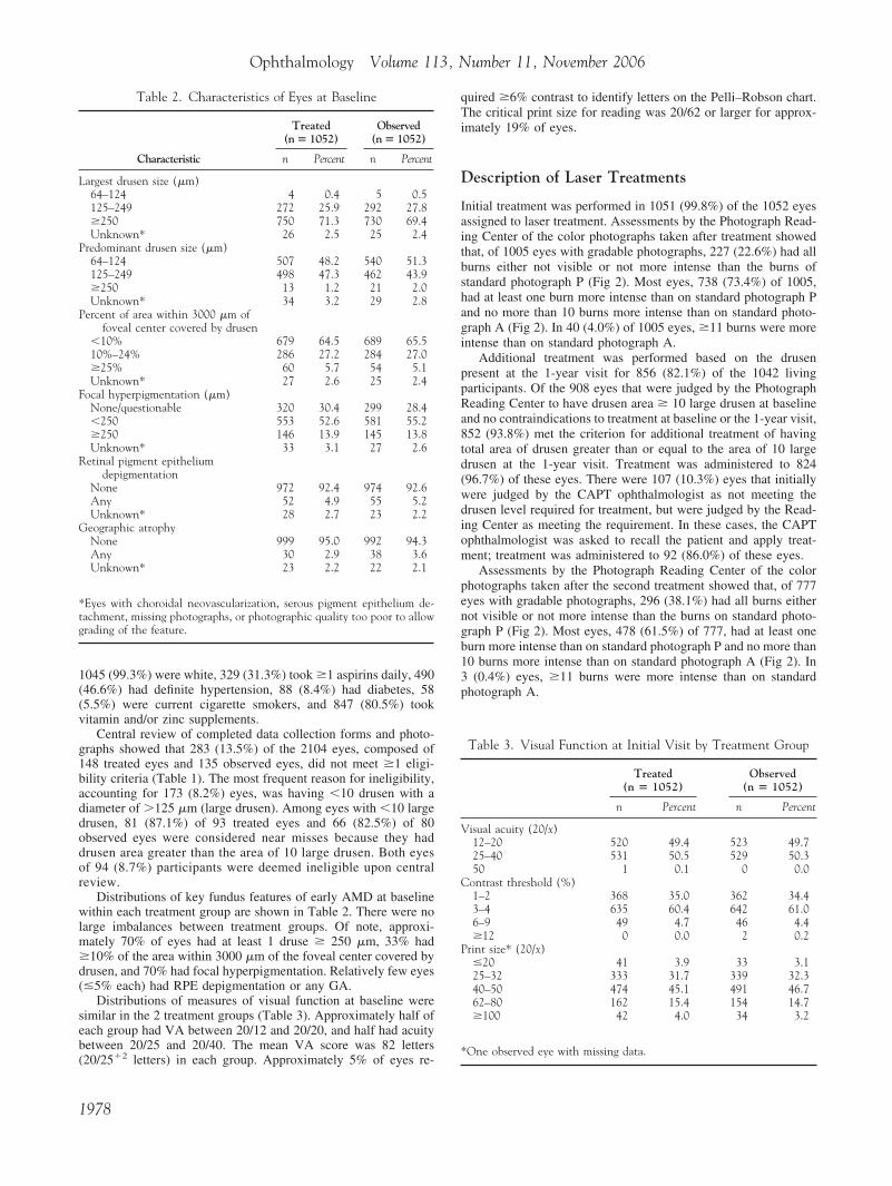
Table 2. Characteristics of Eyes at Baseline
Treated(n � 1052)
Observed(n � 1052)
Characteristic n Percent n Percent
Largest drusen size (�m)64–124 4 0.4 5 0.5125–249 272 25.9 292 27.8�250 750 71.3 730 69.4Unknown* 26 2.5 25 2.4
Predominant drusen size (�m)64–124 507 48.2 540 51.3125–249 498 47.3 462 43.9�250 13 1.2 21 2.0Unknown* 34 3.2 29 2.8
Percent of area within 3000 �m offoveal center covered by drusen
�10% 679 64.5 689 65.510%–24% 286 27.2 284 27.0�25% 60 5.7 54 5.1Unknown* 27 2.6 25 2.4
Focal hyperpigmentation (�m)None/questionable 320 30.4 299 28.4�250 553 52.6 581 55.2�250 146 13.9 145 13.8Unknown* 33 3.1 27 2.6
Retinal pigment epitheliumdepigmentation
None 972 92.4 974 92.6Any 52 4.9 55 5.2Unknown* 28 2.7 23 2.2
Geographic atrophyNone 999 95.0 992 94.3Any 30 2.9 38 3.6Unknown* 23 2.2 22 2.1
*Eyes with choroidal neovascularization, serous pigment epithelium de-tachment, missing photographs, or photographic quality too poor to allowgrading of the feature.
Table 3. Visual Function at Initial Visit by Treatment Group
Treated(n � 1052)
Observed(n � 1052)
n Percent n Percent
Visual acuity (20/x)12–20 520 49.4 523 49.725–40 531 50.5 529 50.350 1 0.1 0 0.0
Contrast threshold (%)1–2 368 35.0 362 34.43–4 635 60.4 642 61.06–9 49 4.7 46 4.4�12 0 0.0 2 0.2
Print size* (20/x)�20 41 3.9 33 3.125–32 333 31.7 339 32.340–50 474 45.1 491 46.762–80 162 15.4 154 14.7�100 42 4.0 34 3.2
*One observed eye with missing data.
Ophthalmology Volume 113, Number 11, November 2006
1978

Description of Follow-up
Through 5 years of follow-up, 5891 (97.2%) visits were completedof the 6061 6-month and annual visits scheduled for survivingCAPT participants. This percentage was relatively stable overtime, with 1020 (97.2%) of 1049 6-month visits, 1035 (99.3%) of1042 1-year visits, 1008 (98.1%) of 1027 2-year visits, 970(96.5%) of 1005 3-year visits, 941 (96.1%) of 979 4-year visits,and 917 (95.6%) of 959 5-year visits completed. An additional 251visits within the first 5 years were not completed because of 93participant deaths. For 78 (1.3%) of the 5891 completed visits,information on VA was obtained under nonprotocol conditionssuch as in a participant’s home or in the office of an ophthalmol-ogist who was not part of the CAPT. Among 503 participantseligible for a sixth year of follow-up, 457 (90.9%) consented toextended follow-up, of whom 439 (96.1%) completed the 6-yearvisit.
Reduction in Area of Drusen
At each follow-up visit, a higher proportion of treated eyes thanobserved eyes had a reduction of �50% in the area of the drusenwithin 3000 �m of the foveal center present at baseline (Fig 3).The proportion among treated eyes increased from 14.2% at 6months to 34.3% at 2 years to 41.8% at 5 years. The proportionamong observed eyes also increased over time, from 1.2% at 6months to 8.6% at 2 years to 31.2% at 5 years.
Visual Acuity
By 5 years after enrollment, the mean VA score had decreased byapproximately 2 lines to 73 letters (20/40�3) in each group(Table 4). Among the 917 participants completing the 5-year visit,291 (31.7%) treated eyes and 278 (30.3%) observed eyes had VAof 20/20 or better.
At 5 years after enrollment, 188 (20.5%) treated eyes and 188(20.5%) observed eyes had VA scores �3 lines worse than at theinitial visit (P � 1.00), yielding a difference of 0.0% (95%confidence interval [CI], �3.2% to 3.2%; Fig 4). Controlling forthe presence of focal hyperpigmentation provided similar resultsfor the difference between treatment groups (P � 0.89). Excludingeyes that were deemed ineligible by the Photograph ReadingCenter resulted in similar results; 21.2% of treated eyes and 20.7%of observed eyes had a loss of �3 lines. The largest differencebetween treated and observed eyes was at 3 years, when among
970 participants, 95 (9.8%) treated eyes and 121 (12.5%) observedeyes had scores �3 lines worse than at the initial visit (P � 0.04).The mean difference in change in VA between treated and ob-served eyes at 3 years was 1.1 letters (P � 0.02). Examination oftreatment group differences in subgroups defined by use of anti-oxidant vitamins and/or zinc at baseline and by use of the AREDSformulation of vitamins and zinc during follow-up did not identifyany significant differences between treatment groups.
Contrast Threshold and Critical Print Size
Both treated and observed eyes required more contrast to read theletters on the Pelli–Robson chart as the time from enrollmentincreased (Table 5). At 5 years, 212 (23.9%) of 888 treated eyesand 182 (20.5%) of 887 observed eyes required twice as muchcontrast (increase of 0.3 log units of contrast) to read letters (P �0.40).
The critical print size increased over time in both treated andobserved eyes (Table 6). At 5 years, 260 (29.6%) of 879 treatedeyes and 249 (28.4%) of 878 observed eyes required a print sizetwice as large (3 logarithm of the minimum angle of resolutionlines) or could not read even the largest print size (P � 0.70).
Incidence of Late Age-Related MacularDegeneration
Late AMD (choroidal neovascularization, end point GA, orserous RPE detachment) developed in 209 treated eyes and 220observed eyes. One hundred fourteen participants developedlate AMD in both eyes during follow-up. The cumulative inci-dence of late AMD was similar in the 2 treatment groupsthrough 6 years (P � 0.51; Fig 5). At 5 years, incidences were19.7% among treated eyes and 20.4% among observed eyes.Choroidal neovascularization developed in 141 treated eyes and141 observed eyes. The cumulative incidence of choroidalneovascularization was similar in the 2 treatment groupsthrough 6 years (P � 0.95; Fig 6). At 5 years, incidences were13.3% among treated eyes and 13.3% among observed eyes.End point GA developed in 74 treated eyes and 78 observedeyes; 8 of these treated eyes and 4 of these observed eyes laterdeveloped choroidal neovascularization. Cumulative incidencesof end point GA were similar in the 2 treatment groups through6 years (P � 0.64; Fig 7). At 5 years, cumulative incidenceswere 7.4% among treated eyes and 7.8% among observed eyes.In addition, a serous detachment of the RPE, in the absence ofapparent choroidal neovascularization, developed in 2 treatedeyes and 5 observed eyes.
Change in VA was associated strongly with the developmentof late AMD but not with treatment group. Among eyes devel-oping late AMD by 5 years, 107 (60.1%) of 178 treated eyesand 100 (54.3%) of 184 untreated eyes lost �3 lines of VA at5 years (P � 0.25). Among eyes that did not develop late AMDby 5 years, 76 (10.4%) of 728 treated eyes and 83 (11.4%) of726 observed eyes lost �3 lines of VA at 5 years (P � 0.50).Lens opacification cannot account for all of the loss in VAamong eyes that did not develop late AMD, because 15 (17.2%)of 87 treated eyes and 13 (15.3%) of 85 untreated eyes knownto be pseudophakic at the initial visit lost �3 lines of VA at 5years.
Adverse Events
There were no reports of burns applied to the foveal avascularzone, breaks in Bruch’s membrane, or hemorrhages at the initialor 1-year treatment. A loss of �6 lines of VA from the initial
19.2
34.3 37.040.7 41.8
46.4
1.2 2.78.6
16.2
24.831.2
35.6
14.2
0
10
20
30
40
50
60
70
80
90
100
6 12 24 36 48 60 72
Months of Follow-up
Treated
Observed
Figure 3. Percentage of eyes with 50% reduction in baseline drusen bytreatment group.
CAPT Research Group � Laser Treatment in Patients with Bilateral Drusen
1979

visit without the development of choroidal neovascularization,serous retinal pigment epithelial detachment, GA, or cataractoccurred in 6 (0.6%) treated eyes and 14 (1.3%) observed eyes.The loss in vision was attributed to a variety of conditionssuch as macular hole (1 treated eye, 1 observed eye), macularedema (1 treated eye, 1 observed eye), and Alzheimer’s disease(1 untreated eye).
Discussion
J. Donald Gass proposed prophylactic photocoagulationtreatment during the asymptomatic stage of AMD 35years ago.36 Since then, ophthalmologists have employedlaser treatment in eyes with drusen in an effort to improvevision, prevent vision loss, or reduce the likelihood ofprogression to late AMD. Published reports on the effectsof treatment have described relatively small numbers ofparticipants observed for varying periods, often with aless than desirable completeness of follow-up. Althoughthese reports have established that various approaches tolaser treatment result in reduction of drusen, the effectson progression to late AMD and loss of VA have beenboth inconsistent and inconclusive. Although reports of
laser treatment providing a better VA outcome by Little,Frennesson, Sarks, Olk, and Scorolli have supported therationale for laser treatment, reports on acceleration ofthe development of choroidal neovascularization in fel-low eyes, development of GA adjacent to laser burns, andaccumulation of foveal deposits after treatment haveraised concerns.9 –13,15,18,19,21,22,37
What has been consistent and conclusive is the poornatural history and visual prognosis of late AMD, espe-cially the neovascular stage. Moreover, until the recentreports on visual outcome after treating neovascularAMD with ranibizumab, the available treatments for neo-vascular AMD have been disappointing.38 In general,even beneficial treatments merely slowed the rate ofanatomic deterioration and accompanying vision loss inmost patients.39 – 42 Results from the AREDS providedevidence that daily use of dietary supplements containinghigh doses of the antioxidant vitamins A, C, and E and ofzinc reduces by 25% the incidence of late AMD andassociated loss of vision.3 However, even if all people athigh risk fully complied with the daily regimen, morethan 200 000 people each year in the United States woulddevelop late AMD.43 Thus, efforts to prevent the devel-
3.3 4.99.8
14.920.5
24.0
2.0 2.96.3
12.516.3
20.524.1
1.80
10
20
30
40
50
60
70
80
90
100
6 12 24 36 48 60 72
Months of Follow-up
Treated
Observed
Figure 4. Percentage of eyes with visual acuity loss of �3 lines by visit by treatment group.
Table 4. Visual Acuity by Follow-up
Visual Acuity(20/x)
12 Months 24 Months 36 Months
Treated Observed Treated Observed Treated Observed
n Percent n Percent n Percent n Percent n Percent n Percent
12–20 492 47.5 484 46.8 464 46.0 408 40.4 391 40.4 365 37.725–40 486 47.0 497 48.0 467 46.4 510 50.6 460 47.4 465 47.950–80 46 4.4 43 4.2 56 5.6 63 6.3 73 7.5 89 9.2
100–160 6 0.6 4 0.4 6 0.6 11 1.1 17 1.8 25 2.6�200 5 0.5 7 0.7 15 1.5 16 1.6 29 3.0 26 2.7
Total (mean) 1035 (20/25�1) 1035 (20/25�1) 1008 (20/25) 1008 (20/25�1) 970 (20/25�2) 970 (20/32�2)P* 0.34 0.01 0.02
*Paired t test.
Ophthalmology Volume 113, Number 11, November 2006
1980

opment of late AMD have continued to be a paramountconcern for more than 3 decades.
The CAPT investigators recognized during the plan-ning stage of the trial that laser treatment could promoteresolution of drusen. Previous studies had provided im-portant information that influenced the CAPT study de-sign and treatment protocol. These included the follow-ing: (1) fellow eyes of participants with unilateral lateAMD have an increased risk of developing choroidalneovascularization after prophylactic laser treatment; (2)laser treatment delivered in a grid or scatter patterntypically did not cause persistent or symptomatic sco-tomata; (3) laser burns could be applied either directly todrusen or adjacent to drusen with no discernible long-term difference in terms of promoting resolution ofdrusen or causing side effects; (4) more intense laserburns were associated with a tendency to develop cho-roidal neovascularization, sometimes at the site of thelaser burn; and (5) laser application could be repeatedafter an interval of 6 to 12 months to promote furtherresolution of drusen without any apparent significantadverse effect.8,9,12,13,15,16,44
Laser treatment in the CAPT had no effect at 5 yearson either VA or the incidence of late AMD (Figs 4, 5).Throughout the follow-up period, incidences of lateAMD were nearly identical in the 2 treatment groups.Although failure to detect a statistically significant dif-ference between treatment groups in some studies may beattributable to low power or bias introduced at baseline
through imbalance between treatment groups or duringfollow-up by missing data, these reasons cannot be ap-plied to the CAPT. With more than 1000 participants, thepower of the study to identify meaningful differencesbetween treatment groups was high, and the 95% CI forthe difference in the proportion with a loss of �3 lines at5 years was �3%. By virtue of having one eye of eachparticipant in each treatment group, all risk factors forlate AMD (age, race, cigarette smoking status, and hy-pertension) were identical in the treatment groups. Ocularcharacteristics were well balanced between the treatmentgroups (Table 2), and there was very little loss to fol-low-up other than patient death.
Laser treatment as applied in the CAPT was onlypartially successful in reducing the extent of drusen in thetreated eyes. Nearly all eyes that did not have contrain-dications to laser treatment qualified for a second treat-ment at 1 year after the initial treatment because ofremaining drusen. Even after 2 treatments, fewer thanhalf of the treated eyes demonstrated a 50% reduction inthe extent of drusen present at baseline. Particularly inthe latter years of follow-up, some of the reduction indrusen present at baseline can be attributed to the naturalcourse of AMD because of the relatively large proportionof observed eyes with drusen reduction (Fig 3).
As delivered in the CAPT, laser treatment was safe inthat there were no adverse events associated with theapplication. In addition, there was no excess incidence ofchoroidal neovascularization among treated eyes during
Time and Treatment Group
48 Months 60 Months 72 Months
Treated Observed Treated Observed Treated Observed
n Percent n Percent n Percent n Percent n Percent n Percent
335 35.6 323 34.3 291 31.7 278 30.3 114 26.0 126 28.7456 48.5 451 47.9 425 46.3 427 46.6 211 48.2 207 47.281 8.6 95 10.1 100 10.9 112 12.2 48 11.0 52 11.833 3.5 35 3.7 37 4.0 45 4.9 24 5.5 22 5.036 3.8 37 3.9 64 7.0 55 6.0 41 9.4 32 7.3941 (20/32�1) 941 (20/32) 917 (20/40�3) 917 (20/40�3) 438 (20/40�1) 439 (20/40�2)
0.30 0.78 0.20
Table 5. Change in Log Contrast Threshold by Follow-up Time and Treatment Group
Change in LogContrast Sensitivity
12 Months 36 Months 60 Months
Treated Observed Treated Observed Treated Observed
n % n % n % n % n % n %
��0.3 (better) 33 3.2 28 2.7 28 2.9 20 2.1 22 2.5 18 2.0�0.15 (better) 186 18.0 168 16.3 144 15.1 140 14.7 103 11.6 103 11.60 515 50.0 547 53.1 396 41.5 381 39.9 290 32.7 313 35.3�0.15 (worse) 243 23.6 240 23.3 276 28.9 292 30.6 261 29.4 271 30.6��0.3 (worse) 54 5.2 48 4.7 110 11.5 121 12.7 212 23.9 182 20.5Total 1031 1031 954 954 888 887P* 0.80 0.03 0.40
*Wilcoxon signed rank test.
CAPT Research Group � Laser Treatment in Patients with Bilateral Drusen
1981

the first years after treatment, as was the case in 3randomized clinical trials when laser treatment was ap-plied to fellow eyes of patients with unilateral lateAMD.12,20 –22
The eligibility criteria for CAPT were designed toidentify participants at high risk for vision loss whomight benefit from an intervention that could reduce thelikelihood of progression from early to late AMD. En-rolling patients with �10 large drusen in each eye, mostof whom also had focal hyperpigmentation, yielded astudy population in which 20% of eyes lost �3 lines ofVA within 5 years.
Reporting at the 2006 Association for Research inVision and Ophthalmology meeting on a multicenter trialof subthreshold infrared laser treatment for patients withbilateral drusen, T. R. Friberg noted a modest beneficialeffect of subthreshold diode infrared laser treatment after24 months in a subgroup of participants whose initialVAs were 20/32 to 20/64 (Invest Ophthalmol Vis Sci47:e-abstract 3538, 2006). Compared with untreatedeyes, treated eyes in this subgroup had a higher percent-age with an increase of �2 lines of VA (31% vs. 19%)
and a lower percentage with a decrease of �2 lines (13%vs. 22%). The CAPT eligibility criteria excluded eyeswith initial VA worse than 20/40; thus, the exactly anal-ogous subgroup cannot be constructed from the CAPTpopulation. When the 385 CAPT participants with initialVA in one or both eyes between 20/32 and 20/40 wereevaluated, the beneficial treatment effect reported byFriberg was not replicated. The CAPT treated and un-treated eyes at 24 months did not differ significantly withrespect to gain of �2 lines (7% vs. 5%) or to loss of �2lines (11% vs. 14%). At 5 years, 33% of both treated eyesand observed eyes in CAPT had lost �2 lines.
In summary, the CAPT was conducted at 22 clinicalcenters involving 1052 participants. Participants were ob-served for at least 5 years after laser treatment. The resultsof this study provide no evidence of a clinically significantbeneficial or harmful effect of preventive laser treatment ineyes with bilateral large drusen at high risk for progressionto late AMD.
Acknowledgments. Data and Safety Monitoring Committee:Daniel Seigel, ScD, Brian P. Conway, MD, Amy Horowitz, DSW,Aaron Kassoff, MD, Christopher Leighton, EdD, Anne Lindblad,
Group 6 12 24 36 48 60 72Treated 0.008 0.030 0.060 0.104 0.156 0.197 0.241Observed 0.012 0.036 0.065 0.104 0.155 0.204 0.254
0
0.05
0.1
0.15
0.2
0.25
0.3
0 6 12 18 24 30 36 42 48 54 60 66 72 78
Months of Follow-up
Prop
ortio
n w
ith L
ate
AM
D
TreatedObserved
p=0.51
Figure 5. Cumulative incidence of late age-related macular degeneration(AMD) by treatment group.
Group 6 12 24 36 48 60 72Treated 0.007 0.026 0.041 0.074 0.111 0.133 0.161Observed 0.011 0.030 0.050 0.070 0.101 0.133 0.160
0
0.05
0.1
0.15
0.2
0.25
0.3
0 6 12 18 24 30 36 42 48 54 60 66 72 78
Months of follow-up
Prop
ortio
n w
ith C
NV
TreatedObserved
P=0.95
Figure 6. Cumulative incidence of choroidal neovascularization (CNV)by treatment group.
Table 6. Change in Critical Print Size by Visit and Treatment Group
Print Size Change(logMAR Lines)
36 Months 60 Months
Treated Observed Treated Observed
n Percent n Percent n Percent n Percent
�3 smaller 65 6.9 45 4.8 46 5.2 35 4.01–2 smaller 193 20.4 195 20.7 147 16.7 162 18.50 change 211 22.3 209 22.2 147 16.7 160 18.21–2 larger 282 29.8 306 32.5 279 31.7 272 31.0�3 larger 185 19.6 181 19.2 241 27.4 226 25.7Could not read† 9 1.0 6 0.6 19 2.2 23 2.6Total 945 942 879 878P* 0.12 0.70
logMAR � logarithm of the minimum angle of resolution.*Wilcoxon signed rank test.†Could not read largest print size.
Ophthalmology Volume 113, Number 11, November 2006
1982

PhD, David Mazur, MD. National Eye Institute: Natalie Kurinij,PhD.
References
1. Bressler SB, Maguire MG, Bressler NM, et al. Relationship ofdrusen and abnormalities of the retinal pigment epithelium tothe prognosis of neovascular macular degeneration. ArchOphthalmol 1990;108:1442–7.
2. Macula Photocoagulation Study Group. Risk factors for cho-roidal neovascularization in the second eye of patients withjuxtafoveal or subfoveal choroidal neovascularization second-ary to age-related macular degeneration. Arch Ophthalmol1997;115:741–7.
3. Age-Related Eye Disease Study Research Group. A random-ized, placebo-controlled, clinical trial of high-dose supple-mentation with vitamins C and E, beta-carotene, and zinc forage-related macular degeneration and vision loss. AREDSreport no. 8. Arch Ophthalmol 2001;119:1417–36.
4. Klein R, Klein BE, Tomany SC, et al. Ten-year incidence andprogression of age-related maculopathy: the Beaver Dam EyeStudy. Ophthalmology 2002;109:1767–79.
5. Cleasby GW, Nakanishi AS, Norris JL. Prophylactic photo-coagulation of the fellow eye in exudative senile maculopathy:a preliminary report. Mod Probl Ophthalmol 1979;20:141–7.
6. Wetzig PC. Treatment of drusen-related aging macular degen-eration by photocoagulation. Trans Am Ophthalmol Soc 1988;86:276–90.
7. Sigelman J. Foveal drusen resorption one year after perifoveallaser photocoagulation. Ophthalmology 1991;98:1379–83.
8. Figueroa MS, Regueras A, Bertrand J. Laser photocoagulationto treat macular soft drusen in age-related macular degenera-tion. Retina 1994;14:391–4.
9. Frennesson IC, Nilsson SE. Laser photocoagulation of softdrusen in early age-related maculopathy (ARM): the one-yearresults of a prospective, randomised trial. Eur J Ophthalmol1996;6:307–14.
10. Sarks SH, Arnold JJ, Sarks JP, et al. Prophylactic perifoveallaser treatment of soft drusen. Aust N Z J Ophthalmol 1996;24:15–26.
11. Guymer RH, Gross-Jendroska M, Owens SL, et al. Laser
treatment in subjects with high-risk clinical features ofage-related macular degeneration: posterior pole appearanceand retinal function. Arch Ophthalmol 1997;115:595–603.
12. Choroidal Neovascularization Prevention Trial ResearchGroup. Laser treatment in eyes with large drusen: short-termeffects seen in pilot randomized clinical trial. Ophthalmology1998;105:11–23.
13. Olk RJ, Friberg TR, Stickney KL, et al. Therapeutic benefitsof infrared (810 nm) diode laser macular grid photocoagula-tion in prophylactic treatment of nonexudative age-relatedmacular degeneration: two-year results of a randomized pilotstudy. Ophthalmology 1999;106:2082–90.
14. Rodanant N, Friberg TR, Cheng L, et al. Predictors of drusenreduction after subthreshold infrared (810 nm) diode lasermacular grid photocoagulation for nonexudative age-relatedmacular degeneration. Am J Ophthalmol 2002;134:577–85.
15. Little HL, Showman JM, Brown BW. A pilot randomizedcontrolled study on the effect of laser photocoagulation of con-fluent soft macular drusen. Ophthalmology 1997;104:623–31.
16. Ho AC, Maguire MG, Yoken J, et al. Laser-induced drusenreduction improves visual function at one year. Ophthalmol-ogy 1999;106:1367–73, discussion 1374.
17. Frennesson CI. Prophylactic laser treatment in early age-related maculopathy: an 8-year follow-up in a randomizedpilot study shows a reduced incidence of exudative complica-tions. Acta Ophthalmol Scand 2003;81:449–54.
18. Scorolli L, Corazza D, Morara M, et al. Argon laser vs.subthreshold infrared (810-nm) diode laser macular grid pho-tocoagulation in nonexudative age-related macular degenera-tion. Can J Ophthalmol 2003;38:489–95.
19. Hyver SW, Schatz H, McDonald HR, Johnson RN. A case ofvisual acuity loss following laser photocoagulation for macu-lar drusen [letter]. Arch Ophthalmol 1997;115:554–5.
20. Choroidal Neovascularization Prevention Trial Research Group.Laser treatment in fellow eyes with large drusen. Updatedfindings from a pilot randomized clinical trial. Ophthalmology2003;110:971–8.
21. Friberg TR, Musch DC, Lim JI, et al. Prophylactic treatmentof age-related macular degeneration report number 1: 810-nanometer laser to eyes with drusen. Unilaterally eligiblepatients. Ophthalmology 2006;113:612–22.
22. Owens SL, Bunce C, Brannon AJ, et al. Prophylactic lasertreatment hastens choroidal neovascularization in unilateralage-related maculopathy: final results of the Drusen LaserStudy. Am J Ophthalmol 2006;141:276–81.
23. Complications of Age-Related Macular Degeneration Preven-tion Trial Study Group. The Complications of Age-RelatedMacular Degeneration Prevention Trial (CAPT): rationale,design and methodology. Clin Trials 2004;1:91–107.
24. Complications of Age-Related Macular Degeneration Preven-tion Trial Research Group. Baseline characteristics, the 25-item National Eye Institute Visual Functioning Questionnaire,and their associations in the Complications of Age-RelatedMacular Degeneration Prevention Trial (CAPT). Ophthalmol-ogy 2004;111:1307–16.
25. Early Treatment Diabetic Retinopathy Study: design and base-line patient characteristics. ETDRS report number 7. Ophthal-mology 1991;98(suppl):741–56.
26. National Eye Institute. Age-Related Eye Disease Study. Manualof operations (MOP). Examination procedures. 2000:7-1–12.Available at: https://web.emmes.com/study/areds/mopfiles/chp7_mop.pdf. Accessed August 9, 2006.
27. Pelli DG, Robson JG, Wilkins AJ. The design of a new letterchart for measuring contrast sensitivity. Clin Vis Sci 1988;2:187–99.
28. Mansfield JS, Legge GE, Bane MC. Psychophysics of reading.
Group 6 12 24 36 48 60 72Treated 0.001 0.004 0.019 0.036 0.052 0.074 0.099Observed 0.001 0.004 0.014 0.034 0.056 0.078 0.105
0
0.05
0.1
0.15
0.2
0.25
0.3
0 6 12 18 24 30 36 42 48 54 60 66 72 78
Months of follow-up
Prop
ortio
n w
ith G
eogr
aphi
c A
trop
hy
TreatedObserved
P=0.64
Figure 7. Cumulative incidence of end point geographic atrophy bytreatment group.
CAPT Research Group � Laser Treatment in Patients with Bilateral Drusen
1983

XV: font effects in normal and low vision. Invest OphthalmolVis Sci 1996;37:1492–501.
29. Klein R, Davis MD, Magli YL, et al. The Wisconsin Age-Related Maculopathy Grading System. Ophthalmology 1991;98:1128–34.
30. Bird AC, Bressler NM, Bressler SB, International ARM Ep-idemiological Study Group. An international classification andgrading system for age-related maculopathy and age-relatedmacular degeneration. Surv Ophthalmol 1995;39:367–74.
31. Holz FG, Wolfensberger TJ, Piguet B, et al. Bilateral maculardrusen in age-related macular degeneration: prognosis and riskfactors. Ophthalmology 1994;101:1522–8.
32. Zeger SL, Liang KY. Longitudinal data analysis for discreteand continuous outcomes. Biometrics 1986;42:121–30.
33. Wei LJ, Lin DY, Weissfeld L. Regression analysis of multi-variate incomplete failure time data by modeling marginaldistributions. J Am Stat Assoc 1989;84:1065–73.
34. O’Brien PC, Fleming TR. A multiple testing procedure forclinical trials. Biometrics 1979;35:549–56.
35. Lan KK, DeMets DL. Discrete sequential boundaries for clin-ical trials. Biometrika 1983;70:649–53.
36. Gass JD. Photocoagulation of macular lesions. Trans AmAcad Ophthalmol Otolaryngol 1971;75:580–608.
37. Ruiz-Moreno JM, De La Vega C, Zarbin MA. Macular atrophyafter photocoagulation of soft drusen. Retina 2003;23:315–21.
38. Heier JS, Antoszyk AN, Pavan PR, et al. Ranibizumab fortreatment of neovascular age-related macular degeneration. Aphase I/II multicenter, controlled, multidose study. Ophthal-mology 2006;113:633–42.
39. Macular Photocoagulation Study Group. Laser photocoagula-tion of subfoveal neovascular lesions of age-related maculardegeneration: updated findings from two clinical trials. ArchOphthalmol 1993;111:1200–9.
40. Treatment of Age-Related Macular Degeneration with Photo-dynamic Therapy (TAP) Study Group. Photodynamic therapyof subfoveal choroidal neovascularization in age-related mac-ular degeneration with verteporfin: two-year results of 2 ran-domized clinical trials—TAP report 2. Arch Ophthalmol2001;119:198–207.
41. Verteporfin in Photodynamic Therapy Study Group. Vertepor-fin therapy of subfoveal choroidal neovascularization in age-related macular degeneration: two-year results of a random-ized clinical trial including lesions with occult with no classicchoroidal neovascularization—Verteporfin in PhotodynamicTherapy report 2. Am J Ophthalmol 2001;131:541–60.
42. Gragoudas ES, Adamis AP, Cunningham ET Jr, et al. Pe-gaptanib for neovascular age-related macular degeneration.N Engl J Med 2004;351:2805–16.
43. Age-Related Eye Disease Study Research Group. Potential pub-lic health impact of Age-Related Eye Disease Study results.AREDS report no. 11. Arch Ophthalmol 2003;121:1621–4.
44. Kaiser RS, Berger JW, Maguire MG, et al. Laser burn intensityand the risk for choroidal neovascularization in the CNVPTFellow Eye Study. Arch Ophthalmol 2001;119:826–32.
Appendix: The Complications of Age-Related Macular Degeneration PreventionTrial (CAPT) Research Group
Retinal Consultants of Arizona, Mesa and SunCity, ArizonaDonald W. Park, MD, Pravin V. Dugel, MD, Allen B.Thach, MD, Siru Adhikari, Christina Alvarado, Jennifer
Blaisdell, Jennifer Cavanagh, Jennifer Cornelius, ElenaMarcos, Kaz Tysiac, Norma Jimenez, Adriana Falcon, Sha-ron Kosecki, Carol Slagle, Cheri Tuttle, Scott E. Bohnen,Brian M. Manor, John Martin, Anne C. Monday.
West Coast Retina, San Francisco, CaliforniaRobert N. Johnson, MD, Everett Ai, MD, H. Richard Mc-Donald, MD, Margaret Stolarczuk, OD, Pat Wood, LVN,CCRS, Kevan Curren, COA, Irina Rozenfeld, MD, BrandiTeske, COA, Marsha Apushkin, Silvia Linares, Kelly De-Boer, Sarah Huggans, Jeremy Miller, John Uy.
University of South Florida Eye Institute, Tampa,FloridaPeter Reed Pavan, MD, Karina K. Billiris, MD, BurtonGoldstein, MD, Mohan Iyer, MD, Matthew M. Menosky,MD, Jonathan Mines, MD, Scott E. Pautler, MD, Sharon M.Millard, RN, COT, Susan Sherouse, COT, Michelle D.West, COT, Steve Carlton, Wyatt Saxon.
Emory Eye Center, Atlanta, GeorgiaPaul Sternberg, Jr, MD, Thomas Aaberg, Sr, MD, BakerHubbard III, MD, David Saperstein, MD, Lindy DuBois,MEd, MMSC, Ann Ervin, MPH, Judy Brower, MMSC, CO,Jayne Brown, Gail Browne, Gabriela Burian, NatalieSchmitz, Rhonda Waldron, MMSc, COMT, James Gilman,CRA, Bob A. Myles.
Northwestern University, Chicago, IllinoisAlice Lyon, MD, Susan Anderson-Nelson, MD, Lee M.Jampol, MD, David V. Weinberg, MD, Annie Muñana, RN,Zuzanna Rozenbajgier, MA, Lori Kaminski, RN, Jill Koe-cher, Laima O’Donnell, Renata Swigost, Lisa Volland, RN,Marsha Apushkin, Alexander Habib, Pamela Hulvey,Jonathan Shankle, James Yuhr.
Illinois Retina Associates, Harvey and Skokie,IllinoisDavid Orth, MD, Jack Cohen, MD, Matthew MacCumber,MD, Pauline Merrill, MD, Celeste Figliulo, Liz Porcz, Car-rie L. Violetto, CMA, Tana N. Drefcinski, Hope P. Ne-nadov, Laurie Rago, Donald Doherty, Marian McVicker,David Nash.
University of Iowa Hospitals & Clinic, Iowa City,IowaJames C. Folk, MD, H. Culver Boldt, MD, Karen M. Gehrs,MD, Stephen R. Russell, MD, Rachael Ivins, CCRC, StevenA. Wallace, Connie Hinz, COT, Michael Harker, Ed Hef-fron, Stefani Karakas.
Ophthalmology & Visual Sciences at theUniversity of Lousiville, Louisville, KentuckyCharles C. Barr, MD, Steve Bloom, MD, Brian Kritchman,MD, Greg Whittington, PsyS, Rhonda Bowyer, Dee Den-
Ophthalmology Volume 113, Number 11, November 2006
1984

ning, COT, Janice Goatley, Janet Nutting, Judy Swartz,Evelyn Temple, Wendy Wilson, COT.
Ophthalmic Consultants of Boston, Boston,MassachusettsJeffrey Heier, MD, Albert R. Frederick, Jr, MD, Michael G.Morley, MD, Trexler Topping, MD, Tammy Hanner, COA,Molly Doherty, Heather L. Davis, Linda Beal, COA, SeanMahoney, COA, Robin A. Ty, Cullen Mike Jones, COA,Elna Rapp, RN, COT.
Wilmer Ophthalmological Institute, Johns HopkinsUniversity, Baltimore, MarylandSusan B. Bressler, MD, Andrew P. Schachat, MD, DantePieramici, MD, Neil M. Bressler, MD, Warren Doll, COA,Ellen Greenberg, COT, Robert A. Jurao, COA, Deborah F.Donohue, COA, Mary Frey, Siobhan Sheehan, Tracey Por-ter, Judith E. Belt, Dennis R. Cain, CRA, Rachel Falk,Charles M. Herring, Jacquelyn M. McDonald.
Associated Retinal Consultants, Royal Oak,MichiganMichael Trese, MD, Antonio Capone, MD, Bruce R. Gar-retson, MD, Tarek S. Hassan, MD, Alan J. Ruby, MD,Michelle M. Kulak, RN, Pat Manatrey, RN, Tammy Osen-toski, RN, Linda Vandell, RN, Kristi Cumming, RN, MSN,Beth Mitchell, RN, Mary Zajechowski, COT, Craig Bridges,Patricia Siedlak, Patricia Streasick, Lynette Szydlowski.
Mayo Clinic, Rochester, MinnesotaColin A. McCannel, MD, Helmut Buettner, MD, John M.Pach, MD, Dennis M. Robertson, MD, Margaret J. Ruszc-zyk, CCRA, Jean Burrington, Kathleen LeBarron, COA,Cindy A. Stephan, COA, Thomas Link, Jay A. Rostvold.
Barnes Retina Institute, St. Louis, MissouriGilbert Grand, MD, Kevin Blinder, MD, Nancy M. Holekamp,MD, Daniel P. Joseph, MD, PhD, Travis A. Meredith, MD, GauravShah, MD, Julie A. Binning, COT, CCRS, Ginny S. Nobel, COT,Cindy M. Wright, Linda Boyd, COA, Janel Gualdoni, COT, PamLight, CCRC, Nancy Soueidan, RN, Bryan Barts, Jon Dahl, Tim-othy Holle, Matt Raeber, John Mark Rogers.
Southeast Clinical Research Associates, Charlotte,North CarolinaAndrew N. Antoszyk, MD, David J. Browning, MD, PhD, ToniaEllsmore, CRC, Jennifer V. Helms, CCRC, Lori Lundy, COMT,Alison H. Stallings, Loraine Clark, Sandy Efird, COT, MarkEvans, Fereshteh Jarrahi, Kara Mundy, Heather Murphy, Tisha L.O’Marah, Jennifer Wike, COA, Patricia Woodland, Linda Davis,Mike McOwen.
Retina-Vitreous Center, Edison and Lakewood,New JerseySteven R. Leff, MD, Eric Friedman, MD, Stuart N. Green, MD, BruceKeyser, MD, Miriam Kushner, MD, David L. Yarian, MD, Cheryl
Hambrock, RN, Linda Wagner, COT, Marge Lucido, Donna Coffey,RN, Melinda Geddes, Thea Tantum, COT, Finn Andersen, AlexSchlosser.
Retina Associates of Cleveland, Cleveland andLakewood, OhioLawrence J. Singerman, MD, David Miller, MD, Robert Mittra,MD, Michael Novak, MD, Scott Pendergast, MD, Jeffrey H.Stockfish, MD, Hernando Zegarra, MD, Scott D. Marella, CCRP,COA, Stephanie A. Schura, COT, Sheila Smith-Brewer, CRA,COMT, Kimberly DuBois, CCRP, COA, Jacqueline L. Hursky,COA, Mary Ilc, COT, Sheri L. Joyce, COA, Vivian Tanner, COT,John DuBois, CRA, Gregg Greanoff, CRA, David S. Lehnhardt,COA.
Ohio State University, Columbus, OhioFrederick H. Davidorf, MD, Robert Chambers, DO, LouisChorich, MD, Cynthia S. Taylor, CCRC, Jill Salerno, COA, MaryT. Deringer, COA, Jill Milliron, COA, Jerilyn Perry, COT, ABO,Scott Savage, EMT-A.
Retina Northwest, Portland, OregonRichard F. Dreyer, MD, Colin Ma, MD, Patricia Bartholomew,CCRC, Harold L. Crider, COT, Marcia R. Kopfer, COT, JeanetteR. Larson, COMT, Cindy Armstrong, Debra DeShazer, SteveHobbs, Wendy Raunig, COT, Katie Reichenberger, StephanieSchmidt, Don Sitts, Howard “Dan” Daniel, Milt Johnson, R.Joseph Logan, Harry J. Wohlsein, Jr.
Casey Eye Institute, Portland, OregonMichael L. Klein, MD, David J. Wilson, MD, Susan K. Nolte,Patricia D. Lindstrom, COT, Susan Pope, COT, Debora R. Vahr-enwald, COT, Jessica Gaultney, Ellen Redenbo, Patrick Rice,Peter Steinkamp, Patrick Wallace.
University of Pennsylvania, Philadelphia,PennsylvaniaJuan E. Grunwald, MD, Jeffrey W. Berger, MD, PhD, AlexanderJ. Brucker, MD, Josh Dunaief, MD, PhD, Stuart L. Fine, MD,Allen Ho, MD, Albert M. Maguire, MD, Michael Tolentino, MD,Sharon Decker, Emily Hall, Jennifer Levin, MD, Monique N.McRay, Gretchen Warley, MSW, Stacey Boxley, Joan DuPont,CCRC, Claudette Geist, CRA, COT, Tatyana Metelitsina, MD,Michele Sheehan, COMT, Cheryl Devine, CRA, Deborah Elkins,William Nyberg, RBP, CRA, Laurel Weeney, CRA.
Texas Retina Associates, Dallas and Arlington,TexasGary Edd Fish, MD, Rajiv Anand, MD, Rand Spencer, MD, JeanArnwine, Jeff Harris, Nancy Resmini, Marilyn Andrews, SallyArceneaux, COA, Barbara McCarty, Dustin Pierce, Rubye Rollins,Brenda Sanchez, Hank A. Aguado, Bob Boleman, Penny B. El-lenich, John G. King.
University of Wisconsin, Madison, Madison,WisconsinSuresh R. Chandra, MD, Michael Altaweel, MD, Barbara Blodi, MD,Justin Gottlieb, MD, Michael Ip, MD, T. Michael Nork, MD, Erika D.
CAPT Research Group � Laser Treatment in Patients with Bilateral Drusen
1985

Soderling, Wendy Walker, COA, Jennie Perry-Raymond, MargoBlatz, Jennifer M. Buechner, Shelly Olson, Alyson Pohlman, BarbSoderling, Gene Knutson, Denise Krolnik, John Peterson.
CAPT Chairman’s Office, University ofPennsylvania, Philadelphia, PennsylvaniaStuart L. Fine, MD, Marilyn Katz.
CAPT Coordinating Center, University ofPennsylvania, Philadelphia, PennsylvaniaMaureen G. Maguire, PhD, Kate Atkins, Mary Brightwell-Arnold,Sandra Harkins, Christine D. Holmes, Andrew James, MS, MargaretJewell, Alexander Khvatov, Chengcheng Liu, MS, Lori O’Brien,Kathy McWilliams, CCRP, Ellen Peskin, MA, CCRP, Renee Rees,PhD, Susan Ryan, N. Nefertiti Stanford, Karen Taylor, ClaressaWhearry, Gui-Shuang Ying, PhD.
CAPT Photograph Reading Center, University ofPennsylvania, Philadelphia, Pennsylvania
Judith Alexander, Jeffrey Berger, MD, PhD, Robert Stoltz, MD,PhD, Steven Begley, Keith Elsner, Allen Ho, MD, Noreen Ja-vornik, MS, Kristin Mathias, MS, Bojidar Madjarov, MD, E.Revell Martin, Renee Zawacki.
Manuscript Writing Team
Maureen G. Maguire, PhD, Gui-Shuang Ying, PhD, Judith Alex-ander, David J. Browning, MD, PhD, Ellen Peskin, MA, James C.Folk, MD, Andrew N. Antoszyk, MD, Juan E. Grunwald, MD,Michael L. Klein, MD, Alice Lyon, MD, Colin A. McCannel, MD,Robert A Stoltz, MD, PhD, Michael Trese, MD, Stuart L. Fine,MD.
Ophthalmology Volume 113, Number 11, November 2006
1986