Embed Size (px)
Citation preview
CENTRAL LASER FACILITY Annual Report 2010 - 201136
LASERS FOR SCIENCE FACILITY PROGRAMME Biology
Biology
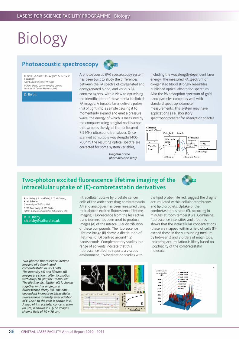
Photoacoustic spectroscopy
A photoacoustic (PA) spectroscopy systemhas been built to study the differencesbetween the PA spectra of oxygenated anddeoxygenated blood, and various PAcontrast agents, with a view to optimisingthe identification of these media in clinicalPA images. A tunable laser delivers pulses(ns) of light into a sample causing it tomomentarily expand and emit a pressurewave, the energy of which is measured bythe computer using a digital oscilloscopethat samples the signal from a focused 7.5 MHz ultrasound transducer. Oncescanned at multiple wavelengths (400-700nm) the resulting optical spectra arecorrected for some system variables,
including the wavelength-dependent laserenergy. The measured PA spectrum ofoxygenated blood strongly resemblespublished optical absorption spectrum.Also the PA absorption spectrum of goldnano-particles compares well withstandard spectrophotometermeasurements. This system may haveapplications as a laboratoryspectrophotometer for absorption spectra.
D. Birtill1, A. Shah1,2, M. Jaeger1,2, A. Gertsch1,J. Bamber1
(1Joint Department of Physics)
(2CRUK-EPSRC Cancer Imaging Centre,Institute of Cancer Research, UK)
D. Birtill
48
Two-photon excited fluorescence lifetime imaging of theintracellular uptake of (E)-combretastatin derivatives
Intracellular uptake by prostate cancercells of the anticancer drug combretastatinA4 and analogues has been measured usingmultiphoton excited fluorescence lifetimeimaging. Fluorescence from the less activetrans isomers has been used to produceimages (A) of the intracellular distributionof these compounds. The fluorescencelifetime image (B) shows a distribution oflifetimes (C, D) centred around 1.2nanoseconds. Complementary studies in arange of solvents indicate that thisfluorescence lifetime reports a viscousenvironment. Co-localisation studies with
the lipid probe, nile red, suggest the drug isaccumulated within cellular membranesand lipid droplets. Uptake of thecombretastatin is rapid (E), occurring inminutes at room temperature. Combiningfluorescence intensities and lifetimesshows that the intracellular concentrations(these are mapped within a field of cells (F))exceed those in the surrounding mediumby between 2 and 3 orders of magnitude,indicating accumulation is likely based onlipophilicity of the combretastatinmolecule.
R. H. Bisby, J. A. Hadfield, A. T. McGown, K. M. Scherer (University of Salford, UK)
S. W. Botchway, A. W. Parker (STFC, Rutherford Appleton Laboratory, UK)
R. H. [email protected]
49
Diagram of thephotoacoustic setup
Two-photon fluorescence lifetimeimaging of a fluorinatedcombretastatin in PC-3 cells. The intensity (A) and lifetime (B)images are shown after incubationwith drug (10 μM) for 10 minutes.The lifetime distribution (C) is showntogether with a single pixelfluorescence decay (D). The time-dependent increase in intracellularfluorescence intensity after additionof E-CA4F to the cells is shown in E.A map of intracellular concentration(in μM) is shown in F. (The imagesshow a field of 70 x 70 μm)
LASERS FOR SCIENCE FACILITY PROGRAMME Biology
CENTRAL LASER FACILITY Annual Report 2010 - 2011 37
The plant secretoryome: protein-protein interactionsin the higher plant secretory pathway
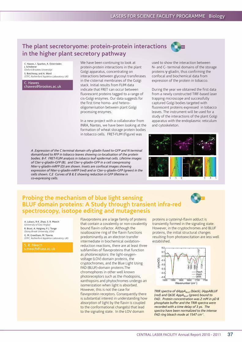
We have been continuing to look atprotein-protein interactions in the plantGolgi apparatus, concentrating oninteractions between glycosyl transferasesin the cisternal membranes of the Golgistack. Initial results from FLIM dataindicate that FRET can occur betweenfluorescent proteins tagged to a range ofcis-Golgi enzymes. Our data suggests forthe first time homo- and hetero-oligomerisation between plant Golgiprocessing enzymes.
In a new project with a collaborator fromINRA, Nantes, we have been looking at theformation of wheat storage protein bodiesin tobacco cells. FRET-FLIM (Figure) was
used to show the interaction between N- and C- terminal domains of the storageproteins γ-gliadin, thus confirming theconfocal and biochemical data fromexpression of the protein in tobacco.
During the year we obtained the first datafrom a newly constructed TIRF-based lasertrapping microscope and successfullycaptured Golgi bodies targeted withfluorescent proteins expressed in tobaccoleaves. The instrument will be used for astudy of the interactions of the plant Golgiapparatus with the endoplasmic reticulumand cytoskeleton.
C. Hawes, I. Sparkes, A. Osterrieder, J. Schoberer (Oxford Brookes University)
S. Botchway, and A. Ward (STFC, Rutherford Appleton Laboratory, UK)
C. Hawes [email protected]
50
A .Expression of the C terminal domain ofγ-gliadin fused to-GFP and N terminaldomainfused to RFP in tobacco leaves showing co-localisation of the proteinbodies. B-F. FRET-FLIM analysis in tobacco leaf epidermal cells. Lifetime imagesof Cter-γ-gliadin-GFP (B), and Cter-γ-gliadin-GFP in a cell coexpressing Nter-γ-gliadin-mRFP (D) are shown. Insets are confocal images showingexpression of Nter-γ-gliadin-mRFP (red) and or Cter-γ-gliadin-GFP (green) in thecells shown. C,E Curves of B & E showing reduction in GFP lifetime in co-expressing cells.
Flavoproteins are a large family of proteinsthat contain a covalently or non-covalentlybound flavin cofactor. Although theisoalloxazine ring of the flavin functionspredominantly as an electron transferintermediate in biochemical oxidation-reduction reactions, there are at least threesubfamilies of flavoproteins that functionas photoreceptors: the light-oxygen-voltage (LOV) domain proteins, thecryptochromes, and the Blue Light UsingFAD (BLUF)-domain proteins.Thechromophores in other well knownphotoreceptors such as the rhodopsins,xanthopsins and phytochromes undergo anisomerization when light is absorbed.However, this is not the case forflavoprotein receptors. Consequently thereis substantial interest in understanding howabsorption of light by the flavin is coupledto the conformational change(s) that leadto the signaling state. In the LOV domain
proteins a cysteinyl-flavin adduct istransiently formed in the signaling state.However, in the cryptochromes and BLUFproteins, the initial structural changesresulting from photoexcitation are less wellestablished.
A. Lukacs, R-K. Zhao, S. R. Meech(University of East Anglia)
R. Brust, A. Haigney, P. J. Tonge(Stony Brook University, USA)
G. M. Greetham, M. Towrie(STFC, Rutherford Appleton Laboratory, UK)
S. R. [email protected]
TRIR spectra of dAppABLUF (black), lAppABLUF(red) and Q63E AppABLUF (green) bound toFAD. Protein concentration was 2 mM in pD 8phosphate buffer and the TRIR spectra wererecorded with a time delay of 3 ps. Thespectra have been normalized to the intenseFAD ring bleach mode at 1547 cm-1.
Probing the mechanism of blue light sensingBLUF domain proteins: A Study through transient infra-redspectroscopy, isotope editing and mutagenesis
51
CENTRAL LASER FACILITY Annual Report 2010 - 201138
LASERS FOR SCIENCE FACILITY PROGRAMME Biology
Kinetically stable metal complexes for multimodality PET/SPECTand optical fluorescence microscopy probed in vitro by FLIM
J. R. Dilworth, S. Faulkner M. W. Jones and P. A. Waghorn(University of Oxford, UK)
S. I. Pascu and R. Arrowsmith(University of Bath, UK)
S. W. Botchway and A. Parker
(STFC, Rutherford Appleton Laboratory, UK)
S. I. [email protected]
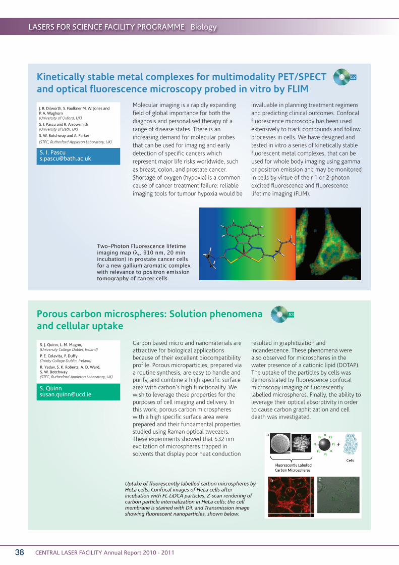
Molecular imaging is a rapidly expandingfield of global importance for both thediagnosis and personalised therapy of arange of disease states. There is anincreasing demand for molecular probesthat can be used for imaging and earlydetection of specific cancers whichrepresent major life risks worldwide, suchas breast, colon, and prostate cancer.Shortage of oxygen (hypoxia) is a commoncause of cancer treatment failure: reliableimaging tools for tumour hypoxia would be
invaluable in planning treatment regimensand predicting clinical outcomes. Confocalfluorescence microscopy has been usedextensively to track compounds and followprocesses in cells. We have designed andtested in vitro a series of kinetically stablefluorescent metal complexes, that can beused for whole body imaging using gammaor positron emission and may be monitoredin cells by virtue of their 1 or 2-photonexcited fluorescence and fluorescencelifetime imaging (FLIM).
Two-Photon Fluorescence lifetimeimaging map (λex 910 nm, 20 minincubation) in prostate cancer cellsfor a new gallium aromatic complexwith relevance to positron emissiontomography of cancer cells
52
Porous carbon microspheres: Solution phenomenaand cellular uptake
Carbon based micro and nanomaterials areattractive for biological applicationsbecause of their excellent biocompatibilityprofile. Porous microparticles, prepared viaa routine synthesis, are easy to handle andpurify, and combine a high specific surfacearea with carbon’s high functionality. Wewish to leverage these properties for thepurposes of cell imaging and delivery. Inthis work, porous carbon microsphereswith a high specific surface area wereprepared and their fundamental propertiesstudied using Raman optical tweezers.These experiments showed that 532 nmexcitation of microspheres trapped insolvents that display poor heat conduction
resulted in graphitization andincandescence. These phenomena werealso observed for microspheres in thewater presence of a cationic lipid (DOTAP).The uptake of the particles by cells wasdemonstrated by fluorescence confocalmicroscopy imaging of fluorescentlylabelled microspheres. Finally, the ability toleverage their optical absorptivity in orderto cause carbon graphitization and celldeath was investigated.
S. J. Quinn, L. M. Magno, (University College Dublin, Ireland)
P. E. Colavita, P. Duffy (Trinity College Dublin, Ireland)
R. Yadav, S. K. Roberts, A. D. Ward, S. W. Botchway(STFC, Rutherford Appleton Laboratory, UK)
53
Uptake of fluorescently labelled carbon microspheres byHeLa cells. Confocal images of HeLa cells afterincubation with FL-LiDCA particles. Z-scan rendering ofcarbon particle internalization in HeLa cells; the cellmembrane is stained with DiI. and Transmission imageshowing fluorescent nanoparticles, shown below.

LASERS FOR SCIENCE FACILITY PROGRAMME Biology
CENTRAL LASER FACILITY Annual Report 2010 - 2011 39
Total internal reflection and single molecule fluorescencemicroscopy in plant cells
Total internal reflection fluorescencemicroscopy (TIRFM) has been proven to bean extremely powerful technique in animalcell research for generating high contrastimages and dynamic protein conformationinformation. However, there has long beena perception that TIRF is not feasible inplant cells because the cell wall wouldrestrict the penetration of the evanescentfield and lead to scattering of illumination.By comparative analysis of epifluorescenceand TIRF in root cells, we demonstrate forthe first time that TIRF can generate highcontrast images, superior to otherapproaches from intact plant cells. We alsoshow that TIRF imaging is not only possibleat the plasma membrane level, but also insubcellular organelles, for example thenucleus, due to the presence of the centralvacuole. Importantly, we demonstrate thatthis is TIRF excitation, and not TIRF-likeexcitation described as variable-angleepifluorescence microscopy (VAEM) andshow how to distinguish the twotechniques in practical microscopy. TheseTIRF images show the highest signal-to-
background ratio and we show that theycan be used for single moleculemicroscopy. Rare protein events, whichwould otherwise be masked by theaverage molecular behaviour, cantherefore be detected, including theconformations and oligomerisation statesof interacting proteins and signallingnetworks in vivo. The demonstration of theapplication of TIRFM and single-moleculeanalysis to plant cells therefore opens up anew range ofpossibilities forplant cellimaging.
G. Vizcay-Barrena, Z. A. Wilson(University of Nottingham, UK)
S. E. D. Webb, M. L. Martin-Fernandez(STFC, Rutherford Appleton Laboratory, UK)
S. E. D. [email protected]
54
Analysis ofplasmamembrane andcytoskeletonmarkers byEpifluorescenceand TIRFmicroscopy inroots.
We have used confocal microscopy andFRET-FLIM to investigate the localisationand protein interactions involved withinthe mammalian Target of Rapamycin(mTOR) signaling pathway. mTOR proteinkinase is a central regulator of cellulargrowth, metabolism and proliferation in alleukaryotes. The mTOR pathway isderegulated in many human diseases suchas cancer and type 2 diabetes. Rapamycin,an anticancer drug used to inhibit mTOR, isin clinical trials for cancer treatment but asyet how exactly rapamycin perturbsthe functions of mTOR is notcompletely understood.
In the present study we have investigatedthe localisation of mTOR and its molecularinteraction with raptor using FRET-FLIMtechnology in live HeLa cells (an immortalcell line derived from cervical cancer cells).Effect of rapamycin on the localisation ofmTOR and its interaction with raptor wasinvestigated. It was found that rapamycindid not dissociate the mTOR-raptorinteraction.
R. B. Yadav, C. D. Stubbs, A. W. Parker,S. W. Botchway
(STFC, Rutherford Appleton Laboratory, UK)
V. Iadevaia, C. G. Proud(School of Biological Sciences)
C. D. [email protected]
Rapamycin treatment affects the localisationof EGFP-mTOR in HeLa cells. Confocal imagesof HeLa cells expressing EGFP-mTOR A) notreatment and B) rapamycin (100 nM)treatment for 24 h.
Rapamycin does not affect the interaction between mTORand raptor but causes increased nuclear levels of highlyexpressed mTOR in HeLa cells
55

CENTRAL LASER FACILITY Annual Report 2010 - 201140
LASERS FOR SCIENCE FACILITY PROGRAMME Chemistry
Chemistry
LIAD-fs: A novel method for studies of neutralbiomolecules in the gas phase
Intense femtosecond laser pulses provideunique tools for influencing and observingmolecular dynamics on ultrashorttimescales.
We report here on recent experimentalstudies of femtosecond laser interactionswith molecules of biological interest, usingthe UFL2 laser from the EPSRC laser loanpool. In a novel experimental approach wehave adopted Laser Induced AcousticDesorption to produce a gas phase targetof neutral molecules, which are thenexposed to an fs laser pulse, withsubsequentionisation/fragmentationproducts being mass-analysed inour KEIRA ion trap.
Using critical laser pulse parameters, suchas intensity and pulse duration, we havedemonstrated control over the molecularfragmentation and ionisation processes fora range of amino acids, small peptides andDNA bases. This technique has providedinsight into the dynamics of suchbiomolecules in intense laser fields andprovides a valuable platform for futurestudies where ultrashort pulses promise toenable observation and control of ultrafastprocesses in biomolecules.
C. R. [email protected]
56
C. R. Calvert, O. Kelly, L. Belshaw, M. J. Duffy, R. B. King, I. D. Williamsand J. B. Greenwood(Queen’s University Belfast)
T. J. Kelly and J. T. Costello(Dublin City University)
Photoacoustic stimulated raman spectroscopy (PARS)for trace detection of molecular hydrogen
Sensitive and selective detection ofmolecular hydrogen (H2) is very relevant,for example for a future hydrogeneconomy (use of hydrogen gas as fuel), orto detect traces of H2 in metallurgy (H2 cancompromise steel enclosures).Spectroscopic detection of H2, however, isnotoriously difficult to achieve. We have set up two different schemes of
stimulated Raman photoacoustic detectionof H2, one with a Raman shifter and asecond scheme with a tuneable dye laser,to generate stimulating Raman excitation.In addition, green Nd:YAG light served asRaman pump radiation. Both beams werefocused into a cell with a gas mixture to beanalysed. H2 absorbed radiation due to thestimulated Raman effect, andphotoacoustic signals were picked up by amicrophone.
Excellent detection linearity and detectionlimits in the ppm range for H2 in 1 atm airwere achieved. The schemes are thussuitable for reliable detection of H2 inambient air and gas mixtures.
57
C. L. Spencer, V. Watson and M. Hippler(University of Sheffield, UK)
Schematic of LIAD-fs technique forstudying biomolecules in the gas phase.The sample is deposited on a Ta foil (a)which is then back irradiated using a UVns pulse (b). A femtosecond laser (c) isthen interacted with the resultingbiomolecular plume, with chargedproducts extracted and analysedelectrostatically (d).
Photograph showingthe red Stokes, greenRayleigh, and blue/UVanti-Stokes radiationproduced from the H2Raman shifter, dispersedby a prism.

LASERS FOR SCIENCE FACILITY PROGRAMME Chemistry
CENTRAL LASER FACILITY Annual Report 2010 - 2011 41
Dynamics of chemical and photochemical reactions in solution
Transient broadband IR absorption spectrahave been obtained of the HCN products ofbimolecular reactions of CN radicals withcyclohexane in solution in variouschlorinated organic solvents. The spectrareveal that the HCN is initially formed withvibrational excitation in both its C−Hstretching and bending vibrational modes,and that this mode-specific vibrationalexcitation relaxes through coupling to thesolvent with solvent-dependent timeconstants of 130 - 270 ps. The early time
reaction dynamics are remarkably similarto those previously reported for gas-phasereactions of CN radicals withhydrocarbons, despite the presence of asolvent. Complementary dynamicalcalculations have been performed that,when combined with the experimentalresults, provide acute insights concerningthe influence of the solvent on thefundamental mechanisms of chemicalreactions in solution.
S. J. Greaves, R. A. Rose, T. A. A. Oliver, M. N. R. Ashfold, A. J. Orr-Ewing(University of Bristol, UK)
M. Towrie, G. M. Greetham, I. P. Clark(STFC, Rutherford Appleton Laboratory, UK)
58
Picosecond time-resolved infrared spectroscopyof arylpentazole
P. Portius, M. Davis(University of Sheffield, UK)
M. Towrie(STFC, Rutherford Appleton Laboratory, UK)
59
Design of the flow system for low temperaturepicosecond-TRIR as integrated in the ULTRA facility; V1-V4 solenoid valves applying N2 pressure (0.3 bar) orreleasing pressure from the reservoir bottles B1 and B2, H1and H2 heat exchange coils, T1-T3 thermocouples, Ccommercial variable temperature spectroscopic cell. Thearrows indicate the direction of flow when V1(in)&V4(out) areopen and V2&V3 closed.
Nitrogen-rich molecules are important forchemical energy storage and have a majoradvantage over conventional solutionsto the problem of chemical energy storage,since their decomposition releases almostexclusively the environmentally friendlydinitrogen.[1] In search for stablecompounds with high nitrogen content,methods have to be found which allow theintroduction of nitrogen in syntheticprocedures. The most relevant of thesehave traditionally been the coordination ofthe azido anion (N3
-) to main groupelements[2] or the derivatisation oftetrazoles[1, 3] (R-CN4-R’). Meanwhile, thescope of these methods has been largelyrealized and forms the basis of varioussuccessful applications.
[1] Steinhauser, G.; Klapoetke, T. M., Angew.Chem. Int. Ed. 2008, 47, 2-20.
[2] a) Portius, P.; Filippou, A. C.; Schnakenburg,G.; Davis, M.; Wehrstedt, K.-D., Angew.Chem. Int. Ed. 2010, 49, 8013-8016; b)Mueller, J., Coord. Chem. Rev. 2002, 235,105-119; c) Banert, K.; Joo, Y.-H.; Ru�ffer, T.;Walfort, B.; Lang, H., Angew. Chem. 2007,119, 1187-1190.
[3] a) Singh, R. P.; Verma, R. D.; Meshri, D. T.;Shreeve, J.M., Angew. Chem. Int. Ed. 2006,45, 3584-3601; b) Stierstorfer, J.; Tarantik, K. R.; Klapoetke, T. M., Chemistry--AEuropean Journal 2009, 15, 5775-5792.

CENTRAL LASER FACILITY Annual Report 2010 - 201142
LASERS FOR SCIENCE FACILITY PROGRAMME Chemistry
Check
Isotopic hydration of cellobiose: vibrational spectroscopyand dynamical simulations
J. P. Simons, N. Mayorkas(University of Oxford, UK)
B. G. Davis(Chemistry Research Laboratory, UK)
E. . Cocinero(University of Bilbao, Spain)
M. Pincu, R. B. Gerber(University of California, USA)
B. Brauer(The Hebrew University, Israel)
60
Interactions with water can influence boththe conformation and the bio-activity ofcarbohydrates. If sugars didn’t changeshape, life would be radically different andsome, perhaps many, biological processesjust would not work anymore. The factorswhich dictate the conformation ofcellobiose, the basic building block ofcellulose, remain something of an enigma.To address this issue, water moleculeshave been added to a range of ‘natural’carbohydrates, including cellobiose , undercontrolled conditions in a cold molecularbeam and their hydrated molecularstructures have been interrogated throughinfrared laser spectroscopy. A new andoriginal ‘twist’ to this approach has beenintroduced by using heavy water (D2O)which allows the infrared spectraassociated with the carbohydrate (OHmodes) and its hydration shell (OD modes)to be separately identified, analysed andinterpreted.
Experimental and (DFT)computed IR spectra and thecorresponding hydrogen-bonded molecular structures ofphenyl β cellobioside (top),phenyl β cellobioside•D2O(middle) and phenyl βcellobioside•(D2O)2(bottom).
Laser tweezers have become increasinglypopular in the study of atmospheric aerosolscience because of the unique way thatindividual micron-scale aerosol dropletscan be trapped and retained for periods ofseveral hours. Recent work has highlightedthat conventional single-beam gradientoptical trapping becomes unstable whenthe particle diameter is reduced to below 2 microns. This finding has a significantimpact, for aerosol science, as sub-micronaerosol particulates are abundant in theatmosphere. Using a counter-propagatinglaser trapping configuration, particles canbe captured with sizes from about 0.4 µmto more than 6 µm. Alternative methodsfor characterizing particle size arediscussed and initial studies into a Ramanspectra based technique show promise andindicate that sizing of sub-micron aerosoldroplets is realistic. The extension in thetrapping diameter range for aerosolparticles will have application indetermining atmospheric aerosol reactionand light scattering properties.
A. Ward(STFC, Rutherford Appleton Laboratory,UK)
M. King(University of London, UK)
Examples ofdroplet images inoptical focus whenusing the counter-propagating lasertweezersconfiguration
Optical trapping of sub-micron liquid aerosol droplets 61

LASERS FOR SCIENCE FACILITY PROGRAMME Physics
CENTRAL LASER FACILITY Annual Report 2010 - 2011 43
Physics
Ultrafast manipulation of photon transportand molecular beams
We used a fs-pulsed laser for two differentexperiments opening new avenues towardsthe ultrafast manipulation of massiveparticles in gas-phase beams and of themesoscopic transport of light. Wedemonstrated for the first time amechanical focusing effect by off-resonantdipole interaction acting on the centre ofmass motion of large neutral moleculessuch as tetra-phenyl-porphyrin (TPP).Focusing is enabled by interactions of asingle molecule with many weak lightpulses.
The mesoscopic transport of light througha random media consisting of a layer ofvery strongly scattering GaP nanowires in
which the mean free path is much shorterthan the depth of the layer was alsoinvestigated. The statistical distribution oftransmitted intensities is compared to thenormal Rayleigh distribution in which thereare an infinite number of transmissionmodes. A significant decrease in thenumber of independent transmissionmodes to 25 can be seen in the figure.
O. L. [email protected]
62
T. Strudley, P. Venn, H. Ulbricht, O. L. Muskens(University of Southampton, UK)
Distribution of angular intensitiesnormalised to ensemble average (Sab).