Embed Size (px)
Citation preview

CLINICAL CASE REPORT
Late presentation of RPE65 retinopathy in three siblings
Moustafa Magliyah . Amjad Ameen Saifaldein . Patrik Schatz
Received: 25 September 2019 / Accepted: 30 December 2019
� The Author(s) 2020
Abstract
Purpose Gene therapy for RPE65 retinopathy has
been recently approved. The purpose of this study was
to assess retinal structure and function in 3 siblings
presenting with late-stage RPE65 retinopathy and to
assess the unmet need for such therapy in Saudi
Arabia.
Methods Search of the retinal dystrophy registry at
King Khaled Eye Specialist Hospital and clinical
examination including multimodal retinal imaging,
full-field electroretinography (ERG), dark adapted
full-field stimulus sensitivity thresholds, and molecu-
lar genetic testing in 3 patients.
Results Nine (9) patients were identified with bial-
lelic RPE65 mutations, corresponding to a prevalence
rate of 9/187 = 5% among early onset retinal dystro-
phies. Of these, 3 siblings (2 male and 1 female) with
RPE65 retinopathy were assessed in detail, because of
an unusual, late presentation. They were all over
30 years old at the time of their most recent visits and
had non-recordable ERGs. The 2 male siblings
presented with poor vision and paracentral loss of
the inner segment ellipsoid (ISe) and focal attenuation
of the outer nuclear layer (ONL) in the macula. On the
other hand, the female sibling presented with 20/100
vision with preserved foveal ISe and intact ONL
throughout the macula and significantly lower light
sensitivity thresholds compared to her male siblings. A
homozygous missense p.Arg91Trpmutation inRPE65
was identified in all. All patients were eligible for gene
therapy, demonstrating a central retinal thickness of
more than 100 microns on repeated examinations.
Conclusions RPE65 retinopathy seems to be rela-
tively common on the Arabian peninsula, and in
addition it may be underdiagnosed. To the best of our
knowledge, this is the first detailed presentation,
including multimodal retinal imaging and electro-
physiological assessment, of such patients from this
region. Patients with late presentation of RPE65
retinopathy may be eligible for gene therapy, in terms
of remaining retinal function and structural
Electronic supplementary material The online version ofthis article (https://doi.org/10.1007/s10633-019-09745-z) con-tains supplementary material, which is available to authorizedusers.
M. Magliyah � P. Schatz (&)
Vitreoretinal Division, King Khaled Eye Specialist
Hospital, Riyadh, Saudi Arabia
e-mail: [email protected]
M. Magliyah
Ophthalmology Department, Prince Mohammed Medical
City, Aljouf, Saudi Arabia
A. A. Saifaldein
Ophthalmology Department, King Faisal Medical
Complex, Taif, Saudi Arabia
P. Schatz
Department of Ophthalmology, Clinical Sciences, Skane
University Hospital, Lund University, Lund, Sweden
123
Doc Ophthalmol
https://doi.org/10.1007/s10633-019-09745-z(0123456789().,-volV)(0123456789().,-volV)

preservation. The therapeutic window of such therapy
remains to be determined.
Keywords RPE65 � Gene therapy � Multimodal
retinal imaging
Introduction
Leber’s congenital amaurosis (LCA) is a group of
congenital retinal dystrophies which usually present
before 6 months of age with severe visual impairment
and nystagmus [1–5]. Inheritance is autosomal reces-
sive; however, dominant inheritance has been reported
in cases with CRX and IMPDH1 mutations [6–11].
Mutations in several genes have been shown to
cause recessive LCA (reviewed in https://www.ncbi.
nlm.nih.gov/books/NBK1298/). Recessive RPE65
retinopathy has traditionally been grouped with LCA;
however, it differs because useful vision and central
retinal structure may be preserved for several years
[12]. Together with slow progression, this makes it a
potential candidate for gene therapy [13]. RPE65
encodes the isomerohydrolase of the visual cycle, and
dysfunction of this enzyme leads to an insufficient
regeneration of the chromophore linked to opsin in the
photoreceptors. This also leads to a slowly progressive
degeneration of the retina. Typically the full-field
ERG is non-recordable even at the time of initial
diagnosis and therefore other form of assessment of
progression, or of therapeutic response after gene
replacement therapy, is needed, such as visual fields,
visual acuity, full-field sensitivity testing (FST) and
multimodal retinal imaging, including assessment of
preservation of retinal layers and retinal thickness.
In this study, we describe the clinical, electrophys-
iological and molecular genetic findings in 3 siblings
who presented with late-stage RPE65 retinopathy, in
light of the recently approved gene therapy for this
condition. To the best of our knowledge, such
assessment including detailed analysis of retinal
structure and function has not been described for this
disease in the Arabian peninsula. The recently
approved gene replacement therapy is expensive and
resource demanding and therefore may require signif-
icant planning and coordination. Thus in addition, we
estimate the prevalence of this condition in the region,
in preparation for future gene therapy.
Methods
Informed consent was obtained, and the study was
approved by an institutional review board at King
Khaled Eye Specialist Hospital, which is a nation-
wide tertiary referral centre, and at times also accepts
referred patients from neighbouring countries. Oph-
thalmic examination including multimodal retinal
imaging and full-field electroretinography (ERG)
was done as described by us and others previously
[14, 15]. Goldman visual fields were performed using
objects V4 and III4.
ERG (Nicolet Biomedical Instruments, Madison,
Wisconsin, USA) was obtained as follows, in dark
adapted and light adapted state according to ISCEV
standards [15], with a few modifications as follows.
Dark adaptation was performed for 30 min, and
dilatation of the pupils was obtained with topical
cyclopentolate 1% and metaoxedrine 2.5%. After
topical anaesthesia, a Burian Allen bipolar contact
lens was placed on the cornea and a ground electrode
was applied to the forehead. Responses were obtained
stimulating with single full-field flash (30 ms) with
blue light (0.81 cd s/m2: rod response) and with white
light (10.02 cd s/m2: combined rod–cone response).
In addition, the dark adapted cone response was
measured after stimulation with dim red light
(3.93 cd s/m2). Photopic responses were obtained
with a background illumination of 3.4 cd s/m2 in
order to saturate the rods.
Dark adapted full-field stimulus thresholds (FST)
were assessed using white light and the Espion
ColorDomeTM system (Diagnosys LLC). FST mea-
sures the light sensitivity over the whole visual field
and is therefore not affected by nystagmus. A mean-
ingful change has been suggested as 10 dB or 1
log. Results were presented as log candela seconds per
square meter (log cd s/m2).
A targeted next-generation sequencing (NGS) was
performed using two panels in two different labora-
tories; a retinal dystrophy panel at molecular genetics
laboratory at King Faisal Specialist Hospital (KFSH)
[16], and a LCA panel in the Bioscientia Human
Genetics laboratory (Bioscientia, Ingelheim, Ger-
many) which includes GUCY2D, RPE65, SPATA7,
AIPL1, LCA5, RPGRIP1, CRX, CRB1, CEP290,
IMPDH1, RD3, RDH12, LRAT, MERTK and TULP1.
Finally, we searched a newly established retinal
dystrophy registry at King Khaled Eye Specialist
123
Doc Ophthalmol

Hospital, which includes most patients seen in
specially designed ‘‘retinal dystrophy clinics’’ since
2014, at the time of which electronical medical records
were implemented in the hospital, for ‘‘RPE65’’, in
order to estimate the prevalence of RPE65 retinopathy
in the region.
Results
The retinal dystrophy registry at this time included a
total of 789 patients. Of these, 187 patients for whom
NGS had been carried out were diagnosed with early
onset retinal dystrophy. Nine (9) of these patients had
biallelic RPE65 mutations, arriving at an estimated
prevalence rate of 9/187 = 5% among early onset
retinal dystrophies.
Three of the affected patients, 3 siblings, had an
atypical late presentation of the disease, while the
other 6 patients had all been diagnosed before reaching
10 years of age. The 3 patients were further examined
in detail, because of the unusual late presentation, with
the aim of assessing the extent, if any, of remaining
retinal function. The patients were belonging to a
family of 8 siblings (3 brothers and 5 sisters) with a
positive history of parental consanguinity. All three, 2
male and 1 female (patients 1, 2 and 3), aged 32,
36 years and 34 years old, respectively, at most recent
follow-up, were homozygous for the c.271C[T
(p.Arg91Trp) mutation in RPE65. The mutations were
confirmed by 2 independent molecular genetic insti-
tutions (KFSH for patients 1 and 3 and Bioscientia for
patient 2); however, other family members were not
available for mutation segregation analysis or clinical
examination.
All 3 had experienced nystagmus and night blind-
ness since early childhood, with gradual and progres-
sive visual loss. They presented 4 years ago with best
corrected visual acuity (BCVA) of 20/400 in both eyes
of patients 1 and 2, and 20/80 in patient 3. Patients 1
and 2 showed prominent peripheral retinal pigmenta-
tion, with less peripheral changes in patient 3. Central
macular retinal layers showed signs of paracentral loss
of the inner segment ellipsoid (ISe) and outer nuclear
layer (ONL) compromise, more severe in patients 1
and 2, whereas the ONL was preserved throughout the
macula in patient 3 (Figs. 1, 2, 3). ERG was non-
recordable in all (Fig. 4 demonstrates the ERG
obtained from Patient 1, and Supplemental document
1 demonstrates the ERGs of Patients 2 and 3). After
4 years of follow-up, BCVA was reduced to light
perception (LP) in both eyes of the male patients and
to 20/100 in the female patient.
In patient 3, an ETDRS OCT map was possible in
the right eye on 2 separate occasions, separated by
30 months, showing an apparent decline of the central
subfield thickness from 252 to 196 lm, while the
visual acuity remained stable during the same period,
ranging between 20/100 and 20/80 during a total of 7
visits. Retinal thickness was well above 100 lm in all
subfields, in both eyes. In the other 2 patients, a
quantitative approach over time including analysis of
potential changes of retinal thickness was not possible
because of insufficient fixation during obtaining OCT
images in most of the visits, due to nystagmus;
however, available thickness measurements from
single-line horizontal transfoveal scans demonstrated
a retinal thickness well above 100 lm along the
scanned lines.
Visual fields were severely constricted to less than
10 degrees with the largest object in Patients 1 and 2,
while Patient 3 had constricted fields to less than 10
degrees with the III4 object and less than 60 degrees
horizontally and less than 50 degrees vertically with
the largest V4 object. Mean (of right and left eyes for
each patient) FST thresholds were 2.7, 1.9 and
- 1.8 log cd s/m2, for Patients 1, 2 and 3,
respectively.
Fundus autofluorescence demonstrated a general-
ized loss of autofluorescence, where the discs
appeared relatively bright on a dark background;
however, a trace of preserved autofluorescence could
be noticed within the vascular arcades, compared to
the periphery, especially in Patient 3 (Supplemental
document 2).
Discussion
We describe a late presentation of RPE65 retinopathy
in 3 siblings with the homozygous p.Arg91Trp
mutation. King Khaled Eye Specialist Hospital is a
major tertiary referral centre on the Arabian peninsula,
and we have so far identified 9 patients with RPE65
retinopathy during our experience of more than
5 years in the hospital. This estimates the minimum
prevalence of RPE65 retinopathy among retinal dys-
trophies at 5% which is higher by a factor of 5
123
Doc Ophthalmol

compared to a previous study in the US population
[17]. This may be due to a high rate of consanguineous
marriage. Furthermore, the condition may be under-
recognized and underdiagnosed due to limited avail-
ability to perform genetics testing, cost and failure to
recognize the phenotype. However with the recently
approved gene therapy for this condition, it is likely
that the awareness of this condition, among ophthal-
mologists and patients alike, will increase, which may
lead to identification of significantly greater number of
patients.
We assessed 3 of these patients further, carrying the
p.Arg91Trp mutation, which has been described
previously in RPE65 retinopathy [18–20]. Table 1
shows the phenotypic features of all patients with
p.Arg91Trp mutation in RPE65 including the three
siblings in this study. Features in common for these
patients include nystagmus, hyperopia, and non-
recordable ERGs. Ambulatory vision was maintained
in the female sibling (20/100 OU), similar to two
patients described by Thompson et al. [20]. This could
be due to a preserved RPE65 activity which can
produce low amounts of 11-cis retinal in patients with
the missense p.Arg91Trp mutation, allowing for better
cone and rod function compared to patients with
RPE65 null mutations, as suggested in experimental
work with mice with corresponding knock-in muta-
tion, compared toRPE65 null mice [21]. This may also
account for some degree of preservation of fundus
autofluorescence (Supplemental document 2). On the
other hand, the 2 male patients in this study, similar to
one of the patients described by Thompson et al., had
poorer vision, which was noted also in the patient
described by Habibi et al. [18] and was correlated with
foveal atrophy on SD-OCT. The integrity of the ONL
is a prominent indicator of photoreceptors function
and was advised to be included in the evaluation for
gene therapy [22].
Recently, Russell et al. [23] conducted a phase 3
randomized controlled trial evaluating the efficacy and
safety of gene therapy for patients with RPE65
retinopathy, in which four patients were compound
Fig. 1 A and B colour fundus photographs of both eyes of a
32-year-old male patient (patient 1) with the homozygous
c.271C[T (p.Arg91Trp) mutation in RPE65, showing periph-
eral pigment depositions (bone spicules), attenuated blood
vessels and oval shaped hyperopic discs with temporal pallor.
Spectral domain optical coherence tomography (SD-OCT) of
both eyes C and D shows paracentral loss of the inner segment
ellipsoid (ISe) and focal attenuation of the outer nuclear layer
(ONL)
123
Doc Ophthalmol

heterozygous for the Arg91Trp mutation (and the
mutations IVS7 ? 2T[A, Trp402Stop [2 patients]
and Arg91Gln, respectively). Table 2 shows that for
this subgroup of patients, the average improvement in
BCVA was 10 letters, which was similar to the mean
for all patients (9 letters), while the average of
illuminance level difference for passing the multilu-
minance mobility test (MLMT, the primary outcome
of the study) was slightly less than the illuminance
level difference mean for all patients (Table 2) [23].
In this study, in spite of late presentation, all 3
patients with the homozygous Arg91Trp mutation in
RPE65 maintained a central retinal thickness of more
than 100 microns and also fulfilled other eligibility
criteria, including the level of visual acuity, for gene
replacement therapy. On the other hand, there was
variability regarding the stage of disease, as observed
by, for example, visual fields and remaining autoflu-
orescence, among the 3 patients. Patient 3 presented
with the least advanced disease stage, based on these
assessments. Furthermore, based on the FST results,
Patient 3 might be the best candidate, because of
relatively preserved dark adapted sensitivity thresh-
olds, presenting results that imply a sensitivity in the
upper range compared to other patients with ‘‘LCA’’
who were examined previously in the literature by this
modality, albeit the thresholds were significantly ele-
vated compared to normal subjectswhowere reported to
have a mean of -4.3 log cd s/m2 [24]. Thus, gene
replacement therapy should be primarily be considered
for Patient 3 once available in Saudi Arabia. This
illustrates that FST is more appropriate than ERG as a
measure of eligibility for and therapeutic response to
currently available gene augmentation therapy, where
the gene and its viral vector are delivered by subretinal
injection in a limited area of the central retina only, and
thus any improvement of retinal function in such a
limited area may not be detected using ERG. Thus,
albeit the ERG is a very powerful tool and is essential in
diagnosing retinal dystrophies, themethod has a limited
sensitivity, and needs to be complemented with other
more sensitive measures when evaluating potential
treatments for retinal dystrophies.
Limitations of this study include the few number of
patients examined and lack of mutation segregation
analysis and clinical examination of other family
members. On the other hand, this is the first report of
clinical findings including multimodal retinal imaging
Fig. 2 A and B colour fundus photographs of both eyes of a
36-year-old male (patient 2), sibling of patient 1, with the
homozygous c.271C[T (p.Arg91Trp) mutation in RPE65
showing bone spicules, attenuated blood vessels and oval
shaped hyperopic discs with temporal pallor. Spectral domain
optical coherence tomography (SD-OCT) of both eyes C and
D shows paracentral loss of the inner segment ellipsoid (ISe) and
attenuation of the outer nuclear layer (ONL)
123
Doc Ophthalmol
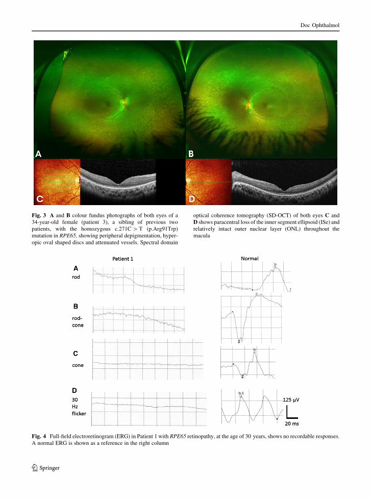
Fig. 3 A and B colour fundus photographs of both eyes of a
34-year-old female (patient 3), a sibling of previous two
patients, with the homozygous c.271C[T (p.Arg91Trp)
mutation in RPE65, showing peripheral depigmentation, hyper-
opic oval shaped discs and attenuated vessels. Spectral domain
optical coherence tomography (SD-OCT) of both eyes C and
D shows paracentral loss of the inner segment ellipsoid (ISe) and
relatively intact outer nuclear layer (ONL) throughout the
macula
Fig. 4 Full-field electroretinogram (ERG) in Patient 1 with RPE65 retinopathy, at the age of 30 years, shows no recordable responses.
A normal ERG is shown as a reference in the right column
123
Doc Ophthalmol
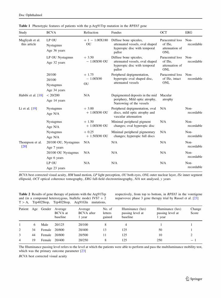
Table 1 Phenotypic features of patients with the p.Arg91Trp mutation in the RPE65 gene
Study BCVA Refraction Fundus OCT ERG
Magliyah et al.
this article
LP OU
Nystagmus
Age 36 years
? 1 - 1.00X180
OU
Diffuse bone spicules,
attenuated vessels, oval shaped
hyperopic disc with temporal
pallor
Paracentral loss
of ISe,
attenuation of
ONL
Non-
recordable
LP OU Nystagmus
Age 32 years
? 3.50
- 1.00X90 OU
Diffuse bone spicules,
attenuated vessels, oval shaped
hyperopic disc with temporal
pallor
Paracentral loss
of ISe,
attenuation of
ONL
Non-
recordable
20/100
20/100
Nystagmus
Age 34 years
? 1.75
- 1.00X90
OU
Peripheral depigmentation,
hyperopic oval shaped disc,
attenuated vessels
Paracentral loss
of ISe, intact
ONL
Non-
recordable
Habibi et al. [18] \ 20/200
Age 14 years
N/A Depigmented deposits in the mid
periphery, Mild optic atrophy,
Narrowing of the vessels
Macular
atrophy
Li et al. [19] Nystagmus
Age N/A
? 3.00
? 1.00X90 OU
Peripheral depigmentation, oval
discs, mild optic atrophy and
vascular attenuation
N/A Non-
recordable
Nystagmus
Age N/A
? 1.50
? 1.00X90 OU
Minimal peripheral pigment
changes; oval hyperopic disc
N/A Non-
recordable
Nystagmus
Age N/A
? 0.25
? 1.50X90 OU
Minimal peripheral pigmentary
changes; hyperopic full discs
N/A Non-
recordable
Thompson et al.
[20]
20/100 OU, Nystagmus
Age 7 years
N/A N/A N/A Non-
recordable
20/100 OU Nystagmus
Age 6 years
N/A N/A N/A Non-
recordable
LP OU
Age 23 years
N/A N/A N/A Non-
recordable
BCVA best corrected visual acuity, HM hand motion, LP light perception, OU both eyes, ONL outer nuclear layer, ISe inner segment
ellipsoid, OCT optical coherence tomography, ERG full-field electroretinography, N/A not analysed, y years
Table 2 Results of gene therapy of patients with the Arg91Trp
and (in a compound heterozygous, biallelic mode) IVS7 ? 2
T[A, Trp402Stop, Trp402Stop, Arg91Gln mutations,
respectively, from top to bottom, in RPE65 in the voretigene
neparvovec phase 3 gene therapy trial by Russel et al. [23]
Patient Age Gender Average
BCVA at
baseline
Average
BCVA after
1 year
No. of
letters
gained
Illuminance (lux)
passing level at
baseline
Illuminance (lux)
passing level at
1 year
Change
Score
1 6 Male 20/125 20/100 8 4 1 1
2 34 Female 20/800 20/400 13 125 50 1
3 44 Female 20/800 20/500 11 125 10 2
4 19 Female 20/400 20/250 8 125 250 - 1
The Illuminance passing level refers to the level at which the patients were able to perform and pass the multiluminance mobility test,
which was the primary outcome parameter [23]
BCVA best corrected visual acuity
123
Doc Ophthalmol
in late-stage RPE65 retinopathy due to this specific
homozygous mutation, confirming eligibility for gene
therapy for some patients, even with late presentation.
A further novelty with this study is that RPE65
retinopathy may be relatively common in Saudi Arabia,
which may necessitate mobilization of economical
resources and further diagnostic and operational skill in
ophthalmic health care in the region. The therapeutic
window for such therapy remains to be determined.
Acknowledgements Open access funding provided by Lund
University.
Authors’ contribution MM, AS, and PS designed the study;
MM, AS, and PS conducted the study; and MM, AS, and PS
were involved in collection, management, analysis, and
interpretation of the data. MM, AS, and PS were involved in
preparation, review and final approval of the manuscript. We
thank Mr Adolph Cabanas at Design and Publications, King
Khaled Eye Specialist Hospital, for skillful technical assistance
with preparation of figures. Part of this material was presented at
the Euretina meeting in Paris on September 5–8, 2019.
Funding No funding was received for this research.
Compliance with ethical standards
Conflict of interest Each of the authors declares that they
have no conflicts of interest.
Statement of human rights All procedures performed in
studies involving human participants were in accordance with
the ethical standards of the IRB of King Khaled Eye Specialist
Hospital and with the 1964 Helsinki Declaration and its later
amendments.
Statement on the welfare of animals This article does not
contain any studies with animals performed by any of the
authors.
Informed consent Informed consent was obtained from all
individual participants included in the study.
Open Access This article is licensed under a Creative Com-
mons Attribution 4.0 International License, which permits use,
sharing, adaptation, distribution and reproduction in any med-
ium or format, as long as you give appropriate credit to the
original author(s) and the source, provide a link to the Creative
Commons licence, and indicate if changes were made. The
images or other third party material in this article are included in
the article’s Creative Commons licence, unless indicated
otherwise in a credit line to the material. If material is not
included in the article’s Creative Commons licence and your
intended use is not permitted by statutory regulation or exceeds
the permitted use, you will need to obtain permission directly
from the copyright holder. To view a copy of this licence, visit
http://creativecommons.org/licenses/by/4.0/.
References
1. Heckenlively JR, Foxman SG, Parelhoff ES (1988) Retinal
dystrophy and macular coloboma. Doc Ophthalmol
68:257–271
2. Mizuno K, Takei Y, Sears ML et al (1977) Leber’s con-
genital amaurosis. Am J Ophthalmol 83:32–42
3. Noble KG, Carr RE (1978) Leber’s congenital amaurosis: a
retrospective study of 33 cases and a histopathological study
of one case. Arch Ophthalmol 96:818–821
4. Margolis S, Scher BM, Carr RE (1977) Macular colobomas
in Leber’s congenital amaurosis. Am J Ophthalmol
83:27–31
5. Francois J (1968) Leber’s congenital tapeto-retinal degen-
eration. Int Ophthalmol Clin 8:929–947
6. Heckenlively JR (1988) Autosomal dominant retinitis pig-
mentosa. In: Retinitis Pigmentosa. JB Lippincott Co,
Philadelphia, pp 125–149
7. Sorsby A, Williams CE (1960) Retinal aplasia as a clinical
entity. BMJ 1:293–297
8. Sohocki MM, Sullivan LS, Mintz-Hittner HA et al (1998) A
range of clinical phenotypes associated with mutations in
CRX, a photoreceptor transcription-factor gene. Am J Hum
Genet 63:1307–1315
9. Rivolta C, Berson EL, Dryja TP (2001) Dominant Leber
congenital amaurosis, cone-rod degeneration, and retinitis
pigmentosa caused by mutant versions of the transcription
factor CRX. Hum Mutat 18:488–498
10. Arcot Sadagopan K, Battista R, Keep RB, Capasso JE,
Levin AV (2015) Autosomal-dominant Leber congenital
amaurosis caused by a heterozygous CRX mutation in a
father and son. Ophthalmic Genet 36:156–159
11. Bowne SJ, Sullivan LS, Mortimer SE et al (2006) Spectrum
and frequency of mutations in IMPDH1 associated with
autosomal dominant retinitis pigmentosa and leber con-
genital amaurosis. Invest Ophthalmol Vis Sci 47:34–42
12. Jacobson SG, Aleman TS, Cideciyan AV et al (2007)
Human cone photoreceptor dependence on RPE65 iso-
merase. Proc Natl Acad Sci USA 104:15123–15128
13. Cai X, Conley SM, Naash MI (2009) RPE65: role in the
visual cycle, human retinal disease, and gene therapy.
Ophthalmic Genet 30:57–62
14. Schatz P, Abdalla Elsayed MEA, Khan AO (2017) Multi-
modal imaging in CABP4-related retinopathy. Ophthalmic
Genet 38:459–464
15. McCulloch DL, Marmor M, Brigell MG et al (2015) ISCEV
Standard for full-field clinical electroretinography (2015
update). Doc Ophthalmol 130:1–12
16. Saudi Mendeliome Group (2015) Comprehensive gene
panels provide advantages over clinical exome sequencing
for Mendelian diseases. Genome Biol 16:134
17. Stone EM, Andorf JL, Whitmore SS, DeLuca AP, Giaca-
lone JC, Streb LM, Braun TA, Mullins RF, Scheetz TE,
Sheffield VC, Tucker BA (2017) Clinically focused
molecular investigation of 1000 consecutive families with
inherited retinal disease. Ophthalmology 124:1314–1331
18. Habibi I, Chebil A, Falfoul Y et al (2016) Identifying
mutations in Tunisian families with retinal dystrophy. Sci
Rep 6:37455
123
Doc Ophthalmol
19. Li Y,WangH, Peng J et al (2008)Mutation survey of known
LCA genes and loci in the Saudi Arabian population. Invest
Ophthalmol Vis Sci 50:1336–1343
20. Thompson DA, Gyurus P, Fleischer LL et al (2000)
Genetics and phenotypes of RPE65 mutations in inherited
retinal degeneration. Invest Ophthalmol Vis Sci
41:4293–4299
21. Samardzija M, von Lintig J, Tanimoto N et al (2008)
ARG91TRP mutation in Rpe65 leads to milder early-onset
retinal dystrophy due to the generation of low levels of
11-cis-retinal. Hum Mol Genet 17:281–292
22. Jacobson SG, Cideciyan AV, Aleman TS et al (2008)
Photoreceptor layer topography in children with leber
congenital amaurosis caused by RPE65 mutations. Invest
Ophthalmol Vis Sci 49:4573–4577
23. Russell S, Bennett J, Wellman JA et al (2017) Efficacy and
safety of voretigene neparvovec (AAV2-hRPE65v2) in
patients with RPE65-mediated inherited retinal dystrophy: a
randomised, controlled, open-label, phase 3 trial. Lancet
390:849–860
24. Klein M, Birch DG (2009) Psychophysical assessment of
low visual function in patients with retinal degenerative
diseases (RDDs) with the Diagnosys full-field stimulus
threshold (D-FST). Doc Ophthalmol 119:217–224
Publisher’s Note Springer Nature remains neutral with
regard to jurisdictional claims in published maps and
institutional affiliations.
123
Doc Ophthalmol


![The Guide - Diabetic Retinopathy - Vision Lossvisionloss.org.au/wp-content/uploads/2016/05/The... · the guide [diabetic retinopathy] What is Diabetic Retinopathy? Diabetic Retinopathy](https://img.pdfslide.net/doc/110x75/5e3ed00bf9c32e41ea6578a8/the-guide-diabetic-retinopathy-vision-the-guide-diabetic-retinopathy-what.jpg)