Embed Size (px)
Citation preview
ORIGINAL RESEARCHpublished: 04 September 2015doi: 10.3389/fncel.2015.00348
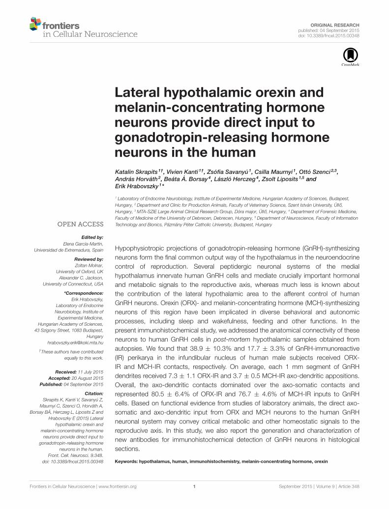
Lateral hypothalamic orexin andmelanin-concentrating hormoneneurons provide direct input togonadotropin-releasing hormoneneurons in the humanKatalin Skrapits 1†, Vivien Kanti 1†, Zsófia Savanyú 1, Csilla Maurnyi 1, Ottó Szenci 2,3,András Horváth 2, Beáta Á. Borsay 4, László Herczeg 4, Zsolt Liposits 1,5 andErik Hrabovszky 1*
1 Laboratory of Endocrine Neurobiology, Institute of Experimental Medicine, Hungarian Academy of Sciences, Budapest,Hungary, 2 Department and Clinic for Production Animals, Faculty of Veterinary Science, Szent István University, Üll ´́o,Hungary, 3 MTA-SZIE Large Animal Clinical Research Group, Dóra major, Üll ´́o, Hungary, 4 Department of Forensic Medicine,Faculty of Medicine of the University of Debrecen, Debrecen, Hungary, 5 Department of Neuroscience, Faculty of InformationTechnology and Bionics, Pázmány Péter Catholic University, Budapest, Hungary
Edited by:Elena García-Martín,
Universidad de Extremadura, Spain
Reviewed by:Zoltan Molnar,
University of Oxford, UKAlexander C. Jackson,
University of Connecticut, USA
*Correspondence:Erik Hrabovszky,
Laboratory of EndocrineNeurobiology, Institute ofExperimental Medicine,
Hungarian Academy of Sciences,43 Szigony Street, 1083 Budapest,
†These authors have contributedequally to this work.
Received: 11 July 2015Accepted: 20 August 2015
Published: 04 September 2015
Citation:Skrapits K, Kanti V, Savanyú Z,
Maurnyi C, Szenci O, Horváth A,Borsay BÁ, Herczeg L, Liposits Z and
Hrabovszky E (2015) Lateralhypothalamic orexin and
melanin-concentrating hormoneneurons provide direct input to
gonadotropin-releasing hormoneneurons in the human.
Front. Cell. Neurosci. 9:348.doi: 10.3389/fncel.2015.00348
Hypophysiotropic projections of gonadotropin-releasing hormone (GnRH)-synthesizingneurons form the final common output way of the hypothalamus in the neuroendocrinecontrol of reproduction. Several peptidergic neuronal systems of the medialhypothalamus innervate human GnRH cells and mediate crucially important hormonaland metabolic signals to the reproductive axis, whereas much less is known aboutthe contribution of the lateral hypothalamic area to the afferent control of humanGnRH neurons. Orexin (ORX)- and melanin-concentrating hormone (MCH)-synthesizingneurons of this region have been implicated in diverse behavioral and autonomicprocesses, including sleep and wakefulness, feeding and other functions. In thepresent immunohistochemical study, we addressed the anatomical connectivity of theseneurons to human GnRH cells in post-mortem hypothalamic samples obtained fromautopsies. We found that 38.9 ± 10.3% and 17.7 ± 3.3% of GnRH-immunoreactive(IR) perikarya in the infundibular nucleus of human male subjects received ORX-IR and MCH-IR contacts, respectively. On average, each 1 mm segment of GnRHdendrites received 7.3 ± 1.1 ORX-IR and 3.7 ± 0.5 MCH-IR axo-dendritic appositions.Overall, the axo-dendritic contacts dominated over the axo-somatic contacts andrepresented 80.5 ± 6.4% of ORX-IR and 76.7 ± 4.6% of MCH-IR inputs to GnRHcells. Based on functional evidence from studies of laboratory animals, the direct axo-somatic and axo-dendritic input from ORX and MCH neurons to the human GnRHneuronal system may convey critical metabolic and other homeostatic signals to thereproducive axis. In this study, we also report the generation and characterization ofnew antibodies for immunohistochemical detection of GnRH neurons in histologicalsections.
Keywords: hypothalamus, human, immunohistochemistry, melanin-concentrating hormone, orexin
Frontiers in Cellular Neuroscience | www.frontiersin.org 1 September 2015 | Volume 9 | Article 348
Skrapits et al. LHA input to GnRH neurons
Introduction
In all mammals including the human, neurons synthesizingtype-I gonadotropin-releasing hormone (GnRH) form thefinal common output way from the hypothalamus in theneuroendocrine control of reproduction. Failure of these cellsto finish their prenatal migration from the olfactory placodeto the forebrain results in hypogonadotropic hypogonadismcharacterized by the absence of puberty and reproductivecapacity (Schwanzel-Fukuda and Pfaff, 1989). The decapeptideGnRH is secreted into the hypophysial portal circulationin an episodic manner (Carmel et al., 1976; Clarke andCummins, 1982) to regulate the synthesis and secretion of thetwo gonadotroph hormones follicle-stimulating hormone andluteinizing hormone (LH) by the anterior pituitary (Belchetzet al., 1978).
Hormonal and metabolic signals influencing the reproductiveaxis at the hypothalamic level either regulate GnRH cellsdirectly or act on the neuronal circuitry upstream from theGnRH neuron. Anatomical information about the neuronalsystems regulating human GnRH neurons via afferentconnections have been summarized in recent review articles(Dudás and Merchenthaler, 2006; Hrabovszky and Liposits,2013). Such afferents originate from hypothalamic as well asextrahypothalamic sources and use monoamines, amino acidsand a variety of neuropeptides for neuronal transmission (Dudásand Merchenthaler, 2006; Hrabovszky and Liposits, 2013).
Most peptide neurotransmitters identified within neuronalafferents to human GnRH neurons are synthesized in distinctcell populations of the medial hypothalamus (Hrabovszky andLiposits, 2013) and include neuropeptide Y (Dudás et al., 2000),substance P (Dudás and Merchenthaler, 2002a; Hrabovszkyet al., 2013), galanin (Dudás and Merchenthaler, 2004a),corticotropin-releasing hormone (Dudás and Merchenthaler,2002b), kisspeptin (Hrabovszky et al., 2010, 2011, 2012b;Molnár et al., 2012), neurokinin B (Hrabovszky et al.,2011, 2012b; Molnár et al., 2012), endorphins (Dudás andMerchenthaler, 2004b), enkephalins (Dudás and Merchenthaler,2003), dynorphins (Dahl et al., 2009), RF-amide relatedpeptide/gonadotropin-inhibiting hormone (Ubuka et al., 2009)and cocaine- and amphetamine-regulated transcript (Skrapitset al., 2014).
The lateral hypothalamic area (LHA) is crucially involved inthe control of diverse behavioral and autonomic processes,including respiration (Burdakov et al., 2013), sleep andwakefulness (Konadhode et al., 2015), feeding and drinking(Kunii et al., 1999; Watts et al., 2007; Burdakov et al.,2013) and reproduction (Pu et al., 1998; Wu et al., 2009).This site hosts orexin (ORX)- and melanin-concentratinghormone (MCH)-synthesizing neurons, among other cell-types. Both ORX and MCH neurons send wide projectionsin different species. Of particular interest, these fibersabundantly innervate the preoptic region, where GnRH-immunoreactive (IR) neuronal cell bodies reside, and themediobasal hypothalamus, where the hypophysiotropic GnRHterminals are found (Bittencourt et al., 1992; Peyron et al., 1998;Date et al., 1999; Nambu et al., 1999). The ORX-containing
fibers in rats (Campbell et al., 2003) and sheep (Iqbal et al.,2001) and the MCH-IR fibers in male mice (Ward et al.,2009) and female rats (Williamson-Hughes et al., 2005)were reported to provide direct inputs to GnRH neurons.Electrophysiological studies on GnRH-GFP mice have alsoshown that GnRH neurons possess functional receptors forthese neuropeptides (Campbell et al., 2003; Wu et al., 2009;Gaskins and Moenter, 2012). These peptidergic inputs fromthe LHA to GnRH neurons may significantly contribute tothe functional link between the regulation of food intakeand reproduction (López et al., 2010). The issue of whetherpeptidergic cell populations of the LHA play any rolein the afferent control of human GnRH neurons requiresclarification.
In the present study, we investigated the putativeprojections of lateral hypothalamic ORX and MCH neuronsto preoptic/medial hypothalamic GnRH cells in humans byperforming immunohistochemical studies of post-mortemhypothalamic samples obtained at autopsies. Investigation ofthese anatomical links with dual-labeling immunohistochemistryhas been supplemented with quantitative analyses to determine:(i) the percentages of GnRH-IR perikarya receiving ORX-IRand MCH-IR contacts; (ii) the mean incidences of ORX-IR andMCH-IR afferent contacts on GnRH-IR cell bodies; (iii) theaverage number of axo-dendritic contacts per 1 mm segment ofGnRH dendrites; and (iv) the relative incidences of axo-somaticvs. axo-dendritic contacts on GnRH-IR neuronal elements.We also report the generation and characterization of severalnew antibodies capable of recognizing GnRH neurons inimmunohistochemical assays.
Materials and Methods
Human SubjectsHuman hypothalamic tissue samples from five male (ages 21–78years) and two female (ages 56 and 59 years) subjects who diedfrom sudden causes of death were obtained at autopsy from theForensic Medicine Department of the University of Debrecen.Permission was obtained from the Regional Committee ofScience and Research Ethics (DEOEC RKEB/IKEB: 3183-2010).The history of patients and autopsy diagnoses did not indicateprevious neurological and endocrine disorders.
Tissue Preparation for ImmunohistochemistryAutopsies were carried out within 48 h after death. Hypothalamictissue blocks were dissected out, rinsed with running tap waterand then, immersion-fixed in 4% formaldehyde in 0.1 Mphosphate buffered saline (PBS; pH 7.4) for 14 days. Then,the blocks were cut in half in the midsagittal plane, trimmed,infiltrated with 20% sucrose (5 days, 4◦C) and cryo-sectionedcoronally at 30 µm with a Leica SM 2000R freezing microtome(Leica Microsystems, Nussloch GmbH, Germany), as describedearlier (Hrabovszky et al., 2010, 2011, 2012b, 2013; Molnár et al.,2012; Skrapits et al., 2014). The sections were stored permanentlyin anti-freeze solution (30% ethylene glycol; 25% glycerol; 0.05Mphosphate buffer; pH 7.4) at−20◦C.
Frontiers in Cellular Neuroscience | www.frontiersin.org 2 September 2015 | Volume 9 | Article 348
Skrapits et al. LHA input to GnRH neurons
Animal Tissues Used to Test the Performanceof Newly-Developed GnRH AntibodiesAdult male CD1 mice (N = 2) and Wistar rats (N = 2) were usedfrom local breeding colonies of the Medical Gene TechnologyUnit of the Institute of Experimental Medicine. They weredeeply anesthetized with a cocktail of ketamine (25 mg/kg),xylavet (5 mg/kg) and pipolphen (2.5 mg/kg) in saline andsacrificed by transcardiac perfusion with 10 ml of a 0.1 M PBS,followed by 4% paraformaldehyde in 0.1 M PBS. The brainswere removed, postfixed for 1 h in the same fixative, infiltratedwith 20% sucrose overnight and then, snap-frozen on dry-ice. Preoptic/hypothalamic blocks were dissected and 30-µm-thick coronal sections were prepared on a freezing microtome(Leica). All experiments were carried out in accordance withthe Council Directive of 24 November 1986 of the EuropeanCommunities (86/609/EEC) and approved by the AnimalWelfare Committee of the Institute of Experimental Medicine(No. A5769-01).
Tissue Pretreatments for ImmunohistochemistryPrior to immunohistochemistry, the sections were rinsed in PBSand pretreated with a mixture of 0.5% H2O2 and 0.5% Triton X-100 for 30 min. In case of human tissues, this was followed byantigen retrieval using 0.1 M citrate buffer (pH 6.0) at 80◦C for30 min.
Experiment 1: Generation and Characterizationof GnRH and hGAP1 Antibodies in Different HostSpeciesA previously characterized reference GnRH antiserum(EH#1018) has been generated in guinea pig against type-1(mammalian) GnRH conjugated to bovine thyroglobulin with1-ethyl-3-(3-dimethylaminopropyl) carbodiimide in 100 mMMES buffer (pH 4.7; Hrabovszky et al., 2011). Here we usedthe same antigen preparation to raise GnRH antibodies inone rat and two sheep. In addition, another antigen constructwas used to raise polyclonal antibodies in a mouse against a14-amino acid segment of the human GnRH-associated peptide1 (hGAP1).
Rat GnRH Antibodies (EH#1044)One rat (#1044) was immunized intraperitoneally (i.p.) with100 µg antigen/injection in 300 µl volume. The ratio of theaqueous phase and the adjuvant was always 1:9. The firstinjection was carried out with Freund’s complete adjuvant. Toinduce ascites production, 500 µl Pristane (Sigma, St. Louis,MO, USA) was injected i.p. to each rat on day 6. Subsequentboosts were given i.p. with Freund’s incomplete adjuvant at3-week intervals. Ascites (EH#1044) was collected 8 days afterbooster injections and tested with immunohistochemistry aswell as immunofluorescence at various dilutions on tissuesections of the mouse medial preoptic area (containing GnRHcell bodies), organum vasculosum of the lamina terminalis(containing many GnRH cell bodies and processes) andeminentia mediana (containing the hypophysiotropic GnRHaxon terminals).
Sheep GnRH Antibodies (EH#2000 and EH#2001)To generate GnRH antibodies in sheep, two one-year-oldgimmers were immunized subcutaneously with 1000 µg(sheep #2001) and 2000 µg (sheep #2000) antigen/injection,respectively. The ratio of the aqueous phase and theadjuvant was 1:1 in 2 ml volumes. Subsequent boosts weregiven monthly. Eight days after each booster injection,serum samples were withdrawn from the jugular vein andtested with immunohistochemistry on mouse and humanpreoptic/hypothalamic tissue sections. The sheep wereexsanguinated after 3 months when appropriate serum titerswere achieved. The blood was allowed to clot and the seraof the two sheep (EH#2000 and EH#2001) were collected bycentrifugation. Sodium azide was added at 0.1% and then, theantiserum aliquots were frozen and transferred to −20◦C forlong-term storage.
Mouse Antibodies (EH#1001) Against hGAP1A 14-amino acid synthetic peptide (FECTTHQPRSPLRD)corresponding to amino acids 61–74 of the human proGnRH1and to amino acids 24–37 of hGAP1 (accession: P01148) wasused as hapten. The homologous mouse and rat sequencesexhibit two amino acid replacements (V for T at position 65and W for Q at position 67). Four mg peptide was conjugatedto 25 mg bovine serum albumin with 12 mg 1-ethyl-3-(3-dimethylaminopropyl) carbodiimide in 4ml 100mMMES buffer(pH 4.7). Unconjugated peptides were removed with dialysisagainst 0.1 M PBS (pH 7.6; 3 × 8 h; Hrabovszky et al., 2011).The conjugate was used to immunize two mice i.p. with thesame doses and schedule as described above for the generation ofGnRH antibodies in rats. Ascites (EH#1001) was collected 8 daysafter boosts and tested on human and rat preoptic/hypothalamicsections.
Characterization of the New GnRH and hGAP1AntibodiesThe new GnRH and hGAP1 antibodies were testedfor labeling specificity using several approaches. Theseincluded use of the biotinylated secondary antibody(donkey biotin-SP-anti-rat, sheep or mouse IgG; JacksonImmunoResearch Laboratories, West Grove, PA, USA;1:500)-ABC method (ABC Elite reagent; Vector, Burlingame,CA, USA; 1:1000), with nickel-diaminobenzidine in theperoxidase developer. In addition, fluorochrome-conjugatedsecondary antibodies (Jackson ImmunoResearch Laboratories)were used for immunofluorescent experiments. Negativecontrols to demonstrate the specificity of immunolabelingincluded the omission of the primary antibodies from theimmunohistochemical protocol or the overnight preabsorptionof the antibody working solutions with 1 µg/ml of thesynthetic peptides used for conjugation. As a positivecontrol, the new antibodies were used in parallel assayson neighboring test sections of mice or humans, and thelabeling patterns were compared to those obtained withthe reference guinea pig GnRH antiserum (EH#1018;Hrabovszky et al., 2011). Immunofluorescent dual-labelingexperiments also assessed the co-labeling of GnRH perikarya
Frontiers in Cellular Neuroscience | www.frontiersin.org 3 September 2015 | Volume 9 | Article 348
Skrapits et al. LHA input to GnRH neurons
and fibers with the different antibodies using secondaryantibodies conjugated to distinct fluorochromes (JacksonImmunoResearch Laboratories; FITC, 1:250; Cy3, 1:1000).Section mounting, coverslipping and the microscopic analysesfollowing peroxidase- and fluorescent immunolabeling werecarried out as described below for experiments 2 and 3,respectively.
Experiment 2: Dual-ImmunoperoxidaseDetection of ORX-IR and MCH-IR Inputs toGnRH-Synthesizing NeuronsFirst, every 24th section from each individual was incubated ina goat polyclonal ORX B antiserum (sc-8071; C-19, 1:50,000;Santa Cruz Biotech Inc., Santa Cruz, CA, USA; Bullmannet al., 2010) for 48 h at 4◦C. A second set of sections wasreacted similarly with a goat polyclonal proMCH antiserum(sc-14509; C-20, 1:4000; Santa Cruz, CA, USA; Whiddon andPalmiter, 2013). The primary antibodies were reacted withbiotinylated secondary antibodies (donkey biotin-SP-anti-goatIgG; Jackson ImmunoResearch Laboratories; 1:500) and the ABCElite reagent (Vector; 1:1000) for 60 min each. The peroxidasesignal was visualized with nickel-intensified diaminobenzidinechromogen and then, post-intensified with silver-gold (Lipositset al., 1984).
Following the detection of ORX-IR or MCH-IR neurons,the sections were incubated overnight in polyclonal GnRHantibodies raised in one of three different host species (guineapig, 1:30,000, rat, 1:30,000 and sheep, 1:30,000), or alternatively,in the newly generated hGAP1 (1: 30,000) antibodies. Theprimary antibodies were reacted with biotinylated secondaryantibodies (Jackson ImmunoResearch Laboratories; 1:500;1 h) and the ABC Elite reagent (Vector; 1:1000; 1 h) andthen, the peroxidase signal was visualized with browndiaminobenzidine chromogen. The sections were mountedon microscope slides from Elvanol, air-dried, dehydratedwith 95% (5 min), followed by 100% (2 × 5 min) ethanol,cleared with xylene (2 × 5 min) and coverslipped with DPXmounting medium (Sigma). Representative light microscopicimages were prepared with an AxioCam MRc 5 digitalcamera mounted on a Zeiss AxioImager M1 microscopeand using the AxioVision 4.6 software (Carl Zeiss, Göttingen,Germany).
Experiment 3: Confocal Analysis of ORX-IR andMCH-IR Inputs to GnRH-Synthesizing NeuronsThese studies were performed to confirm the presence of directcontacts between the ORX-IR and MCH-IR axons and theGnRH-IR perikarya and/or processes at the confocal level.Two sets of sections were incubated in the sc-8071 ORXB (1:2000) and the sc-14509 proMCH (1:500) antibodies,respectively, for 48 h at 4◦C. The goat primary antibodieswere reacted with Cy3-conjugated donkey anti-goat IgG(Jackson ImmunoResearch Laboratories; 1:1000; 5 h). To detectGnRH, the guinea pig GnRH antibodies (EH#1018; 1:3000)were used for 48 h at 4◦C, followed by FITC-conjugateddonkey anti-guinea pig antibodies (Jackson ImmunoResearch
Laboratories; 1:250; 5 h). The immunofluorescent specimenswere mounted on glass slides from 0.1 M Tris-HCl buffer(pH 7.6) and coverslipped with the aqueous mountingmedium Mowiol. The dual-labeled fluorescent sections wereanalyzed using a Zeiss LSM 780 confocal microscope with a20x/0.8 NA objective and the Zen software (Carl Zeiss). Toillustrate the results, confocal z-stacks were used which alsoshowed the orthogonal side-views of ORX-IR and MCH-IRappositions.
Experiment 4: Quantitative Analysis of ORX-IRand MCH-IR Inputs to the Somatic and DendriticCompartments of GnRH Neurons in theInfundibulumTwo series of sections containing the infundibular nucleus(INF) were immunostained with the dual-immunoperoxidasemethod to detect ORX-IR and MCH-IR inputs to GnRHneurons, as described in experiment 2. GnRH neurons werevisualized using the guinea pig antiserum (EH#1018) and severalquantitative aspects of their ORX-IR and MCH-IR innervationwere studied in histological specimens of the five male subjects.The analysis of neuronal appositions was carried out usinga 63× oil-immersion objective. A contact was defined usingstringent criteria that were applied consistently. The axon andthe GnRH profile had to be in the same focus plane withoutany visible intervening gap and instances of partial overlapwere not considered (Hrabovszky et al., 2011, 2012a). First,the percent ratios of GnRH-IR perikarya that received at leastone afferent contact and the average number of axo-somaticcontacts were determined for both the ORX/GnRH and theMCH/GnRH dual-labeling experiments. The analyses included63 and 64 GnRH neurons, respectively. In the absence of a sharptransition between the somatic and dendritic compartments ofthe typically elongated GnRH neurons, ORX-IR and MCH-IR inputs identified on GnRH-IR profiles ticker than 3 µmwere considered axo-somatic. Next, the sections were analyzedto determine the average number of afferent contacts permm of GnRH dendrite. A total length of 28.385 mm GnRHdendrites found in eight sections of five male individuals wereanalyzed for ORX-IR inputs and 13.894 mm GnRH dendritesin 10 sections of five male individuals for MCH-IR inputs. Afinal analysis determined the percentages of the encounteredORX/GnRH (N = 219) and MCH/GnRH (N = 67) appositionsthat targeted the dendritic vs. the somatic compartment ofGnRH neurons. Beaded GnRH-IR processes which had axonalrather than dendritic appearances as well as GnRH processeswithin or close to the infundibular stalk were excluded from thisanalysis.
Results
Experiment 1: Generation and Characterizationof GnRH and hGAP1 Antibodies in DifferentSpeciesThe immunohistochemical detection of GnRH with thenew rat (EH#1044; Figure 1A) and sheep (EH#2000 and
Frontiers in Cellular Neuroscience | www.frontiersin.org 4 September 2015 | Volume 9 | Article 348
Skrapits et al. LHA input to GnRH neurons
FIGURE 1 | Immunohistochemical characterization of GnRH and hGAP1 antibodies. The validation of immunohistochemical labeling with the newly generatedrat GnRH (EH#1044; A,F,I), sheep GnRH (EH#2000; B,L,O) and mouse hGAP1 (EH#1001; D,E,R) antibodies used a reference guinea pig GnRH antiserum(EH#1018; C,G,J,M,P,S) as positive control in light (A–E) and confocal (F–T) microscopic specimens. Light micrographs of nickel-diaminobenzidine-stainedsections illustrate the distribution of GnRH-IR cell bodies in the medial preoptic area (mPOA) of the mouse using the new rat (EH#1044; 1:200,000; A) and sheep(EH#2000; 1:200,000; B) GnRH antibodies and the guinea pig (positive control) antiserum (EH#1018; 1:200,000; C). Note that all three antibodies reveal typicaldistribution patterns of preoptic GnRH neurons. Antibodies #1001 raised in mouse ascites fluid against the hGAP1 can label intensely the scattered GnRH-IR cellbodies (D) and their processes (E) in the human hypothalamus, including its infundibular nucleus (INF). A series of dual-immunofluorescent experiments confirms thatthe new antibodies recognize the same perikarya (arrowheads) and axons (arrows) as the reference guinea pig GnRH antiserum. Panels (F–H) illustrate structuresdual-labeled with the rat (1:5000)/guinea pig (1:5000) antiserum combination around the organum vasculosum of the lamina terminalis (OVLT) and also in theeminentia mediana (EM; I–K) where hypophysiotropic GnRH axons project. Panels (L–Q) reveal the dual-labeling of GnRH-IR structures in the mPOA (L–N) and theEM (O–Q) using the sheep (1:10,000)/guinea pig (1:5000) antiserum combination. Similarly, GnRH neurons of the human can be dual-labeled with the combined useof the mouse hGAP1 (1:5000) and the guinea pig GnRH (1:5000) antibodies (R–T). Scale bars = 100 µm in (A–C), 16 µm in (D,E), 25 µm in (F–T).
EH#2001; Figure 1B) GnRH antibodies and the referenceguinea pig GnRH antiserum (EH#1018; Figure 1C) revealedtypical distribution patterns and morphology of GnRH-IRcell bodies in the medial preoptic area of mice. IntenselyIR neurons were also visualized in the human, but notthe rodent, hypothalmi using the newly-generated mouse
antibodies against hGAP1 (EH#1001; Figures 1D,E). Innegative control experiments, all labeling was eliminated ifthe primary antibodies were omitted from the procedureor preabsorbed overnight with 1 µg/ml of the peptide usedfor conjugation (not shown). In addition, positive controlexperiments using dual-immunofluorescent labeling with two
Frontiers in Cellular Neuroscience | www.frontiersin.org 5 September 2015 | Volume 9 | Article 348
Skrapits et al. LHA input to GnRH neurons
different fluorochromes verified that the new rat (Figures 1F,I)and sheep (Figures 1L,O) GnRH antibodies and the mousehGAP1 antibodies (Figure 1R) co-labeled the same cell bodiesand fibers (Figures 1H,K,N,Q,T) that were recognized by theguinea pig reference GnRH antibodies EH#1018 (Hrabovszkyet al., 2011; Figures 1G,J,M,P,S).
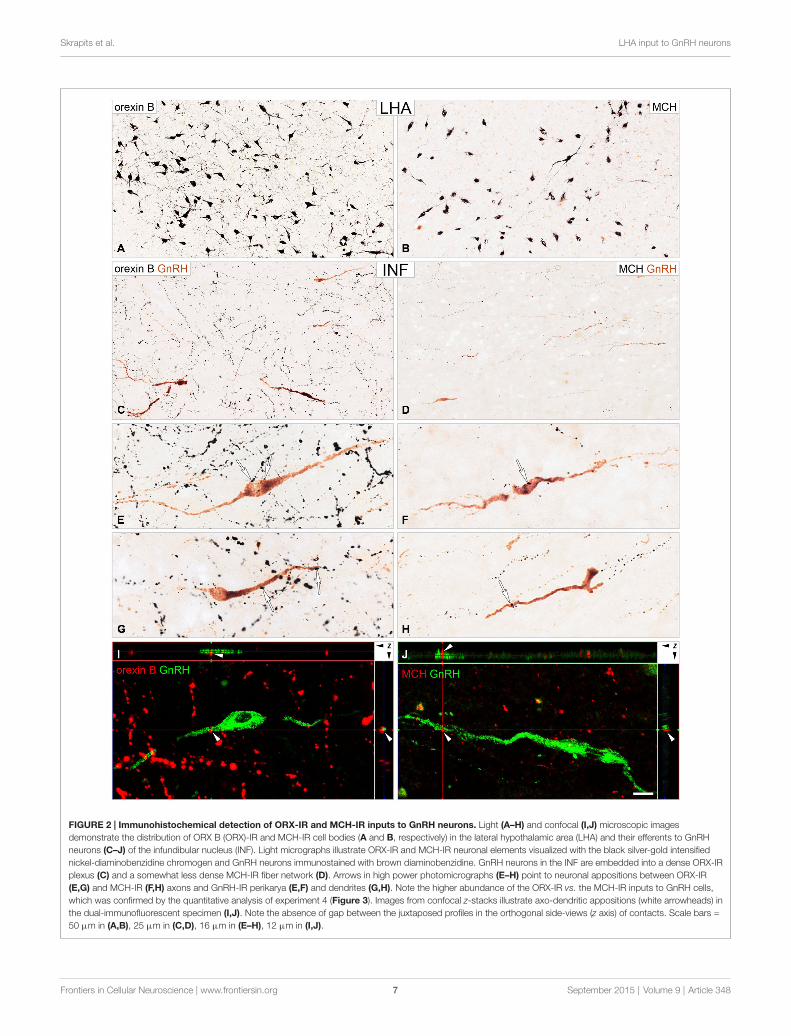
Experiment 2: Dual-ImmunoperoxidaseDetection of ORX-IR and MCH-IR Inputs toGnRH-Synthesizing NeuronsFollowing the dual-immunoperoxidase labeling of hypothalamicsections, the silver-gold intensified nickel-diaminobenzidinechromogen visualized intensely-labeled ORX-IR (Figure 2A)and MCH-IR (Figure 2B) cell bodies in the LHA. Themajority of GnRH cells labeled with brown diaminobenzidinechromogen were located in preoptic/medial hypothalamicregions; the same sites also received peptidergic axonalprojections from the LHA (Figures 2C,D). High-powerlight microscopic analysis of the double-labeled preopticand hypothalamic sections revealed ORX-IR (Figures 2E,G)and MCH-IR (Figures 2F,H) axonal appositions on subsetsof the GnRH-IR perikarya (Figures 2E,F) and dendrites(Figures 2G,H).
Experiment 3: Confocal Analysis of ORX-IR andMCH-IR Inputs to GnRH-Synthesizing NeuronsThe existence of direct ORX-IR and MCH-IR inputs to GnRHneurons was analyzed and confirmed with confocal microscopy.The orthogonal side-views of z-stacks reconstructed frommultiple optical slices ruled out that overlaps are mistakenlyconsidered appositions (Figures 2I,J).
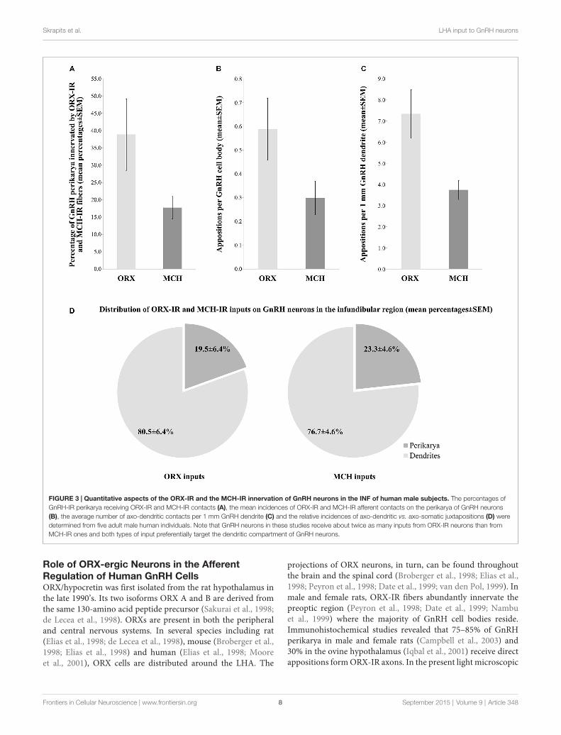
Experiment 4: Quantitative Analysis of ORX-IRand MCH-IR Inputs to the Somatic and DendriticCompartments of GnRH NeuronsThe number and heterogeneity of cases available for thesestudies did not allow us to address the issues of the putativesexual dimorphism and age-related changes in the innervationpatterns. The quantitative light microscopic analysis of inputsto GnRH neurons of the INF was restricted to the dual-peroxidase stained specimen of male subjects and results wereexpressed as mean ± SEM of the five individuals. Thesestudies have revealed ORX-IR contacts on 38.9 ± 10.3% andMCH-IR contacts on 17.7 ± 3.3% of GnRH-IR perikaryain the INF (Figure 3A). On average, GnRH-IR cell bodiesreceived 0.6 ± 0.1 ORX-IR and 0.3 ± 0.07 MCH-IR inputs(Figure 3B). ORX-IR and MCH-IR appositions were moretypically encountered on the processes of GnRH neurons. Aquantitative analysis (from which axon-like varicose processesof GnRH cells were excluded) identified 7.3 ± 1.1 ORX-IR and3.7 ± 0.5 MCH-IR axo-dendritic appositions on each 1 mmdendrite of GnRH neurons in the INF (Figure 3C). Overall,the axo-dendritic contacts represented the dominant form ofinputs to GnRH cells, representing 80.5 ± 6.4% of the ORX-IRand 76.7 ± 4.6% of the MCH-IR inputs that were encountered(Figure 3D).
Discussion
This immunohistochemical study provides neuroanatomicalevidence for the direct innervation of human GnRH cellsby ORX and MCH neurons of the lateral hypothalamus.We show that both types of inputs preferentially targetthe dendritic compartment of GnRH neurons. In addition,we describe the generation and characterization of newpolyclonal rat and sheep GnRH and mouse hGAP1 antibodiessuitable for the detection of GnRH-IR perikarya andprocesses.
Generation of New Antibodies to Detect GnRHNeuronsIn this study, we report the preparation and specificitytesting of new antibodies allowing the immunohistochemicalvisualization of GnRH perikarya and their processes. Theavailability of such antibodies from optional host specieswill be particularly valuable in future immunofluorescentmultiple-labeling experiments. Although antibodies against themammalian form of GnRH decapeptide are readily availablefrom commercial sources, many of such products require thecolchicine pretreatment of the experimental rodents to achievethe sufficient visualization of the GnRH cell bodies. Thistechnical limitation accounts for the wide use in publicationsof excellent private GnRH antisera; one of them (rabbit anti-GnRH; LR-1) has been made generously available for thewide scientific community by Dr. R. A. Benoit (Montréal,Canada). In a previous study, we have also used the LR-1 reference antibodies as a positive control to demonstratethe specificity of immunolabeling obtained with our twonewly produced polyclonal guinea pig GnRH antisera EH#1018and EH#1035 (Hrabovszky et al., 2011). In the presentwork, we report the generation of GnRH antibodies in twoadditional host species. At optimal dilutions, all of the newantibodies can recognize the cell bodies of GnRH neuronsin immunohistochemical assays, in addition to visualizingtheir axonal and dendritic processes. The rat (EH#1044) andsheep (EH#2001 and EH#2000) GnRH antibodies as well asthe mouse antibodies against hGAP1 (EH#1001) were equallysuitable to visualize the perikarya of GnRH neurons in humansections. Test sections from rodents also exhibited excellentsomatic labeling of GnRH neurons in the organum vasculosumof the lamina terminalis/preoptic regions using the rat andsheep antibodies against GnRH, but not the mouse antibodiesdirected against the hGAP1 sequence. Thus, the two aminoacids substituted in the rodent GAP1 sequence appear tobe important antigen determinants of the hGAP1 fragmentwe used to generate EH#1001. Our positive control studiesestablished that the immunohistochemical/immunofluorescentimages obtained using the new antibodies were similar to thoseprovided by the use of our reference guinea pig GnRH antiserum(EH#1018). Indeed, in dual-immunofluorescent studies weconfirmed that the new antibodies reacted specifically withthe same neuronal elements as did the reference GnRHantiserum, if appropriate working dilutions were chosenfor each.
Frontiers in Cellular Neuroscience | www.frontiersin.org 6 September 2015 | Volume 9 | Article 348
Skrapits et al. LHA input to GnRH neurons
FIGURE 2 | Immunohistochemical detection of ORX-IR and MCH-IR inputs to GnRH neurons. Light (A–H) and confocal (I,J) microscopic imagesdemonstrate the distribution of ORX B (ORX)-IR and MCH-IR cell bodies (A and B, respectively) in the lateral hypothalamic area (LHA) and their efferents to GnRHneurons (C–J) of the infundibular nucleus (INF). Light micrographs illustrate ORX-IR and MCH-IR neuronal elements visualized with the black silver-gold intensifiednickel-diaminobenzidine chromogen and GnRH neurons immunostained with brown diaminobenzidine. GnRH neurons in the INF are embedded into a dense ORX-IRplexus (C) and a somewhat less dense MCH-IR fiber network (D). Arrows in high power photomicrographs (E–H) point to neuronal appositions between ORX-IR(E,G) and MCH-IR (F,H) axons and GnRH-IR perikarya (E,F) and dendrites (G,H). Note the higher abundance of the ORX-IR vs. the MCH-IR inputs to GnRH cells,which was confirmed by the quantitative analysis of experiment 4 (Figure 3). Images from confocal z-stacks illustrate axo-dendritic appositions (white arrowheads) inthe dual-immunofluorescent specimen (I,J). Note the absence of gap between the juxtaposed profiles in the orthogonal side-views (z axis) of contacts. Scale bars =50 µm in (A,B), 25 µm in (C,D), 16 µm in (E–H), 12 µm in (I,J).
Frontiers in Cellular Neuroscience | www.frontiersin.org 7 September 2015 | Volume 9 | Article 348
Skrapits et al. LHA input to GnRH neurons
FIGURE 3 | Quantitative aspects of the ORX-IR and the MCH-IR innervation of GnRH neurons in the INF of human male subjects. The percentages ofGnRH-IR perikarya receiving ORX-IR and MCH-IR contacts (A), the mean incidences of ORX-IR and MCH-IR afferent contacts on the perikarya of GnRH neurons(B), the average number of axo-dendritic contacts per 1 mm GnRH dendrite (C) and the relative incidences of axo-dendritic vs. axo-somatic juxtapositions (D) weredetermined from five adult male human individuals. Note that GnRH neurons in these studies receive about twice as many inputs from ORX-IR neurons than fromMCH-IR ones and both types of input preferentially target the dendritic compartment of GnRH neurons.
Role of ORX-ergic Neurons in the AfferentRegulation of Human GnRH CellsORX/hypocretin was first isolated from the rat hypothalamus inthe late 1990’s. Its two isoforms ORX A and B are derived fromthe same 130-amino acid peptide precursor (Sakurai et al., 1998;de Lecea et al., 1998). ORXs are present in both the peripheraland central nervous systems. In several species including rat(Elias et al., 1998; de Lecea et al., 1998), mouse (Broberger et al.,1998; Elias et al., 1998) and human (Elias et al., 1998; Mooreet al., 2001), ORX cells are distributed around the LHA. The
projections of ORX neurons, in turn, can be found throughoutthe brain and the spinal cord (Broberger et al., 1998; Elias et al.,1998; Peyron et al., 1998; Date et al., 1999; van den Pol, 1999). Inmale and female rats, ORX-IR fibers abundantly innervate thepreoptic region (Peyron et al., 1998; Date et al., 1999; Nambuet al., 1999) where the majority of GnRH cell bodies reside.Immunohistochemical studies revealed that 75–85% of GnRHperikarya in male and female rats (Campbell et al., 2003) and30% in the ovine hypothalamus (Iqbal et al., 2001) receive directappositions formORX-IR axons. In the present light microscopic
Frontiers in Cellular Neuroscience | www.frontiersin.org 8 September 2015 | Volume 9 | Article 348
Skrapits et al. LHA input to GnRH neurons
studies, we provided evidence that high percentages (∼40%)of GnRH cell bodies in the INF of the human hypothalamusalso receive direct ORX-ergic inputs. The innervation of GnRHneurons was confirmed at the confocal level. The isoforms of theORX receptor (OX) mediating the putative effects of ORXs tohuman GnRH neurons have not been addressed in our study.
The ORX receptors (OX1 and OX2) use complex signaltransduction processes in which the extracellular Ca2+
concentration plays a more critical role than the cytoplasmicCa2+-stores. Right amount of extracellular calcium is essentialfor ORX binding, and Ca2+ influx also amplifies the responsesmediated by OXs (Johansson et al., 2007; Kukkonen andLeonard, 2014). In rats, 85% of GnRH cells express the OX1receptor form (Campbell et al., 2003) which is known tobind preferentially ORX A (Sakurai et al., 1998), whereas theOX2 receptor which binds the two ORX forms with similaraffinity (Sakurai et al., 1998) has not been reported on GnRHcells so far. Multiple sites and mechanisms of action maycomplicate the interpretation of the variable reproductive effectsof ORXs in vivo. ORX receptors are not only expressed inthe brain but also in peripheral endocrine organs such as thepituitary, testis, epididymis, seminal vesicle, ovary and placenta(Jöhren et al., 2001; Karteris et al., 2004). In addition, ORXsalso influence reproduction via acting at multiple hypothalamicsites. The LH surge of the estrogen plus progesterone-treatedovariectomized rats is inhibited by direct injections of ORXA into the mediobasal hypothalamus and the medial preopticarea and stimulated by injections placed around the organumvasculosum of the lamina terminalis (Small et al., 2003).ORX effects are influenced by the sex steroid levels of theexperimental animal. Accordingly, central administration ofORXs either stimulate or suppress the pulsatile secretion ofLH in ovariectomized rats, depending on the presence or theabsence, respectively, of ovarian sex steroids (Pu et al., 1998;Tamura et al., 1999). ORX A causes more dramatic effectsthan ORX B, suggesting the involvement of the OX1 receptorform (Tamura et al., 1999). Of note, the steroid-dependentbimodal effects of ORXs are reminiscent to similar observationsusing another orexigenic peptide neuropeptide Y (McDonald,1990). Results of electrophysiological studies indicate thatthe direct actions of ORXs on GnRH neurons are inhibitory.ORX A suppressed the firing frequency (both bursts andsingle spikes) of GnRH neurons via acting on OX1 receptor(Gaskins and Moenter, 2012). This suppression was differentin the presence and absence of estrogen; in ovariectomizedmice ORX A reduced GnRH activity in both the morning andevening, whereas GnRH activity was only reduced in the eveningusing ovariectomized mice treated with estradiol (Gaskins andMoenter, 2012).
One possible physiological relevance of the ORX system infertility is linked to the adaptation of the reproductive axis to themetabolic state (Silveyra et al., 2007; López et al., 2010). It has alsobeen proposed that ORXs may connect reproduction with thesleep-wake cycle (López et al., 2010). The pulsatile release of LH isdiminished in ORX-deficient narcoleptic men (Kok et al., 2004),although they show normal reproductive capacity (Taheri et al.,2002), similarly to the prepro-ORX knockout mice (Chemelli
et al., 1999). The direct axo-somatic and axo-dendritic inputfrom ORX neurons to the human GnRH neuronal system mayrepresent an important anatomical route in the communicationchannel whereby the LHA regulates the reproductive axis.
Functional Significance of MCH in the AfferentRegulation of GnRH NeuronsMCH is a cyclic 19-amino-acid peptide originally isolated fromthe teleost pituitary (Kawauchi et al., 1983) and later detectedalso in the central nervous system of various species, includingthe human (Mouri et al., 1993). Neuronal cell bodies synthesizingMCH were localized similarly to the medial forebrain bundle-LHA, subzona incerta and the perifornical area in rodents(Skofitsch et al., 1985; Bittencourt et al., 1992), sheep (Tillet et al.,1996) and monkeys (Bittencourt et al., 1998). The dominantdistribution of MCH-IR neurons in the LHA/perifornical regionwe observed in human male individuals was in agreementwith previous findings (Pelletier et al., 1987; Mouri et al.,1993), except that we have not detected labeled cell bodies inthe periventricular region, unlike one previous study (Pelletieret al., 1987). In different species, the axons arising from MCHneurons abundantly innervate the hypothalamic preoptic andperiventricular areas, the medial septum/diagonal band of Broca,the lateral septum, the LHA, the arcuate/premamillary areaand the eminentia mediana (Bittencourt et al., 1992, 1998;Tillet et al., 1996; Elias et al., 1998; Chiocchio et al., 2001;Gallardo et al., 2004; Williamson-Hughes et al., 2005). Here,we found that the MCH-IR projections innervated the preopticand infundibular regions where large subsets of GnRH neuronsare located in humans (Dudás et al., 2000; Hrabovszky andLiposits, 2013). In line with earlier observations in laboratoryrodents (Williamson-Hughes et al., 2005; Ward et al., 2009;Wu et al., 2009), our present immunohistochemical experimentsprovided evidence for direct axo-somatic and axo-dendriticconnections between human MCH and GnRH neurons. Wenote that this innervation was about 50% less heavy thanthe ORX-ergic input what we studied in histological specimenprocessed in parallel. This difference can have biological(more ORX than MCH fibers innervating GnRH cells) aswell as technical (e.g., better ORX vs. MCH antibody quality)explanations.
Functional studies of rodents revealed that GnRH/LH releasecan be influenced by exogenous MCH. As also noted above forORXs, the effect of MCH is bimodal, depending on estrogenlevels; MCH stimulates and suppresses GnRH/LH secretionunder conditions of high (Gonzalez et al., 1997; Murrayet al., 2000a,b; Chiocchio et al., 2001) and low (Tsukamuraet al., 2000) estrogens, respectively, in vitro, as well as afterintracerebroventricular administration of the peptide. Estrogensregulate MCH and MCHR1 protein expression negatively, asshown in ovariectomized rats treated with estradiol and in intactfemales during proestrus (Santollo and Eckel, 2013). The reducedMCH andMCHR1 protein detected with immunohistochemistryand in immunoblots, respectively, do not seem to result fromthe direct effects of estradiol on MCH neurons, since estradiol-treatment does not change MCH or MCHR1 mRNA expressionin cultured hypothalamic neurons (Santollo and Eckel, 2013).
Frontiers in Cellular Neuroscience | www.frontiersin.org 9 September 2015 | Volume 9 | Article 348
Skrapits et al. LHA input to GnRH neurons
Furthermore, MCH neurons do not express the alpha estrogenreceptor isoform (Muschamp and Hull, 2007; Santollo and Eckel,2013).
MCH acts via the G-protein coupled receptor MCHR1 (SLC-1/GPR24; Bächner et al., 1999; Chambers et al., 1999; Lemboet al., 1999; Saito et al., 1999; Shimomura et al., 1999). MCHR1can activate multiple signaling pathways via coupling to differentintracellular effector proteins Gi, Go and Gq (Hawes et al.,2000). While MCH can cause increased cytoplasmic calciumand excitation of MCH receptor expressing non-neuronal cells(Gao and van den Pol, 2002), most studies found that MCHinhibits neuronal firing (Pissios et al., 2006). Accordingly, MCHattenuates the calcium currents and inhibits neuronal activity inlateral hypothalamic neurons due to the Gi coupling of MCHR1(Gao and van den Pol, 2001, 2002). Similarly, MCH was alsofound to inhibit GnRH neuronal activity in mice and interruptthe persistent excitatory effect of kisspeptins on GnRH cells (Wuet al., 2009).
Our study has not addressed the presence of MCH receptorsin GnRH cells. Of note, a second MCH receptor form (MCHR2)with high-affinity MCH binding has also been identified inhuman (An et al., 2001; Sailer et al., 2001; Wang et al., 2001).Unlike MCHR1, MCHR2 couples only to the Gq protein,indicating its involvement in stimulatory neurotransmission.Therefore, the clarification of the receptor subtype/s in humanGnRH cells may have important functional implications.
Overall, anatomical evidence in this study suggests that MCHhas a direct effect on human GnRH neurons. Similarly to ORXs,the presence of MCH in neuronal afferents to human GnRH cellsmay play an important role in linking energy balance and otherLHA-related functions to reproduction.
The Dominance of Dendritic Over Somatic Inputsto GnRH NeuronsResults of early electron microscopic experiments in rats andmonkeys established that the cell body of GnRH neuronsreceives only very few (2–12) synaptic inputs (Witkin andSilverman, 1985; Witkin et al., 1991, 1995). More recentstudies of biocytin-filled mouse GnRH neurons revealed lengthyGnRH dendrites with numerous dendritic spines which receivedexcitatory synaptic inputs quite abundantly (Campbell et al.,2005). These newer observations suggest that GnRH neuronsare more heavily innervated than thought previously (Witkinand Silverman, 1985; Witkin et al., 1991, 1995) and themajority of inputs may target the dendritic compartment.The preferential innervation of the dendritic vs. the somaticcompartment may also be the case in the human. Results
of recent studies from our laboratory have established thatthe human GnRH dendrite receives very dense GABAergicand glutamatergic innervation (Hrabovszky et al., 2012a). Arecently proposed anatomical model of the neurosecretoryGnRH cell suggests that the hypophysiotropic projections ofGnRH neurons exhibit several dendritic rather than axonalproperties and receive abundant synaptic innervation (Herdeet al., 2013). While this observation has not been extendedto the human yet, the hypophysiotropic projections of humanGnRH neurons are often even much longer than in rodents,and may offer large membrane surfaces to receive synapticinputs. In addition to dendrites, hypophysiotropic GnRH axonprojections in the vicinity of the infundibular stalk whichapproach and surround the portal vasculature may servefor additional sites of neuronal interactions (Hrabovszky andLiposits, 2013; Borsay et al., 2014). Of note, we have recentlyshown that the lengthy hypophysiotropic GnRH projectionsintermingle with the plexuses of kisspeptin, neurokinin Band substance P neurons and establish occasional axo-axonalappositions with these systems (Borsay et al., 2014). In thepresent study, we observed themajority (77–81%) of ORX/GnRHand MCH/GnRH appositions on the dendritic compartment ofGnRHneurons.While the axo-dendritic inputs seem to representthe dominant form of communication between human MCHand GnRH neurons, significant axo-axonal interactions did notseem to exist in the infundibular stalk and postinfundibulareminence which contained relatively few ORX and MCH fibersin our study. In contrast, the rat eminentia mediana wasreported to contain MCH fibers both in its internal and externallayers (Bittencourt et al., 1992; Chiocchio et al., 2001) andproposed to represent an important site where MCH maystimulate GnRH secretion in this species (Chiocchio et al.,2001).
In summary, our study provides morphological evidence forthe afferent regulation of human GnRH neurons by lateralhypothalamic ORX and MCH neurons.
Author Contributions
KS, VK, ZS, CM, OS, AH, BÁB, LH, ZL and EH conceived anddesigned the experiments and wrote the paper. BÁB and LHcontributed essential research material.
Acknowledgment
This work was supported by grants from theHungarian ScientificResearch Fund (OTKA K83710, K112669, K100722).
References
An, S., Cutler, G., Zhao, J. J., Huang, S. G., Tian, H., Li, W., et al. (2001).Identification and characterization of a melanin-concentrating hormonereceptor. Proc. Natl. Acad. Sci. U S A 98, 7576–7581. doi: 10.1073/pnas.131200698
Bächner, D., Kreienkamp, H., Weise, C., Buck, F., and Richter, D. (1999).Identification of melanin concentrating hormone (MCH) as the natural ligand
for the orphan somatostatin-like receptor 1 (SLC-1). FEBS Lett. 457, 522–524.doi: 10.1016/s0014-5793(99)01092-3
Belchetz, P. E., Plant, T. M., Nakai, Y., Keogh, E. J., and Knobil, E.(1978). Hypophysial responses to continuous and intermittent deliveryof hypopthalamic gonadotropin-releasing hormone. Science 202, 631–633.doi: 10.1126/science.100883
Bittencourt, J. C., Frigo, L., Rissman, R. A., Casatti, C. A., Nahon, J. L., andBauer, J. A. (1998). The distribution of melanin-concentrating hormone in
Frontiers in Cellular Neuroscience | www.frontiersin.org 10 September 2015 | Volume 9 | Article 348
Skrapits et al. LHA input to GnRH neurons
the monkey brain (Cebus apella). Brain Res. 804, 140–143. doi: 10.1016/s0006-8993(98)00662-3
Bittencourt, J. C., Presse, F., Arias, C., Peto, C., Vaughan, J., Nahon, J. L.,et al. (1992). The melanin-concentrating hormone system of the rat brain: animmuno- and hybridization histochemical characterization. J. Comp. Neurol.319, 218–245. doi: 10.1002/cne.903190204
Borsay, B. A., Skrapits, K., Herczeg, L., Ciofi, P., Bloom, S. R., Ghatei, M. A.,et al. (2014). Hypophysiotropic gonadotropin-releasing hormone projectionsare exposed to dense plexuses of kisspeptin, neurokinin B and substance pimmunoreactive fibers in the human: a study on tissues from postmenopausalwomen. Neuroendocrinology 100, 141–152. doi: 10.1159/000368362
Broberger, C., De Lecea, L., Sutcliffe, J. G., and Hökfelt, T. (1998).Hypocretin/orexin- and melanin-concentrating hormone-expressing cellsform distinct populations in the rodent lateral hypothalamus: relationship tothe neuropeptide Y and agouti gene-related protein systems. J. Comp. Neurol.402, 460–474. doi: 10.1002/(sici)1096-9861(19981228)402:4<460::aid-cne3>3.0.co;2-s
Bullmann, T., Härtig, W., Holzer, M., and Arendt, T. (2010). Expression of theembryonal isoform (0N/3R) of the microtubule-associated protein tau in theadult rat central nervous system. J. Comp. Neurol. 518, 2538–2553. doi: 10.1002/cne.22351
Burdakov, D., Karnani, M. M., and Gonzalez, A. (2013). Lateral hypothalamus asa sensor-regulator in respiratory and metabolic control. Physiol. Behav. 121,117–124. doi: 10.1016/j.physbeh.2013.03.023
Campbell, R. E., Grove, K. L., and Smith, M. S. (2003). Gonadotropin-releasinghormone neurons coexpress orexin 1 receptor immunoreactivity and receivedirect contacts by orexin fibers. Endocrinology 144, 1542–1548. doi: 10.1210/en.2002-220958
Campbell, R. E., Han, S. K., and Herbison, A. E. (2005). Biocytin filling ofadult gonadotropin-releasing hormone neurons in situ reveals extensive, spiny,dendritic processes. Endocrinology 146, 1163–1169. doi: 10.1210/en.2004-1369
Carmel, P. W., Araki, S., and Ferin, M. (1976). Pituitary stalk portal bloodcollection in rhesus monkeys: evidence for pulsatile release of gonadotropin-releasing hormone (GnRH). Endocrinology 99, 243–248. doi: 10.1210/endo-99-1-243
Chambers, J., Ames, R. S., Bergsma, D., Muir, A., Fitzgerald, L. R., Hervieu,G., et al. (1999). Melanin-concentrating hormone is the cognate ligand forthe orphan G-protein-coupled receptor SLC-1. Nature 400, 261–265. doi: 10.1038/22313
Chemelli, R. M., Willie, J. T., Sinton, C. M., Elmquist, J. K., Scammell, T., Lee, C.,et al. (1999). Narcolepsy in orexin knockout mice: molecular genetics of sleepregulation. Cell 98, 437–451. doi: 10.1016/S0092-8674(00)81973-X
Chiocchio, S. R., Gallardo, M. G., Louzan, P., Gutnisky, V., and Tramezzani, J. H.(2001). Melanin-concentrating hormone stimulates the release of luteinizinghormone-releasing hormone and gonadotropins in the female rat acting at bothmedian eminence and pituitary levels. Biol. Reprod. 64, 1466–1472. doi: 10.1095/biolreprod64.5.1466
Clarke, I. J., and Cummins, J. T. (1982). The temporal relationship betweengonadotropin releasing hormone (GnRH) and luteinizing hormone (LH)secretion in ovariectomized ewes. Endocrinology 111, 1737–1739. doi: 10.1210/endo-111-5-1737
Dahl, S. K., Amstalden, M., Coolen, L., Fitzgerald, M., and Lehman, M. (2009).Dynorphin immunoreactive fibers contact GnRH neurons in the humanhypothalamus. Reprod. Sci. 16, 781–787. doi: 10.1177/1933719109336619
Date, Y., Ueta, Y., Yamashita, H., Yamaguchi, H., Matsukura, S., Kangawa, K., et al.(1999). Orexins, orexigenic hypothalamic peptides, interact with autonomic,neuroendocrine and neuroregulatory systems. Proc. Natl. Acad. Sci. U S A 96,748–753. doi: 10.1073/pnas.96.2.748
de Lecea, L., Kilduff, T. S., Peyron, C., Gao, X., Foye, P. E., Danielson, P. E., et al.(1998). The hypocretins: hypothalamus-specific peptides with neuroexcitatoryactivity. Proc. Natl. Acad. Sci. U S A 95, 322–327. doi: 10.1073/pnas.95.1.322
Dudás, B., and Merchenthaler, I. (2002a). Close juxtapositions between LHRHimmunoreactive neurons and substance P immunoreactive axons in the humandiencephalon. J. Clin. Endocrinol. Metab. 87, 2946–2953. doi: 10.1210/jc.87.6.2946
Dudás, B., andMerchenthaler, I. (2002b). Close juxtapositions between luteinizinghormone-releasing hormone-immunoreactive neurons and corticotropin-releasing factor-immunoreactive axons in the human diencephalon. J. Clin.Endocrinol. Metab. 87, 5778–5784. doi: 10.1210/jc.2002-020996
Dudás, B., and Merchenthaler, I. (2003). Topography and associations of leu-enkephalin and luteinizing hormone-releasing hormone neuronal systems inthe human diencephalon. J. Clin. Endocrinol. Metab. 88, 1842–1848. doi: 10.1210/jc.2002-021416
Dudás, B., and Merchenthaler, I. (2004a). Bi-directional associations betweengalanin and luteinizing hormone-releasing hormone neuronal systems in thehuman diencephalon. Neuroscience 127, 695–707. doi: 10.1016/j.neuroscience.2004.05.018
Dudás, B., and Merchenthaler, I. (2004b). Close anatomical associations betweenbeta-endorphin and luteinizing hormone-releasing hormone neuronal systemsin the human diencephalon. Neuroscience 124, 221–229. doi: 10.1016/j.neuroscience.2003.11.021
Dudás, B., and Merchenthaler, I. (2006). Three-dimensional representationof the neurotransmitter systems of the human hypothalamus: inputsof the gonadotrophin hormone-releasing hormone neuronal system. J.Neuroendocrinol. 18, 79–95. doi: 10.1111/j.1365-2826.2005.01398.x
Dudás, B., Mihály, A., and Merchenthaler, I. (2000). Topography andassociations of luteinizing hormone-releasing hormone and neuropeptide Y-immunoreactive neuronal systems in the human diencephalon. J. Comp.Neurol. 427, 593–603. doi: 10.1002/1096-9861(20001127)427:4<593::aid-cne7>3.0.co;2-c
Elias, C. F., Saper, C. B., Maratos-Flier, E., Tritos, N. A., Lee, C., Kelly, J., et al.(1998). Chemically defined projections linking the mediobasal hypothalamusand the lateral hypothalamic area. J. Comp. Neurol. 402, 442–459. doi: 10.1002/(sici)1096-9861(19981228)402:4<442::aid-cne2>3.0.co;2-r
Gallardo, M. G., Chiocchio, S. R., and Tramezzani, J. H. (2004). Changes ofmelanin-concentrating hormone related to LHRH release in the medianeminence of rats. Brain Res. 1030, 152–158. doi: 10.1016/j.brainres.2004.10.005
Gao, X. B., and van den Pol, A. N. (2001). Melanin concentrating hormonedepresses synaptic activity of glutamate and GABA neurons from ratlateral hypothalamus. J. Physiol. 533, 237–252. doi: 10.1111/j.1469-7793.2001.0237b.x
Gao, X. B., and van den Pol, A. N. (2002). Melanin-concentrating hormonedepresses L-, N- and P/Q-type voltage-dependent calcium channels in ratlateral hypothalamic neurons. J. Physiol. 542, 273–286. doi: 10.1113/jphysiol.2002.019372
Gaskins, G. T., and Moenter, S. M. (2012). Orexin a suppresses gonadotropin-releasing hormone (GnRH) neuron activity in the mouse. Endocrinology 153,3850–3860. doi: 10.1210/en.2012-1300
Gonzalez, M. I., Baker, B. I., and Wilson, C. A. (1997). Stimulatoryeffect of melanin-concentrating hormone on luteinising hormone release.Neuroendocrinology 66, 254–262. doi: 10.1159/000127246
Hawes, B. E., Kil, E., Green, B., O’neill, K., Fried, S., and Graziano, M. P.(2000). The melanin-concentrating hormone receptor couples to multiple Gproteins to activate diverse intracellular signaling pathways. Endocrinology 141,4524–4532. doi: 10.1210/en.141.12.4524
Herde, M. K., Iremonger, K. J., Constantin, S., and Herbison, A. E. (2013).GnRH neurons elaborate a long-range projection with shared axonal anddendritic functions. J. Neurosci. 33, 12689–12697. doi: 10.1523/jneurosci.0579-13.2013
Hrabovszky, E., Borsay, B. A., Racz, K., Herczeg, L., Ciofi, P., Bloom, S. R., et al.(2013). Substance P immunoreactivity exhibits frequent colocalization withkisspeptin and neurokinin B in the human infundibular region. PLoS One8:e72369. doi: 10.1371/journal.pone.0072369
Hrabovszky, E., Ciofi, P., Vida, B., Horvath, M. C., Keller, E., Caraty, A., et al.(2010). The kisspeptin system of the human hypothalamus: sexual dimorphismand relationship with gonadotropin-releasing hormone and neurokinin Bneurons. Eur. J. Neurosci. 31, 1984–1998. doi: 10.1111/j.1460-9568.2010.07239.x
Hrabovszky, E., and Liposits, Z. (2013). Afferent neuronal control of type-Igonadotropin releasing hormone neurons in the human. Front. Endocrinol.(Lausanne) 4:130. doi: 10.3389/fendo.2013.00130
Hrabovszky, E., Molnár, C. S., Nagy, R., Vida, B., Borsay, B. A., Rácz, K., et al.(2012a). Glutamatergic and GABAergic innervation of human gonadotropin-releasing hormone-I neurons. Endocrinology 153, 2766–2776. doi: 10.1210/en.2011-2106
Hrabovszky, E., Sipos, M. T., Molnár, C. S., Ciofi, P., Borsay, B. A., Gergely,P., et al. (2012b). Low degree of overlap between kisspeptin, neurokinin Band dynorphin immunoreactivities in the infundibular nucleus of young male
Frontiers in Cellular Neuroscience | www.frontiersin.org 11 September 2015 | Volume 9 | Article 348
Skrapits et al. LHA input to GnRH neurons
human subjects challenges the KNDy neuron concept. Endocrinology 153,4978–4989. doi: 10.1210/en.2012-1545
Hrabovszky, E., Molnár, C. S., Sipos, M., Vida, B., Ciofi, P., Borsay, B. A., et al.(2011). Sexual dimorphism of kisspeptin and neurokinin B immunoreactiveneurons in the infundibular nucleus of aged men and women. Front.Endocrinol. (Lausanne) 2:80. doi: 10.3389/fendo.2011.00080
Iqbal, J., Pompolo, S., Sakurai, T., and Clarke, I. J. (2001). Evidence that orexin-containing neurones provide direct input to gonadotropin-releasing hormoneneurones in the ovine hypothalamus. J. Neuroendocrinol. 13, 1033–1041.doi: 10.1046/j.1365-2826.2001.00719.x
Johansson, L., Ekholm, M. E., and Kukkonen, J. P. (2007). Regulation of OX1orexin/hypocretin receptor-coupling to phospholipase C by Ca2+ influx. Br.J. Pharmacol. 150, 97–104. doi: 10.1038/sj.bjp.0706959
Jöhren, O., Neidert, S. J., Kummer, M., Dendorfer, A., and Dominiak, P. (2001).Prepro-orexin and orexin receptor mRNAs are differentially expressed inperipheral tissues of male and female rats. Endocrinology 142, 3324–3331.doi: 10.1210/en.142.8.3324
Karteris, E., Chen, J., and Randeva, H. S. (2004). Expression of humanprepro-orexin and signaling characteristics of orexin receptors in the malereproductive system. J. Clin. Endocrinol. Metab. 89, 1957–1962. doi: 10.1210/jc.2003-031778
Kawauchi, H., Kawazoe, I., Tsubokawa, M., Kishida, M., and Baker, B. I.(1983). Characterization of melanin-concentrating hormone in chum salmonpituitaries. Nature 305, 321–323. doi: 10.1038/305321a0
Kok, S. W., Roelfsema, F., Overeem, S., Lammers, G. J., Frolich, M., Meinders,A. E., et al. (2004). Pulsatile LH release is diminished, whereas FSH secretionis normal, in hypocretin-deficient narcoleptic men. Am. J. Physiol. Endocrinol.Metab. 287, E630–E636. doi: 10.1152/ajpendo.00060.2004
Konadhode, R. R., Pelluru, D., and Shiromani, P. J. (2015). Neurons containingorexin ormelanin concentrating hormone reciprocally regulate wake and sleep.Front. Syst. Neurosci. 8:244. doi: 10.3389/fnsys.2014.00244
Kukkonen, J. P., and Leonard, C. S. (2014). Orexin/hypocretin receptor signallingcascades. Br. J. Pharmacol. 171, 314–331. doi: 10.1111/bph.12324
Kunii, K., Yamanaka, A., Nambu, T., Matsuzaki, I., Goto, K., and Sakurai,T. (1999). Orexins/hypocretins regulate drinking behaviour. Brain Res. 842,256–261. doi: 10.1016/s0006-8993(99)01884-3
Lembo, P. M., Grazzini, E., Cao, J., Hubatsch, D. A., Pelletier, M., Hoffert, C.,et al. (1999). The receptor for the orexigenic peptide melanin-concentratinghormone is a G-protein-coupled receptor. Nat. Cell Biol. 1, 267–271.
Liposits, Z., Sétáló, G., and Flerkó, B. (1984). Application of the silver-gold intensified 3,3′-diaminobenzidine chromogen to the light and electronmicroscopic detection of the luteinizing hormone-releasing hormone systemof the rat brain. Neuroscience 13, 513–525. doi: 10.1016/0306-4522(84)90245-8
López, M., Tena-Sempere, M., and Dieguez, C. (2010). Cross-talk between orexins(hypocretins) and the neuroendocrine axes (hypothalamic-pituitary axes).Front. Neuroendocrinol. 31, 113–127. doi: 10.1016/j.yfrne.2009.07.001
McDonald, J. K. (1990). Role of neuropeptide Y in reproductive function. Ann.N Y Acad. Sci. 611, 258–272. doi: 10.1111/j.1749-6632.1990.tb48937.x
Molnár, C. S., Vida, B., Sipos, M. T., Ciofi, P., Borsay, B. A., Racz, K., et al. (2012).Morphological evidence for enhanced kisspeptin and neurokinin B signalingin the infundibular nucleus of the aging man. Endocrinology 153, 5428–5439.doi: 10.1210/en.2012-1739
Moore, R. Y., Abrahamson, E. A., and Van Den Pol, A. (2001). The hypocretinneuron system: an arousal system in the human brain. Arch. Ital. Biol. 139,195–205.
Mouri, T., Takahashi, K., Kawauchi, H., Sone, M., Totsune, K., Murakami, O.,et al. (1993). Melanin-concentrating hormone in the human brain. Peptides 14,643–646. doi: 10.1016/0196-9781(93)90158-d
Murray, J. F., Adan, R. A., Walker, R., Baker, B. I., Thody, A. J., Nijenhuis, W. A.,et al. (2000a). Melanin-concentrating hormone, melanocortin receptors andregulation of luteinizing hormone release. J. Neuroendocrinol. 12, 217–223.doi: 10.1046/j.1365-2826.2000.00440.x
Murray, J. F., Mercer, J. G., Adan, R. A., Datta, J. J., Aldairy, C., Moar,K. M., et al. (2000b). The effect of leptin on luteinizing hormone releaseis exerted in the zona incerta and mediated by melanin-concentratinghormone. J. Neuroendocrinol. 12, 1133–1139. doi: 10.1046/j.1365-2826.2000.00577.x
Muschamp, J. W., and Hull, E. M. (2007). Melanin concentrating hormone andestrogen receptor-alpha are coexstensive but not coexpressed in cells of malerat hypothalamus. Neurosci. Lett. 427, 123–126. doi: 10.1016/j.neulet.2007.09.031
Nambu, T., Sakurai, T., Mizukami, K., Hosoya, Y., Yanagisawa, M., and Goto, K.(1999). Distribution of orexin neurons in the adult rat brain. Brain Res. 827,243–260. doi: 10.1016/s0006-8993(99)01336-0
Pelletier, G., Guy, J., Désy, L., Li, S., Eberle, A. N., and Vaudry, H.(1987). Melanin-concentrating hormone (MCH) is colocalized with alpha-melanocyte-stimulating hormone (alpha-MSH) in the rat but not in thehuman hypothalamus. Brain Res. 423, 247–253. doi: 10.1016/0006-8993(87)90846-8
Peyron, C., Tighe, D. K., van den Pol, A. N., de Lecea, L., Heller, H. C., Sutcliffe,J. G., et al. (1998). Neurons containing hypocretin (orexin) project to multipleneuronal systems. J. Neurosci. 18, 9996–10015.
Pissios, P., Bradley, R. L., and Maratos-Flier, E. (2006). Expanding the scales:the multiple roles of MCH in regulating energy balance and other biologicalfunctions. Endocr. Rev. 27, 606–620. doi: 10.1210/er.2006-0021
Pu, S., Jain, M. R., Kalra, P. S., and Kalra, S. P. (1998). Orexins, a novel family ofhypothalamic neuropeptides, modulate pituitary luteinizing hormone secretionin an ovarian steroid-dependent manner. Regul. Pept. 78, 133–136.
Sailer, A. W., Sano, H., Zeng, Z., Mcdonald, T. P., Pan, J., Pong, S. S., et al.(2001). Identification and characterization of a second melanin-concentratinghormone receptor, MCH-2R. Proc. Natl. Acad. Sci. U S A 98, 7564–7569.doi: 10.1073/pnas.121170598
Saito, Y., Nothacker, H. P., Wang, Z., Lin, S. H., Leslie, F., and Civelli, O. (1999).Molecular characterization of the melanin-concentrating-hormone receptor.Nature 400, 265–269. doi: 10.1038/22321
Sakurai, T., Amemiya, A., Ishii, M., Matsuzaki, I., Chemelli, R. M., Tanaka,H., et al. (1998). Orexins and orexin receptors: a family of hypothalamicneuropeptides and G protein-coupled receptors that regulate feeding behavior.Cell 92, 573–585. doi: 10.1016/s0092-8674(00)80949-6
Santollo, J., and Eckel, L. A. (2013). Oestradiol decreases melanin-concentratinghormone (MCH) andMCH receptor expression in the hypothalamus of femalerats. J. Neuroendocrinol. 25, 570–579. doi: 10.1111/jne.12032
Schwanzel-Fukuda, M., and Pfaff, D. W. (1989). Origin of luteinizinghormone-releasing hormone neurons. Nature 338, 161–164. doi: 10.1038/338161a0
Shimomura, Y., Mori, M., Sugo, T., Ishibashi, Y., Abe, M., Kurokawa, T., et al.(1999). Isolation and identification of melanin-concentrating hormone as theendogenous ligand of the SLC-1 receptor. Biochem. Biophys. Res. Commun. 261,622–626. doi: 10.1006/bbrc.1999.1104
Silveyra, P., Catalano, P. N., Lux-Lantos, V., and Libertun, C. (2007). Impact ofproestrous milieu on expression of orexin receptors and prepro-orexin in rathypothalamus and hypophysis: actions of Cetrorelix and Nembutal. Am. J.Physiol. Endocrinol. Metab. 292, E820–E828. doi: 10.1152/ajpendo.00467.2006
Skofitsch, G., Jacobowitz, D. M., and Zamir, N. (1985). Immunohistochemicallocalization of a melanin concentrating hormone-like peptide in the rat brain.Brain Res. Bull. 15, 635–649. doi: 10.1016/0361-9230(85)90213-8
Skrapits, K., Borsay, B. A., Herczeg, L., Ciofi, P., Bloom, S. R., Ghatei, M. A.,et al. (2014). Colocalization of cocaine- and amphetamine-regulated transcriptwith kisspeptin and neurokinin B in the human infundibular region. PLoS One9:e103977. doi: 10.1371/journal.pone.0103977
Small, C. J., Goubillon, M. L., Murray, J. F., Siddiqui, A., Grimshaw, S. E.,Young, H., et al. (2003). Central orexin A has site-specific effects on luteinizinghormone release in female rats. Endocrinology 144, 3225–3236. doi: 10.1210/en.2002-0041
Taheri, S., Zeitzer, J. M., and Mignot, E. (2002). The role of hypocretins (orexins)in sleep regulation and narcolepsy. Annu. Rev. Neurosci. 25, 283–313. doi: 10.1146/annurev.neuro.25.112701.142826
Tamura, T., Irahara, M., Tezuka, M., Kiyokawa, M., and Aono, T. (1999). Orexins,orexigenic hypothalamic neuropeptides, suppress the pulsatile secretion ofluteinizing hormone in ovariectomized female rats. Biochem. Biophys. Res.Commun. 264, 759–762. doi: 10.1006/bbrc.1999.1573
Tillet, Y., Batailler, M., and Fellmann, D. (1996). Distribution of melanin-concentrating hormone (MCH)-like immunoreactivity in neurons of thediencephalon of sheep. J. Chem. Neuroanat. 12, 135–145. doi: 10.1016/s0891-0618(96)00195-0
Frontiers in Cellular Neuroscience | www.frontiersin.org 12 September 2015 | Volume 9 | Article 348
Skrapits et al. LHA input to GnRH neurons
Tsukamura, H., Thompson, R. C., Tsukahara, S., Ohkura, S., Maekawa, F.,Moriyama, R., et al. (2000). Intracerebroventricular administration of melanin-concentrating hormone suppresses pulsatile luteinizing hormone release inthe female rat. J. Neuroendocrinol. 12, 529–534. doi: 10.1046/j.1365-2826.2000.00482.x
Ubuka, T., Morgan, K., Pawson, A. J., Osugi, T., Chowdhury, V. S., Minakata, H.,et al. (2009). Identification of human GnIH homologs, RFRP-1 and RFRP-3and the cognate receptor, GPR147 in the human hypothalamic pituitary axis.PLoS One 4:e8400. doi: 10.1371/journal.pone.0008400
van den Pol, A. N. (1999). Hypothalamic hypocretin (orexin): robust innervationof the spinal cord. J. Neurosci. 19, 3171–3182.
Wang, S., Behan, J., O’neill, K., Weig, B., Fried, S., Laz, T., et al. (2001).Identification and pharmacological characterization of a novel humanmelanin-concentrating hormone receptor, mch-r2. J. Biol. Chem. 276,34664–34670. doi: 10.1074/jbc.m102601200
Ward, D. R., Dear, F. M., Ward, I. A., Anderson, S. I., Spergel, D. J., Smith,P. A., et al. (2009). Innervation of gonadotropin-releasing hormone neuronsby peptidergic neurons conveying circadian or energy balance information inthe mouse. PLoS One 4:e5322. doi: 10.1371/journal.pone.0005322
Watts, A. G., Salter, D. S., and Neuner, C. M. (2007). Neural network interactionsand ingestive behavior control during anorexia. Physiol. Behav. 91, 389–396.doi: 10.1016/j.physbeh.2007.04.010
Whiddon, B. B., and Palmiter, R. D. (2013). Ablation of neurons expressingmelanin-concentrating hormone (MCH) in adult mice improves glucosetolerance independent of MCH signaling. J. Neurosci. 33, 2009–2016. doi: 10.1523/jneurosci.3921-12.2013
Williamson-Hughes, P. S., Grove, K. L., and Smith, M. S. (2005). Melaninconcentrating hormone (MCH): a novel neural pathway for regulation
of GnRH neurons. Brain Res. 1041, 117–124. doi: 10.1016/j.brainres.2004.11.066
Witkin, J. W., Ferin, M., Popilskis, S. J., and Silverman, A. J. (1991). Effects ofgonadal steroids on the ultrastructure of GnRH neurons in the rhesus monkey:synaptic input and glial apposition. Endocrinology 129, 1083–1092. doi: 10.1210/endo-129-2-1083
Witkin, J. W., O’Sullivan, H., and Silverman, A. J. (1995). Novel associationsamong gonadotropin-releasing hormone neurons. Endocrinology 136,4323–4330. doi: 10.1210/en.136.10.4323
Witkin, J. W., and Silverman, A. J. (1985). Synaptology of luteinizing hormone-releasing hormone neurons in rat preoptic area. Peptides 6, 263–271. doi: 10.1016/0196-9781(85)90050-6
Wu, M., Dumalska, I., Morozova, E., van den Pol, A., and Alreja, M. (2009).Melanin-concentrating hormone directly inhibits GnRH neurons and blockskisspeptin activation, linking energy balance to reproduction. Proc. Natl. Acad.Sci. U S A 106, 17217–17222. doi: 10.1073/pnas.0908200106
Conflict of Interest Statement: The authors declare that the research wasconducted in the absence of any commercial or financial relationships that couldbe construed as a potential conflict of interest.
Copyright © 2015 Skrapits, Kanti, Savanyú, Maurnyi, Szenci, Horváth, Borsay,Herczeg, Liposits and Hrabovszky. This is an open-access article distributed underthe terms of the Creative Commons Attribution License (CC BY). The use,distribution and reproduction in other forums is permitted, provided the originalauthor(s) or licensor are credited and that the original publication in this journalis cited, in accordance with accepted academic practice. No use, distribution orreproduction is permitted which does not comply with these terms.
Frontiers in Cellular Neuroscience | www.frontiersin.org 13 September 2015 | Volume 9 | Article 348