Embed Size (px)
Citation preview

Layer-Specific Generation and Propagation of Seizures in Slicesof Developing Neocortex: Role of Excitatory GABAergic Synapses
Sylvain Rheims, Alfonso Represa, Yehezkel Ben-Ari, and Yuri ZilberterInstitut de Neurobiologie de la Mediterranee, Institut National de la Sante et de la Recherche Medicale U901, Universite de laMediterranee, Marseille, France
Submitted 26 March 2008; accepted in final form 3 May 2008
Rheims S, Represa A, Ben-Ari Y, Zilberter Y. Layer-specific gener-ation and propagation of seizures in slices of developing neocortex: roleof excitatory GABAergic synapses. J Neurophysiol 100: 620–628, 2008.First published May 21, 2008; doi10.1152/jn.90403.2008. The neonatalperiod is critical for seizure susceptibility, and neocortical networksare central in infantile epilepsies. We report that application of4-aminopyridine (4-AP) to immature (P6–P9) neocortical slices gen-erates layer-specific interictal seizures (IISs) that transform afterrecurrent seizures to ictal seizures (ISs). During IISs, cell-attachedrecordings show action potentials in interneurons and pyramidal cellsin L5/6 and interneurons but not pyramidal neurons in L2/3. However,L2/3 pyramidal neurons also fire during ISs. Using single N-methyl-D-aspartate (NMDA) channel recordings for measuring the cell restingpotential (Em), we show that transition from IISs to ISs is associatedwith a gradual Em depolarization of L2/3 and L5/6 pyramidal neuronsthat enhances their excitability. Bumetanide, a NKCC1 co-transporterantagonist, inhibits generation of IISs and prevents their transforma-tion to ISs, indicating the role excitatory GABA in epilepsies. There-fore deep layer neurons are more susceptible to seizures than super-ficial ones. The initiating phase of seizures is characterized by IISsgenerated in L5/6 and supported by activation of both L5/6 interneu-rons and pyramidal cells. IISs propagate to L2/3 via activation of L2/3interneurons but not pyramidal cells, which are mostly quiescent atthis phase. In superficial layers, a persistent increase in excitability ofpyramidal neurons caused by Em depolarization is associated with atransition from largely confined GABAergic IIS to ictal events thatentrain the entire neocortex.
I N T R O D U C T I O N
The neonatal period is critical for seizure susceptibility. Theincidence of seizures is indeed higher in neonates than in olderchildren or adults (Mizrahi 1999). Neonates with seizures areat risk for death, and survivors are at risk for neurologicalimpairment, developmental delay, and later epilepsy (Bye et al.1997; Dixon et al. 2002; Rennie and Boylan 2003; Scher et al.1993; Tekgul et al. 2006). Several observations suggest that ahigh incidence of seizures during neonatal period may becaused by intrinsic properties of the neonatal brain. For in-stance, 10–25% of infants present normal examination andnormal MRI (Lombroso 1996; Scher et al. 1993; Tekgul et al.2006; Tharp 2002) despite the high prevalence of cerebrallesions (Bye et al. 1997; Lombroso 1996; Rennie and Boylan2003; Scher et al. 1993; Tekgul et al. 2006; Tharp 2002). Manyinfants, including those suffering from cerebral lesions, expe-rience a seizure-free period of months to years, suggesting thatearly seizures may be self-limited possibly because of the
developmental alterations in neuronal excitability (Tharp2002).
Although neocortical networks are central in infantile epi-lepsies (Mizrahi 1999; Mizrahi and Clancy 2000), our under-standing of epilepsies in the developing brain comes fromstudies on the immature hippocampus (for review, Ben-Ari2002; Ben-Ari et al. 2004; Cossart et al. 2005; Le Van Quyenet al. 2006; Lienekugel et al. 1999). GABA excites immatureneurons in the hippocampus (for review, Ben-Ari 2002; Ben-Ari et al. 2007) and neocortex (Owens et al. 1996, Rheims et al.2008; Yamada et al. 2004), but while this action directlyimpacts seizures in hippocampus (Dzhala and Staley 2003;Dzhala et al. 2005; Khalilov et al. 1999, 2005; Khazipov et al.2004; Lienekugel et al. 1997), there is no evidence that this isthe case for neocortical neurons.
Here we explore the mechanisms of initiation and develop-ment of paroxysmal oscillations in deep and superficial neo-cortical layers (L2/3 and L5/6) of neonatal mice. We report that4-aminopyridine (4-AP) triggers seizures that initially are in-terictal (IIS), generated by L5/6 neurons. Interictal eventspropagate to L2/3 via activation of L2/3 interneurons but notpyramidal cells. With the recurrence of seizures, there is atransition from interictal to ictal events (ISs), which are main-tained by activity of interneurons and pyramidal cells in bothdeep and superficial layers. This transition is associated with agradual depolarization of Em in both L2/3 and L5/6 pyramidalneurons. Therefore our results show that the layer specificexcitatory action of GABA plays an important role in thegeneration of seizures. In addition, recurrent seizures induce apersistent increase in neuron excitability that underlies thetransition from interictal to ictal paroxysmal activity entrainingthe entire cortical network.
M E T H O D S
Slices preparation
Brain slices were prepared from postnatal day (P)6 to P17 Swissmice of both sexes. P0 was the day of birth. All animal protocolsconformed to the French Public Health Service policy and theINSERM guidelines on the use of laboratory animals. Animals weredecapitated, and brains were removed. Saggital slices (300–400 �m)were cut by a Microm slicer (International) using ice-cold oxygenatedmodified artificial cerebrospinal fluid (mACSF) containing 0.5 mMCaCl2 and 7 mM MgSO4, and with Na� replaced by an equimolarconcentration of choline. Slices were transferred to a normal ACSFcontaining (in mM) 126 NaCl, 3.5 KCl, 1.2 NaH2PO4, 26 NaHCO3,
Address for reprint requests and other correspondence: S. Rheims, Institutde Neurobiologie de la Mediterranee, INSERM U901, Universite de la Medi-terranee, Marseille, France (E-mail: [email protected]).
The costs of publication of this article were defrayed in part by the paymentof page charges. The article must therefore be hereby marked “advertisement”in accordance with 18 U.S.C. Section 1734 solely to indicate this fact.
J Neurophysiol 100: 620–628, 2008.First published May 21, 2008; doi10.1152/jn.90403.2008.
620 0022-3077/08 $8.00 Copyright © 2008 The American Physiological Society www.jn.org
by 10.220.33.1 on February 17, 2017
http://jn.physiology.org/D
ownloaded from

1.3 MgCl2, 2.0 CaCl2, and 10 D-glucose, pH 7.4, at room temperature(20–22°C) for �1 h before use.
Electrophysiology
For recordings, slices were placed into a conventional fully sub-merged chamber superfused with ACSF (32–34°C) at a rate of 2–3ml/min. Cell types were identified by IR-DIC video microscopy.Patch-clamp recordings were performed using dual EPC-9 or EPC-10amplifiers (HEKA Elektronik). Pipettes (resistance of 3.5–8 MOhm)were pulled from borosilicate glass capillaries. Patch-clamp record-ings in the cell-attached configuration were performed using thefollowing pipette solutions (in mM): 1) for single GABA channels,140 NaCl, 2.5 KCl, 2 CaCl2, 1 MgCl2, 10 HEPES, and 0.01 GABA,pH adjusted to 7.3 by NaOH; and 2) for single N-methyl-D-aspartate(NMDA) channels, 140 NaCl, 2.5 KCl, 2 CaCl2, 10 HEPES, 0.01NMDA, and 0.01 glycine, pH adjusted to 7.3 by NaOH. Analysis ofcurrents through single channels and current-voltage relationshipswere performed using Clampfit 9.2 (Axon Instruments, Union City,CA) as described previously (Tyzio et al. 2003, 2006). Patch-clamprecordings in the whole cell configuration were performed usingpipette solution containing (in mM) 115 potassium gluconate, 20 KCl,4 ATP_Mg, 10 Na_phosphocreatine, 0.3 GTP_Na, 10 HEPES, andbiocytin 0.5%, pH 7.3 adjusted by NaOH. Similar solution has beenused for recordings of action potentials.
Field potentials were recorded using electrodes made from boro-silicate glass capillaries filled with ACSF. Signals were amplifiedusing DAM8A (World Precision Instruments).
Continuous recordings were digitized (10 kHz) on-line using aDigidata 1322 (Axon Instruments) and analyzed off-line with Clamfit9.0 (Axon Instruments). Noncontinuous recordings were digitized (10or 50 KHz) on-line and analyzed off-line (Igor Wavemetrics, LakeOswego, OR).
Pharmacology
Drugs were purchased from Tocris (gabazine, NBQX, D-APV,1,3-dipropyl-8-cyclopentylxanthine, Carbachol), Sigma (4-AP, biocy-tin, picrotoxin, bumetanide), and Roche Pharmaceutical Division(diazepam).
Experimental model
Among a variety of experimental models for seizure induction, weselected the 4-action potential (AP) model for the following reasons:1) a relatively low concentration of 4-AP (50 �M) readily inducedseizure activity in neonatal neocortex; 2) 4-AP did not affect theneurotransmitter receptors involved in seizure generation; and3) 4-AP increased the level of synaptic activity and thus the entireexcitability of neuronal network. The last effect can be explained bywidening of APs caused by a partial block of K� channels that resultsin a bigger Ca2� influx into presynaptic terminals and thus higherneurotransmitter release probability (Zilberter, unpublished data).
Histological processing
To show biocytin-injected cells, slices were immerged in a fixativesolution of paraformaldehyde (4%) and glutaraldehyde (0.2%) over-night at 4°C after electrophysiological recording. To increase pene-tration of the reagents used for biocytin detection, slices were quicklyfrozen on dry ice and thawed in phosphate buffer. Slices were rinsedin 0.05 M Tris-buffered saline (TBS), pH 7.4, containing 0.3% TritonX-100 for 30 min and incubated overnight at 4°C in an avidin–biotin–peroxidase solution prepared in TBS according to the manufacturer’srecommendation (Vectastain Elite ABC; Vector Laboratories, Burlin-game, CA). After a 30-min wash in TBS and a 10-min rinse in Trisbuffer (TB), pH 7.6, slices were processed for 15 min in 0.06%
3-3-diaminobenzidine tetrahydrochloride and 0.01% hydrogen perox-ide diluted in TB. The slices were rinsed in TB for 30 min, mountedon gelatin-coated slides, dehydrated, and coverslipped with permount.Stained cells were reconstructed using a camera lucida. Axonal sizeand number of branching nodes were analyzed using the analysissoftware National Institutes of Health ImageJ 1.33d (Wayne Rasband,National Institutes of Health, Bethesda, MD).
Random stimulation of nerve fibers
Nerve fibers were stimulated 3–5 times each for 30 s via a bipolarmetal extracellular electrode (50 �m) positioned either in L1 or whitematter. Each stimulus consisted of a random sequence of 0.2-ms pulses(minimal interpulse interval, 20 ms) for 500 ms.
Statistical analysis
Group measures are expressed as mean � SE; error bars alsoindicate SE. The statistical significance of differences was assessedwith the Student’s t-test. The level of significance was set at P � 0.05.
R E S U L T S
Neocortical seizures are initiated in L5/6
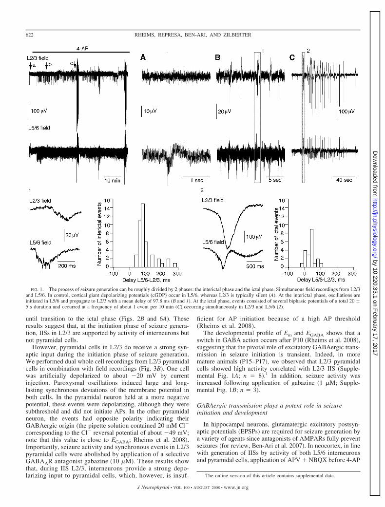
About 3 min after application of 4-AP (50 �M), we ob-served an increase in frequency of cortical giant depolarizingpotentials (cGDP) occurrence in L5/6 (Fig. 1): 3.7 � 0.8/minin control and 17.8 � 3.6/min in the presence of 4-AP (n �18). In L2/3, field recordings showed the initial episodes ofparoxysmal activity (IISs) 7 � 3 min following application of4-AP (Fig. 1). IISs in L2/3 occurred at a frequency of 2.6 �0.8/min, always synchronously with those simultaneously re-corded in L5/6 (n � 16). By contrast, some IISs in L5/6 werenot associated with oscillations in L2/3, suggesting that theywere generated in deep layers and propagated to superficialones (Fig. 1, inset b). Analysis of a delay between synchronousL2/3 and L5/6 IIS confirmed this suggestion: L5/6 oscillationsalways preceded those in L2/3, with a mean delay of 97.8 �8.5 ms (Fig. 1). After exposures to 4-AP exceeding 19 � 7min, an additional pattern of IS appeared (Fig. 1). These eventsconsisted of several biphasic potentials (20 � 5-s duration, 1event per 10 min; Fig. 1, inset c). ISs occurred simultaneouslyin all layers. Oscillations in L2/3 preceded sometimes those inL5/6, and the delay between L5/6 and L2/3 ictal-like eventswas 33.2 � 10.2 ms. Therefore the process of seizure gener-ation can be roughly divided by two phases: 1) an initiatingphase characterized by interictal seizures generated in L5/6 andpropagating to L2/3 and 2) an ictal seizure phase manifested byoscillations of the entire neocortical network.
Interneurons trigger seizures in L2/3
To identify the neuronal subtypes preferentially involved inthe IIS pattern generation, we recorded (cell-attached) inter-neuron-pyramidal cell pairs in combination with field record-ings in both L2/3 and L5/6 (Fig. 2). In L5/6, both interneuronsand pyramidal cells showed the AP activity in control associ-ated with bursts of firing corresponding to cGDPs (Fig. 2B).Both cell types were involved in generation of IISs (Fig. 2B,n � 6). In contrast, shortly after application of 4-AP, someL2/3 interneurons showed high activity correlated with L2/3IIS (Figs. 2A and 3A, n � 27), whereas pyramidal cellsremained mostly quiescent (n � 11). They remained quiescent
621SEIZURE INITIATION AND PROPAGATION IN THE NEONATAL NEOCORTEX
J Neurophysiol • VOL 100 • AUGUST 2008 • www.jn.org
by 10.220.33.1 on February 17, 2017
http://jn.physiology.org/D
ownloaded from
until transition to the ictal phase (Figs. 2B and 6A). Theseresults suggest that, at the initiation phase of seizure genera-tion, IISs in L2/3 are supported by activity of interneurons butnot pyramidal cells.
However, pyramidal cells in L2/3 do receive a strong syn-aptic input during the initiation phase of seizure generation.We performed dual whole cell recordings from L2/3 pyramidalcells in combination with field recordings (Fig. 3B). One cellwas artificially depolarized to about �20 mV by currentinjection. Paroxysmal oscillations induced large and long-lasting synchronous deviations of the membrane potential inboth cells. In the pyramidal neuron held at a more negativepotential, these events were depolarizing, although they weresubthreshold and did not initiate APs. In the other pyramidalneuron, the events had opposite polarity indicating theirGABAergic origin (the pipette solution contained 20 mM Cl�
corresponding to the Cl� reversal potential of about �49 mV;note that this value is close to EGABA; Rheims et al. 2008).Importantly, seizure activity and synchronous events in L2/3pyramidal cells were abolished by application of a selectiveGABAAR antagonist gabazine (10 �M). These results showthat, during IIS L2/3, interneurons provide a strong depo-larizing input to pyramidal cells, which, however, is insuf-
ficient for AP initiation because of a high AP threshold(Rheims et al. 2008).
The developmental profile of Em and EGABA shows that aswitch in GABA action occurs after P10 (Rheims et al. 2008),suggesting that the pivotal role of excitatory GABAergic trans-mission in seizure initiation is transient. Indeed, in moremature animals (P15–P17), we observed that L2/3 pyramidalcells showed high activity correlated with L2/3 IIS (Supple-mental Fig. 1A; n � 8).1 In addition, seizure activity wasincreased following application of gabazine (1 �M; Supple-mental Fig. 1B; n � 3).
GABAergic transmission plays a potent role in seizureinitiation and development
In hippocampal neurons, glutamatergic excitatory postsyn-aptic potentials (EPSPs) are required for seizure generation bya variety of agents since antagonists of AMPARs fully preventseizures (for review, Ben-Ari et al. 2007). In neocortex, in linewith generation of IISs by activity of both L5/6 interneuronsand pyramidal cells, application of APV � NBQX before 4-AP
1 The online version of this article contains supplemental data.
FIG. 1. The process of seizure generation can be roughly divided by 2 phases: the interictal phase and the ictal phase. Simultaneous field recordings from L2/3and L5/6. In control, cortical giant depolarizing potentials (cGDP) occur in L5/6, whereas L2/3 is typically silent (A). At the interictal phase, oscillations areinitiated in L5/6 and propagate to L2/3 with a mean delay of 97.8 ms (B and 1). At the ictal phase, events consisted of several biphasic potentials of a total 20 �5 s duration and occurred at a frequency of about 1 event per 10 min (C) occurring simultaneously in L2/3 and L5/6 (2).
622 RHEIMS, REPRESA, BEN-ARI, AND ZILBERTER
J Neurophysiol • VOL 100 • AUGUST 2008 • www.jn.org
by 10.220.33.1 on February 17, 2017
http://jn.physiology.org/D
ownloaded from

prevented seizure induction in all experiments (n � 7). How-ever, neither APV alone nor NBQX blocked seizures after theirinitiation (n � 6). Furthermore, the mixture of these antago-nists terminated seizures in only one of four experiments. Thissuggests that glutamatergic EPSPs provide the excitatory drivefor interneurons required for their synchronization during cor-tical seizures. Afterward, network oscillations may be sup-ported solely by the interneuron activity.
To test this issue further, we enhanced the efficacy ofGABAergic transmission by 5 �M diazepam (DZP), a benzo-diazepine modulator of GABAARs. In five of eight experi-ments, application of 4-AP � DZP on the background ofAPV � NBQX induced spontaneous IISs in L2/3 and synchro-nous depolarizations in whole cell recorded neurons (Fig. 4A).Extracellular stimulation by a single electrical shock (0.2 ms)also induced pronounced field oscillations, which were com-pletely terminated by gabazine (Fig. 4B).
In addition, shifting the action of GABAergic transmissionfrom excitatory to inhibitory may block seizure development.Bumetanide (10 �M, n � 8; Fig. 5A), which reduces [Cl�]iantagonazing NKCC1, reduced seizures in L5/6 and almostcompletely blocked seizures in L2/3. In L5/6, the seizurefrequency was 33.7 � 17.3% of that observed in the presenceof 4-AP alone (P � 0.02). In L2/3, bumetanide completelyblocked seizures in three of four experiments (Fig. 5A). Im-
portantly, bumetanide prevented transition to the ictal phase:4-AP applied on the background of bumetanide failed to induceIS despite the long-lasting (�1 h) applications. IS, however,did occur after wash out of bumetanide (Fig. 5B; n � 2).
We conclude that seizure initiation and seizure propagationfrom deep to superficial layers are supported by the excitatoryGABAergic transmission in the neonatal neocortex.
Interictal to ictal phase transition: the roleof membrane depolarization
Clearly, a seizure development to ictal-like events requiresactivity of the whole neuronal network including the L2/3pyramidal one. Indeed, when seizures developed to the ictal-like mode, both interneurons and pyramidal cells showedpronounced activity synchronous with seizures recorded in alllayers. Even L2/3 pyramidal cells, which were mostly quies-cent during the interictal phase, were highly active at the ictalphase (Fig. 6A) Therefore after transition into ictal-like events,seizures were maintained by activity of pyramidal cells andinterneurons in both deep and superficial layers.
As a possible mechanism of such transition, we suggestedthat the intense synaptic activity during the initial stage ofseizure generation might result in a gradual membrane depo-larization. This would facilitate seizure development by 1) a
FIG. 2. Neuronal activity during seizureinitiation in L2/3 and L5/6. A: simultaneouscell-attached recordings from the L2/3 pyra-midal cell and interneuron in combinationwith field recordings in L2/3. B: simulta-neous cell-attached recordings from L5/6pyramidal cells in combination with fieldrecordings in L5/6.
623SEIZURE INITIATION AND PROPAGATION IN THE NEONATAL NEOCORTEX
J Neurophysiol • VOL 100 • AUGUST 2008 • www.jn.org
by 10.220.33.1 on February 17, 2017
http://jn.physiology.org/D
ownloaded from

decrease in PSP size required for activation of pyramidal cellsand 2) a partial relief of NMDA receptor (NMDAR) channelsfrom Mg2� block. To test this hypothesis, we noninvasivelymeasured Em (using NMDA channel recordings) in the same
L2/3 pyramidal neurons in control, after appearance of IIS andafter observation of the first IS (Fig. 6A). Seizures weremonitored by field recordings to avoid instabilities of themembrane potential during NMDAR channel measurements.
FIG. 3. Synaptic input to L2/3 pyramidalcells during interictal seizures is mostlyGABAergic. A: simultaneous cell-attachedrecordings from the L2/3 pyramidal cell andinterneuron in combination with field re-cordings in L2/3. B: simultaneous whole cellrecordings from 2 L2/3 pyramids in combi-nation with field recordings. Pyramid 1 wasartificially depolarized by current injection.Note the opposite direction of potential de-viations in pyramids synchronous with fieldseizure events. In pyramid 1, hyperpolariz-ing potential deviations indicate theirGABAergic origin (the pipette solution con-tained 20 mM Cl� corresponding to the Cl�
reversal potential of about �49 mV; notethat this value is close to EGABA, see accom-panying paper).
FIG. 4. GABAergic transmission is able toinitiate seizures at L2/3. A: blockade of theglutamatergic transmission by 4 �M NBQX�40 �M APV before application of 4-amino-pyridine (4-AP) prevented seizure initiation(left). However, enhancement of efficacy ofthe GABAergic transmission by 5 �M diaze-pam resulted in the initiation of spontaneousseizures (right). B: under similar conditions asin A, single stimulations (0.2 ms) via the ex-tracellular electrode positioned in L1 inducedstrong oscillations (left), which were preventedby 10 �M gabazine (right).
624 RHEIMS, REPRESA, BEN-ARI, AND ZILBERTER
J Neurophysiol • VOL 100 • AUGUST 2008 • www.jn.org
by 10.220.33.1 on February 17, 2017
http://jn.physiology.org/D
ownloaded from

In all eight neurons tested, Em was significantly depolarizedduring the initial phase of seizure development (�85.6 � 4.1mV in control, and �78.1 � 3 mV after seizure induction, P �0.01; paired t-test) and depolarized further at the ictal phase(�69.9 � 3 mV, P � 0.01). Similarly, we observed that Em ofL5/6 pyramidal cells was significantly depolarized at the ISphase (�77.7 � 2.4 mV in control and –66.8 � 2.05 mV at theictal phase, n � 9, P � 0.03; paired t-test).
To delineate the contribution of synaptic activity into thegradual membrane depolarization, we blocked synaptic trans-mission by a mixture of antagonists (APV � NBQX �picrotoxin) after initiation of IISs. Figure 6C shows that pyra-midal cells were depolarized at the initial phase of seizuredevelopment (�82.3 � 0.8 mV in control, and �74 � 2 mVduring seizures, n � 5, P � 0.01, paired t-test). However, thisdepolarization was completely abolished by application ofantagonists (�86.5 � 1 mV). These results verify also that thecontinuous presence of 4-AP per se does not promote themembrane depolarization.
Bumetanide reduced seizure activity and abolished the per-sistent neuronal depolarization (–68.56 � 1.55 mV at ictalphase and –81.38 � 3.40 mV after bumetanide, n � 6; P �0.03, paired t-test). Interestingly, EGABA measured with singleGABA channel recordings was not modified during seizuredevelopment (EGABA � �51.20 � 3.7 mV in control and
–53.82 � 5.72 mV during seizures, n � 7; P � 0.72, pairedt-test, data not shown).
To verify that the network depolarization is independentof the type of convulsive agent, we applied carbachol (50�M), an agonist of cholinergic receptors, and DPCPX (200nM), an A1 adenosine receptor antagonist. Although lessefficient than 4-AP, this mixture generated a pronouncedincrease in network activity (see inset to Fig. 6E, left; 60min after drug application) and a significant depolarizationof pyramidal cells (�84 � 1.4 mV in control and �72.7 �1.4 mV after seizure induction; n � 5; P � 0.01). Moreover,after blockade of synaptic transmission in the same exper-iments by NBQX � APV � PTX, Em recovered its controlvalue (�81.8 � 0.9 mV).
It was unclear, however, whether the membrane depolariza-tion of 10–15 mV may result in a significant change in thepyramidal cell excitability. To address this issue, we performedwhole cell current-clamp recordings from a L2/3 pyramidalcell held at �80 mV while stimulating nerve fibers in L1 (Fig.6F). Single shocks (0.2 ms) induced polysynaptic subthresholdPSPs in the pyramidal cell (Fig. 6F, top). However, when thepyramidal cell was depolarized by �10 mV, similar extracel-lular stimulation evoked reliable firing of the pyramidal cell(Fig. 6F, bottom).
FIG. 5. Seizure initiation and development are related to high [Cl�]i. A: simultaneous field recordings in L2/3 and L5/6. Bumetanide (10 �M) dramaticallyreduced seizure activity in L5/6 and almost blocked them in L2/3. B: when bumetanide was applied before 4-AP, it prevented seizure development from theinterictal to ictal phase, ictal events occurring only after bumetanide wash out.
625SEIZURE INITIATION AND PROPAGATION IN THE NEONATAL NEOCORTEX
J Neurophysiol • VOL 100 • AUGUST 2008 • www.jn.org
by 10.220.33.1 on February 17, 2017
http://jn.physiology.org/D
ownloaded from
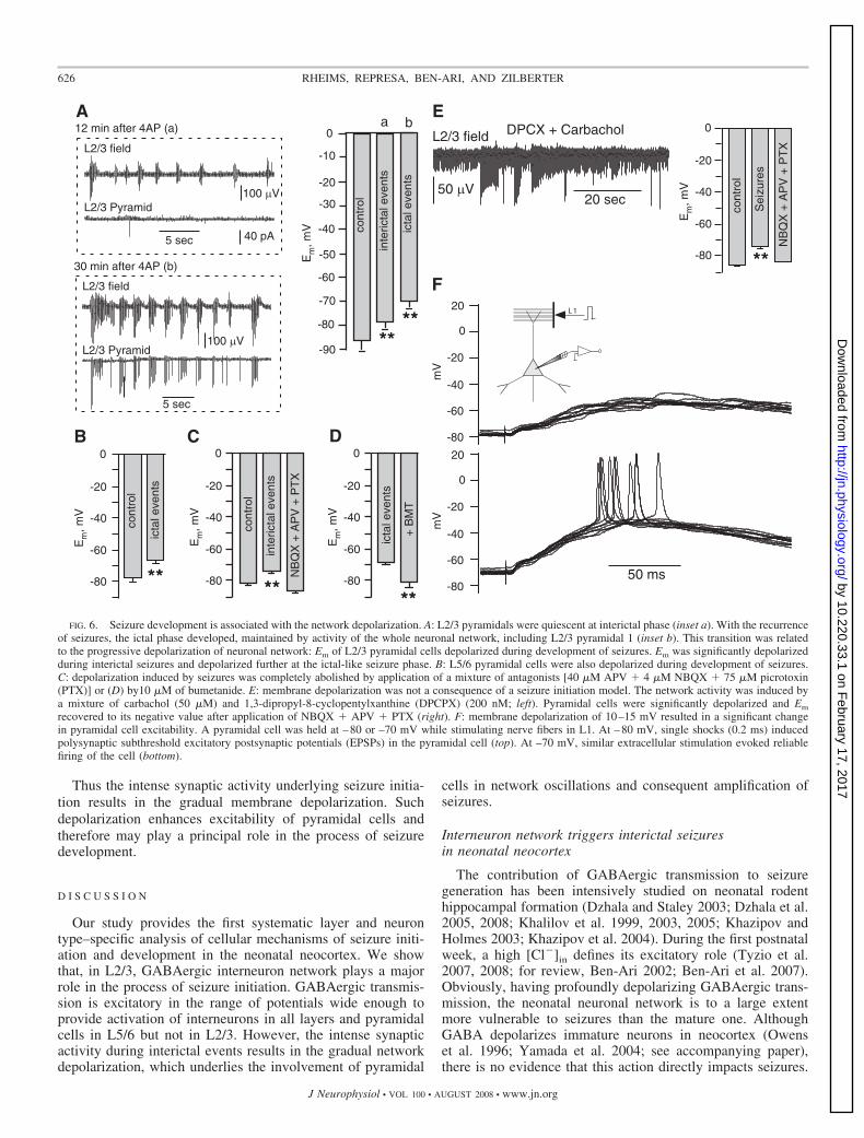
Thus the intense synaptic activity underlying seizure initia-tion results in the gradual membrane depolarization. Suchdepolarization enhances excitability of pyramidal cells andtherefore may play a principal role in the process of seizuredevelopment.
D I S C U S S I O N
Our study provides the first systematic layer and neurontype–specific analysis of cellular mechanisms of seizure initi-ation and development in the neonatal neocortex. We showthat, in L2/3, GABAergic interneuron network plays a majorrole in the process of seizure initiation. GABAergic transmis-sion is excitatory in the range of potentials wide enough toprovide activation of interneurons in all layers and pyramidalcells in L5/6 but not in L2/3. However, the intense synapticactivity during interictal events results in the gradual networkdepolarization, which underlies the involvement of pyramidal
cells in network oscillations and consequent amplification ofseizures.
Interneuron network triggers interictal seizuresin neonatal neocortex
The contribution of GABAergic transmission to seizuregeneration has been intensively studied on neonatal rodenthippocampal formation (Dzhala and Staley 2003; Dzhala et al.2005, 2008; Khalilov et al. 1999, 2003, 2005; Khazipov andHolmes 2003; Khazipov et al. 2004). During the first postnatalweek, a high [Cl�]in defines its excitatory role (Tyzio et al.2007, 2008; for review, Ben-Ari 2002; Ben-Ari et al. 2007).Obviously, having profoundly depolarizing GABAergic trans-mission, the neonatal neuronal network is to a large extentmore vulnerable to seizures than the mature one. AlthoughGABA depolarizes immature neurons in neocortex (Owenset al. 1996; Yamada et al. 2004; see accompanying paper),there is no evidence that this action directly impacts seizures.
-80
-60
-40
-20
0
-10
-50
-70
-30
-90
Em
,mV
********
cont
rol
inte
ricta
lev e
nts
icta
leve
nts
A
L2/3 Pyramid
L2/3 field
5 sec
30 min after 4AP (b)
100 µV
5 sec
100 µV
40 pA
L2/3 field
L2/3 Pyramid
12 min after 4AP (a) a b
****
cont
rol
inte
ricta
leve
nts
NB
QX
+A
PV
+P
TX
-80
-60
-40
-20
0
Em
,mV
B
****-80
-60
-40
-20
0
Em
,mV
cont
rol
Sei
zure
s
NB
QX
+A
PV
+P
TX
DPCX + Carbachol
50 µV20 sec
L2/3 field
-80
-60
-40
-20
0
20
mV
L1
50 ms-80
-60
-40
-20
0
20
mV
F
E
i cta
lev e
nts
cont
rol
****-80
-60
-40
-20
0
Em
,mV
+B
MT
ict a
l eve
nts
-80
-60
-40
-20
0
Em
,mV
****
C D
FIG. 6. Seizure development is associated with the network depolarization. A: L2/3 pyramidals were quiescent at interictal phase (inset a). With the recurrenceof seizures, the ictal phase developed, maintained by activity of the whole neuronal network, including L2/3 pyramidal 1 (inset b). This transition was relatedto the progressive depolarization of neuronal network: Em of L2/3 pyramidal cells depolarized during development of seizures. Em was significantly depolarizedduring interictal seizures and depolarized further at the ictal-like seizure phase. B: L5/6 pyramidal cells were also depolarized during development of seizures.C: depolarization induced by seizures was completely abolished by application of a mixture of antagonists [40 �M APV � 4 �M NBQX � 75 �M picrotoxin(PTX)] or (D) by10 �M of bumetanide. E: membrane depolarization was not a consequence of a seizure initiation model. The network activity was induced bya mixture of carbachol (50 �M) and 1,3-dipropyl-8-cyclopentylxanthine (DPCPX) (200 nM; left). Pyramidal cells were significantly depolarized and Em
recovered to its negative value after application of NBQX � APV � PTX (right). F: membrane depolarization of 10–15 mV resulted in a significant changein pyramidal cell excitability. A pyramidal cell was held at –80 or –70 mV while stimulating nerve fibers in L1. At –80 mV, single shocks (0.2 ms) inducedpolysynaptic subthreshold excitatory postsynaptic potentials (EPSPs) in the pyramidal cell (top). At –70 mV, similar extracellular stimulation evoked reliablefiring of the cell (bottom).
626 RHEIMS, REPRESA, BEN-ARI, AND ZILBERTER
J Neurophysiol • VOL 100 • AUGUST 2008 • www.jn.org
by 10.220.33.1 on February 17, 2017
http://jn.physiology.org/D
ownloaded from

Our results show that GABAergic interneuron network playsa profound role in the process of seizure initiation. The initi-ating phase of seizures is characterized by IISs generated inL5/6 and supported by activation of both L5/6 interneurons andpyramidal cells. IISs propagate to L2/3 via activation of L2/3interneurons but not pyramidal cells, which are mostly quies-cent at this phase. In L2/3, the interneuron network (at leastsome fraction of it) is much more excitable normally than thepyramidal one. For instance, APs can be readily initiated in theintact L2/3 interneurons by a pure GABAergic transmissionthat is not the case for L2/3 pyramidal cells (Rheims et al.2008). The likely reason for this is that, although Em is quitesimilar in L2/3 interneurons and pyramidal cells, the latter havea much more positive AP activation threshold (Rheims et al.2008). Therefore at an early stage, L2/3 IIS consist mostly ofsynchronous firing of interneurons. This firing is reflected inpyramidal cells as strong depolarizations provided by GABAergictransmission (see Fig. 3).
Clearly, glutamatergic transmission is important for theinterneuron excitation since IISs have not been induced by4-AP under blockade of glutamate-activated receptors. How-ever, being induced, IISs are not as sensitive to glutamatereceptors antagonists. This suggests that glutamatergic trans-mission provides interneurons the fraction of initial excitationnecessary to trigger seizures. Because of their lower AP acti-vation threshold (Rheims et al. 2008), L5/6 pyramidal cells aremore excitable than L2/3 ones and are involved in the processof seizure initiation. Therefore they provide the excitatoryinput required to activate the interneuron network. In contrast,it is unlikely that L2/3 pyramidal cells contribute significantlyto this early drive since they are mostly quiescent.
The central role of the interneuron network suggests thatshifting the action of GABAergic transmission from excitatoryto inhibitory may block seizure development. It has beenshown that the NKCC1 blocker bumetanide, which decreases[Cl�]in in immature neurons, blocks hippocampal seizures(Dzhala et al. 2005, 2008) and may thus act in synergy withGABAARs modulators that are recommended as the first linetreatment of neonatal seizures (Bartha et al. 2007; Dzhala et al.2008; Wheless et al. 2005, 2007). As observed in hippocam-pus, bumetanide dramatically reduces neocortical seizure ac-tivity and prevents development from the interictal to ictalphase (see Fig. 5).
Neuronal depolarization underlies transition of networkoscillations to ictal seizures
Seizure development is manifested by transition from inter-ictal to ictal seizure events. This transition is accompanied byinvolvement of L2/3 pyramidal cells into network oscillations(Figs. 2A and 6A). We show that one reason for the activationof L2/3 pyramidal cell firing is depolarization of the wholeneuronal network. The amount of this depolarization correlateswith the time-dependent intensification of seizure events, de-pends on the level of synaptic activity, and is abolished byblockade of synaptic transmission. Importantly, the networkdepolarization that is close to steady state persists between seizureevents and differs from the transient depolarization induced byenhancement of the extracellular K� concentration during sei-zures (Avoli et al. 1996; Hablitz and Heinemann 1987; Luckeet al. 1995; Stringer 1998). Indeed, the clearance of extracellular
K� is only a few seconds longer in the immature brain than in theolder one despite an altered regulation of extracellular K� (Con-nors and Ransom 1984; Stringer 1998). Furthermore, the neuro-nal depolarization was not related to the direct action of 4-APon Em since it was either completely abolished by blockade ofsynaptic transmission or observed with another experimentalmodel (Fig. 6, C and E). The mechanism of network depolar-ization is yet unknown. It is not associated with the intracel-lular accumulation of Cl� ions. It is possible, however, that theintensive synaptic activity during seizures results in an increasein ambient GABA concentration and a consequent membranedepolarization.
We conclude that the increasing involvement of glutamater-gic transmission into network oscillations, powered by neuro-nal depolarization, provides a positive feedback highly ampli-fying seizure generation.
Functional implications
Our results show that, in the neonatal neocortex, seizures areinitiated in deep layers and thereafter propagate to L2/3 wherethey are triggered by synchronization of GABAergic interneu-rons. Although the precise mechanism of such synchronizationremains unclear, we suggest that it may be promoted byspontaneous episodes of hyperactivity (sensory, motor, etc.) atA functionally normal neocortical network. Moreover, whereasthe link between interictal events and epileptogenesis remainsa matter of debate (Avoli et al. 2006; de Curtis and Avanzini2001; Staley and Dudek 2006), our data suggest that IISS andneuronal network depolarization may play a pivotal role insubsequent seizure development and epileptogenesis. Althoughthese conclusions are made for a limited developmental stage(P6–P9), we suggest that the interneuron network may repre-sent a key target for pharmacological interventions aiming toprevent initiation and development of neocortical seizures.However, while developing and administrating such medica-tions, rigorous precautions should be made, given that drugsaffecting GABAergic transmission may be beneficial for theneocortical network but not necessarily for the hippocampalone (see Supplemental Fig. 2).
A C K N O W L E D G M E N T S
We thank I. Jorquera for technical assistance in histological processing.
G R A N T S
This work was supported by Institut National de la Sante et de la RechercheMedicale and grants from Fondation pour la Recherche Medicale, AgenceNationale de la Recherche 2005 Neurosciences, neurologie et psychiatrie andthe European Council Contract LSH-CT-2006-037315 (EPICURE) FP6-The-matic priority LIFESCIHEALTH.
R E F E R E N C E S
Avoli M, Biagini G, de Curtis M. Do interictal spikes sustain seizures andepileptogenesis? Epilepsy Curr 6: 203–207, 2006.
Avoli M, Louvel J, Kurcewicz I, Pumain R, Barbarosie M. Extracellularfree potassium and calcium during synchronous activity induced by 4-aminopyridine in the juvenile rat hippocampus. J Physiol 493: 707–717,1996.
Bartha AI, Shen J, Katz KH, Mischel RE, Yap KR, Ivacko JA, AndrewsEM, Ferriero DM, Ment LR, Silverstein FS. Neonatal seizures: multi-center variability in current treatment practices. Pediatr Neurol 37: 85–90,2007.
Ben-Ari Y. Excitatory actions of gaba during development: the nature of thenurture. Nat Rev Neurosci 3: 728–739, 2002.
627SEIZURE INITIATION AND PROPAGATION IN THE NEONATAL NEOCORTEX
J Neurophysiol • VOL 100 • AUGUST 2008 • www.jn.org
by 10.220.33.1 on February 17, 2017
http://jn.physiology.org/D
ownloaded from

Ben-Ari Y, Gaiarsa JL, Tyzio R, Khazipov R. GABA: a pioneer transmitterthat excites immature neurons and generates primitive oscillations. PhysiolRev 87: 1215–1284, 2007.
Ben-Ari Y, Khalilov I, Represa A, Gozlan H. Interneurons set the tune ofdeveloping networks. Trends Neurosci 27: 422–427, 2004.
Bye AM, Cunningham CA, Chee KY, Flanagan D. Outcome of neonateswith electrographically identified seizures, or at risk of seizures. PediatrNeurol 16: 225–231, 1997.
Connors BW, Ransom BR. Chloride conductance and extracellular potassiumconcentration interact to modify the excitability of rat optic nerve fibres.J Physiol 355: 619–633, 1984.
Cossart R, Bernard C, Ben-Ari Y. Multiple facets of GABAergic neuronsand synapses: multiple fates of GABA signalling in epilepsies. TrendsNeurosci 28: 108–115, 2005.
de Curtis M, Avanzini G. Interictal spikes in focal epileptogenesis. ProgNeurobiol 63: 541–567, 2001.
Dixon G, Badawi N, Kurinczuk JJ, Keogh JM, Silburn SR, Zubrick SR,Stanley FJ. Early developmental outcomes after newborn encephalopathy.Pediatrics 109: 26–33, 2002.
Dzhala VI, Brumback AC, Staley KJ. Bumetanide enhances phenobarbitalefficacy in a neonatal seizure model. Ann Neurol 63: 222–235, 2008.
Dzhala VI, Staley KJ. Excitatory actions of endogenously released GABAcontribute to initiation of ictal epileptiform activity in the developinghippocampus. J Neurosci 23: 1840–1846, 2003.
Dzhala VI, Talos DM, Sdrulla DA, Brumback AC, Mathews GC, BenkeTA, Delpire E, Jensen FE, Staley KJ. NKCC1 transporter facilitatesseizures in the developing brain. Nat Med 11: 1205–1213, 2005.
Hablitz JJ, Heinemann U. Extracellular K� and Ca2� changes duringepileptiform discharges in the immature rat neocortex. Brain Res 433:299–303, 1987.
Khalilov I, Dzhala VI, Ben-Ari Y, Khazipov R. Dual role of GABA in theneonatal rat hippocampus. Dev Neurosci 21: 310–319, 1999.
Khalilov I, Holmes GL, Ben-Ari Y. In vitro formation of a secondaryepileptogenic mirror focus by interhippocampal propagation of seizures. NatNeurosci 6: 1079–1085, 2003.
Khalilov I, Le Van Quyen M, Gozlan H, Ben-Ari Y. Epileptogenic actionsof GABA and fast oscillations in the developing hippocampus. Neuron 48:787–796, 2005.
Khazipov R, Holmes GL. Synchronization of kainate-induced epileptic ac-tivity via GABAergic inhibition in the superfused rat hippocampus in vivo.J Neurosci 23: 5337–5341, 2003.
Khazipov R, Khalilov I, Tyzio R, Morozova E, Ben-Ari Y, Holmes GL.Developmental changes in GABAergic actions and seizure susceptibility inthe rat hippocampus. Eur J Neurosci 19: 590–600, 2004.
Le Van Quyen M, Khalilov I, Ben-Ari Y. The dark side of high-frequencyoscillations in the developing brain. Trends Neurosci 29: 419 – 427,2006.
Lienekugel X, Khalilov I, McLean HA, Caillard O, Gaiarsa J-L, Ben-AriY, Khazipov R. GABA is the principal fast-acting excitatory transmitter inthe neonatal brain. Adv Neurol 79: 189–201, 1999.
Lienekugel X, Medina I, Khalilov I, Ben-Ari Y, Khazipov R. Abstract Ca2�
oscillations mediated by the synergistic excitatory actions of GABA(A) andNMDA receptors in the neonatal hippocampus. Neuron 18: 243–255, 1997.
Lombroso CT. Neonatal seizures: a clinician’s overview. Brain Dev 18: 1–28,1996.
Lucke A, Nagao T, Kohling R, Avoli M. Synchronous potentials andelevations in [K�]o in the adult rat entorhinal cortex maintained in vitro.Neurosci Lett 185: 155–158, 1995.
Mizrahi EM. Acute and chronic effects of seizures in the developing brain:lessons from clinical experience. Epilepsia 40: S42–S50, 1999.
Mizrahi EM, Clancy RR. Neonatal seizures: early-onset seizure syndromesand their consequences for development. Ment Retard Dev Disabil Res Rev6: 229–241, 2000.
Owens DF, Boyce LH, Davies CH, Kriegstein AR. Excitatory GABAresponses in embryonic and neonatal cortical slices demonstrated by gram-icidin perforated-patch recordings and calcium imaging. J Neurosci 16:6414–6423, 1996.
Rennie JM, Boylan GB. Neonatal seizures and their treatment. Curr OpinNeurol 16: 177–181, 2003.
Rheims S, Minlebaev M, Ivanov A, Represa A, Khazipov R, Holmes GL,Ben-Ari Y, Zilberter Y. Excitatory GABA in rodent developing neocortexin vitro. J Neurophysiol doi:10.1152/jn.90402.2008.
Scher MS, Aso K, Beggarly ME, Hamid MY, Steppe DA, Painter MJ.Electrographic seizures in preterm and full-term neonates: clinical corre-lates, associated brain lesions, and risk for neurologic sequelae. Pediatrics91: 128–134, 1993.
Staley KJ, Dudek FE. Interictal spikes and epileptogenesis. Epilepsy Curr 6:199–202, 2006.
Stringer JL. Regulation of extracellular potassium in the developing hip-pocampus. Brain Res Dev Brain Res 110: 97–103, 1998.
Tekgul H, Gauvreau K, Soul J, Murphy L, Robertson R, Stewart J, VolpeJ, Bourgeois B, du Plessis AJ. The current etiologic profile and neurode-velopmental outcome of seizures in term newborn infants. Pediatrics 117:1270–1280, 2006.
Tharp BR. Neonatal seizures and syndromes. Epilepsia 43: 2–10, 2002.Tyzio R, Cossart R, Khalilov I, Minlebaev M, Hubner CA, Represa A,
Ben-Ari Y, Khazipov R. Maternal oxytocin triggers a transient inhibitoryswitch in GABA signaling in the fetal brain during delivery. Science 314:1788–1792, 2006.
Tyzio R, Holmes GL, Ben-Ari Y, Khazipov R. Timing of the developmentalswitch in GABA(A) mediated signalling from excitation to inhibition inCA3 rat hippocampus. Epilepsia 48: 96–105, 2007.
Tyzio R, Holmes GL, Ben-Ari Y, Khazipov R. Timing of the developmentalswitch in GABA(A) mediated signaling from excitation to inhibition in CA3rat hippocampus using gramicidin perforated patch and extracellular record-ings. Epilepsia 5: 96–105, 2007.
Tyzio R, Ivanov A, Bernard C, Holmes GL, Ben-Ari Y, Khazipov R.Membrane potential of CA3 hippocampal pyramidal cells during postnataldevelopment. J Neurophysiol 90: 2964–2972, 2003.
Wheless JW, Clarke DF, Arzimanoglou A, Carpenter D. Treatment ofpediatric epilepsy: European expert opinion, 2007. Epileptic Disord 9:353–412, 2007.
Wheless JW, Clarke DF, Carpenter D. Treatment of pediatric epilepsy:expert opinion, 2005. J Child Neurol 1: S1–S56, 2005.
Yamada KA, Okabe A, Toyoda H, Kilb W, Luhmann HJ, Fukuda A. CI-uptake promoting depolarizing GABA actions in immature rat neocorticalneurones is mediated by NKCC1. J Physiol 557: 829–841, 2004.
628 RHEIMS, REPRESA, BEN-ARI, AND ZILBERTER
J Neurophysiol • VOL 100 • AUGUST 2008 • www.jn.org
by 10.220.33.1 on February 17, 2017
http://jn.physiology.org/D
ownloaded from