Embed Size (px)
Citation preview
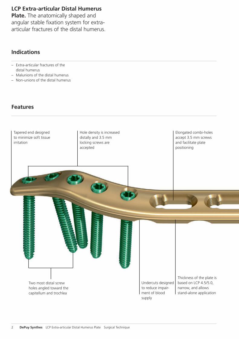
LCP Extra-articular Distal Humerus Plate. The anatomically shaped and angular stable fixation system for extra-articular fractures of the distal humerus.
Surgical Technique
This publication is not intended for distribution in the USA.
Instruments and implants approved by the AO Foundation.

Image intensifier control
WarningThis description alone does not provide sufficient background for direct use of DePuy Synthes products. Instruction by a surgeon experienced in handling these products is highly recommended.
Processing, Reprocessing, Care and MaintenanceFor general guidelines, function control and dismantling of multi-part instruments, as well as processing guidelines for implants, please contact your local sales representative or refer to:http://emea.depuysynthes.com/hcp/reprocessing-care-maintenanceFor general information about reprocessing, care and maintenance of Synthes reusable devices, instrument trays and cases, as well as processing of Synthes non-sterile implants, please consult the Important Information leaflet (SE_023827) or refer to: http://emea.depuysynthes.com/hcp/reprocessing-care-maintenance
LCP Extra-articular Distal Humerus Plate Surgical Technique DePuy Synthes 1
Table of Contents
Introduction
Surgical Technique
Product Information
Bibliography 19
MRI Information 20
LCP Extra-articular Distal Humerus Plate 2
AO Principles 4
Indications 5
Preparation and Approach 6
Surgical Technique 8
Implant Removal 16
Implants 17
Instruments 18
2 DePuy Synthes LCP Extra-articular Distal Humerus Plate Surgical Technique
Undercuts designed to reduce impair-ment of blood supply
Elongated combi-holes accept 3.5 mm screws and facilitate plate positioning
Tapered end designed to minimize soft tissue irritation
Thickness of the plate is based on LCP 4.5/5.0, narrow, and allows stand-alone application
Hole density is increased distally and 3.5 mm locking screws are accepted
Two most distal screw holes angled toward the capitellum and trochlea
LCP Extra-articular Distal Humerus Plate. The anatomically shaped and angular stable fixation system for extra-articular fractures of the distal humerus.
– Extra-articular fractures of the distal humerus
– Malunions of the distal humerus – Non-unions of the distal humerus
Features
Indications
LCP Extra-articular Distal Humerus Plate Surgical Technique DePuy Synthes 3
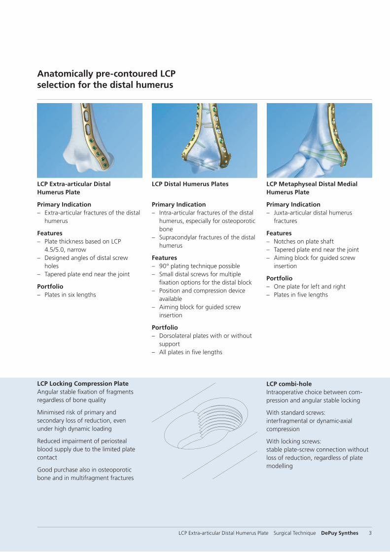
Anatomically pre-contoured LCP selection for the distal humerus
LCP combi-holeIntraoperative choice between com-pression and angular stable locking
With standard screws:interfragmental or dynamic-axial compression
With locking screws: stable plate-screw connection without loss of reduction, regardless of plate modelling
LCP Locking Compression Plate Angular stable fixation of fragments regardless of bone quality
Minimised risk of primary and secondary loss of reduction, even under high dynamic loading
Reduced impairment of periosteal blood supply due to the limited plate contact
Good purchase also in osteoporotic bone and in multifragment fractures
LCP Extra-articular Distal Humerus Plate
Primary Indication – Extra-articular fractures of the distal
humerus
Features – Plate thickness based on LCP
4.5/5.0, narrow – Designed angles of distal screw
holes – Tapered plate end near the joint
Portfolio – Plates in six lengths
LCP Distal Humerus Plates
Primary Indication – Intra-articular fractures of the distal
humerus, especially for osteoporotic bone
– Supracondylar fractures of the distal humerus
Features – 90° plating technique possible – Small distal screws for multiple
fixation options for the distal block – Position and compression device
available – Aiming block for guided screw
insertion
Portfolio – Dorsolateral plates with or without
support – All plates in five lengths
LCP Metaphyseal Distal Medial Humerus Plate
Primary Indication – Juxta-articular distal humerus
fractures
Features – Notches on plate shaft – Tapered plate end near the joint – Aiming block for guided screw
insertion
Portfolio – One plate for left and right – Plates in five lengths
1
4
2
3
4_Priciples_03.pdf 1 05.07.12 12:08
4 DePuy Synthes Expert Lateral Femoral Nail Surgical Technique
AO PRINCIPLES
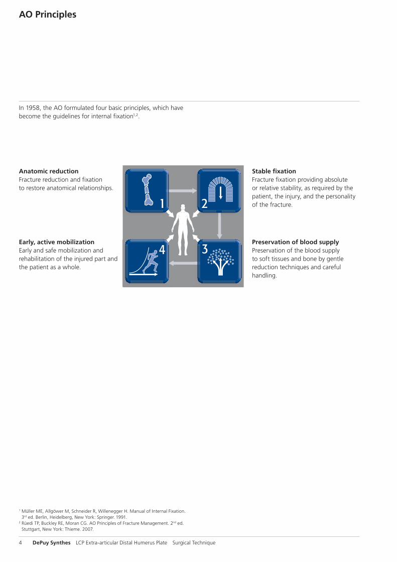
In 1958, the AO formulated four basic principles, which have become the guidelines for internal fixation1, 2.
1 Müller ME, M Allgöwer, R Schneider, H Willenegger. Manual of Internal Fixation. 3rd ed. Berlin Heidelberg New York: Springer. 1991.
2 Rüedi TP, RE Buckley, CG Moran. AO Principles of Fracture Management. 2nd ed. Stuttgart, New York: Thieme. 2007.
Anatomic reductionFracture reduction and fixation to restore anatomical relationships.
Early, active mobilizationEarly and safe mobilization and rehabilitation of the injured part and the patient as a whole.
Stable fixationFracture fixation providing abso-lute or relative stability, as required by the patient, the injury, and the personality of the fracture.
Preservation of blood supplyPreservation of the blood supply to soft tissues and bone by gentle reduction techniques and careful handling.
4 DePuy Synthes LCP Extra-articular Distal Humerus Plate Surgical Technique
AO Principles
1 Müller ME, Allgöwer M, Schneider R, Willenegger H. Manual of Internal Fixation. 3rd ed. Berlin, Heidelberg, New York: Springer. 1991.
2 Rüedi TP, Buckley RE, Moran CG. AO Principles of Fracture Management. 2nd ed. Stuttgart, New York: Thieme. 2007.
Stable fixationFracture fixation providing absolute or relative stability, as required by the patient, the injury, and the personality of the fracture.
Anatomic reductionFracture reduction and fixation to restore anatomical relationships.
Early, active mobilizationEarly and safe mobilization and rehabilitation of the injured part and the patient as a whole.
Preservation of blood supplyPreservation of the blood supply to soft tissues and bone by gentle reduction techniques and careful handling.
In 1958, the AO formulated four basic principles, which have become the guidelines for internal fixation1,2.

LCP Extra-articular Distal Humerus Plate Surgical Technique DePuy Synthes 5
Indications
– Extra-articular fractures of the distal humerus – Malunions of the distal humerus – Non-unions of the distal humerus
LCP Extra-articular Distal Humerus Plate 3.5
0 10 20 30 40 50 60 70 80 90 100mm
For use only with the Original AO System ofInstruments and Implants
Synthes GmbHEimattstrasse 3CH-4436 Oberdorfwww.synthes.com
1.10 Magnification
Right LeftTitanium St. Steel Holes Length (mm) Titanium St. Steel Holes Length (mm)04.104.004 02.104.004 4 122 04.104.024 02.104.024 4 12204.104.006 02.104.006 6 158 04.104.026 02.104.026 6 15804.104.008 02.104.008 8 194 04.104.028 02.104.028 8 19404.104.010 02.104.010 10 230 04.104.030 02.104.030 10 23004.104.012 02.104.012 12 266 04.104.032 02.104.032 12 26604.104.014 02.104.014 14 302 04.104.034 02.104.034 14 302
Rig
ht
Left
4 holes6 holes8 holes10 holes12 holes14 holes
034.
000.
552
3009
0003
©
02/
2009
Syn
thes
, In
c. o
r it
s af
filia
tes
All
rig
hts
res
erve
d
Syn
thes
is a
tra
dem
ark
of
Syn
thes
, In
c. o
r it
s af
filia
tes
20
40
20
40
Ö034.000.552Cä
6 DePuy Synthes LCP Extra-articular Distal Humerus Plate Surgical Technique
2Position patient
Positioning is by surgeon preference. However, the lateral decubitus position is frequently chosen. The arm is rested on a padded bar allowing elbow flexion of 120°.
Preparation and Approach
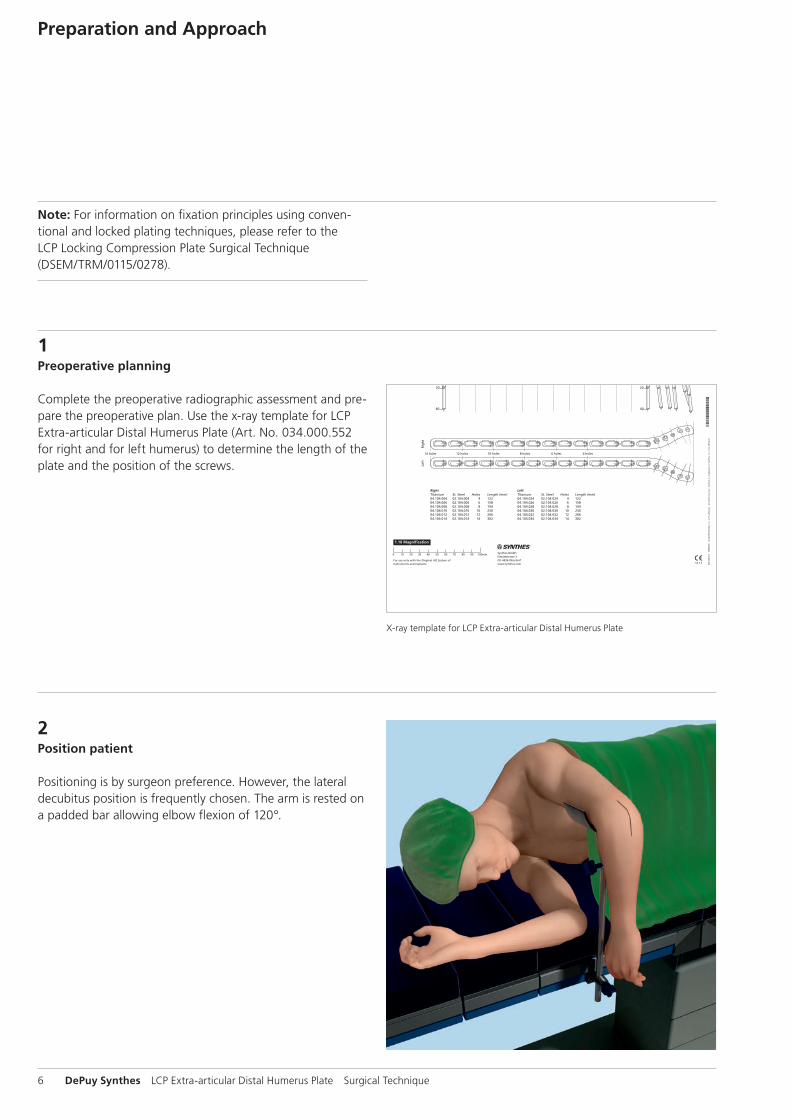
1Preoperative planning
Complete the preoperative radiographic assessment and pre-pare the preoperative plan. Use the x-ray template for LCP Extra-articular Distal Humerus Plate (Art. No. 034.000.552 for right and for left humerus) to determine the length of the plate and the position of the screws.
Note: For information on fixation principles using conventional and locked plating techniques, please refer to the LCP Locking Compression Plate Surgical Technique (DSEM/TRM/0115/0278).
X-ray template for LCP Extra-articular Distal Humerus Plate
LCP Extra-articular Distal Humerus Plate Surgical Technique DePuy Synthes 7
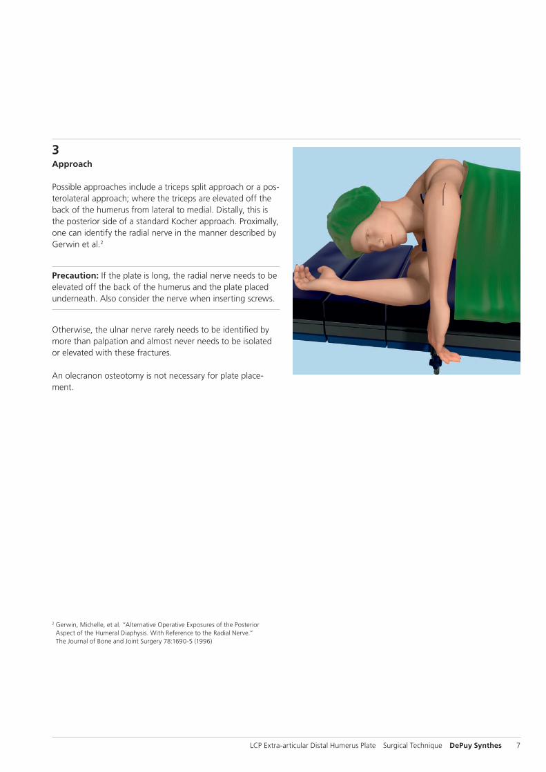
3Approach
Possible approaches include a triceps split approach or a pos-terolateral approach; where the triceps are elevated off the back of the humerus from lateral to medial. Distally, this is the posterior side of a standard Kocher approach. Proximally, one can identify the radial nerve in the manner described by Gerwin et al.2
Precaution: If the plate is long, the radial nerve needs to be elevated off the back of the humerus and the plate placed underneath. Also consider the nerve when inserting screws.
Otherwise, the ulnar nerve rarely needs to be identified by more than palpation and almost never needs to be isolated or elevated with these fractures.
An olecranon osteotomy is not necessary for plate place-ment.
2 Gerwin, Michelle, et al. “Alternative Operative Exposures of the Posterior Aspect of the Humeral Diaphysis. With Reference to the Radial Nerve.” The Journal of Bone and Joint Surgery 78:1690-5 (1996)
8 DePuy Synthes LCP Extra-articular Distal Humerus Plate Surgical Technique
2Determine plate length
Choose a plate length that offers sufficient fixation proximal to the fracture.
Surgical Technique
1Reduce fracture and fix temporarily
Use pointed forceps for temporary fixation in restoring the anatomy. Ensure that forceps will not interfere with subse-quent plate placement.
LCP Extra-articular Distal Humerus Plate Surgical Technique DePuy Synthes 9
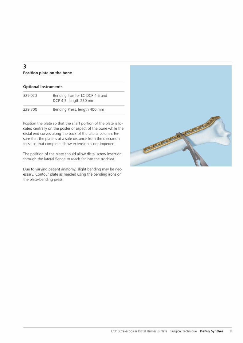
3Position plate on the bone
Optional instruments
329.020 Bending Iron for LC-DCP 4.5 and DCP 4.5, length 250 mm
329.300 Bending Press, length 400 mm
Position the plate so that the shaft portion of the plate is lo-cated centrally on the posterior aspect of the bone while the distal end curves along the back of the lateral column. En-sure that the plate is at a safe distance from the olecranon fossa so that complete elbow extension is not impeded.
The position of the plate should allow distal screw insertion through the lateral flange to reach far into the trochlea.
Due to varying patient anatomy, slight bending may be nec-essary. Contour plate as needed using the bending irons or the plate-bending press.
10 DePuy Synthes LCP Extra-articular Distal Humerus Plate Surgical Technique
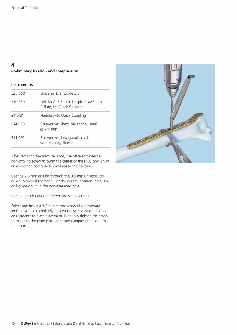
4Preliminary fixation and compression
Instruments
323.360 Universal Drill Guide 3.5
310.250 Drill Bit B 2.5 mm, length 110/85 mm, 2flute, for Quick Coupling
311.431 Handle with Quick Coupling
314.030 Screwdriver Shaft, hexagonal, small, B 2.5 mm
314.020 Screwdriver, hexagonal, small, with Holding Sleeve
After reducing the fracture, apply the plate and insert a non-locking screw through the center of the DCU portion of an elongated combi-hole proximal to the fracture.
Use the 2.5 mm drill bit through the 3.5 mm universal drill guide to predrill the bone. For the neutral position, press the drill guide down in the non-threaded hole.
Use the depth gauge to determine screw length.
Select and insert a 3.5 mm cortex screw of appropriate length. Do not completely tighten the screw. Make any final adjustments to plate placement. Manually tighten the screw to maintain the plate placement and compress the plate to the bone.
Surgical Technique
1
LCP Extra-articular Distal Humerus Plate Surgical Technique DePuy Synthes 11
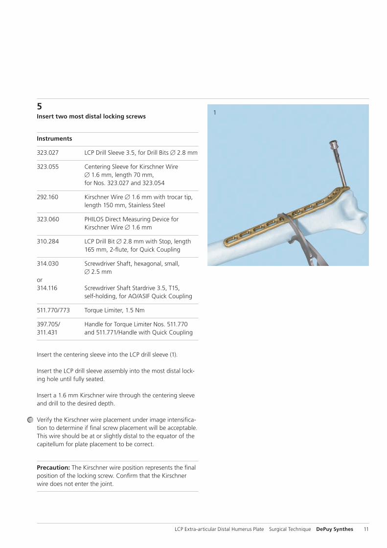
5Insert two most distal locking screws
Instruments
323.027 LCP Drill Sleeve 3.5, for Drill Bits B 2.8 mm
323.055 Centering Sleeve for Kirschner Wire B 1.6 mm, length 70 mm, for Nos. 323.027 and 323.054
292.160 Kirschner Wire B 1.6 mm with trocar tip, length 150 mm, Stainless Steel
323.060 PHILOS Direct Measuring Device for Kirschner Wire B 1.6 mm
310.284 LCP Drill Bit B 2.8 mm with Stop, length 165 mm, 2flute, for Quick Coupling
314.030 Screwdriver Shaft, hexagonal, small, B 2.5 mmor314.116 Screwdriver Shaft Stardrive 3.5, T15,
selfholding, for AO/ASIF Quick Coupling
511.770/773 Torque Limiter, 1.5 Nm
397.705/ Handle for Torque Limiter Nos. 511.770311.431 and 511.771/Handle with Quick Coupling
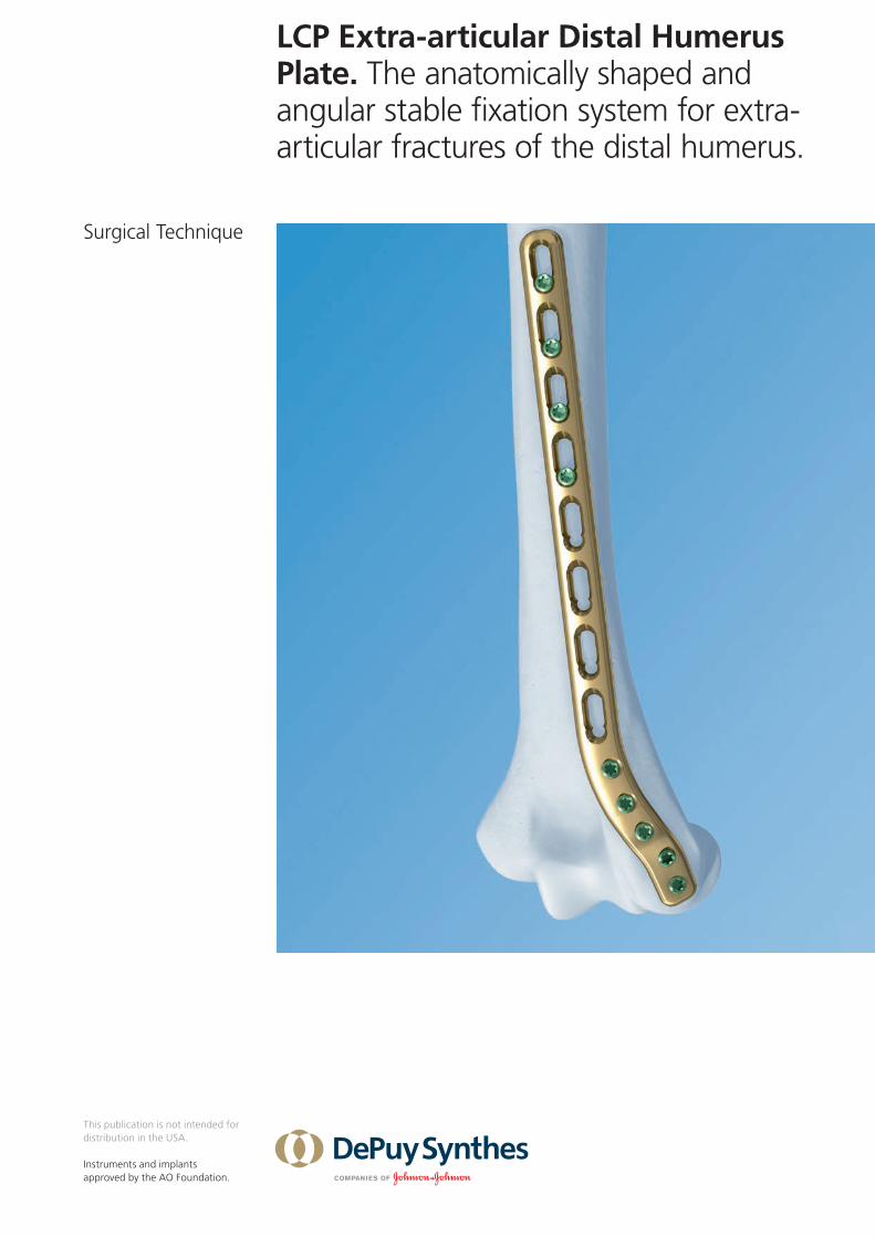
Insert the centering sleeve into the LCP drill sleeve (1).
Insert the LCP drill sleeve assembly into the most distal lock-ing hole until fully seated.
Insert a 1.6 mm Kirschner wire through the centering sleeve and drill to the desired depth.
Verify the Kirschner wire placement under image intensifica-tion to determine if final screw placement will be acceptable. This wire should be at or slightly distal to the equator of the capitellum for plate placement to be correct.
Precaution: The Kirschner wire position represents the final position of the locking screw. Confirm that the Kirschner wire does not enter the joint.
2
3
12 DePuy Synthes LCP Extra-articular Distal Humerus Plate Surgical Technique
Surgical Technique
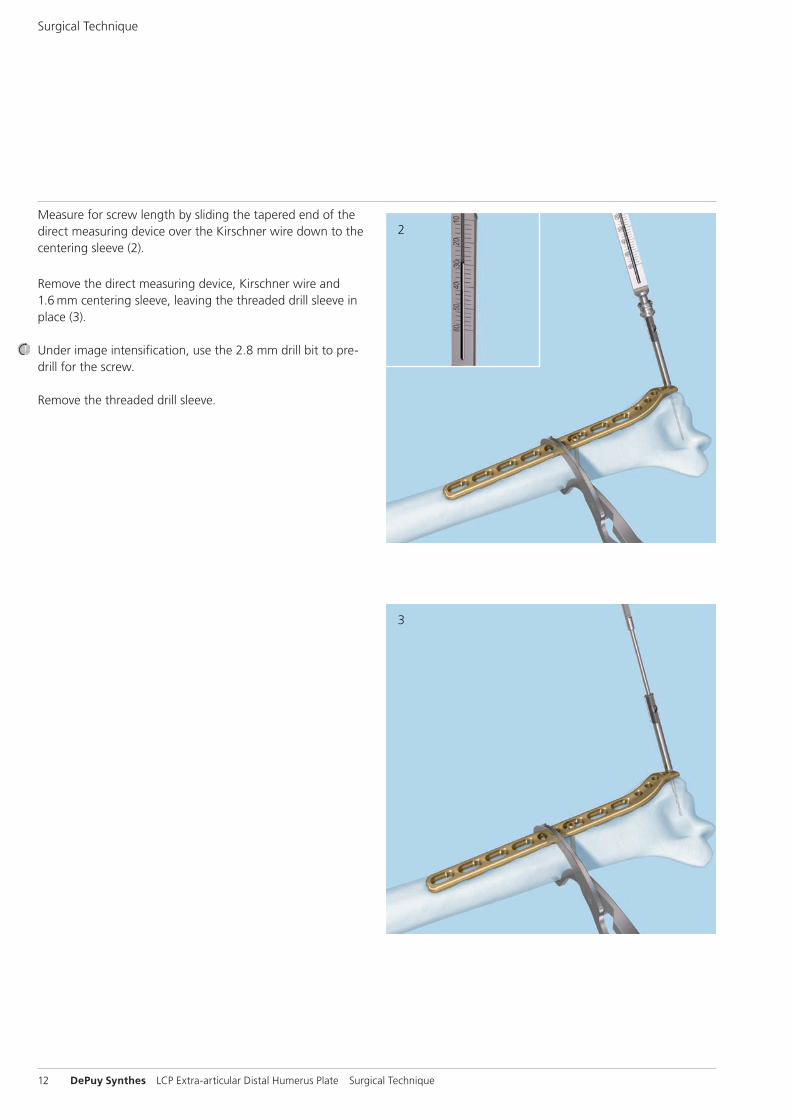
Measure for screw length by sliding the tapered end of the direct measuring device over the Kirschner wire down to the centering sleeve (2).
Remove the direct measuring device, Kirschner wire and 1.6 mm centering sleeve, leaving the threaded drill sleeve in place (3).
Under image intensification, use the 2.8 mm drill bit to pre-drill for the screw.
Remove the threaded drill sleeve.
4
5
LCP Extra-articular Distal Humerus Plate Surgical Technique DePuy Synthes 13
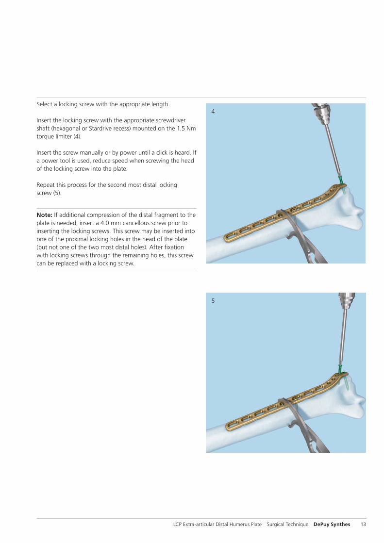
Select a locking screw with the appropriate length.
Insert the locking screw with the appropriate screwdriver shaft (hexagonal or Stardrive recess) mounted on the 1.5 Nm torque limiter (4).
Insert the screw manually or by power until a click is heard. If a power tool is used, reduce speed when screwing the head of the locking screw into the plate.
Repeat this process for the second most distal locking screw (5).
Note: If additional compression of the distal fragment to the plate is needed, insert a 4.0 mm cancellous screw prior to inserting the locking screws. This screw may be inserted into one of the proximal locking holes in the head of the plate (but not one of the two most distal holes). After fixation with locking screws through the remaining holes, this screw can be replaced with a locking screw.
2
1
14 DePuy Synthes LCP Extra-articular Distal Humerus Plate Surgical Technique
Surgical Technique
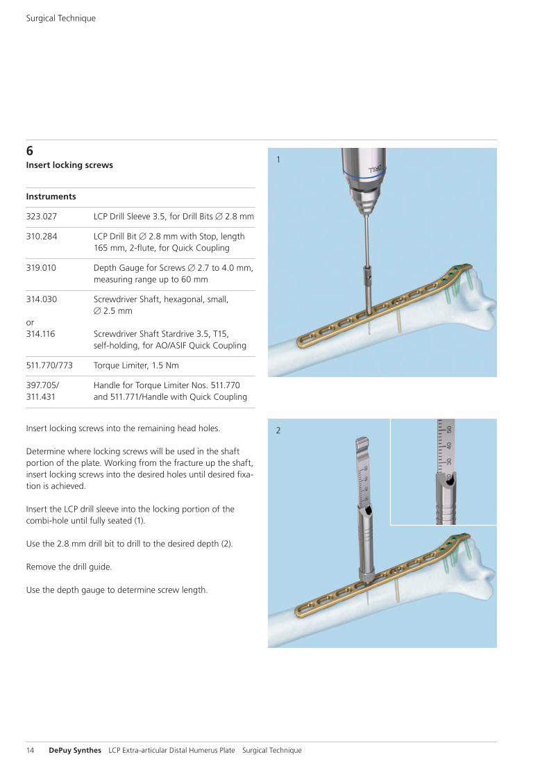
6Insert locking screws
Instruments
323.027 LCP Drill Sleeve 3.5, for Drill Bits B 2.8 mm
310.284 LCP Drill Bit B 2.8 mm with Stop, length 165 mm, 2flute, for Quick Coupling
319.010 Depth Gauge for Screws B 2.7 to 4.0 mm, measuring range up to 60 mm
314.030 Screwdriver Shaft, hexagonal, small, B 2.5 mmor314.116 Screwdriver Shaft Stardrive 3.5, T15,
selfholding, for AO/ASIF Quick Coupling
511.770/773 Torque Limiter, 1.5 Nm
397.705/ Handle for Torque Limiter Nos. 511.770311.431 and 511.771/Handle with Quick Coupling
Insert locking screws into the remaining head holes.
Determine where locking screws will be used in the shaft portion of the plate. Working from the fracture up the shaft, insert locking screws into the desired holes until desired fixa-tion is achieved.
Insert the LCP drill sleeve into the locking portion of the combi-hole until fully seated (1).
Use the 2.8 mm drill bit to drill to the desired depth (2).
Remove the drill guide.
Use the depth gauge to determine screw length.
3
LCP Extra-articular Distal Humerus Plate Surgical Technique DePuy Synthes 15
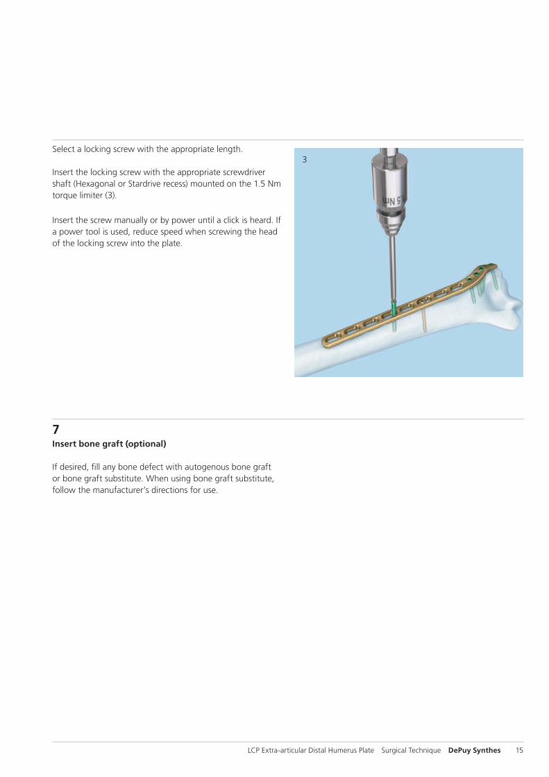
Select a locking screw with the appropriate length.
Insert the locking screw with the appropriate screwdriver shaft (Hexagonal or Stardrive recess) mounted on the 1.5 Nm torque limiter (3).
Insert the screw manually or by power until a click is heard. If a power tool is used, reduce speed when screwing the head of the locking screw into the plate.
7Insert bone graft (optional)
If desired, fill any bone defect with autogenous bone graft or bone graft substitute. When using bone graft substitute, follow the manufacturer’s directions for use.
16 DePuy Synthes LCP Extra-articular Distal Humerus Plate Surgical Technique
Instruments
314.030 Screwdriver Shaft, hexagonal, small, B 2.5 mmor314.116 Screwdriver Shaft Stardrive 3.5, T15,
selfholding, for AO/ASIF Quick Coupling
309.520 Extraction Screw, conical, for Screws B 2.7, 3.5 and 4.0 mm
309.521 Extraction Screw for Screws B 3.5 mm
311.430 Handle with Quick Coupling, length 110 mm
311.440 T Handle with Quick Coupling
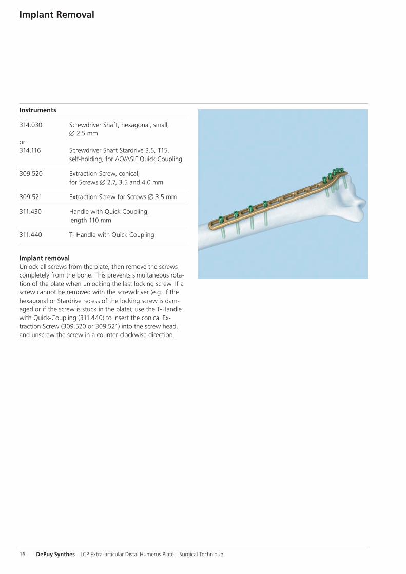
Implant removal Unlock all screws from the plate, then remove the screws completely from the bone. This prevents simultaneous rota-tion of the plate when unlocking the last locking screw. If a screw cannot be removed with the screwdriver (e.g. if the hexagonal or Stardrive recess of the locking screw is dam-aged or if the screw is stuck in the plate), use the T-Handle with QuickCoupling (311.440) to insert the conical Ex-traction Screw (309.520 or 309.521) into the screw head, and unscrew the screw in a counter-clockwise direction.
Implant Removal
LCP Extra-articular Distal Humerus Plate Surgical Technique DePuy Synthes 17
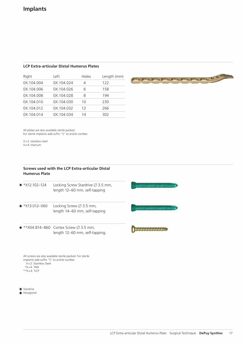
Implants
LCP Extra-articular Distal Humerus Plates
Right Left Holes Length (mm)
0X.104.004 0X.104.024 4 122
0X.104.006 0X.104.026 6 158
0X.104.008 0X.104.028 8 194
0X.104.010 0X.104.030 10 230
0X.104.012 0X.104.032 12 266
0X.104.014 0X.104.034 14 302
Screws used with the LCP Extra-articular Distal Humerus Plate
*X12.102–124 Locking Screw Stardrive B 3.5 mm, length 12–60 mm, selftapping
StardriveHexagonal
All plates are also available sterile packed. For sterile implants add suffix “S” to article number.
X = 2: stainless steelX = 4: titanium
*X13.012–060 Locking Screw B 3.5 mm, length 14–60 mm, self-tapping
**X04.814–860 Cortex Screw B 3.5 mm, length 12–60 mm, self-tapping,
All screws are also available sterile packed. For sterile implants add suffix “S” to article number. X = 2: Stainless Steel *X = 4: TAN**X = 4: TiCP
18 DePuy Synthes LCP Extra-articular Distal Humerus Plate Surgical Technique
Instruments
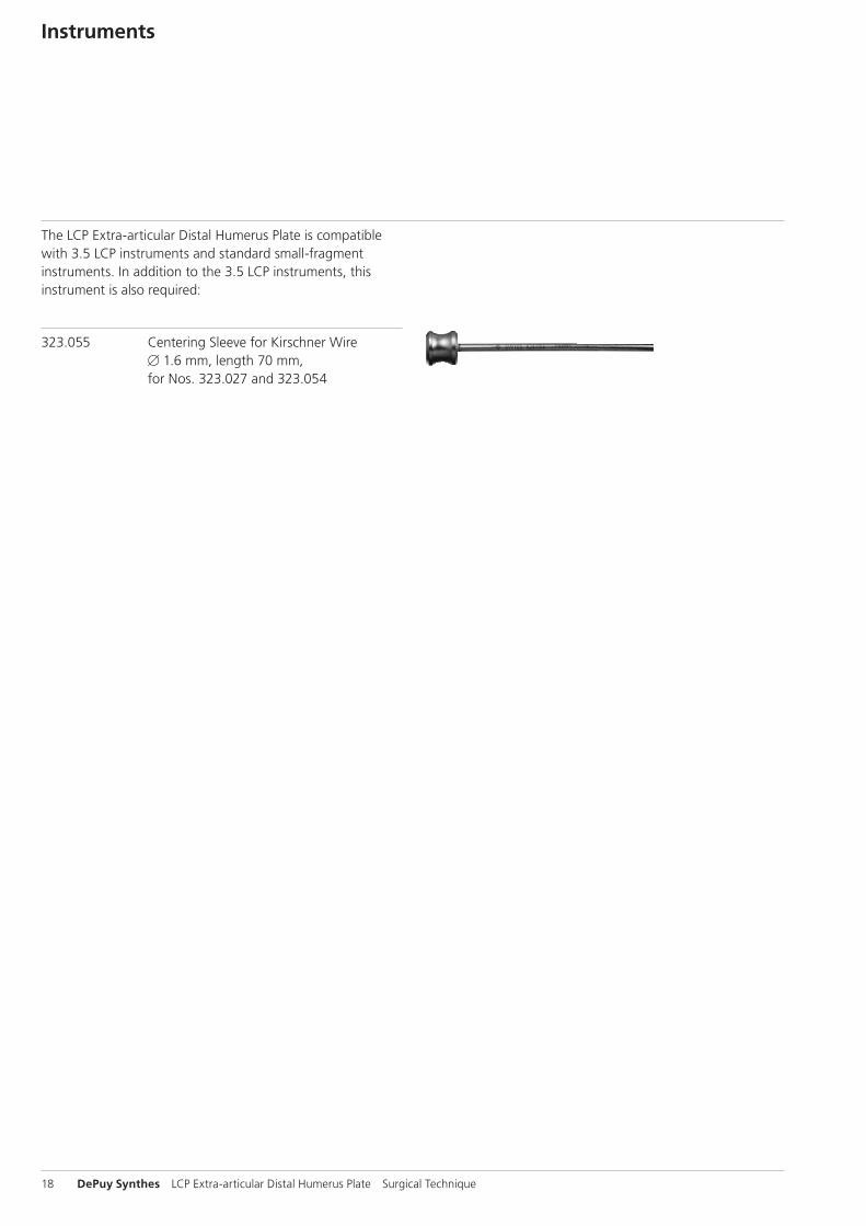
The LCP Extra-articular Distal Humerus Plate is compatible with 3.5 LCP instruments and standard small-fragment instruments. In addition to the 3.5 LCP instruments, this instrument is also required:
323.055 Centering Sleeve for Kirschner Wire B 1.6 mm, length 70 mm, for Nos. 323.027 and 323.054
LCP Extra-articular Distal Humerus Plate Surgical Technique DePuy Synthes 19
Bibliography
Levy JC “An alternative method of osteosynthesis for distal humeral shaft fractures.” J Orthop Trauma. 2005 Jan; 19(1):43-7
Gerwin, Michelle, et al. “Alternative Operative Exposures of the Posterior Aspect of the Humeral Diaphysis. With Ref-erence to the Radial Nerve.” The Journal of Bone and Joint Surgery 78:1690-5 (1996)
McCormack R.G. “Fixation of fractures of the shaft of the humerus by dynamic compression plate or intramedullary nail. A prospective, randomised trial.” J Bone Joint Surg Br. 2000 Sep; 82(7):1085-6
Müller ME, Allgöwer M, Schneider R, Willenegger H (1995) Manual of Internal Fixation. 3rd, expanded and completely revised ed. 1991. Berlin, Heidelberg, New York:Springer
Schemitsch EH “Biomechanical evaluation of methods of internal fixation of the distal humerus.” J Orthop Trauma. 1994 Dec; 8(6):468-75
20 DePuy Synthes LCP Extra-articular Distal Humerus Plate Surgical Technique
MRI Information
Torque, Displacement and Image Artifacts according to ASTM F 2213-06, ASTM F 2052-06e1 and ASTM F2119-07Nonclinical testing of worst case scenario in a 3 T MRI system did not reveal any relevant torque or displacement of the construct for an experimentally measured local spatial gradient of the magnetic field of 3.69 T/m. The largest image artifact extended approximately 169 mm from the construct when scanned using the Gradient Echo (GE). Testing was conducted on a 3 T MRI system.
Radio-Frequency-(RF-)induced heating according to ASTM F2182-11aNon-clinical electromagnetic and thermal testing of worst case scenario lead to peak temperature rise of 9.5 °C with an average temperature rise of 6.6 °C (1.5 T) and a peak temperature rise of 5.9 °C (3 T) under MRI Conditions using RF Coils [whole body averaged specific absorption rate (SAR) of 2 W/kg for 6 minutes (1.5 T) and for 15 minutes (3 T)].
Precautions: The above mentioned test relies on non-clini-cal testing. The actual temperature rise in the patient will depend on a variety of factors beyond the SAR and time of RF application. Thus, it is recommended to pay particular attention to the following points: – It is recommended to thoroughly monitor patients under-
going MR scanning for perceived temperature and/or pain sensations.
– Patients with impaired thermo regulation or temperature sensation should be excluded from MR scanning proce-dures.
– Generally it is recommended to use a MR system with low field strength in the presence of conductive implants. The employed specific absorption rate (SAR) should be reduced as far as possible.
– Using the ventilation system may further contribute to reduce temperature increase in the body.


0123
Synthes GmbHEimattstrasse 34436 OberdorfSwitzerlandTel: +41 61 965 61 11Fax: +41 61 965 66 00www.depuysynthes.com
Not all products are currently available in all markets.
This publication is not intended for distribution in the USA.
All surgical techniques are available as PDF files at www.depuysynthes.com/ifu ©
DeP
uy S
ynth
es T
raum
a, a
div
isio
n of
Syn
thes
Gm
bH. 2
015.
A
ll rig
hts
rese
rved
. 03
6.0
00.
786
DSE
M/T
RM
/081
5/0
445
(1)
04/
16