Embed Size (px)
Citation preview
Mock Strawberry (Duchesnea indica, Fig. 2) of the Rose family (Rosaceae) is an introduced perennial plant consisting of trifoliate basal leaves with long petioles that develop from a crown of roots. The petioles have appressed white hairs. Each blunt-tipped leaflet is broadly ovate or obovate, spanning about 1½" in length and 1" across. The middle leaflet is wedge-shaped at the base. The margins of these leaflets are coarsely crenate-serrate, and they have conspicuous pinnate venation. The upper leaflet surfaces are medium to dark green and hairless. Occasionally, light green to reddish purple stolons develop from the crown that are long and slender. They have appressed hairs and form new plantlets at their tips.
Two microscopy methods were used to study Orange rust morphology. Samples were mounted on an aluminum stub using a sticky pad and observed with a Hitachi T-1000 scanning electron microscope. Light microscopy observations were performed with a Nikon light microscope. Slides were prepared by scraping the infected areas of the leaves in a drop of water on a microscopic slide. Digital photographs were captured with both microscopes.
Orange rust is a disease of blackberry and black raspberry. Purple raspberry may also become infected, but red raspberry is known to be resistant. There are two genera of Orange rust fungi that develop very similar symptoms on host plants. The scientific name given to the form on black raspberry is Arthuriomyces peckianus, while the form more common on blackberry is known as Gymnoconia nitens. Orange rust is one of the most serious diseases of susceptible brambles in the Northeast, adversely affects blossoming and fruit set (Cornell, 2011). Orange rust does not kill the host plants, however it affects the whole plant body and once plants are heavily infected they become worthless and do not recover. There are no effective fungicides for control of Orange rust at this time (Cornell, 2011). By late May or early June in the North East, lower leaf surfaces of infected leaves become covered with blister-like masses of yellow-orange spores. These spores spread the disease to other plants. Leaves that become heavily infected may die, and infected shoots will appear weak, spindly and have very few, or no thorns. Orange rust is autoecious, meaning that it lacks an alternate host and completes its life cycle on one host (Fig. 1).
LEARN BY DOING: MICROSCOPIC STUDIES OF A PLANT PARASITE, ORANGE RUST, GYMNOCONIA SP. (PHRAGMIDIACEAE)
Marcello A. Monterrosa and Camelia Maier Department of Biology
Introduction
The Parasite: Orange Rust
SEM Observations
The Host: Mock Strawberry
Materials and Methods
Abstract Microscopy is an exciting, hands-on, active learning resource for students that can inspire a lifelong interest in science. As part of the Quality Enhancement Plan entitled ‘Pioneering Pathways: Learn by Doing’ at our University, an ecology class project was developed for use of different forms of microscopy to study the ecological relationships between Mock strawberry and its parasite. Orange rust is a common disease of both wild and cultivated Rosaceae. Samples of Mock strawberry leaves infected with the fungus were observed using light and electron microscopes. Microscopy analyses revealed the presence of spores with spiky surfaces in leaf lesions. This project helped me 1) develop scanning electron microscopy skill and 2) understand the adaptations of Orange rust for dissemination and the parasitic relationship between Mock strawberry and Orange rust.
References Anikster, Y. et al. Morphology, life cycle biology, and DNA sequence analysis of rust fungi on garlic and chives from California. Phytopathology 94.6 (2004): 569-577. Hernandezl, J. R., Aime, M. C. and Henkel't T. W. The rust fungi (Uredinales) of Guyana. Fitopatol Bras 18.437 (1993). Kunkel, L.O. Further studies of the orange rusts of Rubus in the United States. Bulletin of the Torrey Botanical Club (1916): 559-569. Laundon, G.F. and Rainbow, A.F. Gymnoconia nitens. C.M.I. Descr. Pathog. Fungi Bact. 201 (1969): 1-2. Mims, C. and Richardson, E. Ultrastructure of teliospores and promycelium and basidiospore formation in the four-spored form of Gymnoconia nitens, one of the causes of orange rust of Rubus. Can J Bot 85.10 (2007): 926-934. Mims, C. and Richardson, E. Ultrastructure of basidium and basidiospore formation in the rust fungus Gymnoconia nitens. Microscopy and Microanalysis 13.S02 (2007): 268-269. Orange Rust. Ithaca: Cornell University, 1999. Petersen, R. H. The rust fungus life cycle. The Botanical Review 40.4 (1974): 453-513.
Figure 2. Mock Duchesnea indica (Rosaceae). A. Healthy plant with fruit B. Leaf
specimen infected with Orange Rust.
Arthuriomyces peckianus (Howe) Cummins & Y.Hirats. (1983) Kingdom: Fungi Division: Basidiomycota Class: Pucciniomycetes Order: Pucciniales Family: Phragmidiaceae Genus: Arthuriomyces Species: A. peckianus
Gymnoconia nitens (Schwein.) F.Kern & Thurst. (1929) Kingdom: Fungi Division: Basidiomycota Class: Pucciniomycetes Order: Pucciniales Family: Phragmidiaceae Genus: Gymnoconia Species: G. nitens
Light Microscopy Observations
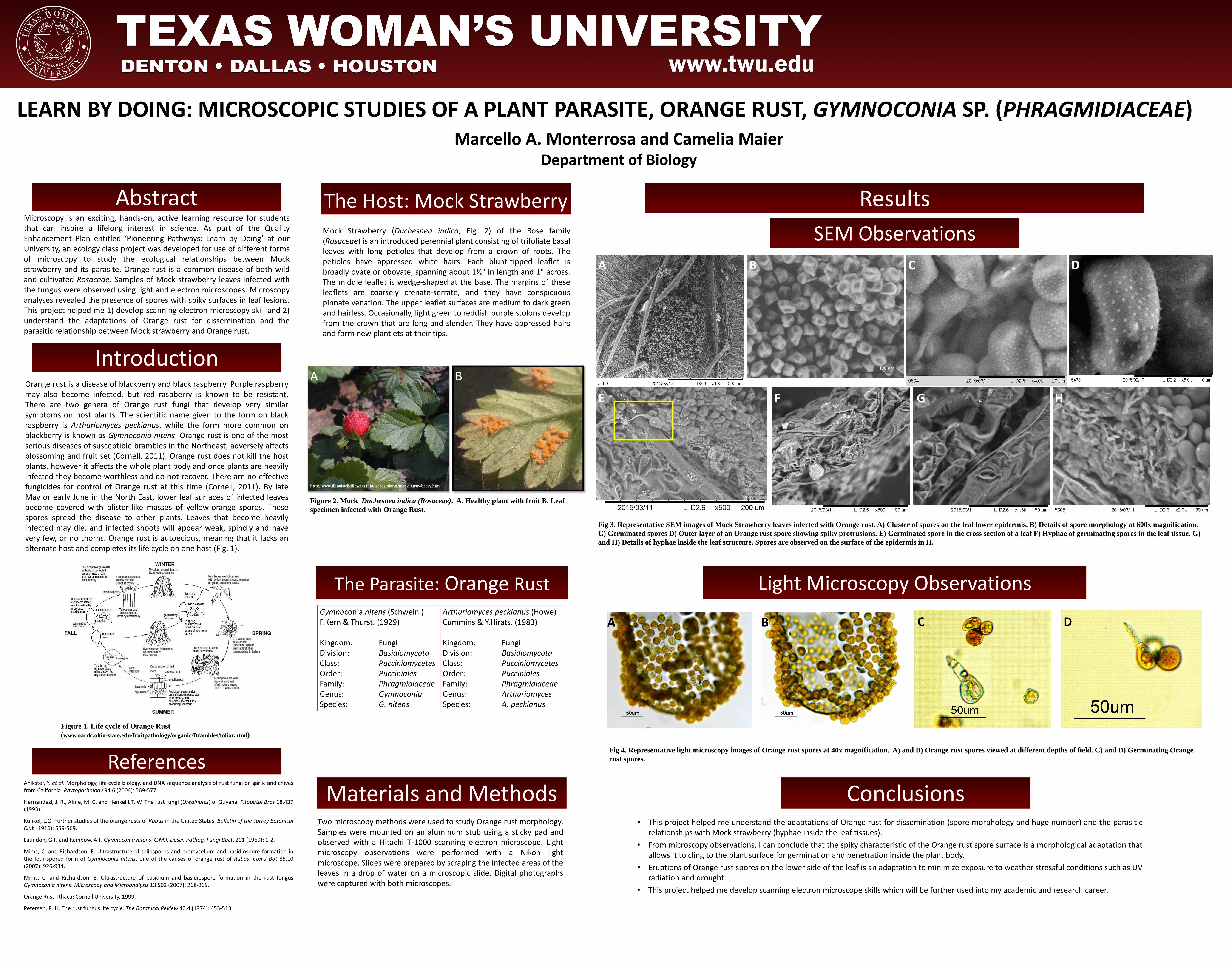
Fig 3. Representative SEM images of Mock Strawberry leaves infected with Orange rust. A) Cluster of spores on the leaf lower epidermis. B) Details of spore morphology at 600x magnification.
C) Germinated spores D) Outer layer of an Orange rust spore showing spiky protrusions. E) Germinated spore in the cross section of a leaf F) Hyphae of germinating spores in the leaf tissue. G)
and H) Details of hyphae inside the leaf structure. Spores are observed on the surface of the epidermis in H.
Fig 4. Representative light microscopy images of Orange rust spores at 40x magnification. A) and B) Orange rust spores viewed at different depths of field. C) and D) Germinating Orange
rust spores.
• This project helped me understand the adaptations of Orange rust for dissemination (spore morphology and huge number) and the parasitic relationships with Mock strawberry (hyphae inside the leaf tissues).
• From microscopy observations, I can conclude that the spiky characteristic of the Orange rust spore surface is a morphological adaptation that allows it to cling to the plant surface for germination and penetration inside the plant body.
• Eruptions of Orange rust spores on the lower side of the leaf is an adaptation to minimize exposure to weather stressful conditions such as UV radiation and drought.
• This project helped me develop scanning electron microscope skills which will be further used into my academic and research career.
Conclusions
http://www.illinoiswildflowers.info/weeds/plants/mock_strawberry.htm
A B
Results
100 um
Figure 1. Life cycle of Orange Rust
(www.oardc.ohio-state.edu/fruitpathology/organic/Brambles/foliar.html)
A C D
E F G H
A B C D
B