Embed Size (px)
Citation preview
1
Lecture (1)
Introduction and language of dermatology
Lecture outline: 1. Function and structure of the skin. 2. Approach to dermatology patient. 3. Descriptive terms and morphology of skin lesions. 4. Important signs and investigations. 5. Topical therapy. Done by:
● Abdulrahman Alkaff ● Rakan Barghouthi ● Super Salman
Reviewed by: Faisal Abunohaiah Color index: slides, doctor notes, extra explanation.
2
DOCTOR DID NOT EXPLAIN/MENTION WHAT IS HIGHLIGHTED. THEY WERE ONLY PROVIDED IN THE SLIDE(S)
Function and structure of the skin.
Function: · Barrier to harmful exogenous substance & pathogens. · Prevents loss of water & proteins. · Sensory organ protects against physical injury. · Regulates body temperature. · Important component of immune system. · Vit. D production by absorbing UVB. · Has psychological and cosmetic importance such as hair, nails. Skin structure:
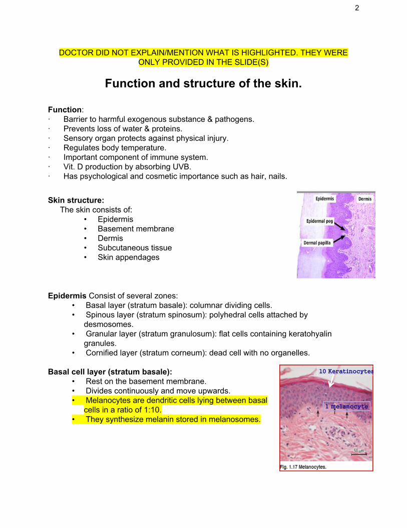
The skin consists of: • Epidermis • Basement membrane • Dermis • Subcutaneous tissue • Skin appendages
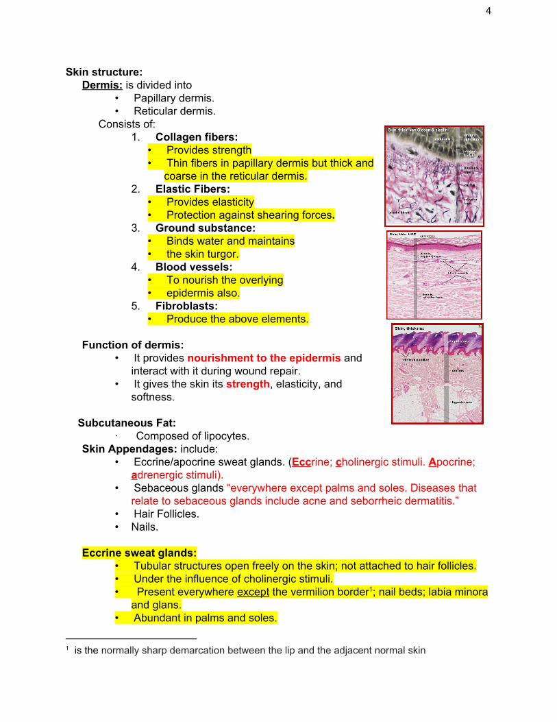
Epidermis Consist of several zones:
• Basal layer (stratum basale): columnar dividing cells. • Spinous layer (stratum spinosum): polyhedral cells attached by
desmosomes. • Granular layer (stratum granulosum): flat cells containing keratohyalin
granules. • Cornified layer (stratum corneum): dead cell with no organelles.
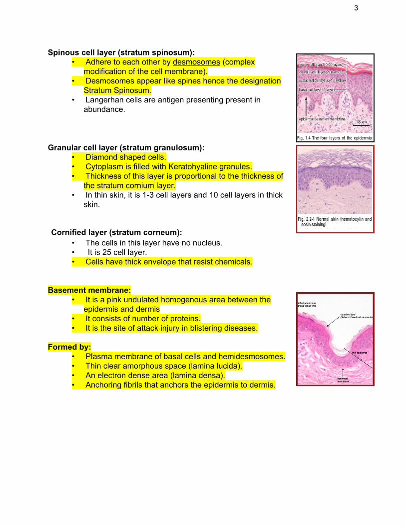
Basal cell layer (stratum basale): • Rest on the basement membrane. • Divides continuously and move upwards. • Melanocytes are dendritic cells lying between basal
cells in a ratio of 1:10. • They synthesize melanin stored in melanosomes.
3
Spinous cell layer (stratum spinosum): • Adhere to each other by desmosomes (complex
modification of the cell membrane). • Desmosomes appear like spines hence the designation
Stratum Spinosum. • Langerhan cells are antigen presenting present in
abundance. Granular cell layer (stratum granulosum):
• Diamond shaped cells. • Cytoplasm is filled with Keratohyaline granules. • Thickness of this layer is proportional to the thickness of
the stratum cornium layer. • In thin skin, it is 1-3 cell layers and 10 cell layers in thick
skin. Cornified layer (stratum corneum):
• The cells in this layer have no nucleus. • It is 25 cell layer. • Cells have thick envelope that resist chemicals.
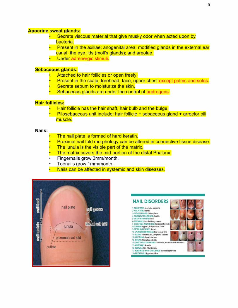
Basement membrane:
• It is a pink undulated homogenous area between the epidermis and dermis
• It consists of number of proteins. • It is the site of attack injury in blistering diseases.
Formed by: • Plasma membrane of basal cells and hemidesmosomes. • Thin clear amorphous space (lamina lucida). • An electron dense area (lamina densa). • Anchoring fibrils that anchors the epidermis to dermis.
4
Skin structure: Dermis: is divided into
• Papillary dermis. • Reticular dermis.
Consists of: 1. Collagen fibers:
• Provides strength • Thin fibers in papillary dermis but thick and
coarse in the reticular dermis. 2. Elastic Fibers:
• Provides elasticity • Protection against shearing forces.
3. Ground substance: • Binds water and maintains • the skin turgor.
4. Blood vessels: • To nourish the overlying • epidermis also.
5. Fibroblasts: • Produce the above elements.
Function of dermis:
• It provides nourishment to the epidermis and interact with it during wound repair.
• It gives the skin its strength, elasticity, and softness.
Subcutaneous Fat:
· Composed of lipocytes. Skin Appendages: include:
• Eccrine/apocrine sweat glands. (Eccrine; cholinergic stimuli. Apocrine; adrenergic stimuli).
• Sebaceous glands “everywhere except palms and soles. Diseases that relate to sebaceous glands include acne and seborrheic dermatitis.”
• Hair Follicles. • Nails.
Eccrine sweat glands:
• Tubular structures open freely on the skin; not attached to hair follicles. • Under the influence of cholinergic stimuli. • Present everywhere except the vermilion border ; nail beds; labia minora 1
and glans. • Abundant in palms and soles.
1 is the normally sharp demarcation between the lip and the adjacent normal skin
5
Apocrine sweat glands: • Secrete viscous material that give musky odor when acted upon by
bacteria. • Present in the axillae; anogenital area; modified glands in the external ear
canal; the eye lids (moll’s glands); and areolae. • Under adrenergic stimuli.
Sebaceous glands:
• Attached to hair follicles or open freely. • Present in the scalp, forehead, face, upper chest except palms and soles. • Secrete sebum to moisturize the skin. • Sebaceous glands are under the control of androgens.
Hair follicles:
• Hair follicle has the hair shaft, hair bulb and the bulge. • Pilosebaceous unit include: hair follicle + sebaceous gland + arrector pili
muscle.
Nails: • The nail plate is formed of hard keratin. • Proximal nail fold morphology can be altered in connective tissue disease. • The lunula is the visible part of the matrix. • The matrix covers the mid-portion of the distal Phalanx. • Fingernails grow 3mm/month. • Toenails grow 1mm/month. • Nails can be affected in systemic and skin diseases.
6
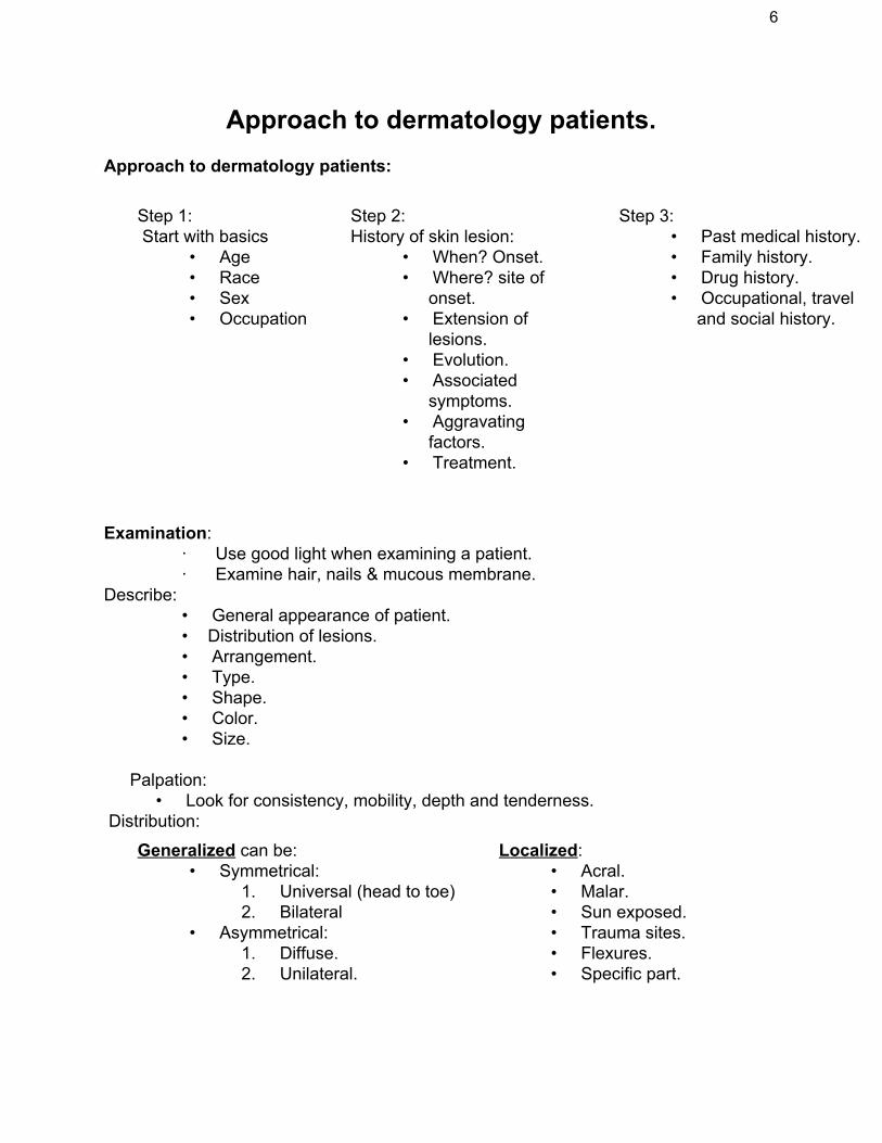
Approach to dermatology patients.
Approach to dermatology patients:
Step 1: Start with basics
• Age • Race • Sex • Occupation
Step 2: History of skin lesion:
• When? Onset. • Where? site of
onset. • Extension of
lesions. • Evolution. • Associated
symptoms. • Aggravating
factors. • Treatment.
Step 3: • Past medical history. • Family history. • Drug history. • Occupational, travel
and social history.
Examination:
· Use good light when examining a patient. · Examine hair, nails & mucous membrane.
Describe: • General appearance of patient. • Distribution of lesions. • Arrangement. • Type. • Shape. • Color. • Size.
Palpation:
• Look for consistency, mobility, depth and tenderness. Distribution:
Generalized can be: • Symmetrical:
1. Universal (head to toe) 2. Bilateral
• Asymmetrical: 1. Diffuse. 2. Unilateral.
Localized: • Acral. • Malar. • Sun exposed. • Trauma sites. • Flexures. • Specific part.
7
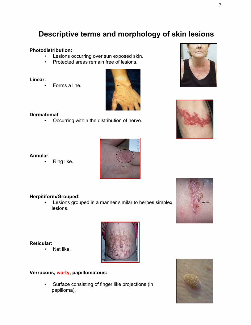
Descriptive terms and morphology of skin lesions Photodistribution:
• Lesions occurring over sun exposed skin. • Protected areas remain free of lesions.
Linear:
• Forms a line. Dermatomal:
• Occurring within the distribution of nerve. Annular:
• Ring like. Herpitiform/Grouped:
• Lesions grouped in a manner similar to herpes simplex lesions.
Reticular:
• Net like. Verrucous, warty, papillomatous:
• Surface consisting of finger like projections (in papilloma).
8
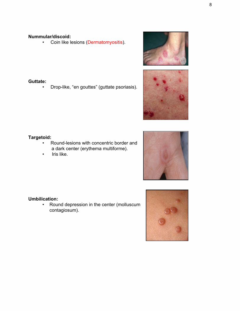
Nummular/discoid:
• Coin like lesions (Dermatomyositis).
Guttate:
• Drop-like, “en gouttes” (guttate psoriasis).
Targetoid:
• Round-lesions with concentric border and a dark center (erythema multiforme). • Iris like.
Umbilication:
• Round depression in the center (molluscum contagiosum).
9
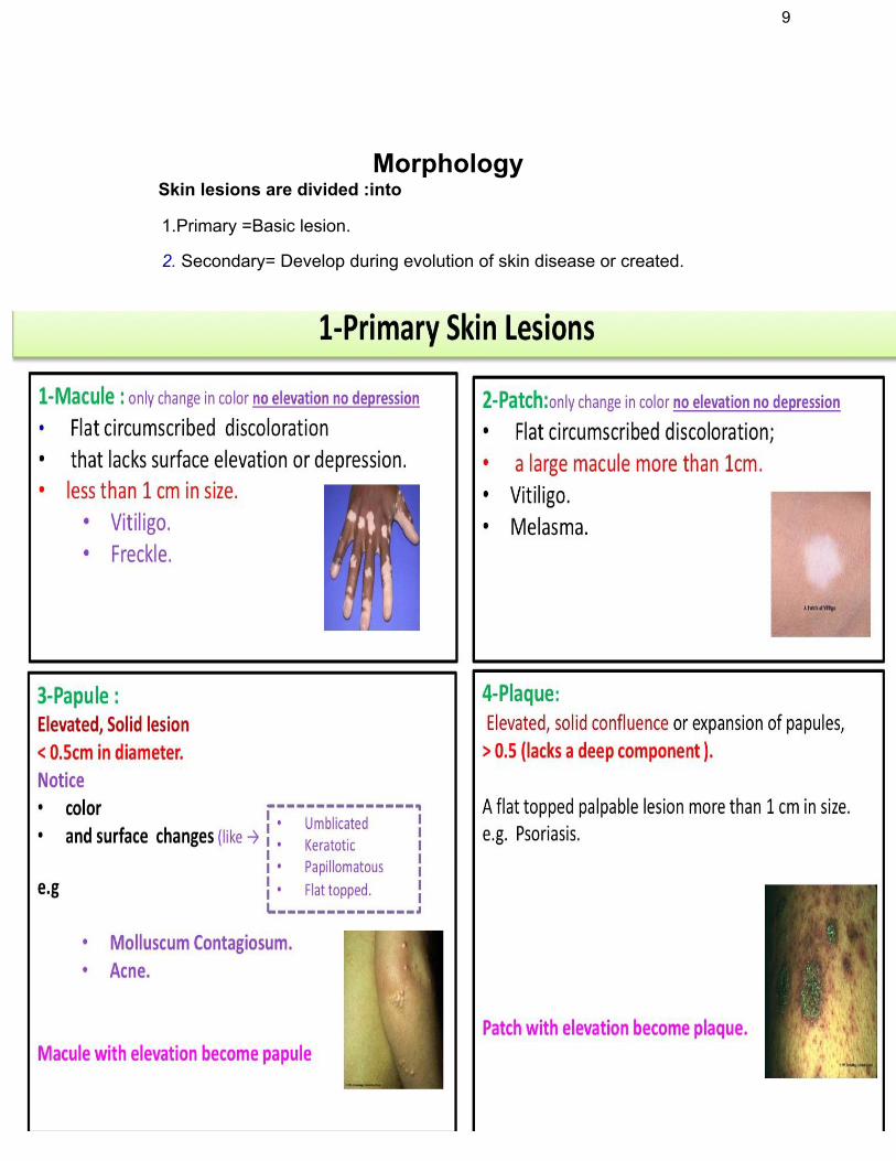
Morphology
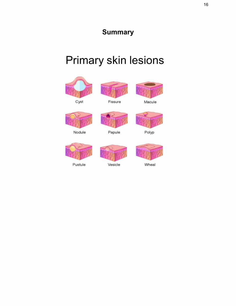
Skin lesions are divided :into
1.Primary =Basic lesion.
2. Secondary= Develop during evolution of skin disease or created.
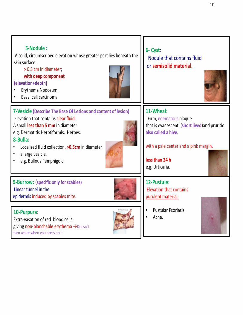
10
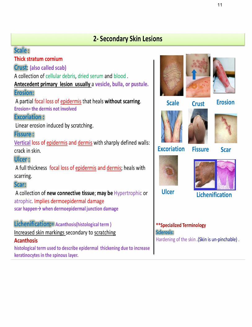
11
12
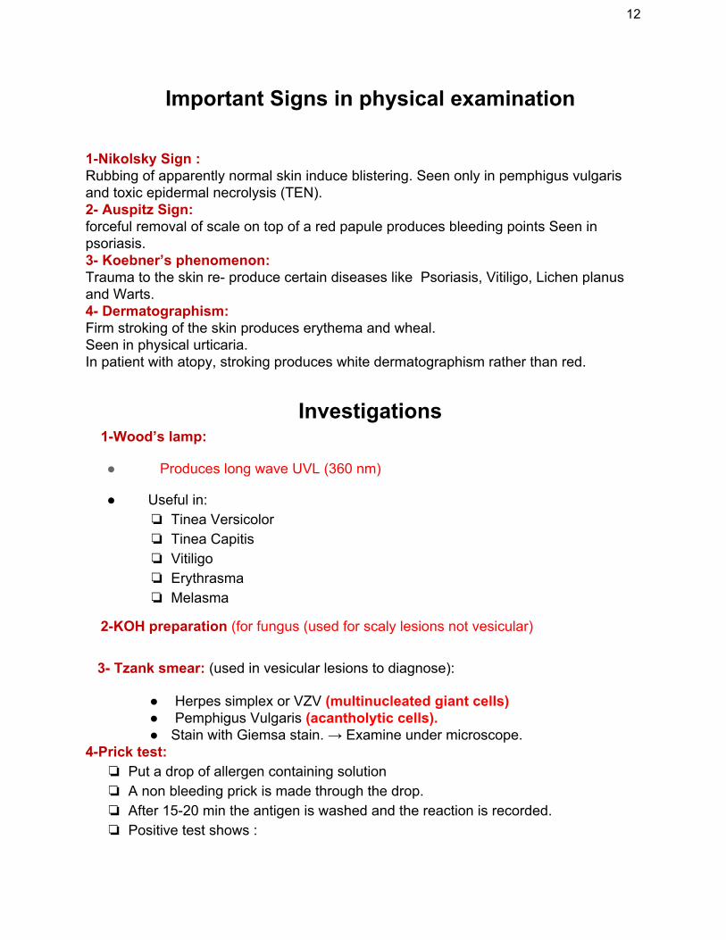
Important Signs in physical examination
1-Nikolsky Sign : Rubbing of apparently normal skin induce blistering. Seen only in pemphigus vulgaris and toxic epidermal necrolysis (TEN). 2- Auspitz Sign: forceful removal of scale on top of a red papule produces bleeding points Seen in psoriasis. 3- Koebner’s phenomenon: Trauma to the skin re- produce certain diseases like Psoriasis, Vitiligo, Lichen planus and Warts. 4- Dermatographism: Firm stroking of the skin produces erythema and wheal. Seen in physical urticaria. In patient with atopy, stroking produces white dermatographism rather than red.
Investigations 1-Wood’s lamp:
● Produces long wave UVL (360 nm)
● Useful in: ❏ Tinea Versicolor ❏ Tinea Capitis ❏ Vitiligo ❏ Erythrasma ❏ Melasma
2-KOH preparation (for fungus (used for scaly lesions not vesicular)
3- Tzank smear: (used in vesicular lesions to diagnose):
● Herpes simplex or VZV (multinucleated giant cells) ● Pemphigus Vulgaris (acantholytic cells). ● Stain with Giemsa stain. → Examine under microscope.
4-Prick test: ❏ Put a drop of allergen containing solution ❏ A non bleeding prick is made through the drop. ❏ After 15-20 min the antigen is washed and the reaction is recorded. ❏ Positive test shows :
13
❖ urticarial reaction at site of prick. ❖ Detects immediate-type IgE mediated reaction. (type 1 hypersensitivity reaction).
❏ Emergency therapeutic measures should be available in case of anaphylaxis.
5-Patch skin test
● Important in Allergic contact dermatitis. (Type 4 cellular immunity) ● Select the most probable substance causing dermatitis. ● Apply the test material over the back. ● Read after 48 & 72 hr. Look for (erythema, edema, vesiculation) ● Positive shows edema and erythema, in severe cases vesicles could present ● Clean skin with alcohol. ● Infiltrate with 1-2% xylocaine with adrenaline. ● Rotate 2-6 mm diameter. ● punch into the lesions. ● Lift specimen and cut at base of lesion. ● Fix in 10% formalin ● For Immunoflourescence →Put in normal saline. (to keep the tissue fresh). ● Suture if 5 mm is used.(if less the 4 mm we do not need to suture it but if more
the 4 mm we need to suture)
6-Direct immunofluorescence:
● Used to diagnose autoimmune diseases e.g. ➢ Pemphigus Vulgaris ➢ Bullous pemphigoid ● Detects immunoglobulin and complement deposits in skin. ● The deposits will give a green fluorescence ● Fluorescence will be noted if immunoglobulin deposits are found intercellular
between the epidermal cells as in pemphigus vulgaris, while found the Basement membrane zone as in bullous pemphigoid.
7- Indirect ImmunoFluorescence : Detect auto antibodies in the serum. It is used: ➢ To confirm a diagnosis ➢ To differentiate between bullous diseases ➢ To monitor disease activity.
14
Topical treatment ★ A wide variety of topical agents are available
★ Delivers the drug to target site.
★ (Golden rule).
• IF the lesion is dry -wet it → How to wet it? Creams, ointments • IF wet -dry it →How to dry it? Using compressors (cloth of water) will cause it to evaporate
★ Topical drugs consist of
1- Active substance: →like steroids, antimicrobial agents.
2- Vehicle: → Is the base in which the active ingredient is dispersed.
Topical steroids side effects: ● Atrophy and striae. ● Telangiectasia and purpura. ● Masking the initial lesion. ● Perioral dermatitis and rosacea or acne. ● Systemic absorption. ● Tachyphylaxis (sudden loss of response).
★ Guidelines regarding steroid use:
● Avoid use for extended periods of time. ● Avoid high potency steroid on flexures and face ● Avoid high potency steroid in children.
★ Examples : ❏ Creams : are mixture of oils and water in which the active substance is
dispersed. white in color useful in folds.and are applied to wet lesions. ❏ Ointments are primarily grease ,useful in dry lesions and Are translucent.
❏ gels are mixtures of propylene glycol and water. Sometimes they contain Alcohol. They are translucent and are best used in wet disorders and hairy regions.
15
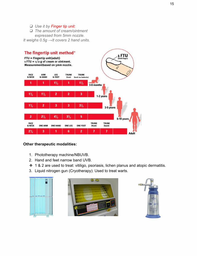
❏ Use it by Finger tip unit: ❏ The amount of cream/ointment
expressed from 5mm nozzle. It weighs 0.5g →It covers 2 hand units.
Other therapeutic modalities:
1. Phototherapy machine/NBUVB. 2. Hand and feet narrow band UVB. ❖ 1 & 2 are used to treat: vitiligo, psoriasis, lichen planus and atopic dermatitis. 3. Liquid nitrogen gun (Cryotherapy): Used to treat warts.
16
Summary