Embed Size (px)
Citation preview

Lecture 15Autoimmunity
Midterm 2 distribution
0
5
10
15
20
25
30
<95
<90
<85
<80
<75
<70
<65
<60
<55
<50
<45
<40
<35
<30
<25
<20
<15
<10 <5
0
Scores
Nu
mb
er o
f stu
den
ts
scores
median 79.5

Autoimmunity
•Types of autoimmunity•Possible mechanisms
•Defects in thymic tolerance, AIRE•Defects in downregulation of immune responses
•FasL•CTLA4
•Role of inflammation/adjuvants/ costimulation•Suppressor/regulatory T cells•Environmental factors
•Cross reactivity•Crosslinking
•Role of MHC

"horror autoxicus"
Paul Ehrlich ca. 1890
Ehrlich, who was studying the ability of antibodies and complement to destroy xenogeneic red blood cells, recognized that if antibodies to self erythrocytes were produced, the results would be devastating.

2-5% of the population
suffers from autoimmune
disease
Autoimmune diseases are tissue and antigen specific

Type I juvenile onset (insulin-dependent) diabetes
Type I diabetes is a T cell-mediated disease in which the islets cells of the pancreas are attacked and killed. The antigen recognized is not known, but there is a very large correlation with the expression of the DR3/4 HLA types. The culprit appears to be a variant of the linked DQ-beta gene lacking a charged aspartic acid at position 57.
Diabetic autoimmunity is characterized by self-reactive B and T lymphocytes that target a set of proteins expressed in pancreatic cells. Proinsulin (PI), IA2, GAD65 and 67, and islet cell autoantigen of 69 kDa (ICA69) are the major examples. These target self-Ags are not islet cell specific, and neither is diabetic autoimmunity; signs of celiac and thyroid autoimmunity are fairly common in patients, and diabetes-prone NOD mice develop signs of thyroid and Sjögren’s disease. Occasional islet-reactive T cells are found in almost 10% of the general population, but <0.5% of these subjects are likely to develop overt diabetes. Although it is uncertain what expands autoimmune T cell pools and what determines their tissue-destructive potential, access to islet target tissue has been suggested as a critical element in diabetes-prone hosts, despite availability of most relevant autoantigens in other tissues.

Processive insulitis and islet cell destruction in diabetes-prone NOD strain mice
Healthy islets
Lymphocyte infiltrate

Figure 13-34

Some autoantibody-
mediated diseases

Figure 11-2
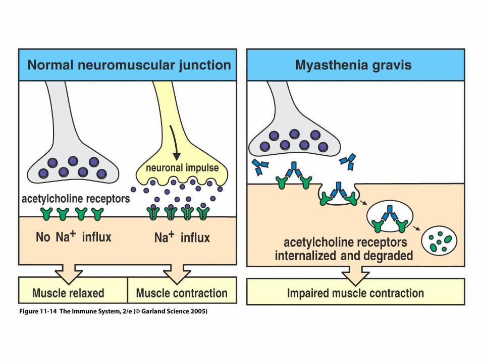
Figure 11-14

Figure 11-5Graves disease involves antibodies to thyroid stimulating hormone receptor

In some cases, it is clear that antibodies are pathogenic

Figure 11-7 part 2 of 2

System lupus erythematosisThe diagnosis of SLE is made when other illnesses have been ruled out, and when a person has 4 of 11 criteria. These criteria include: discoid rash, malar (butterfly) rash, photosensitivity, mouth sores, arthritis, serositis (usually heart or lung inflammation), kidney disorder, neurological problems, blood changes, immune changes, abnormal titre of antinuclear antibody (ANA). It is important to remember that 4 or more of the above need to be present. Sometimes SLE is suspected with fewer symptoms present. For example, if a person has a positive ANA, the diagnosis may not be confirmed unless other symptoms are present. It can be frustrating to not know for sure. The average time from the onset of symptoms and diagnosis is about 3 years.

SLE continuedSLE can damage almost any part or organ of the body. Many people are initially thought to have arthritis because SLE has caused inflammation of the joints. Kidneys are frequently affected. If SLE affects the brain, for example, a person might be initially diagnosed as psychotic, epileptic or suffering from a stroke.
The respiratory system is one of the most frequently involved systems in SLE. Infections present frequent problems. Certain medications used in SLE may predispose a person to unusual kinds of infections.The more serious problems involve lung disease including interstitial lung disease, pulmonary hemorrhage.
Treatable. Treatment of SLE depends on the severity of the case. Some people require only anti-inflammatory medicine (i.e. aspirin) to control pain and swelling. Others may need corticosteroids or cytotoxic medications including azathioprine or cyclophosphamide. In some cases, medications may be used to suppress the immune system. Antimalarial medications are frequently used.

Figure 11-9
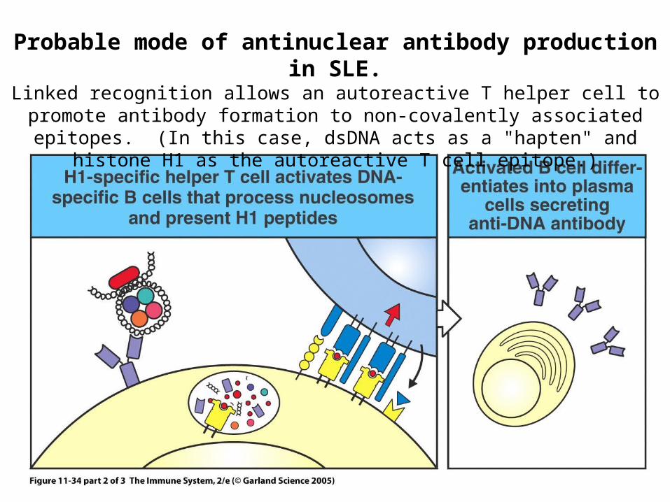
Probable mode of antinuclear antibody production in SLE.Linked recognition allows an autoreactive T helper cell to promote antibody
formation to non-covalently associated epitopes. (In this case, dsDNA acts as a "hapten" and histone H1 as the autoreactive T cell epitope.)
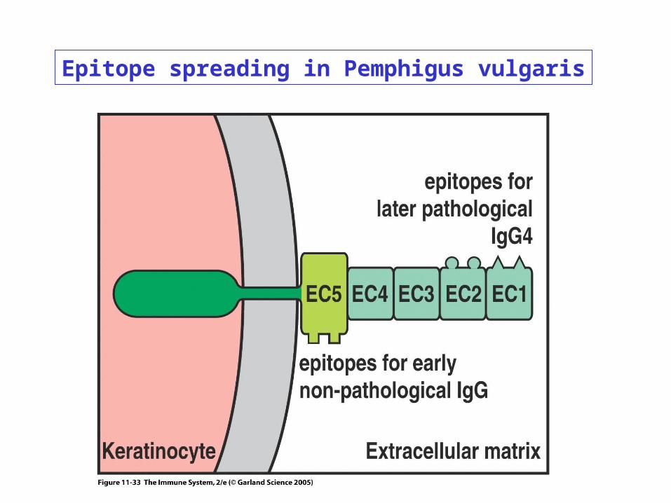
Epitope spreading in Pemphigus vulgaris
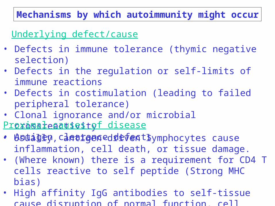
Mechanisms by which autoimmunity might occur
• Defects in immune tolerance (thymic negative selection)• Defects in the regulation or self-limits of immune reactions• Defects in costimulation (leading to failed peripheral tolerance)• Clonal ignorance and/or microbial crossreactivity• Antigen clearance defects
Underlying defect/cause
• Usually, antigen-driven lymphocytes cause inflammation, cell death, or tissue damage.
• (Where known) there is a requirement for CD4 T cells reactive to self peptide (Strong MHC bias)
• High affinity IgG antibodies to self-tissue cause disruption of normal function, cell death, or C' damage.
Proximal causes of disease

Defects in immune tolerance
• AIRE deficiency: failure to delete autoreactive T cells in the thymus.
• The protein is a transcription factor that is believed to turn on many tissue specific genes IN THYMIC MEDULLARY EPITHELIAL CELLS and plays a role in negative selection.

Defects in the regulation or self-limits of immune reactions
These proteins are associated with limiting T cell responses
Protein Expressed by Phenotype of knockout
Fas Activated T cells Lymphoproliferation and autoimmunity
CTLA4 Activated T cells Lymphoproliferation,Regulatory T cells autoimmunity, death
TGF Various, including Massive inflammationRegulatory T cells death
IL10 Th2 cells Inflammatory bowel Regulatory T cells disease

Along with its role in cell killing, FasL is important in eliminating activated T cells
T cells
T cell
T cell

Fas deficiency leads to autoimmune lymphoproliferative syndrome

Fas and FasL deficient mice
•Both types of mice have an identical phenotype
•Lupus erythematosus•Antinuclear antibodies (histone, dsDNA)•Immune complex disease •Glomerulonephritis (kidney inflammation)•Skin disease•Proteinuria (leakage of protein into urine)
•Massive T cell accumulation

FasL expression may play a role in "immune privilege"
FasL expression in brain, eye, placenta, and reproductive organs is believed to contribute to immunological privilege.
Aberrant FasL expression may also be an adaptation of tumors to evade immune surveillance.

Mechanism of Fas Ligand-induced death

Appropriate activation of naïve T cells requires in addition to a ligand for the T cell receptor a second interaction with CD28. This second signal is called "costimulation." Under many conditions, dendritic cells provide this signal during a primary response.
Normal costimulation by signaling through CD28