Embed Size (px)
Citation preview
1
Lecture 19
Secondary phloem (cont’d)
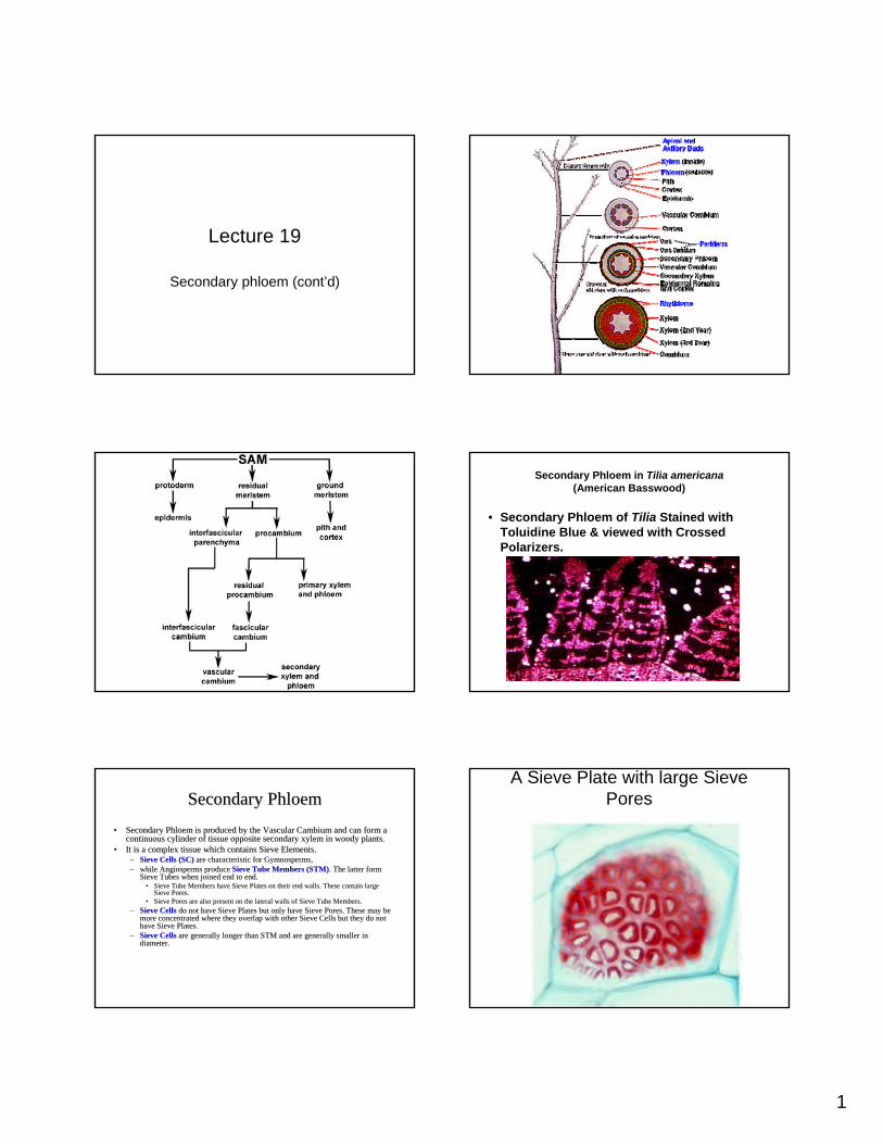
Secondary Phloem in Tilia americana(American Basswood)
• Secondary Phloem of Tilia Stained with Toluidine Blue & viewed with Crossed Polarizers.
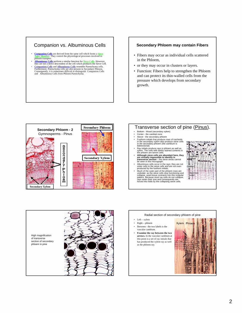
Secondary PhloemSecondary Phloem•• Secondary Phloem is produced by the Vascular Cambium and can forSecondary Phloem is produced by the Vascular Cambium and can form a m a
continuous cylinder of tissue opposite secondary xylem in woody continuous cylinder of tissue opposite secondary xylem in woody plants. plants. •• It is a complex tissue which contains Sieve Elements. It is a complex tissue which contains Sieve Elements.
–– Sieve Cells (SC)Sieve Cells (SC) are characteristic for Gymnosperms, are characteristic for Gymnosperms, –– while Angiosperms produce while Angiosperms produce Sieve Tube Members (STM)Sieve Tube Members (STM). The latter form . The latter form
Sieve Tubes when joined end to end. Sieve Tubes when joined end to end. •• Sieve Tube Members have Sieve Plates on their end walls. These cSieve Tube Members have Sieve Plates on their end walls. These contain large ontain large
Sieve Pores. Sieve Pores. •• Sieve Pores are also present on the lateral walls of Sieve Tube Sieve Pores are also present on the lateral walls of Sieve Tube Members. Members.
–– Sieve CellsSieve Cells do not have Sieve Plates but only have Sieve Pores. These may bdo not have Sieve Plates but only have Sieve Pores. These may be e more concentrated where they overlap with other Sieve Cells but more concentrated where they overlap with other Sieve Cells but they do not they do not have Sieve Plates. have Sieve Plates.
–– Sieve CellsSieve Cells are generally longer than STM and are generally smaller in are generally longer than STM and are generally smaller in diameter. diameter.
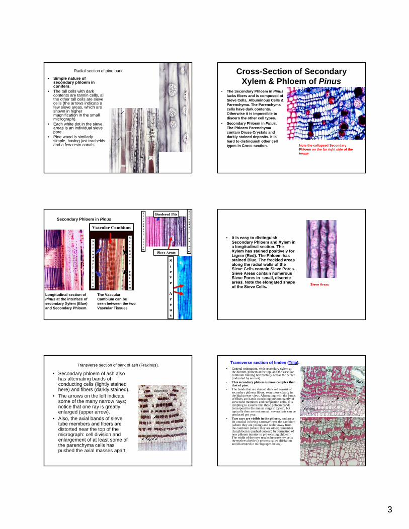
A Sieve Plate with large Sieve Pores
2
Companion vs. Albuminous Cells• Companion Cells are derived from the same cell which forms a Sieve
Tube Member. They control the physiological processes involved in phloem transport.
• Albuminous Cells perform a similar function for Sieve Cells. However, they are not a direct descendent of the cell which produces the Sieve Cell.
• Companion Cells and Albuminous Cells resemble Parenchyma cells. Furthermore, Parenchyma cells are also present in Secondary Phloem. Consequently, it is sometimes difficult to distinguish Companion Cells and Albuminous Cells from Phloem Parenchyma.
Secondary Phloem may contain Fibers
• Fibers may occur as individual cells scattered in the Phloem,
• or they may occur in clusters or layers. • Function: Fibers help to strengthen the Phloem
and can protect its thin-walled cells from the pressure which develops from secondary growth.
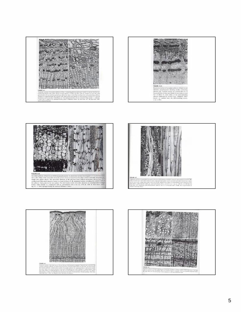
Secondary Phloem - 2 Gymnosperms - Pinus
Transverse section of pine (Pinus). • Bottom - Wood (secondary xylem)• Center - the cambial zone• Above - the secondary phloem• Fusiform initials that produce rows of tracheids
in the secondary xylem also produce sieve cells in the secondary phloem (the cambium is bidirectional)
• Ray initials produce rays in phloem as well as xylem. The cells with darkly stained contents in the phloem are tannin cells.
• Although sieve cells are abundant here, they are virtually impossible to identify in transverse section – the sieve areas cannot be seen in this micrograph.
• Albuminous cells occur in the rays; they are not sister cells to the sieve cells and are not even produced by the fusiform initials.
• Much of the outer part of the phloem rows are undulate: as the sieve cells stop functioning and collapse, axial tissue contracts into this irregular pattern. Because most ray cells do not collapse, rays retain their size and consequently are forced into folds by the collapsing sieve cells.
High magnification of transverse section of secondary phloem in pine
Radial section of secondary phloem of pine• Left – xylem• Right – phloem• Between - the two labels is the
vascular cambium. • Examine the ray between the two
arrows. In the vascular cambium at this point is a set of ray initials that has produced the xylem ray as well as the phloem ray.
3
Radial section of pine bark
• Simple nature of secondary phloem in conifers.
• The tall cells with dark contents are tannin cells, all the other tall cells are sieve cells (the arrows indicate a few sieve areas, which are shown in higher magnification in the small micrograph).
• Each white dot in the sieve areas is an individual sieve pore.
• Pine wood is similarly simple, having just tracheidsand a few resin canals.
Cross-Section of Secondary Xylem & Phloem of Pinus
• The Secondary Phloem in Pinuslacks fibers and is composed of Sieve Cells, Albuminous Cells & Parenchyma. The Parenchyma cells have dark contents. Otherwise it is impossible to discern the other cell types.
• Secondary Phloem in Pinus. The Phloem Parenchyma contain Druse Crystals and darkly stained deposits. It is hard to distinguish other cell types in Cross-section. Note the collapsed Secondary
Phloem on the far right side of the image
Secondary Phloem in Pinus
Longitudinal section of Pinus at the interface of secondary Xylem (Blue) and Secondary Phloem.
The Vascular Cambium can be seen between the two Vascular Tissues
• It is easy to distinguish Secondary Phloem and Xylem in a longitudinal section. The Xylem has stained positively for Lignin (Red). The Phloem has stained Blue. The freckled areas along the radial walls of the Sieve Cells contain Sieve Pores. Sieve Areas contain numerous Sieve Pores in small, discrete areas. Note the elongated shape of the Sieve Cells. Sieve Areas
Transverse section of bark of ash (Fraxinus).
• Secondary phloem of ash also has alternating bands of conducting cells (lightly stained here) and fibers (darkly stained).
• The arrows on the left indicate some of the many narrow rays; notice that one ray is greatly enlarged (upper arrow).
• Also, the axial bands of sieve tube members and fibers are distorted near the top of the micrograph: cell division and enlargement of at least some of the parenchyma cells has pushed the axial masses apart.
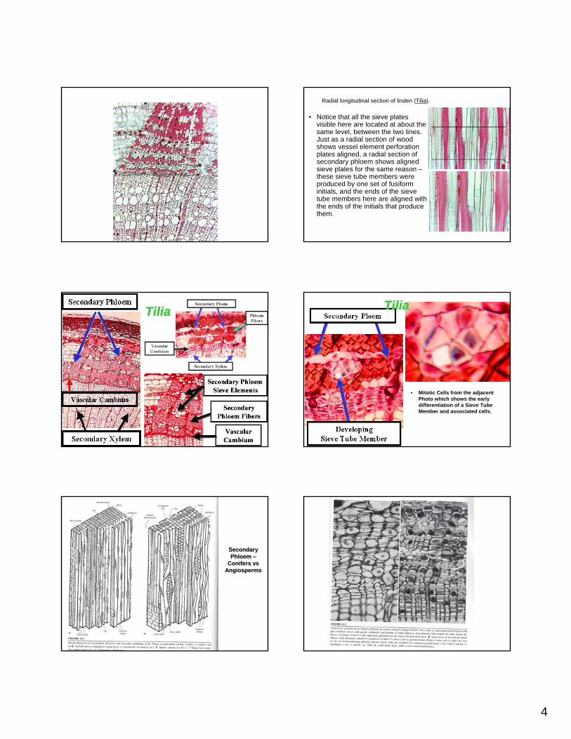
Transverse section of linden (Transverse section of linden (TiliaTilia). ). • General orientation, with secondary xylem at
the bottom, phloem at the top, and the vascular cambium running horizontally across the center (indicated by arrows).
• This secondary phloem is more complex than that of pine.
• The bands that are stained dark red consist of secondary phloem fibers, seen more clearly in the high power view. Alternating with the bands of fibers are bands consisting predominantly of sieve tube members and companion cells. It is tempting to assume that these phloem bands correspond to the annual rings in xylem, but typically they are not annual: several sets can be produced per year.
• Two rays are visible in the phloem, and are a bit unusual in being narrower near the cambium (where they are young) and wider away from the cambium (where they are older; remember that phloem is pushed outward by formation of new phloem interior to pre-existing phloem). The width of the rays results because ray cells themselves divide (a process called dilatation and illustrated in micrographs below).
4
Radial longitudinal section of linden (Tilia).
• Notice that all the sieve plates visible here are located at about the same level, between the two lines. Just as a radial section of wood shows vessel element perforation plates aligned, a radial section of secondary phloem shows aligned sieve plates for the same reason –these sieve tube members were produced by one set of fusiforminitials, and the ends of the sieve tube members here are aligned with the ends of the initials that produce them.
TiliaTilia
• Mitotic Cells from the adjacent Photo which shows the early differentiation of a Sieve Tube Member and associated cells.
TiliaTilia
Secondary Secondary Phloem Phloem ––
Conifers Conifers vsvsAngiospermsAngiosperms
5