Embed Size (px)
Citation preview

8/8/2019 Lecture 2 Classification & Identification
http://slidepdf.com/reader/full/lecture-2-classification-identification 1/46

8/8/2019 Lecture 2 Classification & Identification
http://slidepdf.com/reader/full/lecture-2-classification-identification 2/46
Laboratory procedures employed in theidentification of bacteria
1. Isolation of organism in pure culture2. Bacterial colony morphology3. Microscopic morphology and Staining reaction
Gram Stain Acid fast Stain
4. Biochemical characteristics5. Serological procedure6. Antibiotic sensitivity

8/8/2019 Lecture 2 Classification & Identification
http://slidepdf.com/reader/full/lecture-2-classification-identification 3/46
Isolation of organism in Pure CulturePure culture (axenic culture)
y Population of cells arising from a single cell
y the approach used for the isolation of organism depends
upon the source of clinical specimen
y pure bacterial culture : Blood, spinal fluid and closed abscessesmay yield almost
y mixture of organism : specimen of sputum, stool, materialsfrom the skin and body orifices usually contains
y The specimen is streaked onto solid agar-containing medium so asto separate the bacterial population into individual cells which growas individual colonies
8/8/2019 Lecture 2 Classification & Identification
http://slidepdf.com/reader/full/lecture-2-classification-identification 4/46
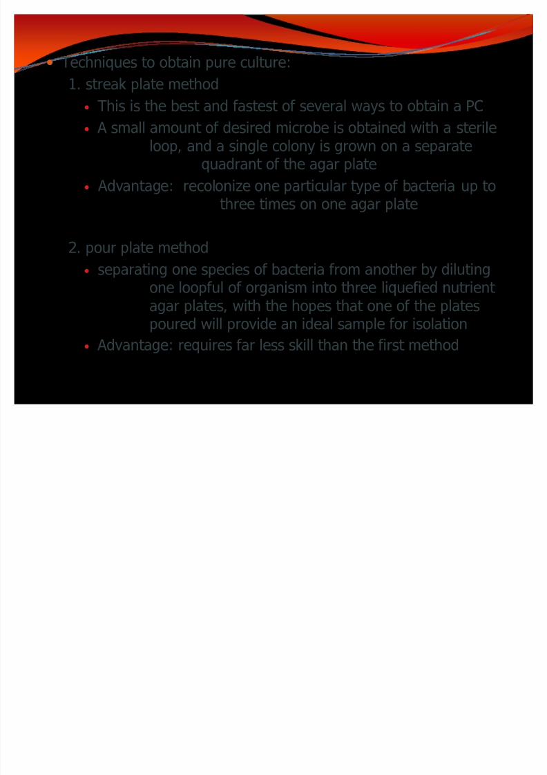
y Techniques to obtain pure culture:
1. streak plate method
y This is the best and fastest of several ways to obtain a PC
y A small amount of desired microbe is obtained with a sterileloop, and a single colony is grown on a separate
quadrant of the agar plate
y
Advantage: recolonize one particular type of bacteria up tothree times on one agar plate
2. pour plate method
y separating one species of bacteria from another by diluting
one loopful of organism into three liquefied nutrient agar plates, with the hopes that one of the platespoured will provide an ideal sample for isolation
y Advantage: requires far less skill than the first method

8/8/2019 Lecture 2 Classification & Identification
http://slidepdf.com/reader/full/lecture-2-classification-identification 5/46
Laboratory Cultivation
y Cultivation is the process of growing microorganisms bytaking bacteria from the infectionsite by some means of specimencollection and growing them in theartificial environment of thelaboratory
y For the in vitro environment of the bacteria, required nutrients
are supplied in a culture mediumy culture - organisms that grow and multiply in or on a culture
media

8/8/2019 Lecture 2 Classification & Identification
http://slidepdf.com/reader/full/lecture-2-classification-identification 6/46
Culture Medium
y is a general term referring to the substanceupon which a microorganism is grown
y Ideally designed to mimic the environment in which theorganism naturally grows
y The most common growth media for microorganisms arenutrient broths and agar plates
y microbiological media currently are categorized as:

8/8/2019 Lecture 2 Classification & Identification
http://slidepdf.com/reader/full/lecture-2-classification-identification 7/46

8/8/2019 Lecture 2 Classification & Identification
http://slidepdf.com/reader/full/lecture-2-classification-identification 8/46
Based on Chemical Composition
y chemically defined medium
- the exact amount of pure chemicals used toformulate the medium is known
y Complex medium
- is composed of a mixture of proteins and extracts inwhich the exact amount of a particular amino
acid, sugar, etc. is not known- Nutrient media contain all the elements that most bacteria need for growth and are non-selective, so theyare used for the general cultivation and maintenance of bacteria kept in laboratory culture collections

8/8/2019 Lecture 2 Classification & Identification
http://slidepdf.com/reader/full/lecture-2-classification-identification 9/46
Functional Types of Media
Supportive or general purpose media- Support the growth of many microorganisms- E.g. Tryptic soy agar
Enriched media- For fastidious organisms- contain nutrients ecologically favorable to the organism to be
isolated- General purpose media supplemented by blood or other special
nutrients Blood agar is an enriched medium in which nutritionally rich
whole blood supplements the basic nutrients Chocolate agar is enriched with heat-treated blood (40-45°C),
which turns brown and gives the medium the color
for which it is named

8/8/2019 Lecture 2 Classification & Identification
http://slidepdf.com/reader/full/lecture-2-classification-identification 10/46
Selective media-Favor the growth of only selected microorganisms and inhibit growth
of others- requirements for growth:
- specific pH- ionic strength- Chemical composition- may contain inhibitors- lack of nutrients for all but the organism in question
eosin-methylene blue agar (EMB) that contains methylene blue
toxic to Gram (+) bacteria, allowing only the growth of Gram (-) bacteria
blood agar (used in strep tests), which contains beef heart blood that becomes transparent in the presence of hemolytic Streptococcus
MacConkey agar for Gram-negative bacteria
Mannitol Salt Agar (MSA) which is selective for Gram (+) bacteria

8/8/2019 Lecture 2 Classification & Identification
http://slidepdf.com/reader/full/lecture-2-classification-identification 11/46
Differential media Distinguish between different groups of microorganisms based
on their biochemical characteristics growing in the presenceof specific nutrients or indicators (such as neutral red, phenolred, eosin y, or methylene blue) added to the medium to
visibly indicate the defining characteristics of a microorganismEx. Blood agar differentiates hemolytic versus non-hemolytic bacteria MacConkey agar - lactose fermenters versus non-fermenters Eosin methylene blue (EMB), which is differential for lactose and sucrose
fermentation Mannitol Salt Agar (MSA), which is differential for mannitol fermentation

8/8/2019 Lecture 2 Classification & Identification
http://slidepdf.com/reader/full/lecture-2-classification-identification 12/46
Bacterial colony morphology
Bacteria grow on solid media as colonies
colony- defined as a visible mass of microorganisms
all originating from a single mother cell,when inoculated into appropriatemedium containing 2% agar and incubated12-24 hours in a favorable atmosphere
- ideally composed of the descendants of a single cell- Colony-forming unit (CFU) 2 or more daughter cells
that do not separate- gross characteristics: size, shape, texture, elevation,
pigmentation, effect on growth medium
- useful in bacterial identification

8/8/2019 Lecture 2 Classification & Identification
http://slidepdf.com/reader/full/lecture-2-classification-identification 13/46
To identify the following colonial characteristics:
8/8/2019 Lecture 2 Classification & Identification
http://slidepdf.com/reader/full/lecture-2-classification-identification 14/46
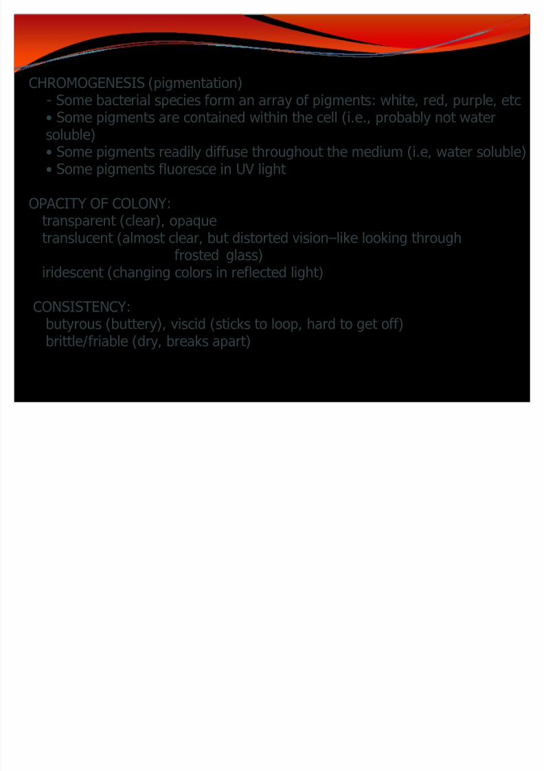
CHROMOGENESIS (pigmentation)
- Some bacterial species form an array of pigments: white, red, purple, etc Some pigments are contained within the cell (i.e., probably not watersoluble) Some pigments readily diffuse throughout the medium (i.e, water soluble) Some pigments fluoresce in UV light
OP ACITY OF COLONY:transparent (clear), opaquetranslucent (almost clear, but distorted visionlike looking through
frosted glass)iridescent (changing colors in reflected light)
CONSISTENC Y:butyrous (buttery), viscid (sticks to loop, hard to get off)brittle/friable (dry, breaks apart)

8/8/2019 Lecture 2 Classification & Identification
http://slidepdf.com/reader/full/lecture-2-classification-identification 15/46
SURFACE OF COLONY:
Smooth (S colonies)- colonies gives the appearance of homogeneity
and uniform texture without appearing as liquid or as mucoidcolonies characteristically isolated from fresh wild type organismsuch as gram- negative enterobacteriaEx. Salmonella, Shigella, Proteus
Mucoid (M colonies) - colonies exhibits a water-like glistening confluent
appearance commonly seem among organism which fromslime layer or capsule. Ex. Klebsiella pneumoniae
Streptococcus pneumoniae
Rough (R colonies) colonies are granulated and rough in appearance,
usually produced by mutant strain that lacks surface protein andpolysaccharide of freshly isolated wild-type parent organism

8/8/2019 Lecture 2 Classification & Identification
http://slidepdf.com/reader/full/lecture-2-classification-identification 16/46
Microscopic morphology
Provide presumptive identification of an organism1. Bacterial morphology
2. Bacterial size3. Staining reaction
Bacterial Morphology Bacterial cell is a fundamental unit of any living organism All its functions are genetically controlled and performed by that
particular cell structure whether it be physiologic orbiochemical
Bacteria and other microorganism are usually transparent, whichmakes the study of the morphologic detail difficult when theyare examined in the natural state
Routinely used to determine: shape, arrangement, and
staining reaction

8/8/2019 Lecture 2 Classification & Identification
http://slidepdf.com/reader/full/lecture-2-classification-identification 17/46
I. Bacterial Shape and Arrangement
y Bacterial Shape
determined by the configuration of the cell wall
detected by brightfield microscopy of stained smear usingoil immersion lens
y Bacterial Arrangement
y is the result of the number of plane division the organism may undergo
and how the cell remain attached afterwardsy divides only across their short axis
y 3 conventional forms :
pherical (cocci) od (bacilli) pirals

8/8/2019 Lecture 2 Classification & Identification
http://slidepdf.com/reader/full/lecture-2-classification-identification 18/46
Spherical (Cocci)
y Shape:y round like a ball, perfect sphere or globe
y Variations :
1. Ovoid shape - both sides rounded ends are
pointedEx. Streptococcus
2. Lancet-shape - one end is pointed, otherend is flat Ex. Pneumococcus
3. Coffee-bean shape - flat on one side,opposite side convex or appear asletter D form
Ex. Neisseria

8/8/2019 Lecture 2 Classification & Identification
http://slidepdf.com/reader/full/lecture-2-classification-identification 19/46
Arrangements:
1. Singly coccus
- occurs as a single spherical cell
2. Chain streptococci- common among ovoid-form resulting from one
plane division with daughter cells remainedattached to one another to form a chainEx. Streptococcus pyogenes
3. Pairs diplococci
- common with lancet-shaped and coffee-bean
shaped spherical resulting from one planedivision with daughter cell separatingEx. Streptococcus pneumoniae
Neisseria gonorrheae

8/8/2019 Lecture 2 Classification & Identification
http://slidepdf.com/reader/full/lecture-2-classification-identification 20/46
4. Clusters staphylococci- common with spherical resulting from many
random plane divisions with daughter cell in
grape-like agglomerationEx. Staphylococcus aureus
5. Tetrads (Packets of 4)- result from 2 plane divisions with daughter
cell separating from one another to formgroup of 4 cellsEx. Micrococcus tetragenous
6. Sarcinae (Packets of 8)- results from 3 plane divisions producingcubical packets of 8 cells
Ex. Sarcina lutea

8/8/2019 Lecture 2 Classification & Identification
http://slidepdf.com/reader/full/lecture-2-classification-identification 21/46
ods (Bacilli)y
Shapey cell appears longer than wide or cylindrical form
y both sides parallel and ends are convex
y varies in actual form depending on the species
y divides only across their short axisy Variations :
1. Clubbed/drumstick shaped swollen on one end
Ex. Clostridium diphtheriae/C. tetani
2. Corset-shape both sides swollen, ends flat orconcave Ex. Bacillus anthracis
3. Fusiform both sides parallel, ends pointed
8/8/2019 Lecture 2 Classification & Identification
http://slidepdf.com/reader/full/lecture-2-classification-identification 22/46
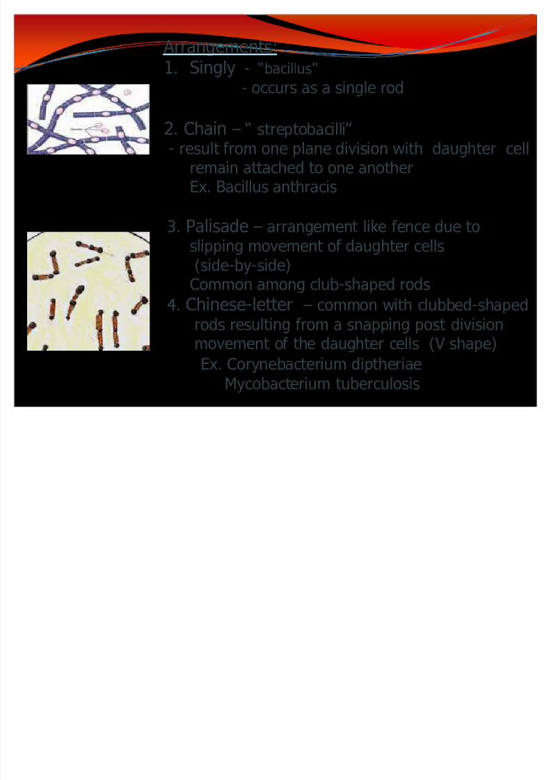
Arrangements:1. Singly - bacillus
- occurs as a single rod
2. Chain streptobacilli- result from one plane division with daughter cell
remain attached to one another
Ex. Bacillus anthracis
3. Palisade arrangement like fence due toslipping movement of daughter cells(side-by-side)Common among club-shaped rods4. Chinese-letter common with clubbed-shapedrods resulting from a snapping post divisionmovement of the daughter cells (V shape)
Ex. Corynebacterium diptheriae
Mycobacterium tuberculosis

8/8/2019 Lecture 2 Classification & Identification
http://slidepdf.com/reader/full/lecture-2-classification-identification 23/46
5. Packets of cigarette arrangement
like bundlesEx. Mycobacterium leprae
6. Serpentine commonly seen with virulent
strain of Mycobacterium tuberculosis

8/8/2019 Lecture 2 Classification & Identification
http://slidepdf.com/reader/full/lecture-2-classification-identification 24/46
I t r i t f r f cilli
Coccobacilli- a rod is s ort & ide / l- t ese f or is i t er ediat e bet een a
s erical and rodEx. aemophil s, r cella
Vibrio - a entl cur e bact eria (comma-shaped)
- it is an int ermediat e bet een a rod and a spiralEx. Vibrio cholerae

8/8/2019 Lecture 2 Classification & Identification
http://slidepdf.com/reader/full/lecture-2-classification-identification 25/46
Spiralsy bacteria with more than one somatic curve
y may be regarded as bacillary forms trusted in the form of a helix
y no characteristic cell arrangement
y most occurs singly
y different specie vary in size, length, rigidity and amplitude of theircoils
y 2 types :
1. Flexible spirals that can contract and relax move by creeping
movement Ex. Spirochetes
y 2. Rigid spirals that cannot contract and relax move byrotation or corkscrew-like motion
Ex. Spirillum

8/8/2019 Lecture 2 Classification & Identification
http://slidepdf.com/reader/full/lecture-2-classification-identification 26/46
SPIRILLUM- whose long axis remains rigid when in
motion
Ex. Campylobacter jejuni
SPIROCHETE whose long axis bends when in motion
Genus Treponema tightly coiled w/ cork screw appearanceEx. Treponema pallidum
Genus Leptospira less tightly coiled w/ sharp hook-like ends
Ex. Leptospira interrogansGenus Borrelia
much less tightly coiled w/c has theappearance of extremely long undulatingbacillary poresEx. Borrelia recurrentis

8/8/2019 Lecture 2 Classification & Identification
http://slidepdf.com/reader/full/lecture-2-classification-identification 27/46
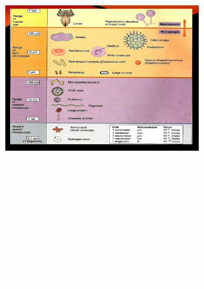
II. Bacterial size
y all linear measurements in microbiology are expressed inmetric units
the basic unit of the metric system is the meter m
y centimeter cm (1/100th of a m)
- the largest unit of length used for measuring microorganismy micrometer µm
- visible only with high powered microscope
- unit of measurement most frequently used in microbiology
y 1µm = 1/1000 of a mmy Cocci = 0.4-2µm
y Bacilli = 0.2-4µm in width by o.5-20µm in length
y Spirals = 1-4µm in length
y nanometer nm - commonly used to measure virus
y Angstrom smallest unit of measurement
8/8/2019 Lecture 2 Classification & Identification
http://slidepdf.com/reader/full/lecture-2-classification-identification 28/46

8/8/2019 Lecture 2 Classification & Identification
http://slidepdf.com/reader/full/lecture-2-classification-identification 29/46
III. Bacterial Staining Reaction
Staining procedure that apply colored chemicals called dyes tospecimen in order to facilitate identification
Stains - salts composed of a positive and negative ion, one of whichis colored (chromophore color bearing ion), which imparts
a color to cell or cell parts by becoming affixed to themthrough a chemical reaction
Basic (cationic) Dyes - chromophore is the positive ion dye Acid (anionic) Dyes - chromophore is the negative ion dye
Bacteria are slightly negative, so are attracted to the positive chromophoreof the BASIC DYE
- Staining procedure was 1st introduced by Paul E hrlich
8/8/2019 Lecture 2 Classification & Identification
http://slidepdf.com/reader/full/lecture-2-classification-identification 30/46
y Preparing smears for staining
1. Smear preparation
- depends on the physical state; if in liquid state spread thesmear out
- Bacteria on slide
2. Air Dry
- preserve the morphology of the bacteria
- allow the smear to adhere to the slide
3. Bacteria are HEAT FIXED to the slide
Heat Fixation
- simultaneously kills the specimen and secures it to the slide
- preserve various cellular component in a natural state withminimal distortion
4. Stain is applied
Staining coloring the microorganisms with a dye
8/8/2019 Lecture 2 Classification & Identification
http://slidepdf.com/reader/full/lecture-2-classification-identification 31/46
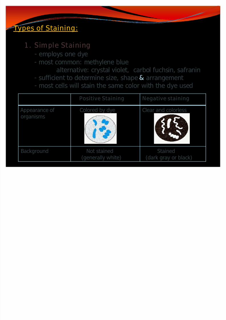
Positive Staining Negative staining
Appearance of organisms
Colored by dye Clear and colorless
Background Not stained
(generally white)
Stained
(dark gray or black)
Types of Staining:
1. Simple Staining- employs one dye
- most common: methylene bluealternative: crystal violet, carbol fuchsin, safranin
- sufficient to determine size, shape arrangement - most cells will stain the same color with the dye used

8/8/2019 Lecture 2 Classification & Identification
http://slidepdf.com/reader/full/lecture-2-classification-identification 32/46
2. Differential Staining
- employs the use of more than one dye added in several stepsand stained structures are differentiated by color as well asshape
- it is based on the relative affinity of different bacterial cells forthe stains used
- enables microbiologist to differentiate one group from anothera) Gram staining - differentiate gram (+) from gram (-)
bacteriab) Acidfast staining - differentiate acidfast from non-acidfast
bacteria

8/8/2019 Lecture 2 Classification & Identification
http://slidepdf.com/reader/full/lecture-2-classification-identification 33/46
Gram-staining
y Hans Christian Gram (1884), a Danish doctor, accidentallystumbled on a method which still forms the basis for theidentification of bacteria
y This staining procedure defines 2 bacterial group
y Those w/c retain the primary dye (gram-positive)y those which are decolorized (gram negative)
y The difference in dye retention is dependent on such physicalproperties as thickness, density, porosity and integrity of thebacterial cell wall, as well to some extent, the chemicalcomposition

8/8/2019 Lecture 2 Classification & Identification
http://slidepdf.com/reader/full/lecture-2-classification-identification 34/46
y The reagents needed:
y Crystal Violet (Primary Stain)
y Iodine Solution/Grams Iodine (Mordant)
Mordant - intensifies the stain or coats a structure to makeit thicker and easier to see after it is stained
- Increase the affinity of a stain to the specimeny Decolorizer (ethanol is a good choice, mixture of acetone
alcohol)
y Safranin (Counterstain)
Counterstain gives contrasting color to the primary stain

8/8/2019 Lecture 2 Classification & Identification
http://slidepdf.com/reader/full/lecture-2-classification-identification 35/46
STEP 2: Flood the entire slide with crystal violet (primary
stain) for 1min. Then rinse with the water.
STEP 3: flood the slide with the iodine solution (mordant)
for 1min. Then rinse with water for 5 seconds. The bacteria
become deeply stained and appear deep purple in color due
to crystal violet-iodine-complex formation
Step 4: addition of the decolorizer, 95% ethyl alcohol
Rinse with water.
Gram (+) cells : purple dye is retained
Gram (-): purple dye is readily removed and appears colorless
STEP 5: Flood the slide with the counterstain, safranin
Again, rinse with water.
Gram (+) cells will incorporate little or no counterstain and will
remain purple in appearance
Gram (-) bacteria take on a pink/red color
Gram Staining
STEP 1: Make a smear. Mounted and heat fixed.

8/8/2019 Lecture 2 Classification & Identification
http://slidepdf.com/reader/full/lecture-2-classification-identification 36/46
PRINCIPLE:
y Gram reaction is based on the structure of the bacterial cell wall
y Gram-positive bacteria
y the peptidoglycan appears to act as a permeability barrier preventingloss of crystal violet-iodine-complex
y When gram-positive bacteria are treated with alcohol, the alcoholcauses coagulation and dehydration of the thick layer of peptidoglycanresulting in shrinkage of pores preventing C VI-complex from escapingand the bacteria remain deep purple
y Reaction to Gram staining is also believed to be asso. with proteincomplex Magnesium ribonucleate which is absent in Gram (-) org.
y Gram Negative bacteria
y peptidoglycan is very thin in gram (-) bacteria and has larger pores
y Alcohol readily penetrates the lipidrich outer layer of the cell wall andextracts enough lipid thus increasing the porosity further
y alcohol more readily removes the deep purple C VI-complex fromgram (-) bacteria thus becomes decolorized
y The outer membrane is then permeabilized by the decolorizer, andthe pink safranin counterstain is trapped by the peptidoglycan layer

8/8/2019 Lecture 2 Classification & Identification
http://slidepdf.com/reader/full/lecture-2-classification-identification 37/46
Divides bacteria into 2 groups
y Gram (+) : violet
y
Gram (-) : red
Dictome of Gram Staining
y All COCCI are Gram Positive except Neisseria group,Moraxella (Branhamella) catarrhalis and Veilonella
y All BACILLI are Gram Negative except the acid fast
organisms (Mycobacterium, Nocardia) , Sporeformers(Bacillus, Clostridum) and Corynebacterium species
y Spirals are difficult to stain but when stained, they are
Gram Negative

8/8/2019 Lecture 2 Classification & Identification
http://slidepdf.com/reader/full/lecture-2-classification-identification 38/46
Acid Fast Stainingy
Acid-fast stain is a useful differential staining procedure that specifically stains all members of the genus mycobacteria
y The walls of certain bacteria contain long chain fatty acids(mycolic acid) lending the property of resistance to decolorizationof basic dyes by acid alcohol; thus called acid fast
y The high lipid and wax content of the mycobacterial cell walls isthought to be the reason for such impermeability
y 2 methods
y Ziehl-Neelsen method
y The procedure utilizes heat and phenol (carbolic acid) to help thepenetration of the dye, carbol fuchsin, to the inside of mycobacterial cells, which are impermeable to basic dyes in routinestains like in Gram staining
y Cold Kinyoun technique
y Instead of heat, this technique uses increasing the concentration of phenol or the inclusion of a detergent in the stain

8/8/2019 Lecture 2 Classification & Identification
http://slidepdf.com/reader/full/lecture-2-classification-identification 39/46
y Divides bacteria into 2 groups
y Acid - Fast organism: red
y Non Acid Fast organism: blue
y The reagents needed
1. Primary stain: Carbol fuchsin
2. Decolorizer: Acid Alcohol
3. Counterstain: Methylene Blue

8/8/2019 Lecture 2 Classification & Identification
http://slidepdf.com/reader/full/lecture-2-classification-identification 40/46
Acid - Fast Staining (Ziehl-Neelsen method)
STEP 2: Flood the entire slide with Carbol Fuchsin.
STEP 3: Using a Bunsen burner, heat the slides slowly until
they are steaming. Acid fast organisms have a very
hydrophobic surface which resist entry of dyes. Heat is used to
enhance penetration and retention of dye
Maintain steaming for 5 minutes by using low or intermittent
heat (i.e. by occasionally passing the flame from the Bunsenburner over the slides) Then rinse the slide with water.
STEP 4: Flood the slide with 3% acid-alcohol and allow to
decolorize for 5 minutes. Throughout the 5 minutes, continue to
flood the slides with 3% acid-alcohol until the slides are clear of
stain visible to the naked eye. Rinse the slide thoroughly withwater and then drain any excess from the slides.
STEP 5: Flood with the counterstain, Methylene Blue Keep
the counterstain on the slides for 1 minute. Rinse with water.
STEP 1: Make a smear. Mounted and heat fixed

8/8/2019 Lecture 2 Classification & Identification
http://slidepdf.com/reader/full/lecture-2-classification-identification 41/46
Positive Staining Negative staining
Capsule
Flagella
Endospore
3. Special Staining- used to color and isolate specific structure of a microorganism like
capsule, flagella, inclusion granule, endospore and etc.

8/8/2019 Lecture 2 Classification & Identification
http://slidepdf.com/reader/full/lecture-2-classification-identification 42/46
Biochemical Test
y various species of organism exhibits characteristicpattern of substrate utilization, metabolic product formation and sugar fermentation
y Enzyme based test based on its reaction with a substrate
y Catalase, oxidase, indole, ureasey Reactions in glucose fermentation broth
y Reactions in lactose fermenation broth
y Starch hydrolysis of test strains
y Nitrate Broth reactions
y 60% of common pathogens can be identified bybiochemical test

8/8/2019 Lecture 2 Classification & Identification
http://slidepdf.com/reader/full/lecture-2-classification-identification 43/46
Serological procedurey
Antigen and antibody determinationy Serological Tests
y Use group specific antiserum isolated from the plasma of animals that have been sensitized to the organism
y The antiserum contains antibody proteins that react withantigens on the unknown organism.
y Procedures: agglutination, precipitation test, hemagglutinationinhibition, complement fixation, ELISA, RIA, Western blot assay
y Advantages:
y Highly specificy Does not usually require the organism to be isolated into pure
culture
y Can be used to identify organisms that cant be grown onmedium

8/8/2019 Lecture 2 Classification & Identification
http://slidepdf.com/reader/full/lecture-2-classification-identification 44/46
Antibiotic sensitivityy
is a term used to describe the susceptibility of bacteria toantibiotics
y Antibiotic susceptibility testing (AST) is usually carried out todetermine which antibiotic will be most successful intreating a bacterial infection in vivo
y Methods of testing:y Broth dilution
- The lower the dilution, the greater the antibiotic content
y Agar dilution
y Disk diffusion- the Kirby-Bauer test for antibiotic susceptibility, called
the disc diffusion test, is a standard that has beenused for years
8/8/2019 Lecture 2 Classification & Identification
http://slidepdf.com/reader/full/lecture-2-classification-identification 45/46
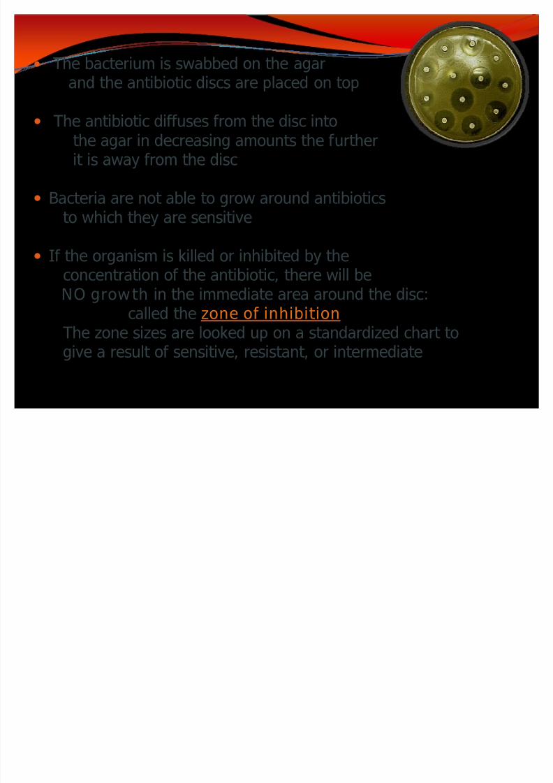
y The bacterium is swabbed on the agarand the antibiotic discs are placed on top
y The antibiotic diffuses from the disc intothe agar in decreasing amounts the furtherit is away from the disc
y Bacteria are not able to grow around antibioticsto which they are sensitive
y If the organism is killed or inhibited by theconcentration of the antibiotic, there will be
NO growth in the immediate area around the disc:called the zone of inhibition
The zone sizes are looked up on a standardized chart togive a result of sensitive, resistant, or intermediate

8/8/2019 Lecture 2 Classification & Identification
http://slidepdf.com/reader/full/lecture-2-classification-identification 46/46