Embed Size (px)
Citation preview

Carcinogenesis

Characteristics of Cancer
Disorder of altered cell differentiation and growthResults in neoplasia (“new growth”)
Growth is uncoordinated and relatively autonomousLacks normal regulatory controls over cell
growth and divisionTends to increase in size and grow after
stimulus ceases or needs of organism are met

Components of Tissue Renewal and Repair
Cell proliferationProcess of cell division Inherent adaptive mechanism for replacing body
cellsCell differentiationProcess of specializationNew cells acquire the structure and function of
cells they replaceApoptosisForm of programmed cell death to eliminate
unwanted cells

Types of Stem Cells
Unipotent: give rise to one type of differentiated cellMuscle satellite cellEpidermal stem cellSpermatogoniumBasal cell of the olfactory epithelium
Oligopotent: produce small number of cells
Pluripotent: give rise to numerous cell types

Determination and Differentiation

The Cell CycleDefinition: The interval between each cell division
Genetic information is duplicated Duplicated chromosomes are appropriately aligned
for distribution between two genetically identical daughter cells
Checkpoints in cycle provide opportunities for monitoring the accuracy of deoxyribonucleic acid (DNA) replication Edited and repaired defects ensure full complement of
genetic information to each daughter cell

Phase of the Cell Cycle
G1 (gap 1): the post mitotic phase DNA synthesis ceases while ribonucleic acid (RNA) and
protein synthesis and cell growth take place
S phase: DNA synthesis occurs, giving rise to two separate sets of chromosomes, one for each daughter cell
G2 (gap 2): the premitotic phase DNA synthesis ceases; RNA and protein synthesis
continues
M phase: the phase of cellular division or mitosis
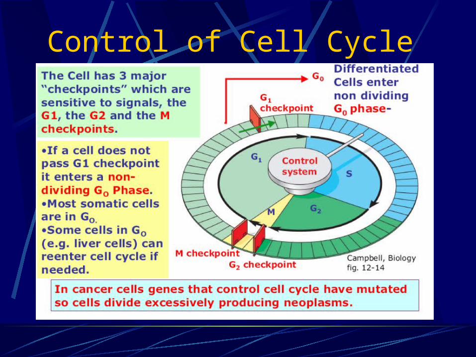
Control of Cell Cycle

Control of Cell CycleThe cell cycle is controlled by many proteins from inside & outside the cell.
Intracellular cyclins and cyclin dependent kinases (CDKs) control the checkpoints.
Extracellular proteins from other cells called Growth Factors signal the target cell to divide.
Binding of growth factors to membrane receptor proteins of the target cell triggers a molecular signaling pathway - a series of proteins which allows the cell to pass the checkpoints of the cell cycle.

Cell Cycle is controlled by genes.

Signal Transduction

Cell Cycle: RegulationCyclines: regulatory proteins, active at specific stages in the cell cycle CDK: cycline dependent kinases CDI: CDK inhibitor

Tumor Suppressor Proteins Inhibit CellDivision & Prevent Cancer
Tumor suppressor proteins are proteins that bind to checkpoint proteins to stop the cell cycle & prevent cell division.
An important function of tumor suppressor proteins is to stop the division of mutated cells until mistakes in DNA are repaired by enzymes.
TS proteins keep most mutations from being passed on to daughter cells & developing into cancer.
If the genes for TS proteins mutate or are deleted cancers may result.
Two important TS proteins are the p53 protein & the RB protein.

Normal (interphase) cells are ‘parked’ in
G1 (G0) and will not proceed to S phase
unless induced.
eg by growth factors
• Therefore normal cells grown in tissue
culture need growth factors in order to
divide (proliferate)
Cancer cells have lost the G1
checkpoint regulation
• Therefore cancer cells will proliferate (in
culture) in the absence of growth factors

Cell Proliferation
DefinitionThe process by which cells divide and
reproduce
RegulationRegulated in normal tissue, so the number of
cells actively dividing equal the number of cells dying or being shed

Two Major Categories of Cells Existing in Humans
Gametes (ovum and sperm) Haploid (containing one set of chromosomes
from one parent) Designed for sexual fusion forming a diploid
cell (containing both sets of chromosomes)
Somatic cell The diploid cell that forms the rest of the
body

Categories of Cell Types of the Body
Well-differentiated neurons and cells of skeletal and cardiac muscle unable to divide and reproduce
Parent or progenitor cells that continue to divide and reproduceBlood cells, skin cells, liver cells
Undifferentiated stem cells that can be triggered to enter cell cycle and produce large numbers of progenitor cells when needed

Types of Tumors
Adenoma: benign tumor of glandular epithelial tissue
Adenocarcinoma: malignant tumor of glandular epithelial tissue
Carcinoma: malignant tumor of epithelial tissue
Osteoma: benign tumor of bone tissue
Sarcoma: malignant tumors of mesenchymal origin
Papillomas: benign microscopic or macroscopic fingerlike projections growing on a surface

Factors differentiating Benign and Malignant Neoplasms
Cell characteristics
Manner of growth
Rate of growth
Potential for metastasizing or spreading
Ability to produce generalized effects
Tendency to cause tissue destruction
Capacity to cause death

Characteristics of Benign Neoplasms
A slow, progressive rate of growth that may come to a standstill or regress
An expansive manner of growth
Inability to metastasize to distant sites
Composed of well-differentiated cells that resemble the cells of the tissue of origin

Characteristics of Malignant Neoplasms
Tend to grow rapidly and spread widely
Have the potential to kill regardless of their original location
Tend to compress blood vessels and outgrow their blood supply, causing ischemia and tissue necrosis
Rob normal tissues of essential nutrients
Liberate enzymes and toxins that destroy tumor tissue and normal tissue

Methods by which Cancer Spreads
Direct invasion and extension
Seeding of cancer cells in body cavities
Metastatic spread through the blood or lymph pathways

Factors Affecting Tumor Growth
The number of cells that are actively dividing or moving through the cell cycle
The duration of the cell cycle
The number of cells that are being lost compared with the number of new cells being produced

Carcinogenesis Hypotheses of the Origin of Neoplasia
1. Oncogenes and Tumor Suppresor Genes
2. Viral Oncogene Hypothesis
3. Epigenetic Hypothesis
4. Failure of Immune Surveillance

1. Oncogenes and Tumor Suppresor Genes
Genes that Control Cell Growth and Replication
Genes control cell division by cytokines.
Three classes of regulatory genes.1. Promotors – Proto-oncogenes
2. Inhibitors – Cancer-suppressor genes – p53
3. DNA stability genes.
Non-lethal Genetic damage lies at the center of carcinogenesis.
Loss/damage to suppressor genes,
Duplication of promotor genes
Loss/damage of DNA stability genes.
Gene Mutations That Cause Cancer
Mutations in 4 types of genes cause CancerProto - oncogenes: genes that code for normal proteins used in cell division Growth factors Growth factor membrane receptors Signaling proteins ras proto- oncogene in 30% of cancers.
Tumor Suppressor genes: gene that code for proteins that help prevent uncontrolled cell division by blocking key steps (e.g. DNA replication).
Retinoblastoma susceptibilty (RB) gene p53 gene in >50% of cancers
DNA stability genes

Oncogenes
Activation
Tumor Suppressor Genes
Inactivation
Differentiation Apoptosis/Proliferation
CANCER
Alterations of Specific Cellular Alterations of Specific Cellular Functions in CancerFunctions in Cancer
Proto-oncogenes
Oncogenes: Viral proteins which interact with the cellular controll
mechanisms to overcome the strict regulation of proliferation (v-ras, v-myc, v-abl, ...)
Proto-Oncogenes: Cellular proteins which correspond to the viral
Oncogenes but which are strictly regulated. Mutations in this genes could transform a cell into a tumor cell (c-ras, c-myc, c-abl, ...).

Tumor genes: Knowledge from Viruses
Proto-oncogenes
TYPES OF ONCOGENES1. Growth factors
2. Growth factors receptors
3. Intracellular signaling transduction factors
Proteins with GTPase activity
Cytoplasmic serine threonine kinases
4. DNA-binding nuclear proteins
5. Cell cycle factors

Growth factors eg IGFGrowth factor receptors Eg erb-2, ret
Signal transducing factorsEg cytoplasmic kinases
DNA binding proteins concerned with transcription
cell cycle proteins eg cyclin D
Relationship between gene products of proto oncogene
Proto-oncogenes
FUNCTION OF ONCOGENES• Cancers have characteristics that indicate, at cellular
level, loss of the normal function of oncogene products consistent with a role in the control of cellular proliferation and differentiation in the process known as signal transduction. It is a complex multistep pathway from the cell membrane, through the cytoplasm to the nucleus.
• Proto oncogenes have been highly conserved during evolution, and the protein products they encode are likely to have essential biological functions.

Oncogenes Are Mutated Proto-oncogenes
A cell can acquire a cancer causing oncogene from A virus A mutation in a proto-oncogene
Oncogenes still code for the proteins needed for cell division but they cause cancer by producing Increased In growth factor Increased In growth factor receptors Increased in signal transduction Increase in activation of transcription

IncreasedIn growth factor
IncreasedIn growth factorreceptors
Increased in signal transduction
Increase in activation of transcription

Cancer causing MutationsProto-oncogenes form oncogenes by
• being misplaced (e.g. by translocation) to a site where the gene is continually expressed resulting in overproduction of a protein that stimulates cell division (e.g. in CML*)
• By mutating to a form that is over expressed.Mutations in Tumor Suppressor genes cause cancer by inactivating the genes.

Tumor-suppressor genes
BIOLOGICAL FUNCTIONS OF TUMOR SUPPRESSOR GENES
1. Growth Inhibitors (e.g., TGF-β; glucocortocoids)
2. Growth Inhibitor Receptors
3. Signal Transduction Protein Inhibitors
4. Transcription Factors of Growth Inhibitors
Tumor-suppressor genesGeneproducts which are normaly responsible for negative controll of transcription and proliferation
Examples: pRb inhibits transcription factors of the E2F-family,
which are needed to get into the S-Phase of the cell cycle (Restriction Point)
p53 induces transcription of the CDK-inhibitor (CDI) p21 which causes a cell cycle arrest (one function)
p53 is found upregulated in cells with a high level of NAdamage


Rb protein

Tumor-suppressor genes
RETINOBLASTOMA• Retinoblastoma (Rb) is a relatively rare, highly
malignant childhood cancer of the developing retinal cells of the eye that usually occurs before the age of 5 years.
• Rb can occur either sporadically (non-hereditary form, ussually involve only one eye), or be familial (hereditary form, more commonly bilateral), which is inherited in an AD manner, and also tend to present at an earlier age.

Retinoblastoma

Retinoblastoma
Two hit hypothesis All cells in the hereditary form have one mutated copy
of the gene RB1,i.e. the mutation is in the germline.

Retinoblastoma
Two hit hypothesisIn the non-hereditary form a mutation in RB1 gene arises as a post-zygotic (somatic) event sometime early in development.

p53 Genep53 senses DNA damage, and induces G1 arrest and induces DNA repair process.
Cell with un-repairable DNA is directed to apoptosis by p53 gene.
“P53 is a guardian of the genome.
Its homozygous loss leads to accumulation of damaged DNA may result in malignancy”
Homozygous loss of p53 is seen in virtually every type of cancer.
Over half of human malignant cells show loss of p53 gene by special tests.

The p53 Tumor Suppressor Protein
The p53 tumor suppressor protein is activated when DNA is damaged. The p53 gene is called the “guardian angel of the genome”
P53 activates genes for proteins that Prevent cell
entering S phase Repair DNA Cause apoptosis
(if DNA is irreparable)

DNA Stability Genes
Monitor and maintain the integrity of the DNA.
Loss of function promotes mutationsDetection of DNA lesions decreasedRepair of damage decreased or improperDecreased apoptosis

Routes to Genetic Instability based on Defective DNA Repair

Carcinogenesis Hypotheses of the Origin of Neoplasia2 – Viral Oncogene HypothesisRNA Retrovirus – produces DNA provirus
DNA provirus containing viral oncogene (v-onc) is introduced, or
DNA provirus without v-onc is inserted adjacent to c-onc in host cell DNA
RNA viruses is thought to have acquired v-onc sequence by recombinant mechanism from animal cells
DNA virusDo not contain viral oncogenesAct by blocking suppressor gene productsExamples – HPV, EBV,HBV

Carcinogenesis Hypotheses of the Origin of Neoplasia3 – Epigenetic HypothesisChanges in the regulation of gene
expression rather than in the genetic apparatus
Pattern of gene expressions responsible for tissue differentiation (ie. epigenetic mechanism) are thought to be heritable

Carcinogenesis Hypotheses of the Origin of Neoplasia4 – Failure of Immune SurveillanceConcepts
Neoplastic changes frequently occur in cells
Altered DNA result in production of neoantigens & tumor-associated antigens
Immune response (cytotoxic) to neoantigens as foreign antigens
Neoplastic cells escaping recognition and destruction become clinical cancers

Causes of Neoplasia
Environmental causes: (Carcinogens)Chemicals Viruses Radiation
Hereditary causes- Genetic defects.
Combination – common.
Obscure defects

Carcinogenesis:

Chemical Carcinogenesis:Chemical Carcinogenesis:
Initiation DNA damage eg.Benzpyrene
Promotion Histologic change – eg.
Turpentine (co-carcinogens)
Malignant transformation: Visible tumor formation –
further DNA damage.

Chemical Carcinogenesis:
Direct Acting Carcinogens:Alkylating Agents: Cyclophosphamide
Procarcinogenes (needs activation)Polycyclinc hydrocarbons – BenzpyreneAromatic amines, dyes - BenzidineNatural products: AflotoxinOthers: Vinyl chloride, turpentine etc.

Viral Oncogenesis:
Insertion of viral nucleic acids mutation
Alterations in Oncogenes, cancer suppressor genes and genes regulating DNA repair resulting in up-regulation of cell division Carcinogenesis.
Nobel Laureates – Varmus and Bishop v-fes, v-sis proto-oncogenes.v-sis sis PDGF Brain tumours.
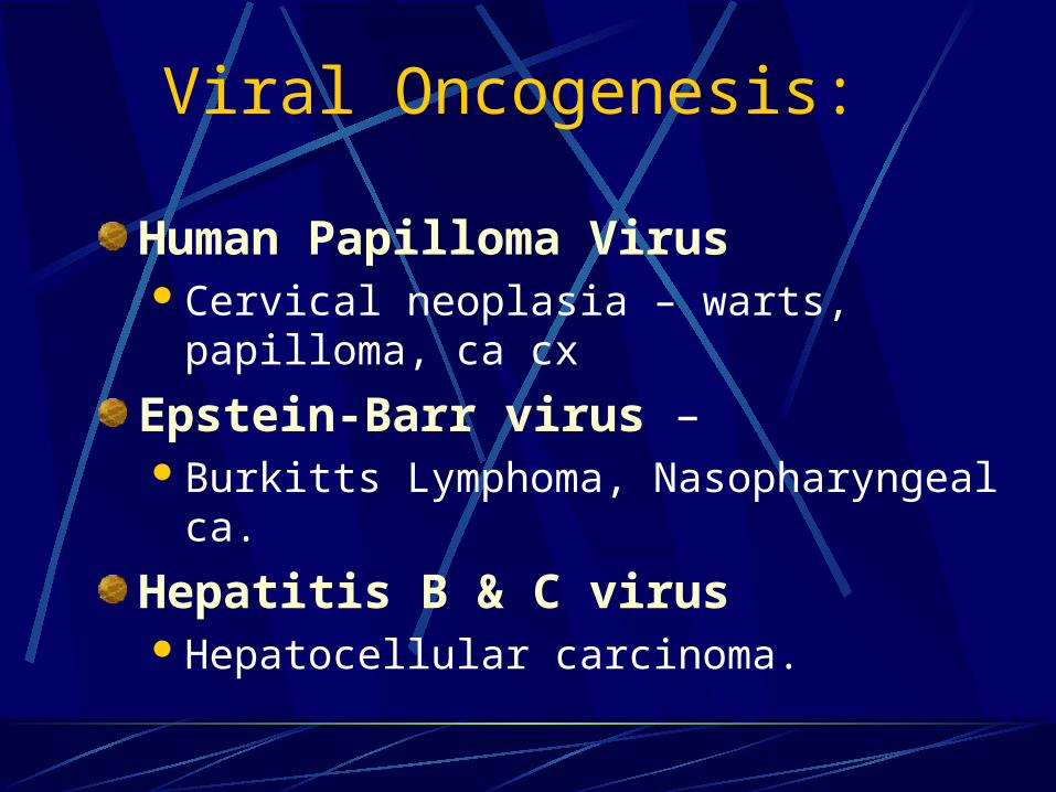
Viral Oncogenesis:
Human Papilloma Virus Cervical neoplasia – warts, papilloma, ca cx
Epstein-Barr virus – Burkitts Lymphoma, Nasopharyngeal ca.
Hepatitis B & C virusHepatocellular carcinoma.

Radiation Carcinogenesis:
Ionizing radiation dysjunction random fusion mutation.
X Ray workers – LeukemiaRadio-isotopes – Thyroid carcinomaAtomic explosion – Skin cancer, Leukemia
Mutations
Neoplasia

Hereditary Causes:
Due to inhereted abnormal genes.
FAP – gene C5, polyposis Adenocarcinoma colon
Retinoblastoma – Rb gene – (C13)
Neuroblastoma – (C17)
Trisomy 21 – Down’s syndrome – Leukemias in infants.

Clinical Manifestations of Cancer
Tissue IntegrityCompressed and eroded blood vessels, ulceration
and necrosis, frank bleeding, and hemorrhage
Cancer CachexiaWeight loss and wasting of body fat and muscle
tissue; profound weakness, anorexia, and anemia
Paraneoplastic SyndromesManifestations in sites not directly affected by the
disease

Molecular Basis of Neoplasia:
Proto-oncogeneProto-oncogene
OncogeneOncogene
V-Onc
V-Onc
Other
Other
Heredity
Heredity
Radiation
Radiation
Chemical
Chemical

Multiple Genetic Changes Cause Cancer
Cancers result from a series of genetic changes in a cell lineage
Some cancers begin with an inherited germ line mutation.
Some inherited cancers follow a dominant pattern, e.g. inherited retinoblastoma caused by a mutation in the Rb tumor supressor gene increases cancer risk 10,000 times.
More than one somatic mutation is often necessary.
Accumulation of mutations over time leads to uncontrolled cell division.
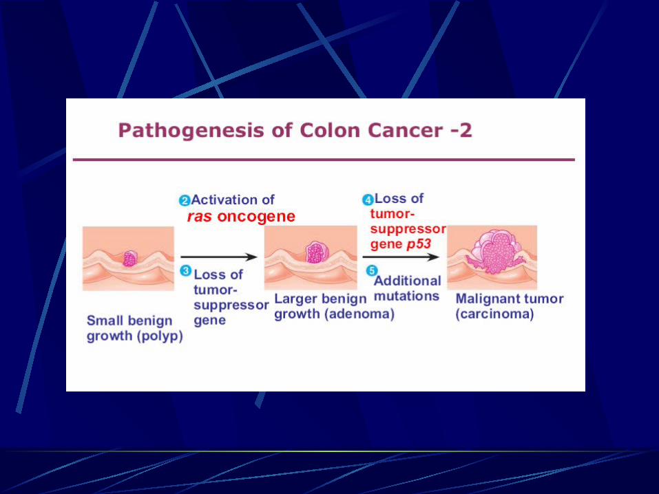
Example: Colon cancer develops in a stepwise fashion.

Multiple Genetic Changes Cause Cancer
Multiple Hits and Multiple Factors Knudson proposed that carcinogenesis requires 2 hits
1st event – initiation Carcinogen = initiator
2nd event – promotion Agent = promoter
Multiple hits occur – 5 or more Each hit produces a change in the genome which is
transmitted to its progeny (ie. clone) Lag period
Time between exposure (first hit) and development of clinically apparent cancer
Altered cell shows no abnormality during lag period

Multiple Genetic Changes Cause Cancer

Multiple Genetic Changes Cause Cancer
1. DNA of a normal cell This piece of DNA is an exact copy of the DNA from which
it came. When the parent cell divided to create two cells, the cell's DNA also divided, creating two identical copies of the original DNA.

2. Mutation of DNAThis DNA has suffered a mutation, either through mis-copying (when its parent cell divided), or through the damaging effects of exposure to radiation or a chemical carcinogen.

3. Genetically altered cell• Body cells replicate through mitosis, they respond to their surrounding
cells and replicate only to replace other cells. Sometimes a genetic mutation will cause a cell and its descendants to reproduce even though replacement cells are not needed.The DNA of the cell highlighted above has a mutation that causes the cell to replicate even though this tissue doesn't need replacement cells at this time or at this place.

4. Spread and second mutationThe genetically altered cells have, over time, reproduced unchecked, crowding out the surrounding normal cells. The growth may contain one million cells and be the size of a pinhead. At this point the cells continue to look the same as the surrounding healthy cells.
After about a million divisions, there's a good chance that one of the new cells will have mutated further. This cell, now carrying two mutant genes, could have an altered appearance and be even more prone to reproduce unchecked.
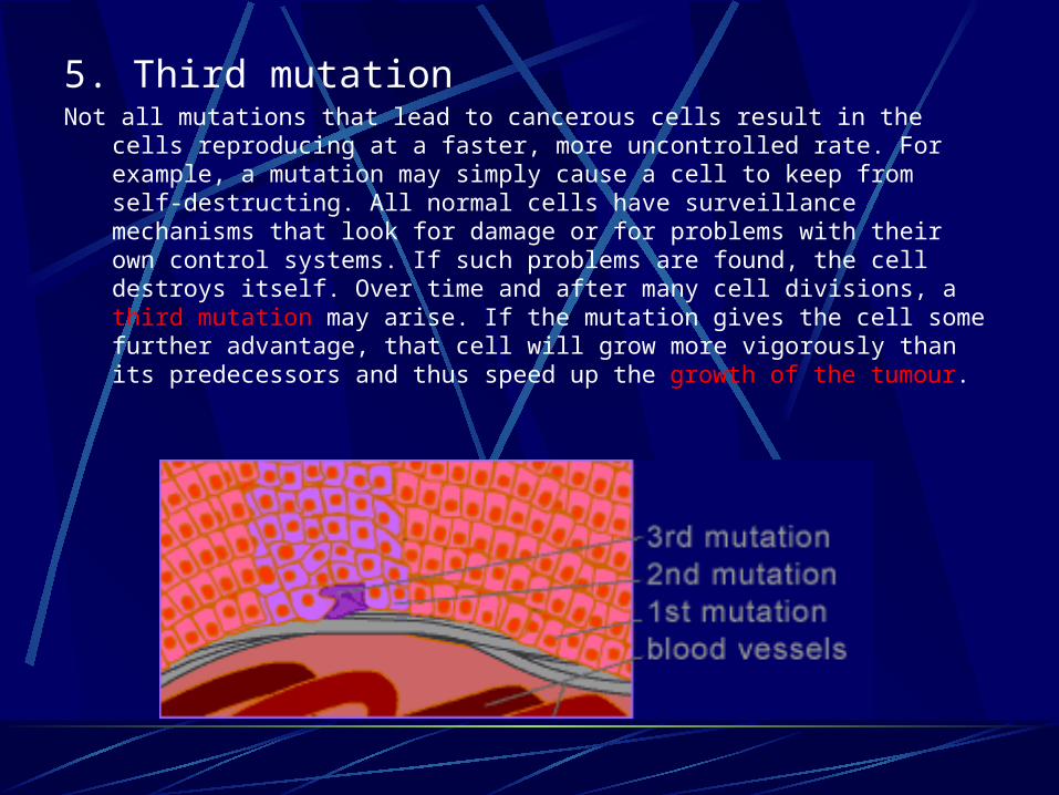
5. Third mutationNot all mutations that lead to cancerous cells result in the cells reproducing at
a faster, more uncontrolled rate. For example, a mutation may simply cause a cell to keep from self-destructing. All normal cells have surveillance mechanisms that look for damage or for problems with their own control systems. If such problems are found, the cell destroys itself. Over time and after many cell divisions, a third mutation may arise. If the mutation gives the cell some further advantage, that cell will grow more vigorously than its predecessors and thus speed up the growth of the tumour.

6. Fourth mutationThe new type of cells grow rapidly, allowing for more opportunities for
mutations. The next mutation paves the way for the development of an even more aggressive cancer.
At this point the tumour is still contained.

7. Breaking through the membraneThe newer, wilder cells created by another mutation are able to push their way through
the epithelial tissue's basement membrane, which is a meshwork of protein that normally creates a barrier. The invasive cells in this tumour are no longer contained.
At this point the cancer is still too small to be detected.

8. AngiogenesisOften during the development of earlier stages of the tumour, or perhaps by the time the tumour has broken through the basement membrane (as pictured above), angiogenesis takes place. Angiogenesis is the recruitment of blood vessels from the network of neighbouring vessels. Without blood and the nutrients it carries, a tumour would be unable to continue growing. With the new blood supply, however, the growth of the tumour accelerates; it soon contains thousand million cells and, now the size of a small grape, is large enough to be detected as a lump

9.Invasion and dispersalThe tumour has now invaded the tissue beyond the basement membrane.
Individual cells from the tumour enter into the network of newly formed blood vessels, using these vessels as highways by which they can move to other parts of the body. A tumour as small as a gram can send out a million tumour cells into blood vessels a day.

10. Tumour cells travel – metastasis
What makes most tumours so lethal is their ability to metastasize -- that is, establish new tumour sites at other locations throughout the body.Secondary tumours.
Metastasis is now underway, as tumour cells from the original cancer growth travel throughout the body. Most of these cells will die soon after entering the blood or lymph circulation.

11. MetastasisTo form a secondary tumour, a tumour cell needs to leave the vessel system and invade tissue. The cell must attach itself to a vessel's wall. Once this is done, it can work its way through the vessel and enter the tissue.
Although perhaps less than one in 10,000 tumour cells will survive long enough to establish a new tumour site, a few survivors can escape and initiate new colonies of the cancer.


Multi-step Theory
Initiation
Promotion
Progression

InitiationAn initiated cell is one in which a chemical carcinogen has interacted with DNA to produce a mutation, often a single base alteration, in the genome.
An initiated cell is not a tumor cell because it has not yet acquired autonomy of growth.
The DNA alteration may remain undetected throughout the life of the organism unless further events stimulate development of a tumor.

Tumor PromotionIn general, tumor promotion can be viewed as the clonal expansion of an initiated cell via altered gene expression that gives the cell a selective growth advantage.
Tumor promoters cause cells to proliferate but not to terminally differentiate, resulting in proliferation of preneoplastic cells (benign lesions).
Unlike initiators, most promoters do not bind covalently to DNA and usually do not cause mutations.

Tumor ProgressionTumor progression describes the process whereby tumors acquire the ability to grow, invade local tissue and establish distant metastases.
Increased genetic instability and karyotypic alterations are hallmarks of progression.
Inherited or acquired mutations in genes such as p53 or DNA mismatch repair can increase the rate of mutation in other genes (mutator phenotype) and, therefore, promote the tumor progression.

Phenotypic characteristics of cancer cells
Immortalization
Transformation
Loss of contact growth inhibition
Autonomy of proliferation
Avoidance of apoptosis
Aberrant differentiation
Induction of angiogenesis

Overview of Carcinogenesis