Embed Size (px)
Citation preview
[Expand]
[Expand]
Lecture - Fetal DevelopmentFrom Embryology
Embryology - 22 Oct 2015 Translate
Introduction
Click Here to play on mobile device
The fetal period (9-36weeks) is about continueddifferentiation of organs andtissues, most importantlythis period is about growthboth in size and weight.
The long Fetal period (4xthe embryonic period) is atime of extensive growth insize and mass as well as ongoing differentiation of organsystems established in the embryonic period and do so atdifferent times. For example, the brain continues to grow anddevelop extensively during this period (and postnatally), therespiratory system differentiates (and completes only justbefore birth), the urogenital system further differentiatesbetween male/female, endocrine and gastrointestinal tractbegins to function.
Note - Some of the content of this lecture has been discussedin earlier systems development lectures.
Lecture - Print PDF
Lecture Objectives
1. Understanding of fetal growth - length and weight2. Understanding of fetal systems development/changes3. Understanding of fetal abnormalities
First Trimester (1 - 12 weeks) - embryonic and early fetalSecond Trimester (13 - 24 weeks) - organ development and function, growthThird Trimester (25 - 40 weeks) - organ function and rapid growth
Lecture Resources
Movies

[Expand]
Fetal length and weight changes
Fetal head (12 weeks)
References
MusculoskeletalOngoing process of ossification.
endochondral and intramembranousgrowth in long bone length
Achondroplasia (common form of short limb dwarfism) thirdtrimester ultrasound very shortened long bones.
increased limb lengthcontinues postnatally
rapid growth during pubertyrelocation of haemopoietic stem cells to bone marrow
[Expand]
[Expand]
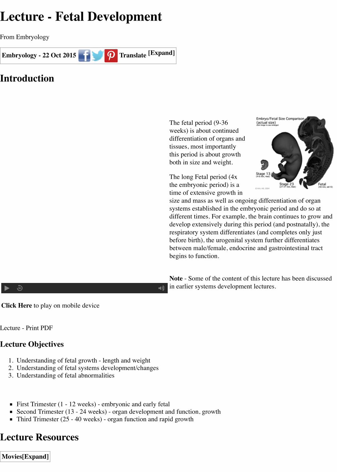
Timeline of events in Human Neural Development
Brain Growth
Endochondral ossification Intramembranous ossification
The collapsed tables below are for information purposes and are not examinable.
Table Of Ossification Of The Bones Of The Superior Extremity
Table Of Ossification Of The Bones Of The Inferior Extremity
Links: Bone Development Timeline | Bone Histology
Fetal Neural
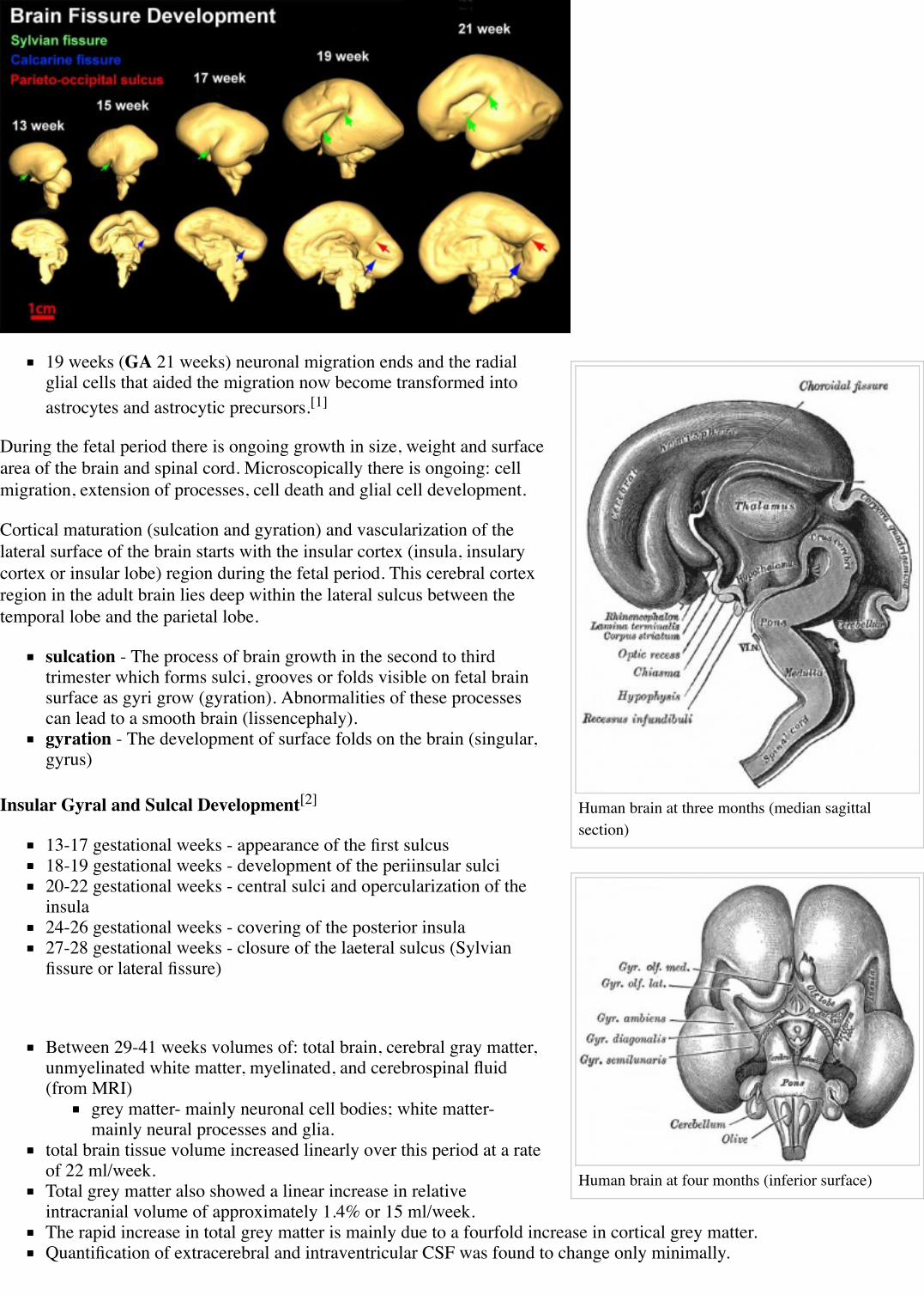
Human brain at three months (median sagittalsection)
Human brain at four months (inferior surface)
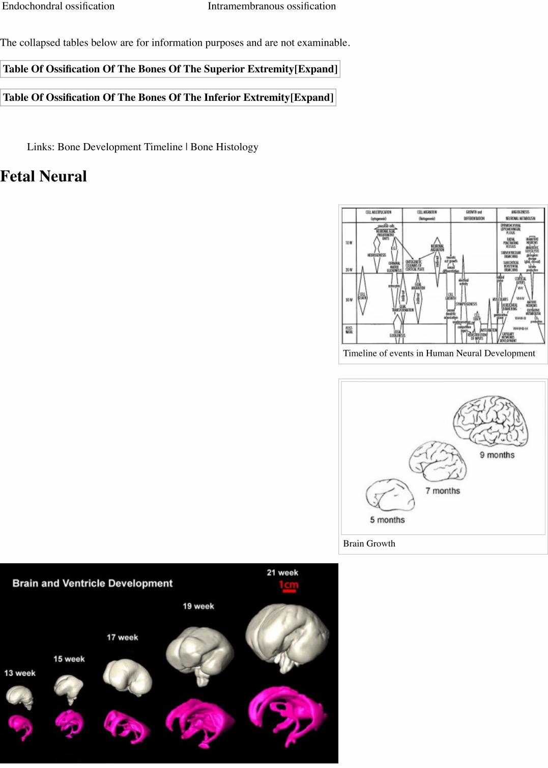
19 weeks (GA 21 weeks) neuronal migration ends and the radialglial cells that aided the migration now become transformed intoastrocytes and astrocytic precursors.[1]
During the fetal period there is ongoing growth in size, weight and surfacearea of the brain and spinal cord. Microscopically there is ongoing: cellmigration, extension of processes, cell death and glial cell development.
Cortical maturation (sulcation and gyration) and vascularization of thelateral surface of the brain starts with the insular cortex (insula, insularycortex or insular lobe) region during the fetal period. This cerebral cortexregion in the adult brain lies deep within the lateral sulcus between thetemporal lobe and the parietal lobe.
sulcation - The process of brain growth in the second to thirdtrimester which forms sulci, grooves or folds visible on fetal brainsurface as gyri grow (gyration). Abnormalities of these processescan lead to a smooth brain (lissencephaly).gyration - The development of surface folds on the brain (singular,gyrus)
Insular Gyral and Sulcal Development[2]
13-17 gestational weeks - appearance of the first sulcus18-19 gestational weeks - development of the periinsular sulci20-22 gestational weeks - central sulci and opercularization of theinsula24-26 gestational weeks - covering of the posterior insula27-28 gestational weeks - closure of the laeteral sulcus (Sylvianfissure or lateral fissure)
Between 29-41 weeks volumes of: total brain, cerebral gray matter,unmyelinated white matter, myelinated, and cerebrospinal fluid(from MRI)
grey matter- mainly neuronal cell bodies; white matter-mainly neural processes and glia.
total brain tissue volume increased linearly over this period at a rateof 22 ml/week.Total grey matter also showed a linear increase in relativeintracranial volume of approximately 1.4% or 15 ml/week.The rapid increase in total grey matter is mainly due to a fourfold increase in cortical grey matter.Quantification of extracerebral and intraventricular CSF was found to change only minimally.

Human brain at five months (outer surface)
(Text - modified from Huppi etal., (1998) Quantitative magnetic resonance imaging of brain development in premature andmature newborns. Ann Neurol 43(2):224-235.)
Neural development will continue after birth with substantial growth,death and reorganization occuring during the postnatally (MH - postnatalnot described in this current lecture)
Links: Neural System - Fetal | Neuroscience - Regionalspecification of the developing brain(http://www.ncbi.nlm.nih.gov/bookshelf/br.fcgi?book=neurosci&part=A1465&rendertype=figure&id=A1466)
Fetal CardiovascularMH - covered in last week's lecture Late Vascular Development.
the 3 septation events (atrial, ventricular andoutflow tract) should be completed by the endof the first trimester.the 3 vascular shunts (foramen ovale, ductusarteriosus, ductus venosus) remain open untilafter birth.
Blood Cells
fetal RBCs contain fetal haemoglobin(hemoglobin F or HbF).fetal neutrophils, monocytes, andmacrophages are produced.mononuclear phagocytes do not mature untilafter birth
FetalCirculationPage | Play
Immune System
maternal placenta transfer of IgG not other immunoglobulin isotypes.fetal lymphocytes (mature T and B cells) produced not activated
MH - see postnatal lecture - maternal milk IgG and IgA antibodies, leukocytes, secretory IgA, lactoferrin, lysozyme, andoligosaccharides and glycoconjugates that are receptor analogs for microbial adhesins and toxins.
Fetal RespiratoryMH - covered also in Lecture - Respiratory Development.
Month 3 to 6 - lungs appear glandular, end month 6 alveolar cells type 2 appear and begin to secrete surfactant.
Month 7 - respiratory bronchioles proliferate and end in alveolar ducts and sacs.
Lung Stages
week 4 - 5 embryonicweek 5 - 17 pseudoglandularweek 16 - 25 canalicularweek 24 - 40 terminal sac
late fetal - 8 years alveolar
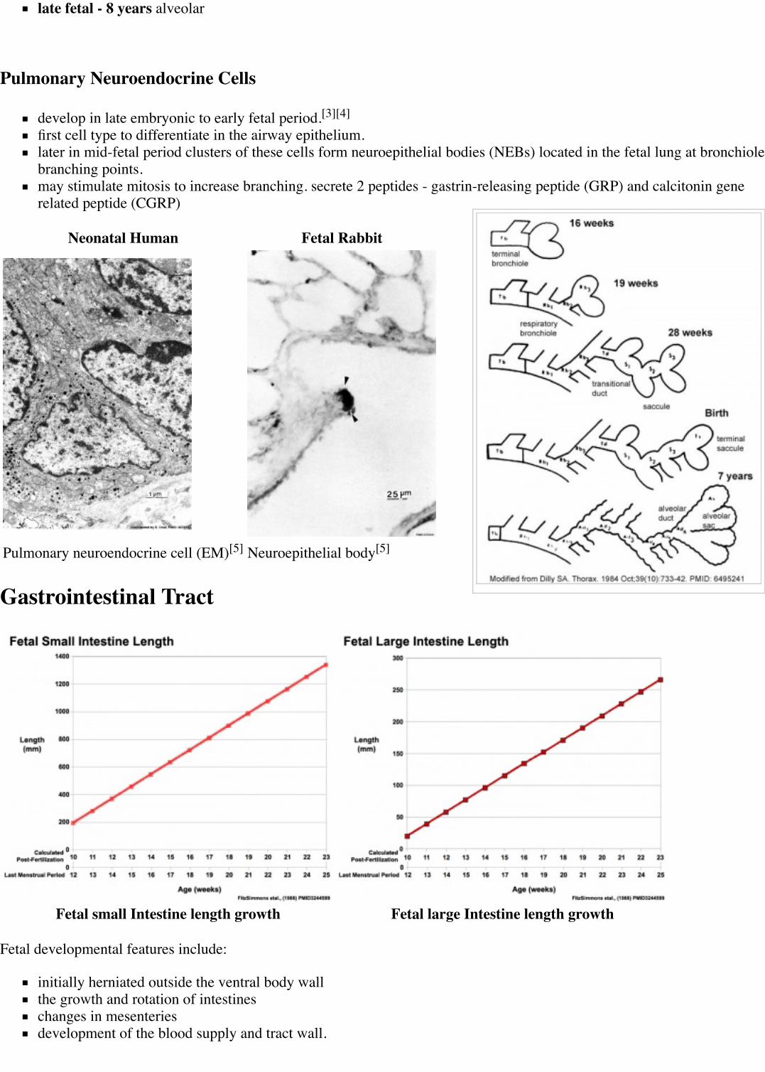
Pulmonary Neuroendocrine Cells
develop in late embryonic to early fetal period.[3][4]
first cell type to differentiate in the airway epithelium.later in mid-fetal period clusters of these cells form neuroepithelial bodies (NEBs) located in the fetal lung at bronchiolebranching points.may stimulate mitosis to increase branching. secrete 2 peptides - gastrin-releasing peptide (GRP) and calcitonin generelated peptide (CGRP)
Neonatal Human Fetal Rabbit
Pulmonary neuroendocrine cell (EM)[5] Neuroepithelial body[5]
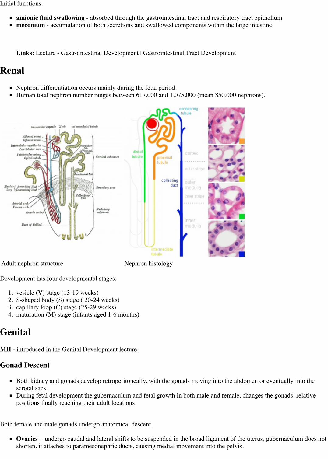
Gastrointestinal Tract
Fetal small Intestine length growth Fetal large Intestine length growth
Fetal developmental features include:
initially herniated outside the ventral body wallthe growth and rotation of intestineschanges in mesenteriesdevelopment of the blood supply and tract wall.
Initial functions:
amionic fluid swallowing - absorbed through the gastrointestinal tract and respiratory tract epitheliummeconium - accumulation of both secretions and swallowed components within the large intestine
Links: Lecture - Gastrointestinal Development | Gastrointestinal Tract Development
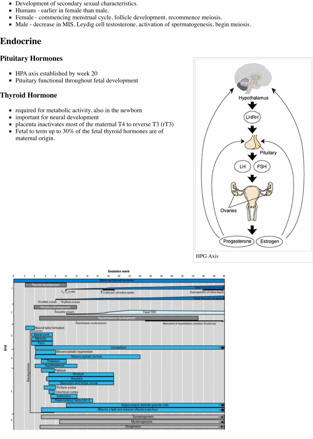
RenalNephron differentiation occurs mainly during the fetal period.Human total nephron number ranges between 617,000 and 1,075,000 (mean 850,000 nephrons).
Adult nephron structure Nephron histology
Development has four developmental stages:
1. vesicle (V) stage (13-19 weeks)2. S-shaped body (S) stage ( 20-24 weeks)3. capillary loop (C) stage (25-29 weeks)4. maturation (M) stage (infants aged 1-6 months)
GenitalMH - introduced in the Genital Development lecture.
Gonad Descent
Both kidney and gonads develop retroperitoneally, with the gonads moving into the abdomen or eventually into thescrotal sacs.During fetal development the gubernaculum and fetal growth in both male and female, changes the gonads’ relativepositions finally reaching their adult locations.
Both female and male gonads undergo anatomical descent.
Ovaries - undergo caudal and lateral shifts to be suspended in the broad ligament of the uterus, gubernaculum does notshorten, it attaches to paramesonephric ducts, causing medial movement into the pelvis.

Testes - two anatomical phases in descent, transabdominal and transinguinal, under the influence of the shorteninggubernaculum.
Beginning
End
Testis DescentPage | Play
The testis (white) lies in the subserous fascia (spotted) a cavityprocessus vaginalis evaginates into the scrotum, and thegubernaculum (green) attached to the testis shortens drawing it intothe scotal sac. As it descends it passes through the inguinal canalwhich extends from the deep ring (transversalis fascia) to thesuperficial ring (external oblique muscle). Descent of the testes intothe scrotal sac begins generally during week 26 and may takeseveral days. The animation shows the path of a single testis.
Gubernaculum - mesenchymal structure occurring associated withgonad development and involved in testes descent. Two factors -insulin-like peptide hormone 3 (INSL3) and androgen, have beenshown to be involved with gubernaculum development.
Incomplete or failed descent can occur unilaterally or bilaterally, is more common in premature births, and can be completedpostnatally.
Data from a recent study of male human fetal (between 10 and 35 weeks) gonad position.
10 to 23 weeks - (9.45%) had migrated from the abdomen and were situated in the inguinal canal24 to 26 weeks - (57.9%) had migrated from the abdomen27 to 29 weeks - (16.7%) had not descended to the scrotum
Postnatal Genital Development
Not completed until puberty.
HPG Axis
Development of secondary sexual characteristics.Humans - earlier in female than male.Female - commencing menstrual cycle, follicle development, recommence meiosis.Male - decrease in MIS, Leydig cell testosterone, activation of spermatogenesis, begin meiosis.
Endocrine
Pituitary Hormones
HPA axis established by week 20Pituitary functional throughout fetal development
Thyroid Hormone
required for metabolic activity, also in the newbornimportant for neural developmentplacenta inactivates most of the maternal T4 to reverse T3 (rT3)Fetal to term up to 30% of the fetal thyroid hormones are ofmaternal origin.

Parathyroid Hormone
newborn has total calcium levels (approx 20 grams) accumulated mainly in the 3rd trimester (weeks 28–40)fetal parathyroid hormone (PTH) potentially available from 10–12 weeks and PTH does not cross the placentafetus relatively hypercalcemic, active transplacental transport of Ca2+ to fetusmaternal serum - calcium ions (Ca2+), inorganic phosphate (Pi) and PTH concentrations are within the non-pregnantnormal range throughout pregnancy.maternal bone turnover increases in the 3rd trimester.
(Based on Endocrinology - Materno—fetal calcium balance (http://www.ncbi.nlm.nih.gov/bookshelf/br.fcgi?book=endocrin&part=A742&rendertype=box&id=A876))
Pancreatic Hormones
maternal diabetes can affect fetal pancreas development (increase in fetal islet beta cells).
Gonadal Hormones
testosterone - required during fetal development for external genital development and internal genital tract in male.estrogens - secreted inactive precursor converted to active form by placenta.
Links: Endocrinology - Control of steroid production in the fetal gonads(http://www.ncbi.nlm.nih.gov/bookshelf/br.fcgi?book=endocrin&part=A972#A1056) | Neuroscience - The Effect ofSex Hormones on Neural Circuitry (http://www.ncbi.nlm.nih.gov/bookshelf/br.fcgi?book=neurosci&part=A2124)
Fetal SizeFetal length increases through both the second and third trimesters.Fetal growth parameters can now be accurately measured by ultrasound: biparietal diameter (BPD), abdominalcircumference (AC) and femoral diaphysis length (FDL).
Head Size
Fetal Graphs: Crown-Rump Length (CRL) | Third trimester CRL | Head Circumference | Head Circumference 2ndTrimester | Liver Weight | Pancreas Weight | Thymus Weight | Small Intestine Length | Large Intestine Length | Lengthand Weight Changes | Fetal Development
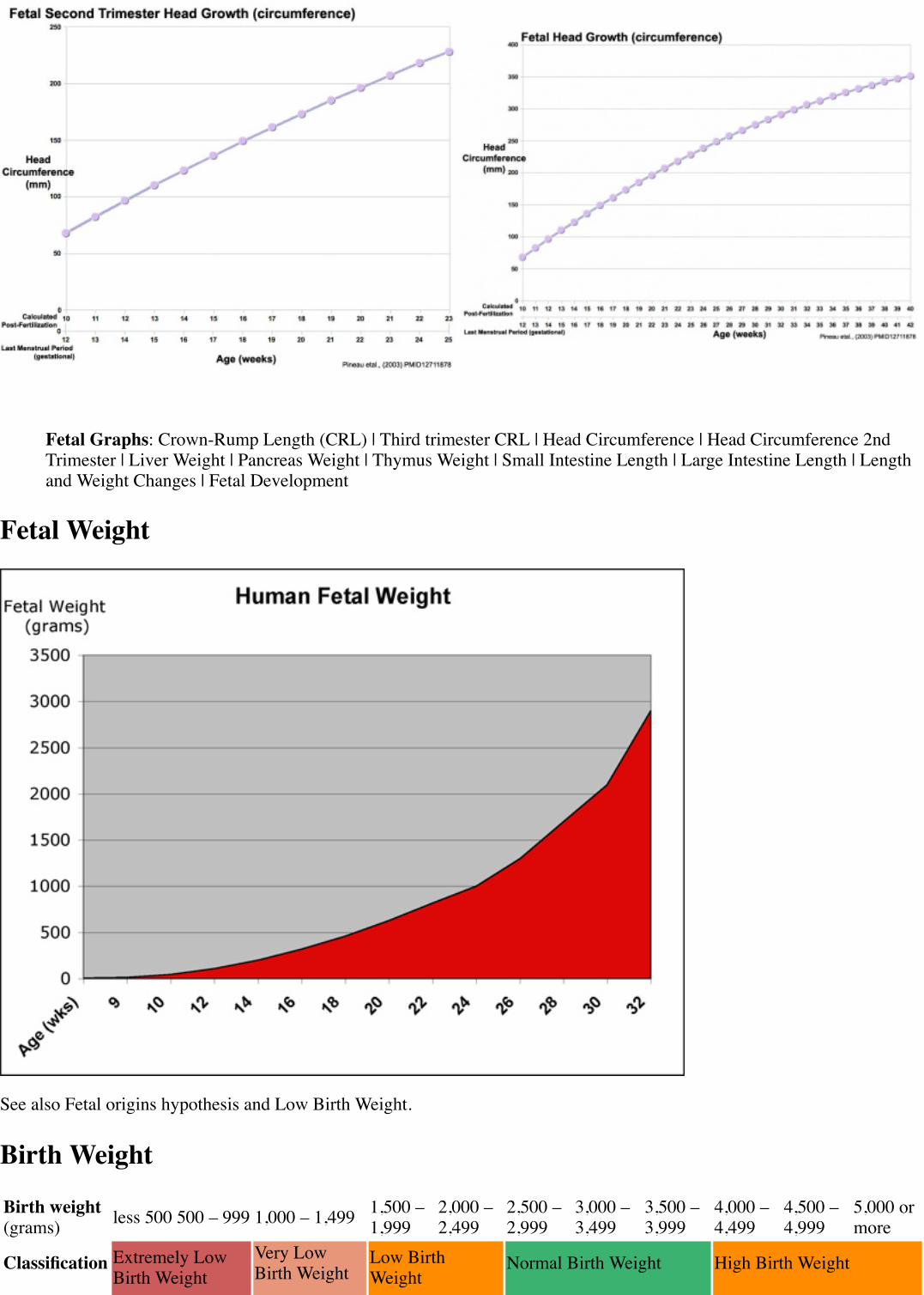
Fetal Weight
See also Fetal origins hypothesis and Low Birth Weight.
Birth Weight
Birth weight(grams) less 500 500 – 999 1,000 – 1,499 1,500 –
1,9992,000 –2,499
2,500 –2,999
3,000 –3,499
3,500 –3,999
4,000 –4,499
4,500 –4,999
5,000 ormore
Classification Extremely LowBirth Weight
Very LowBirth Weight
Low BirthWeight
Normal Birth Weight High Birth Weight
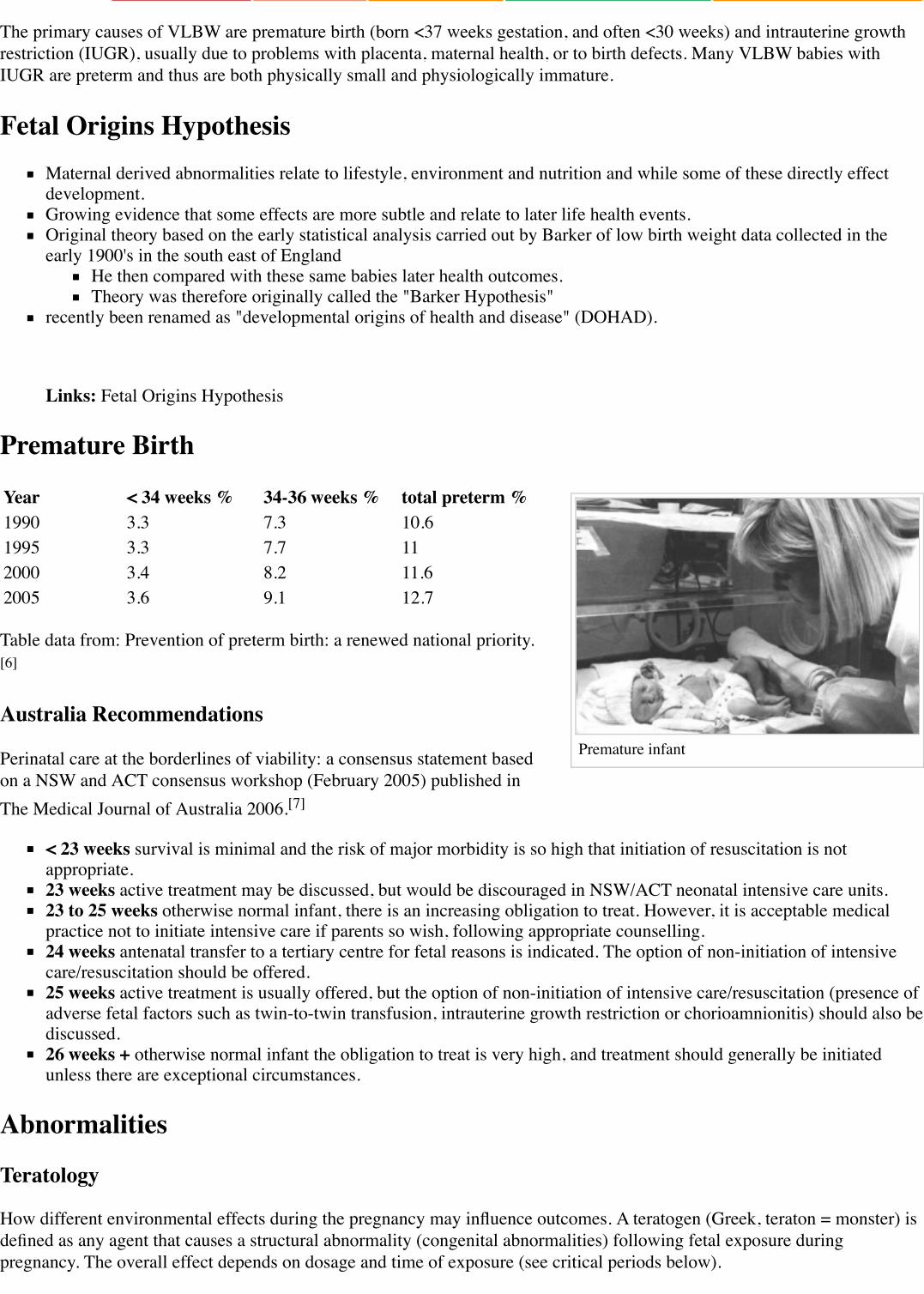
Premature infant
The primary causes of VLBW are premature birth (born <37 weeks gestation, and often <30 weeks) and intrauterine growthrestriction (IUGR), usually due to problems with placenta, maternal health, or to birth defects. Many VLBW babies withIUGR are preterm and thus are both physically small and physiologically immature.
Fetal Origins HypothesisMaternal derived abnormalities relate to lifestyle, environment and nutrition and while some of these directly effectdevelopment.Growing evidence that some effects are more subtle and relate to later life health events.Original theory based on the early statistical analysis carried out by Barker of low birth weight data collected in theearly 1900's in the south east of England
He then compared with these same babies later health outcomes.Theory was therefore originally called the "Barker Hypothesis"
recently been renamed as "developmental origins of health and disease" (DOHAD).
Links: Fetal Origins Hypothesis
Premature Birth
Year < 34 weeks % 34-36 weeks % total preterm %1990 3.3 7.3 10.61995 3.3 7.7 112000 3.4 8.2 11.62005 3.6 9.1 12.7
Table data from: Prevention of preterm birth: a renewed national priority.[6]
Australia Recommendations
Perinatal care at the borderlines of viability: a consensus statement basedon a NSW and ACT consensus workshop (February 2005) published inThe Medical Journal of Australia 2006.[7]
< 23 weeks survival is minimal and the risk of major morbidity is so high that initiation of resuscitation is notappropriate.23 weeks active treatment may be discussed, but would be discouraged in NSW/ACT neonatal intensive care units.23 to 25 weeks otherwise normal infant, there is an increasing obligation to treat. However, it is acceptable medicalpractice not to initiate intensive care if parents so wish, following appropriate counselling.24 weeks antenatal transfer to a tertiary centre for fetal reasons is indicated. The option of non-initiation of intensivecare/resuscitation should be offered.25 weeks active treatment is usually offered, but the option of non-initiation of intensive care/resuscitation (presence ofadverse fetal factors such as twin-to-twin transfusion, intrauterine growth restriction or chorioamnionitis) should also bediscussed.26 weeks + otherwise normal infant the obligation to treat is very high, and treatment should generally be initiatedunless there are exceptional circumstances.
Abnormalities
Teratology
How different environmental effects during the pregnancy may influence outcomes. A teratogen (Greek, teraton = monster) isdefined as any agent that causes a structural abnormality (congenital abnormalities) following fetal exposure duringpregnancy. The overall effect depends on dosage and time of exposure (see critical periods below).
Absolute risk - the rate of occurrence of an abnormal phenotype among individuals exposed to the agent. (e.g. fetal alcoholsyndrome)
Relative risk - the ratio of the rate of the condition among the exposed and the nonexposed. (e.g. smokers risk of having alow birth weight baby compared to non-smokers) A high relative risk may indicate a low absolute risk if the condition is rare.
Mutagen - a chemical or agent that can cause permanent damage to the deoxyribonucleic acid (DNA) in a cell. DNA damagein the human egg or sperm may lead to reduced fertility, spontaneous abortion (miscarriage), birth defects and heritablediseases.
Fetotoxicant - is a chemical that adversely affects the developing fetus, resulting in low birth weight, symptoms of poisoningat birth or stillbirth (fetus dies before it is born).
Synergism - when the combined effect of exposure to more than one chemical at one time, or to a chemical in combinationwith other hazards (heat, radiation, infection) results in effects of such exposure to be greater than the sum of the individualeffects of each hazard by itself.
Toxicogenomics - the interaction between the genome, chemicals in the environment, and disease. Cells exposed to a stress,drug or toxicant respond by altering the pattern of expression of genes within their chromosomes. Based on new genetic andmicroarray technologies.
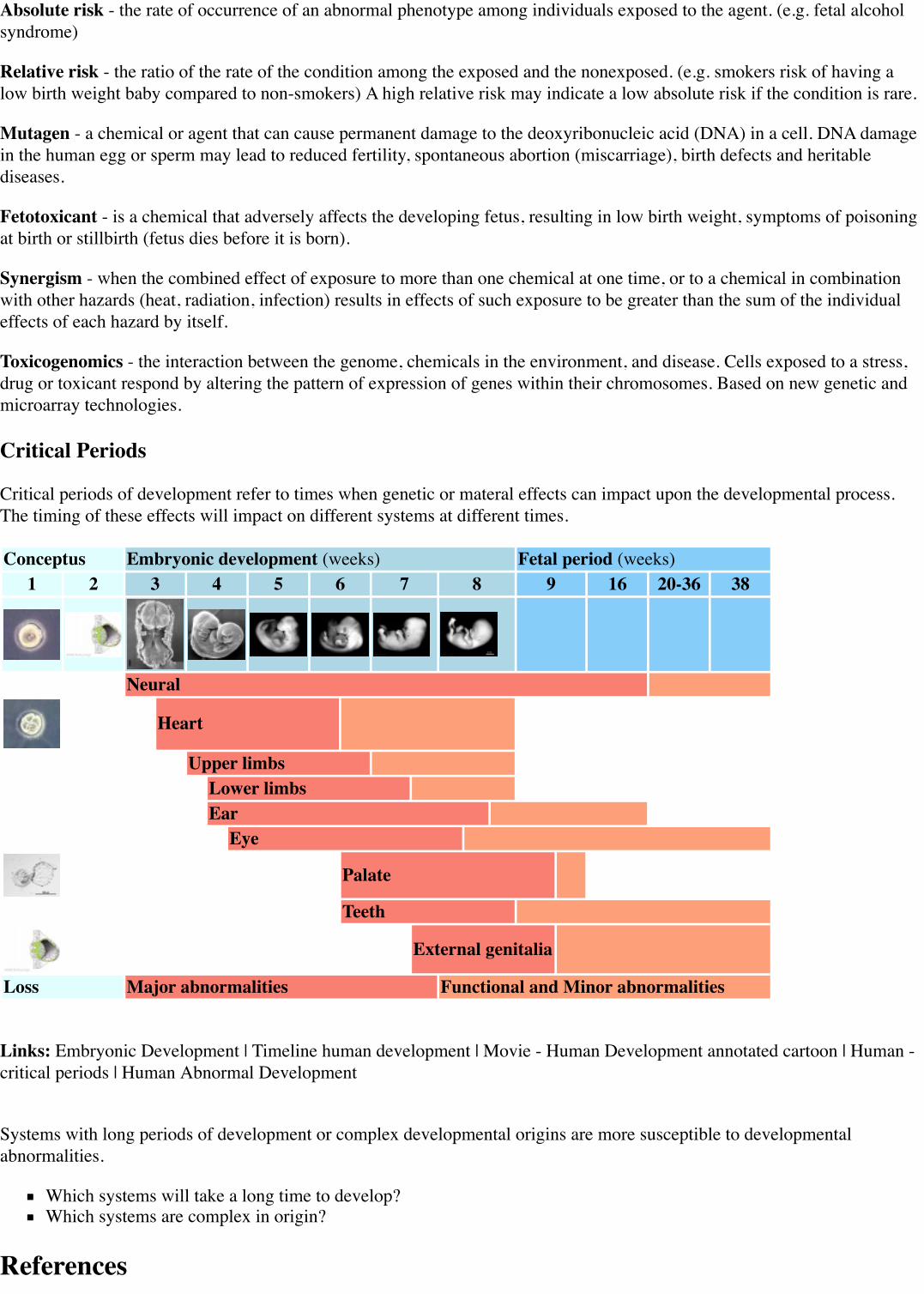
Critical Periods
Critical periods of development refer to times when genetic or materal effects can impact upon the developmental process.The timing of these effects will impact on different systems at different times.
Conceptus Embryonic development (weeks) Fetal period (weeks)1 2 3 4 5 6 7 8 9 16 20-36 38
Neural
Heart
Upper limbsLower limbsEar
Eye
Palate
Teeth
External genitalia
Loss Major abnormalities Functional and Minor abnormalities
Links: Embryonic Development | Timeline human development | Movie - Human Development annotated cartoon | Human -critical periods | Human Abnormal Development
Systems with long periods of development or complex developmental origins are more susceptible to developmentalabnormalities.
Which systems will take a long time to develop?Which systems are complex in origin?
References
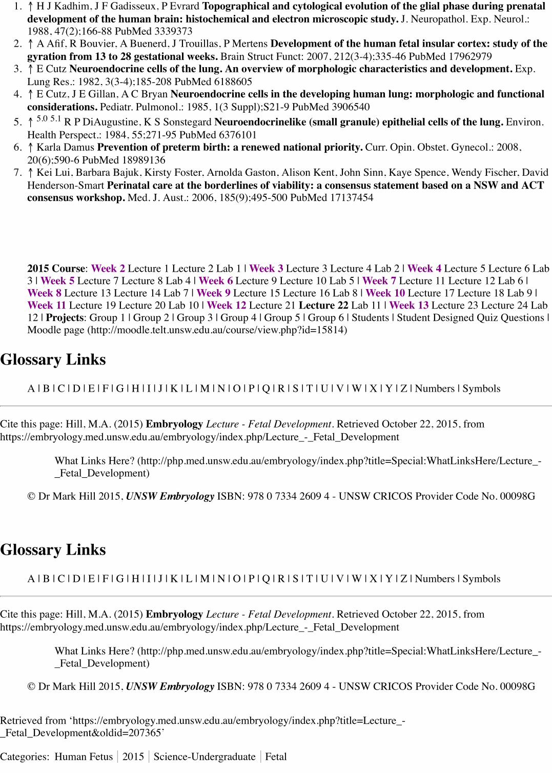
1. ↑ H J Kadhim, J F Gadisseux, P Evrard Topographical and cytological evolution of the glial phase during prenataldevelopment of the human brain: histochemical and electron microscopic study. J. Neuropathol. Exp. Neurol.:1988, 47(2);166-88 PubMed 3339373
2. ↑ A Afif, R Bouvier, A Buenerd, J Trouillas, P Mertens Development of the human fetal insular cortex: study of thegyration from 13 to 28 gestational weeks. Brain Struct Funct: 2007, 212(3-4);335-46 PubMed 17962979
3. ↑ E Cutz Neuroendocrine cells of the lung. An overview of morphologic characteristics and development. Exp.Lung Res.: 1982, 3(3-4);185-208 PubMed 6188605
4. ↑ E Cutz, J E Gillan, A C Bryan Neuroendocrine cells in the developing human lung: morphologic and functionalconsiderations. Pediatr. Pulmonol.: 1985, 1(3 Suppl);S21-9 PubMed 3906540
5. ↑ 5.0 5.1 R P DiAugustine, K S Sonstegard Neuroendocrinelike (small granule) epithelial cells of the lung. Environ.Health Perspect.: 1984, 55;271-95 PubMed 6376101
6. ↑ Karla Damus Prevention of preterm birth: a renewed national priority. Curr. Opin. Obstet. Gynecol.: 2008,20(6);590-6 PubMed 18989136
7. ↑ Kei Lui, Barbara Bajuk, Kirsty Foster, Arnolda Gaston, Alison Kent, John Sinn, Kaye Spence, Wendy Fischer, DavidHenderson-Smart Perinatal care at the borderlines of viability: a consensus statement based on a NSW and ACTconsensus workshop. Med. J. Aust.: 2006, 185(9);495-500 PubMed 17137454
2015 Course: Week 2 Lecture 1 Lecture 2 Lab 1 | Week 3 Lecture 3 Lecture 4 Lab 2 | Week 4 Lecture 5 Lecture 6 Lab3 | Week 5 Lecture 7 Lecture 8 Lab 4 | Week 6 Lecture 9 Lecture 10 Lab 5 | Week 7 Lecture 11 Lecture 12 Lab 6 |Week 8 Lecture 13 Lecture 14 Lab 7 | Week 9 Lecture 15 Lecture 16 Lab 8 | Week 10 Lecture 17 Lecture 18 Lab 9 |Week 11 Lecture 19 Lecture 20 Lab 10 | Week 12 Lecture 21 Lecture 22 Lab 11 | Week 13 Lecture 23 Lecture 24 Lab12 | Projects: Group 1 | Group 2 | Group 3 | Group 4 | Group 5 | Group 6 | Students | Student Designed Quiz Questions |Moodle page (http://moodle.telt.unsw.edu.au/course/view.php?id=15814)
Glossary LinksA | B | C | D | E | F | G | H | I | J | K | L | M | N | O | P | Q | R | S | T | U | V | W | X | Y | Z | Numbers | Symbols
Cite this page: Hill, M.A. (2015) Embryology Lecture - Fetal Development. Retrieved October 22, 2015, fromhttps://embryology.med.unsw.edu.au/embryology/index.php/Lecture_-_Fetal_Development
What Links Here? (http://php.med.unsw.edu.au/embryology/index.php?title=Special:WhatLinksHere/Lecture_-_Fetal_Development)
© Dr Mark Hill 2015, UNSW Embryology ISBN: 978 0 7334 2609 4 - UNSW CRICOS Provider Code No. 00098G
Retrieved from ‘https://embryology.med.unsw.edu.au/embryology/index.php?title=Lecture_-_Fetal_Development&oldid=207365’
Categories: Human Fetus 2015 Science-Undergraduate Fetal

This page was last modified on 22 October 2015, at 16:58.This page has been accessed 8,093 times.


![Qualitative changes in fetal trabecular meshwork fibers … · human fetal eyes (8–36 weeks of gestation), Ruano-Gil et al. [9] also hypothesized that mechanical forces induced](https://img.pdfslide.net/doc/110x75/5b94d13909d3f2e5688daf79/qualitative-changes-in-fetal-trabecular-meshwork-fibers-human-fetal-eyes-836.jpg)