Embed Size (px)
Citation preview
THE SPECIAL SENSES
Lecture Material is adapted from © 2013 Pearson Education, Inc. Human
Anatomy and Physiology
Dr. Henrik Pallos
© 2013 Pearson Education, Inc.
Special Senses
• Special sensory receptors
– Distinct, localized receptor cells in head
• Vision
• Taste
• Smell
• Hearing
• Equilibrium
© 2013 Pearson Education, Inc.
The Eye and Vision
• 70% of body's sensory receptors in eye
• Visual processing by nearly half cerebral cortex
– Primary visual cortex
– Visual association area
• Most of eye protected by cushion of fat and
bony orbit
© 2013 Pearson Education, Inc.
Accessory Structures of the Eye
• Protect the eye and aid eye function
1. Eyebrows
2. Eyelids (palpebrae)
3. Conjunctiva
4. Lacrimal apparatus
5. Extrinsic eye muscles
© 2013 Pearson Education, Inc.
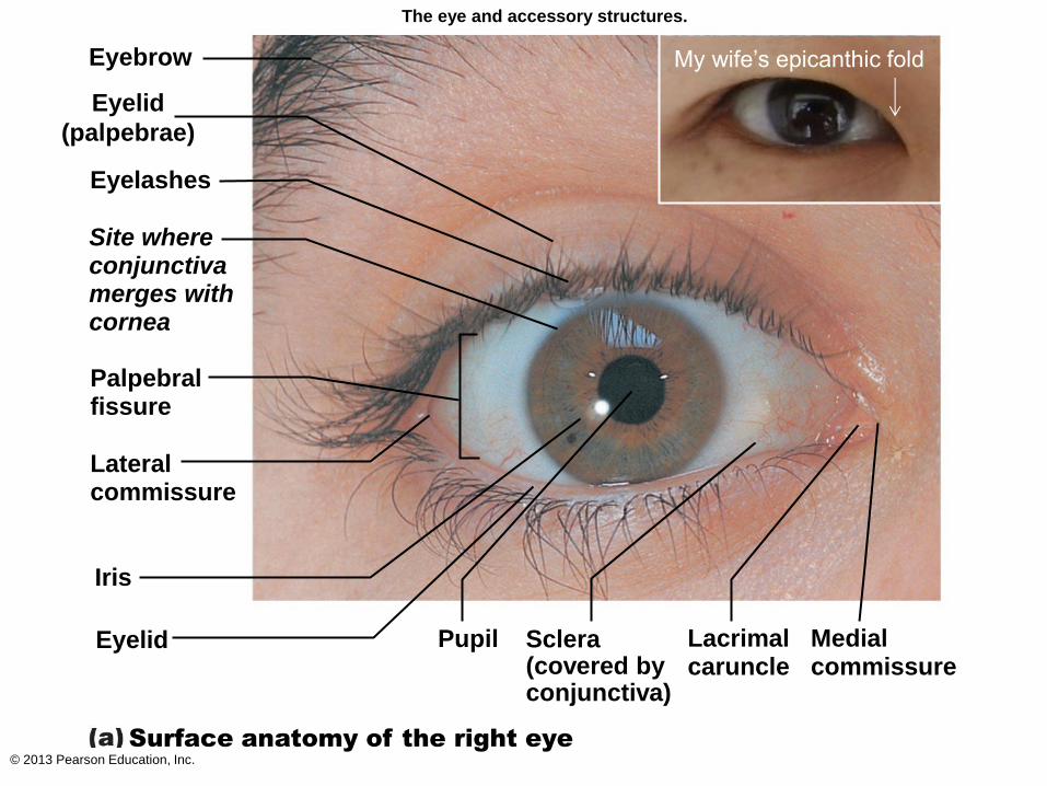
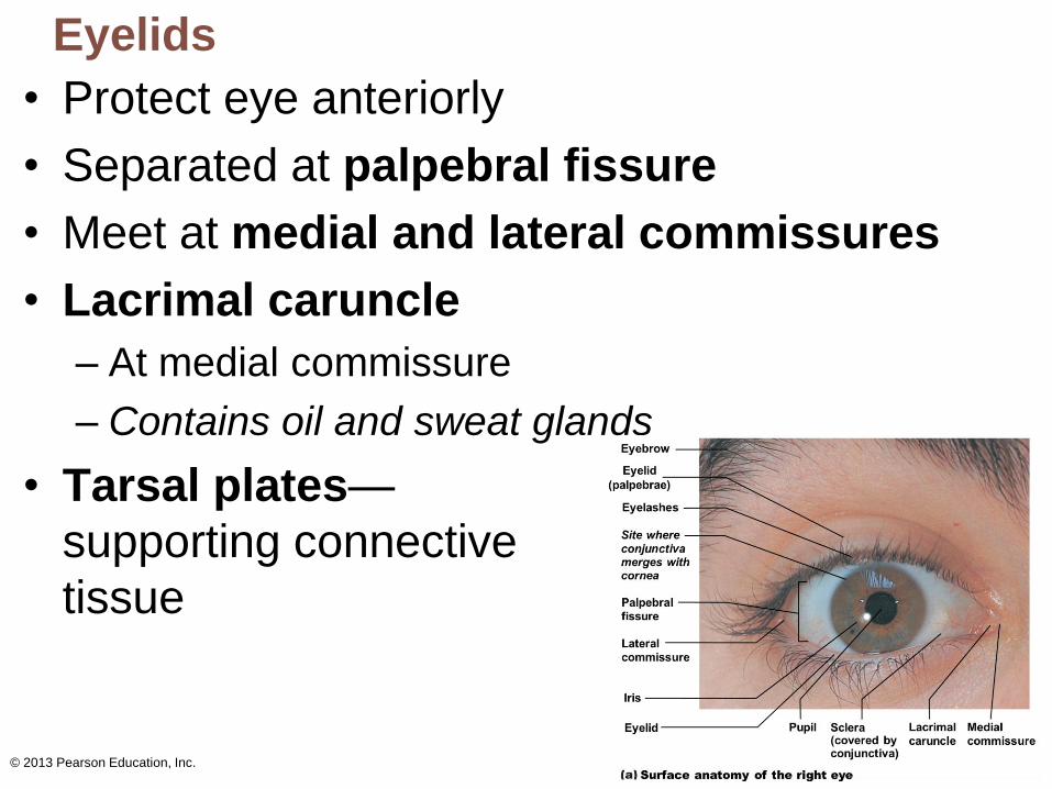
Eyebrow
Eyelid
(palpebrae)
Eyelashes
Site where conjunctiva merges with cornea
Palpebral fissure
Lateral commissure
Iris
Eyelid
Surface anatomy of the right eye
Pupil Sclera (covered by conjunctiva)
Lacrimal caruncle
Medial commissure
The eye and accessory structures.
My wife’s epicanthic fold
© 2013 Pearson Education, Inc.
Eyebrows
• Overlie supraorbital margins
• Function
1. Shade eye from sunlight
2. Prevent perspiration from reaching eye
© 2013 Pearson Education, Inc.
Eyelids
• Protect eye anteriorly
• Separated at palpebral fissure
• Meet at medial and lateral commissures
• Lacrimal caruncle
– At medial commissure
– Contains oil and sweat glands
• Tarsal plates—
supporting connective
tissue
© 2013 Pearson Education, Inc.
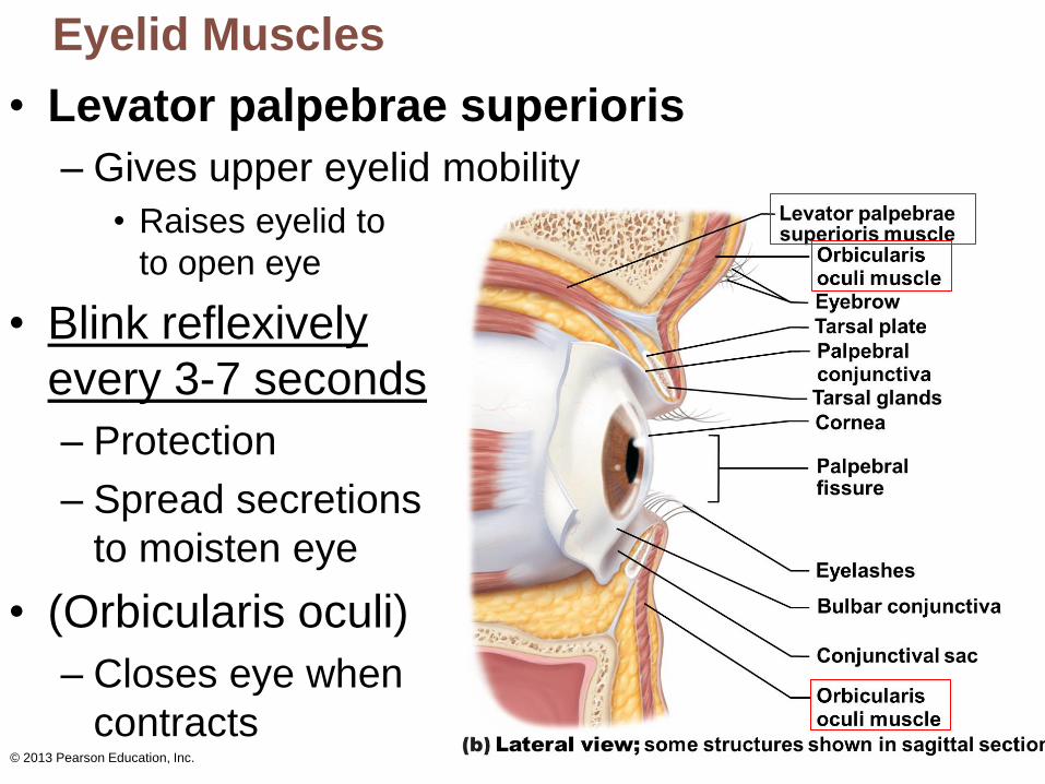
Eyelid Muscles
• Levator palpebrae superioris
– Gives upper eyelid mobility
• Raises eyelid to
to open eye
• Blink reflexively
every 3-7 seconds
– Protection
– Spread secretions
to moisten eye
• (Orbicularis oculi)
– Closes eye when
contracts
© 2013 Pearson Education, Inc.
Eyelids
• Eyelashes
– Nerve endings of follicles initiate reflex blinking
• Lubricating glands associated with eyelids
1. Tarsal (Meibomian) glands
• Modified sebaceous glands
• Oily secretion lubricates lid and eye
2. Ciliary glands between hair follicles
• Modified sweat glands
© 2013 Pearson Education, Inc.
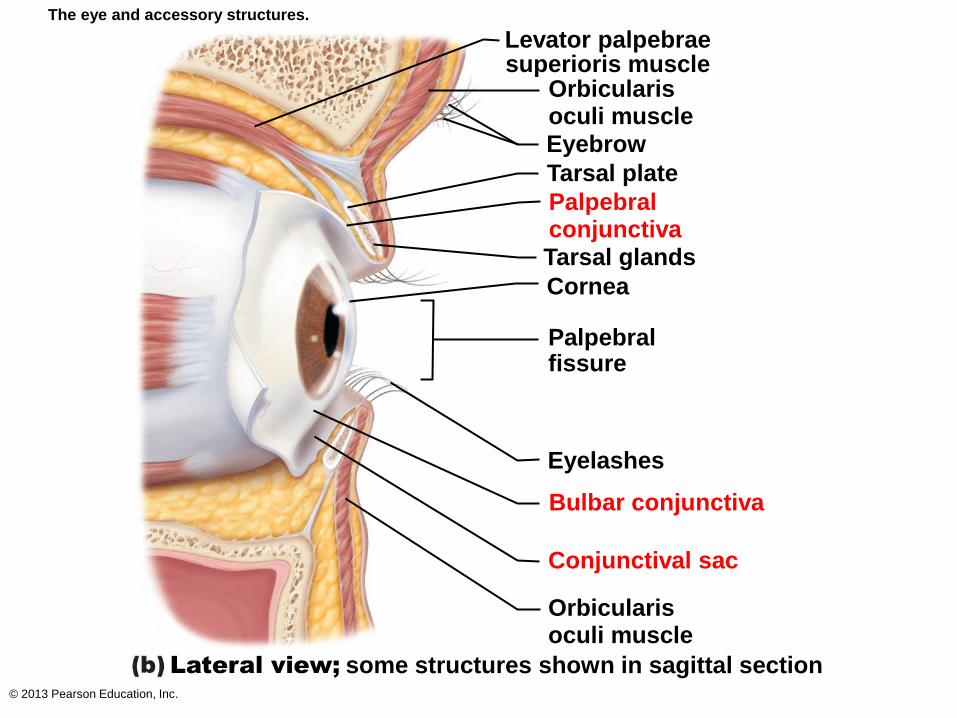
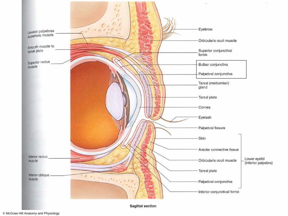
The eye and accessory structures.
Levator palpebrae superioris muscle
Orbicularis oculi muscle Eyebrow
Tarsal plate
Palpebral conjunctiva Tarsal glands
Cornea
Palpebral fissure
Eyelashes
Bulbar conjunctiva
Conjunctival sac
Orbicularis oculi muscle
some structures shown in sagittal section Lateral view;
© McGraw Hill Anatomy and Physiology
© 2013 Pearson Education, Inc.
Conjunctiva
• Transparent mucous membrane
– Produces a lubricating mucous secretion
1. Palpebral conjunctiva lines eyelids
2. Bulbar conjunctiva covers white of eyes
Conjunctival sac between palpebral and
bulbar conjunctiva
- Where contact lens rests
© 2013 Pearson Education, Inc.
Lacrimal Apparatus
• Lacrimal gland and ducts that drain into nasal cavity
• Lacrimal gland in orbit above lateral end of eye
• Lacrimal secretion (tears) – 1ml/day
– Dilute saline solution containing mucus, antibodies, and
lysozyme:
• cleanses and protects the eye surface as it lubricates it
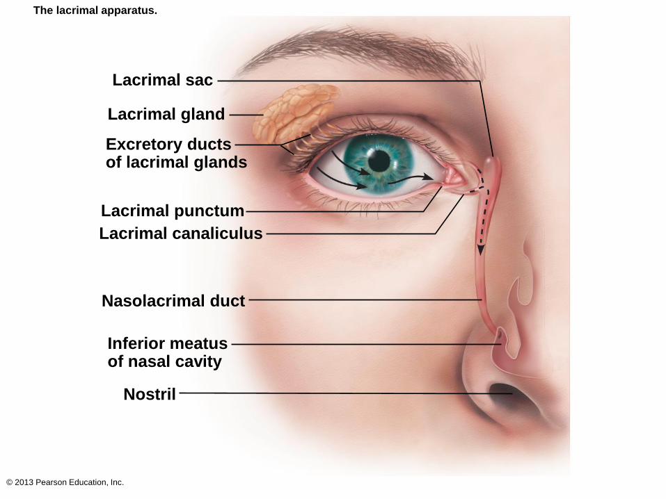
– Blinking spreads tears toward medial commissure
• Tears enter paired lacrimal canaliculi via lacrimal puncta
• Then drain into lacrimal sac and nasolacrimal duct
• Taste of eye drops
– Lacrimal secretion increases:
• Eyes are irritated (washes away irritant)
• Emotional (Why we have, we do not know.)
© 2013 Pearson Education, Inc.
The lacrimal apparatus.
Lacrimal sac
Lacrimal gland
Excretory ducts of lacrimal glands
Lacrimal punctum
Lacrimal canaliculus
Nasolacrimal duct
Inferior meatus of nasal cavity
Nostril
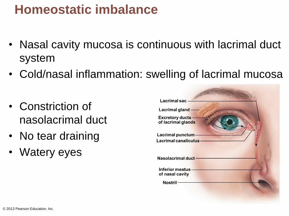
Homeostatic imbalance
• Nasal cavity mucosa is continuous with lacrimal duct
system
• Cold/nasal inflammation: swelling of lacrimal mucosa
• Constriction of
nasolacrimal duct
• No tear draining
• Watery eyes
© 2013 Pearson Education, Inc.
© 2013 Pearson Education, Inc.
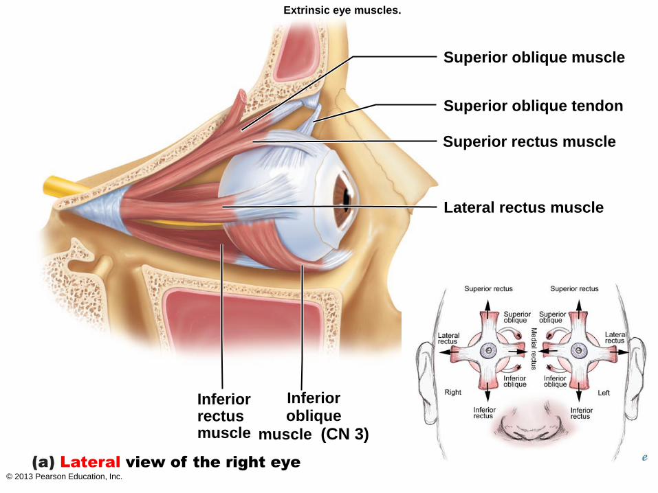
Extrinsic Eye Muscles
• 6 straplike extrinsic eye muscles
– Originate from bony orbit; insert on eyeball
1. enable eye to follow moving objects
2. maintain shape of eyeball
3. hold in orbit
• 4 rectus muscles originate from common tendinous
ring; names indicate movements
– Superior, inferior, lateral, medial rectus muscles
• 2 oblique muscles move eye in vertical plane and
rotate eyeball
– Superior and inferior oblique muscles
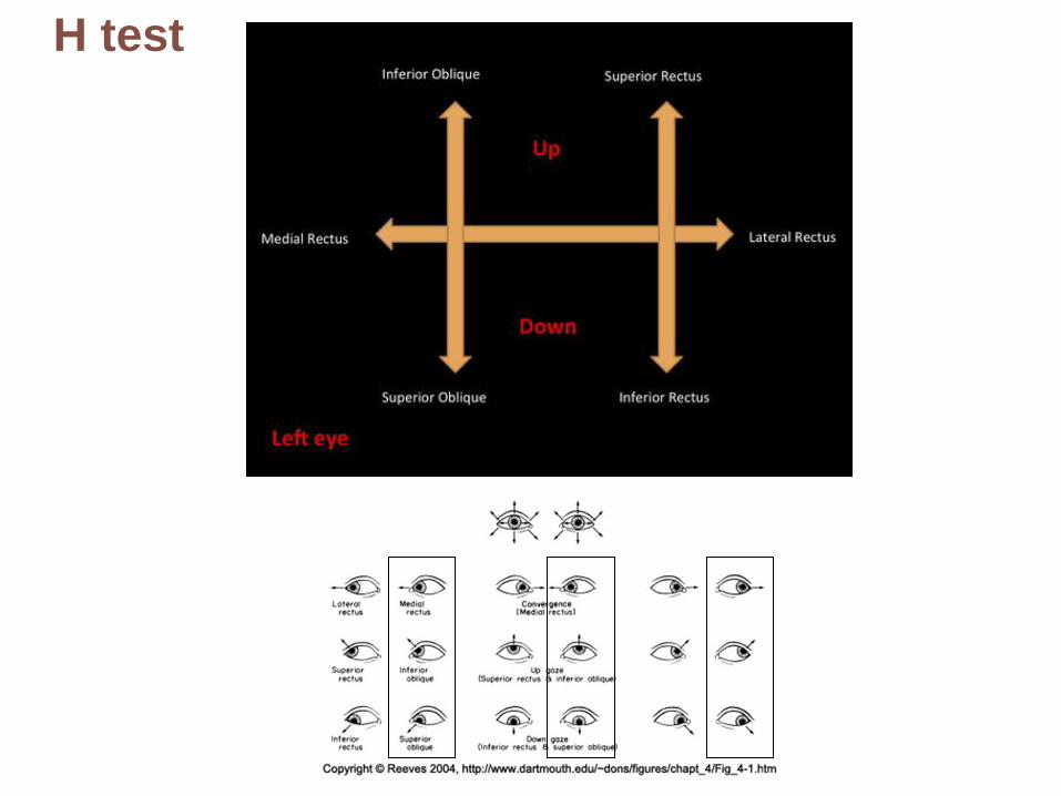
H test
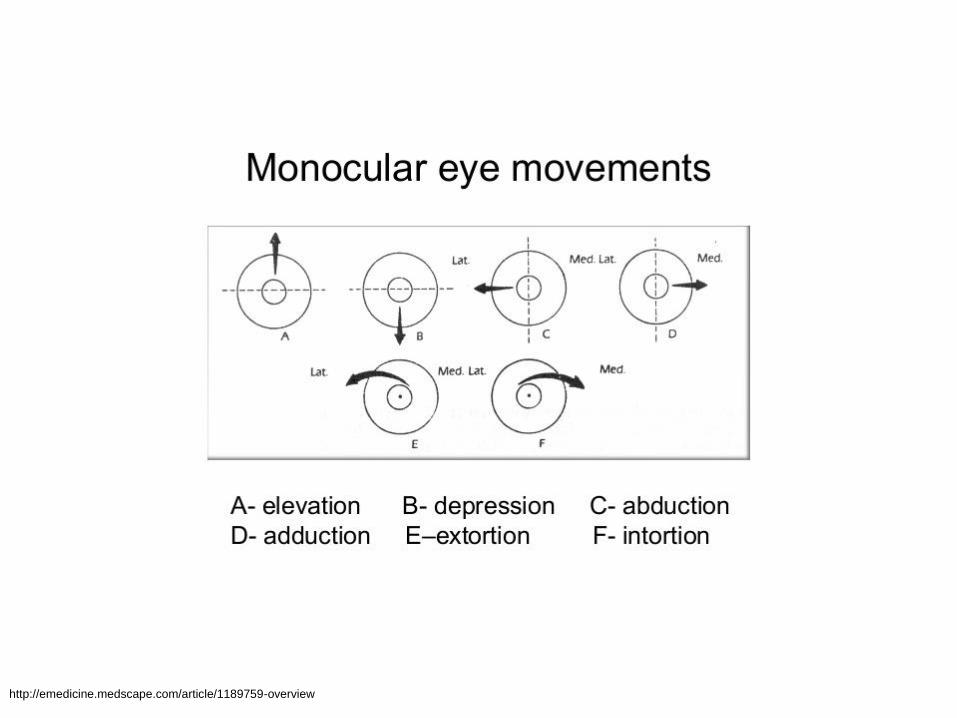
http://emedicine.medscape.com/article/1189759-overview

© 2013 Pearson Education, Inc.
Extrinsic eye muscles.
Trochlea
Axis of rotation of eye
Inferior rectus muscle (CN 3)
Medial rectus muscle (CN 3)
Lateral rectus muscle ( CN 6)
Common
tendinous ring
Superior oblique muscle
(CN 4)
Superior oblique tendon
Superior rectus muscle
(CN 3)
Superior view of the right eye
© 2013 Pearson Education, Inc.
Extrinsic eye muscles.
Superior oblique muscle
Superior oblique tendon
Superior rectus muscle
Lateral rectus muscle
Inferior rectus muscle
Inferior oblique
muscle (CN 3)
Lateral view of the right eye
© 2013 Pearson Education, Inc.
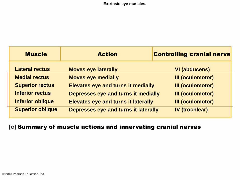
Extrinsic eye muscles.
Muscle Action Controlling cranial nerve
Summary of muscle actions and innervating cranial nerves
Lateral rectus
Medial rectus
Superior rectus
Inferior rectus
Inferior oblique
Superior oblique
Moves eye laterally
Moves eye medially
Elevates eye and turns it medially
Depresses eye and turns it medially
Elevates eye and turns it laterally
Depresses eye and turns it laterally
VI (abducens)
III (oculomotor)
III (oculomotor)
III (oculomotor)
III (oculomotor)
IV (trochlear)
Homeostatic imbalances
• Movements of external muscles of the two
eyes are not perfectly coordinated
• Image is not properly focused on the same
area of the visual field in each eye
• Two images are seen instead of one
© 2013 Pearson Education, Inc.
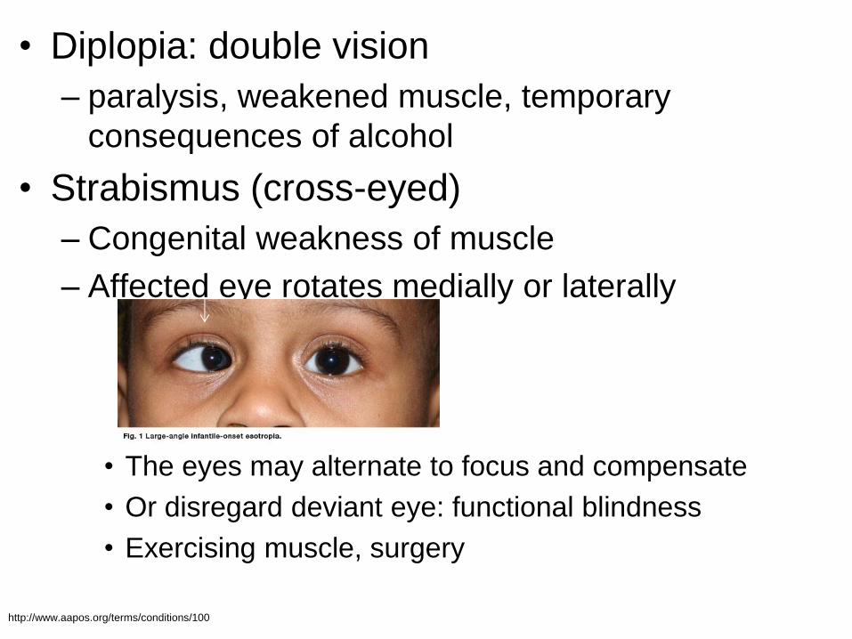
• Diplopia: double vision
– paralysis, weakened muscle, temporary
consequences of alcohol
• Strabismus (cross-eyed)
– Congenital weakness of muscle
– Affected eye rotates medially or laterally
• The eyes may alternate to focus and compensate
• Or disregard deviant eye: functional blindness
• Exercising muscle, surgery
http://www.aapos.org/terms/conditions/100
© 2013 Pearson Education, Inc.
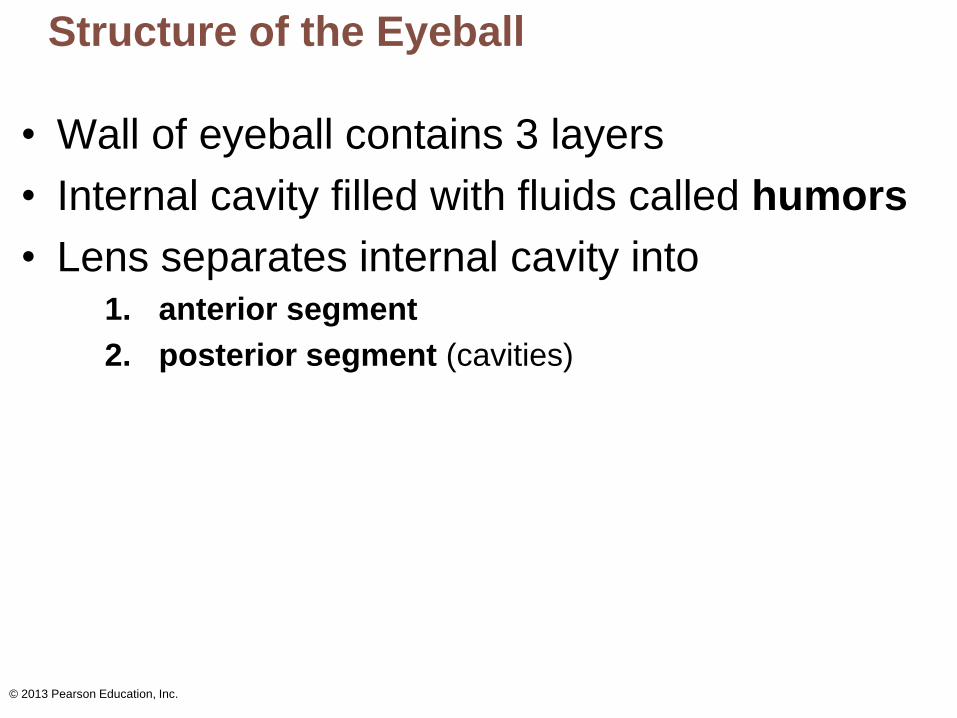
Structure of the Eyeball
• Wall of eyeball contains 3 layers
• Internal cavity filled with fluids called humors
• Lens separates internal cavity into 1. anterior segment
2. posterior segment (cavities)
© 2013 Pearson Education, Inc.
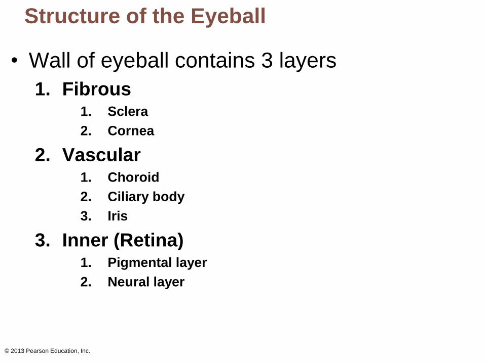
Structure of the Eyeball
• Wall of eyeball contains 3 layers
1. Fibrous 1. Sclera
2. Cornea
2. Vascular 1. Choroid
2. Ciliary body
3. Iris
3. Inner (Retina) 1. Pigmental layer
2. Neural layer
© 2013 Pearson Education, Inc.
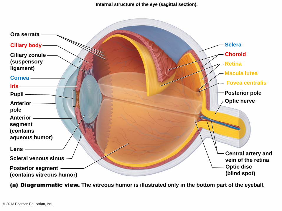
Internal structure of the eye (sagittal section).
Ora serrata
Ciliary body
Ciliary zonule
(suspensory
ligament)
Cornea
Pupil
Anterior
pole
Anterior
segment
(contains
aqueous humor)
Lens
Scleral venous sinus
Posterior segment
(contains vitreous humor)
Diagrammatic view. The vitreous humor is illustrated only in the bottom part of the eyeball.
Sclera
Choroid
Retina
Macula lutea
Fovea centralis
Posterior pole
Optic nerve
Central artery and
vein of the retina
Optic disc
(blind spot)
Iris
© 2013 Pearson Education, Inc.
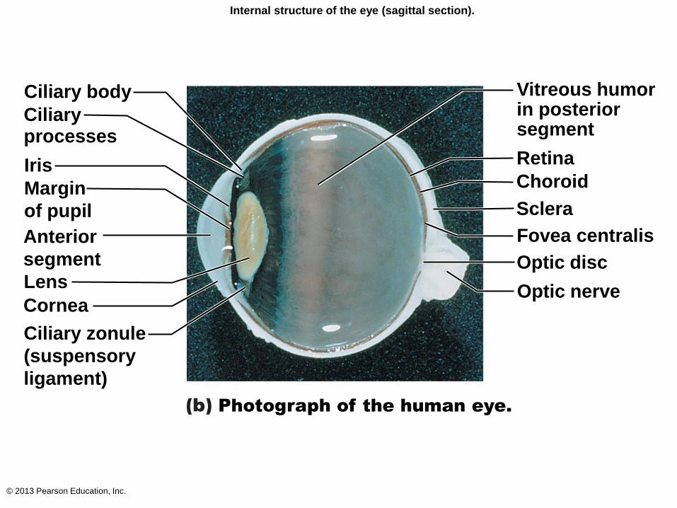
Internal structure of the eye (sagittal section).
Ciliary body
Ciliary processes
Iris
Margin
of pupil
Anterior
segment
Cornea
Ciliary zonule
(suspensory
ligament)
Photograph of the human eye.
Vitreous humor in posterior segment
Choroid
Fovea centralis
Optic disc
Optic nerve Lens
Sclera
Retina
© 2013 Pearson Education, Inc.
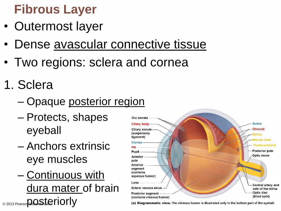
Fibrous Layer
• Outermost layer
• Dense avascular connective tissue
• Two regions: sclera and cornea
1. Sclera
– Opaque posterior region
– Protects, shapes
eyeball
– Anchors extrinsic
eye muscles
– Continuous with
dura mater of brain
posteriorly
© 2013 Pearson Education, Inc.
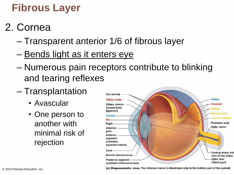
Fibrous Layer
2. Cornea
– Transparent anterior 1/6 of fibrous layer
– Bends light as it enters eye
– Numerous pain receptors contribute to blinking
and tearing reflexes
– Transplantation
• Avascular
• One person to
another with
minimal risk of
rejection
© 2013 Pearson Education, Inc.
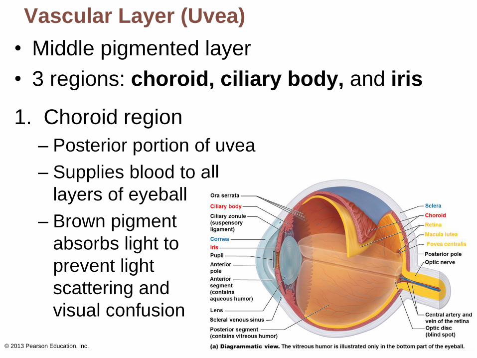
Vascular Layer (Uvea)
• Middle pigmented layer
• 3 regions: choroid, ciliary body, and iris
1. Choroid region
– Posterior portion of uvea
– Supplies blood to all
layers of eyeball
– Brown pigment
absorbs light to
prevent light
scattering and
visual confusion
© 2013 Pearson Education, Inc.
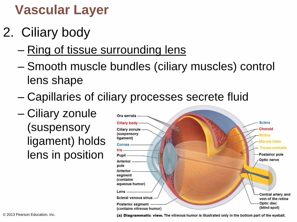
Vascular Layer
2. Ciliary body
– Ring of tissue surrounding lens
– Smooth muscle bundles (ciliary muscles) control
lens shape
– Capillaries of ciliary processes secrete fluid
– Ciliary zonule
(suspensory
ligament) holds
lens in position
© 2013 Pearson Education, Inc.
Vascular Layer
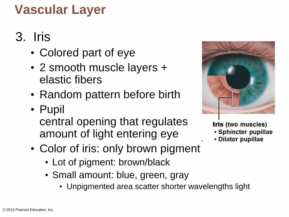
3. Iris
• Colored part of eye
• 2 smooth muscle layers + elastic fibers
• Random pattern before birth
• Pupil central opening that regulates amount of light entering eye
• Color of iris: only brown pigment • Lot of pigment: brown/black
• Small amount: blue, green, gray • Unpigmented area scatter shorter wavelengths light
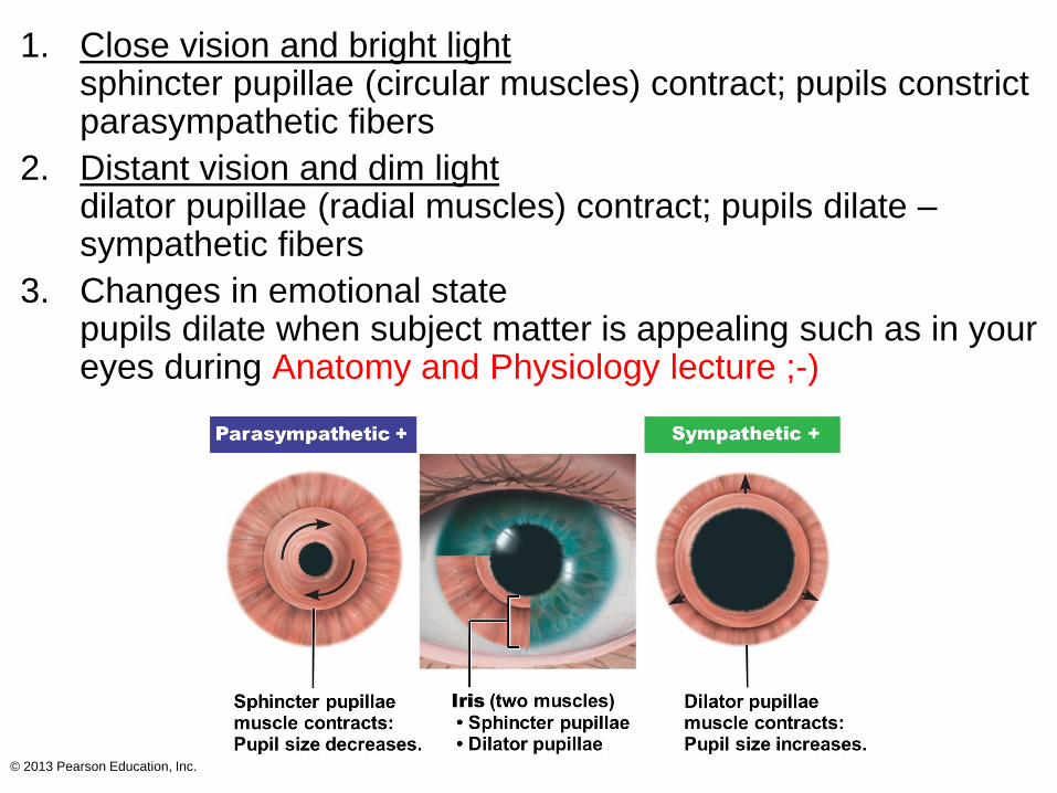
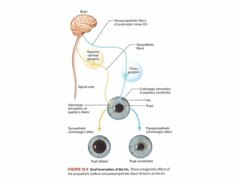
1. Close vision and bright light sphincter pupillae (circular muscles) contract; pupils constrict parasympathetic fibers
2. Distant vision and dim light dilator pupillae (radial muscles) contract; pupils dilate – sympathetic fibers
3. Changes in emotional state pupils dilate when subject matter is appealing such as in your eyes during Anatomy and Physiology lecture ;-)
© 2013 Pearson Education, Inc.
© 2013 Pearson Education, Inc.
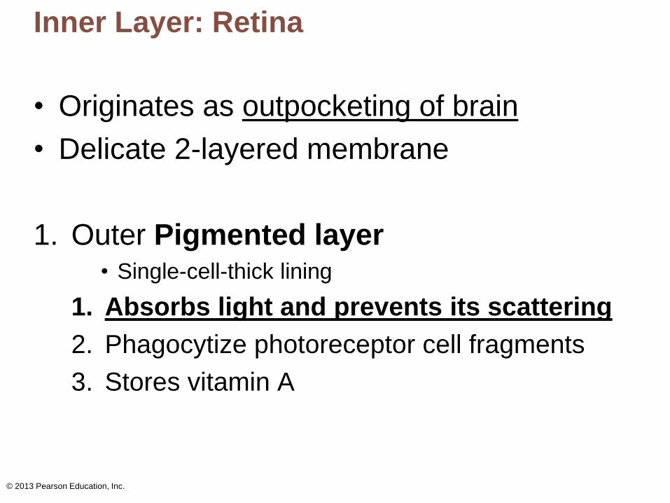
Inner Layer: Retina
• Originates as outpocketing of brain
• Delicate 2-layered membrane
1. Outer Pigmented layer • Single-cell-thick lining
1. Absorbs light and prevents its scattering
2. Phagocytize photoreceptor cell fragments
3. Stores vitamin A
© 2013 Pearson Education, Inc.
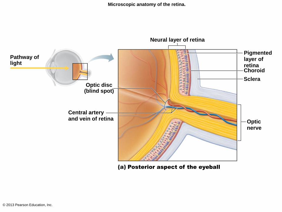
Microscopic anatomy of the retina.
Neural layer of retina
Pathway of light
Optic disc (blind spot)
Central artery and vein of retina
Pigmented layer of retina Choroid
Sclera
Optic nerve
Posterior aspect of the eyeball
© 2013 Pearson Education, Inc.
Inner Layer: Retina
2. Inner Neural layer
– Transparent
– Composed of 3 main types of neurons
1. Photoreceptors
2. Bipolar cells
3. Ganglion cells
– Signals spread from photoreceptors to bipolar cells
to ganglion cells
– Ganglion cell axons exit eye as optic nerve

© 2013 Pearson Education, Inc.
Microscopic anatomy of the retina.
Neural layer of retina
Pathway of light
Optic disc (blind spot)
Central artery and vein of retina
Pigmented layer of retina Choroid
Sclera
Optic nerve
Posterior aspect of the eyeball
© 2013 Pearson Education, Inc.
The Retina
• Optic disc (blind spot)
– Site where optic nerve leaves eye
– Lacks photoreceptors
• Quarter-billion photoreceptors of two types
1. Rods
2. Cones
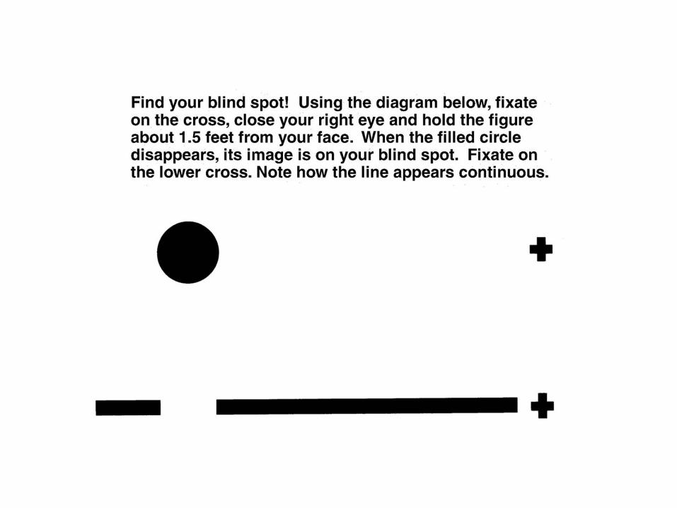
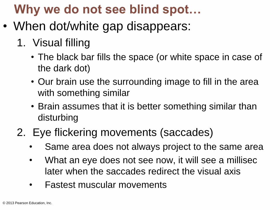
Why we do not see blind spot…
• When dot/white gap disappears:
1. Visual filling
• The black bar fills the space (or white space in case of
the dark dot)
• Our brain use the surrounding image to fill in the area
with something similar
• Brain assumes that it is better something similar than
disturbing
2. Eye flickering movements (saccades)
• Same area does not always project to the same area
• What an eye does not see now, it will see a millisec
later when the saccades redirect the visual axis
• Fastest muscular movements
© 2013 Pearson Education, Inc.
© 2013 Pearson Education, Inc.
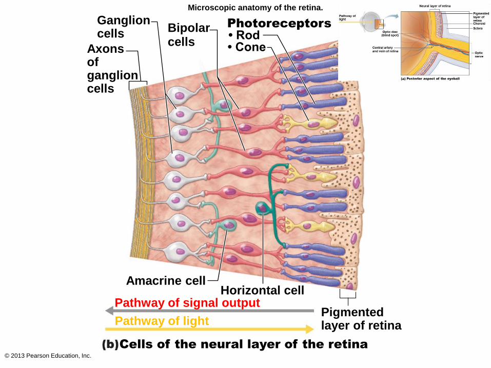
Microscopic anatomy of the retina.
Photoreceptors
• Rod • Cone
Ganglion cells Bipolar
cells Axons of ganglion cells
Amacrine cell Horizontal cell
Pathway of signal output
Pathway of light
Cells of the neural layer of the retina
Pigmented layer of retina
© 2013 Pearson Education, Inc.
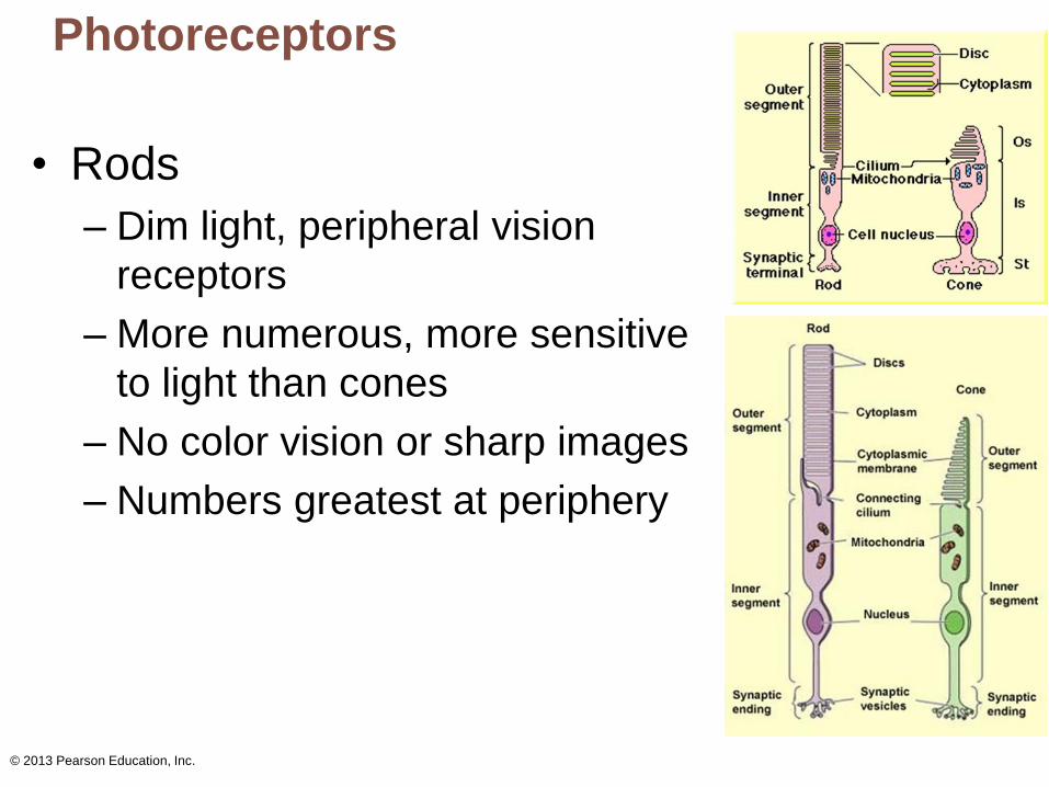
Photoreceptors
• Rods
– Dim light, peripheral vision
receptors
– More numerous, more sensitive
to light than cones
– No color vision or sharp images
– Numbers greatest at periphery
© 2013 Pearson Education, Inc.
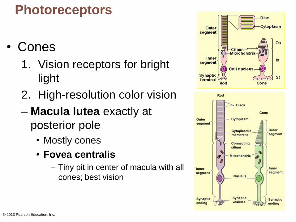
Photoreceptors
• Cones
1. Vision receptors for bright
light
2. High-resolution color vision
– Macula lutea exactly at
posterior pole
• Mostly cones
• Fovea centralis
– Tiny pit in center of macula with all
cones; best vision
© 2013 Pearson Education, Inc.
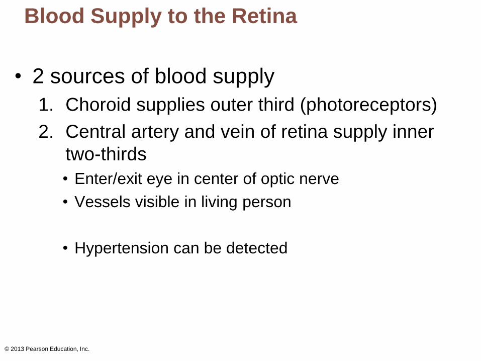
Blood Supply to the Retina
• 2 sources of blood supply
1. Choroid supplies outer third (photoreceptors)
2. Central artery and vein of retina supply inner
two-thirds
• Enter/exit eye in center of optic nerve
• Vessels visible in living person
• Hypertension can be detected
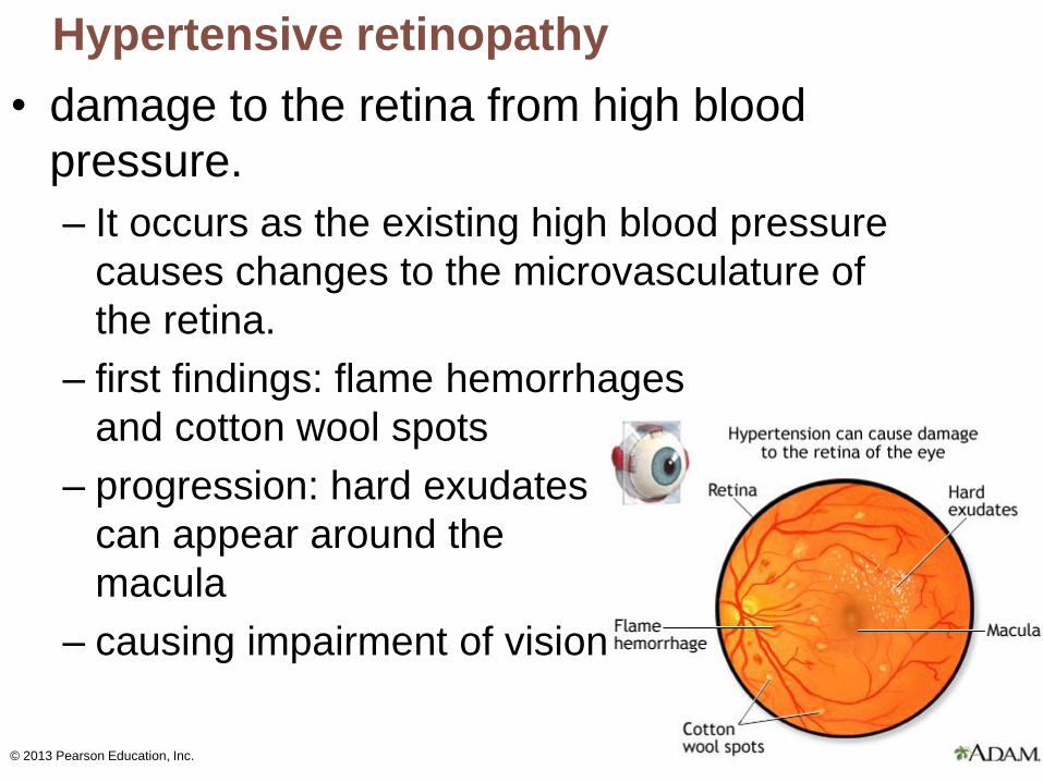
Hypertensive retinopathy
• damage to the retina from high blood
pressure.
– It occurs as the existing high blood pressure
causes changes to the microvasculature of
the retina.
– first findings: flame hemorrhages
and cotton wool spots
– progression: hard exudates
can appear around the
macula
– causing impairment of vision
© 2013 Pearson Education, Inc.
© 2013 Pearson Education, Inc.
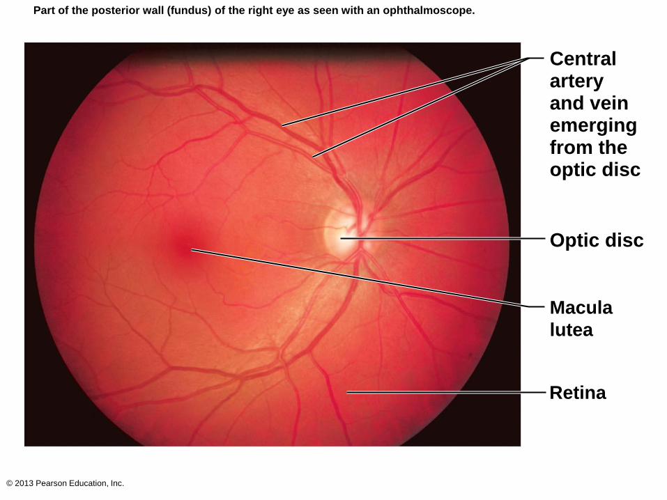
Part of the posterior wall (fundus) of the right eye as seen with an ophthalmoscope.
Central artery and vein emerging from the optic disc
Optic disc
Macula
lutea
Retina
© 2013 Pearson Education, Inc.
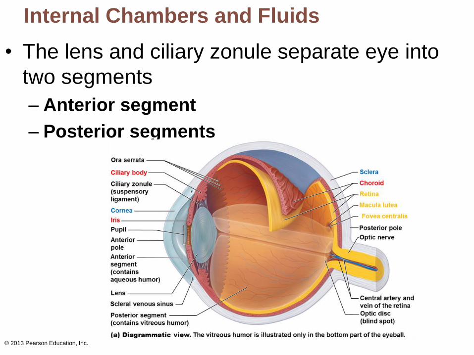
Internal Chambers and Fluids
• The lens and ciliary zonule separate eye into
two segments
– Anterior segment
– Posterior segments
© 2013 Pearson Education, Inc.
Internal Chambers and Fluids
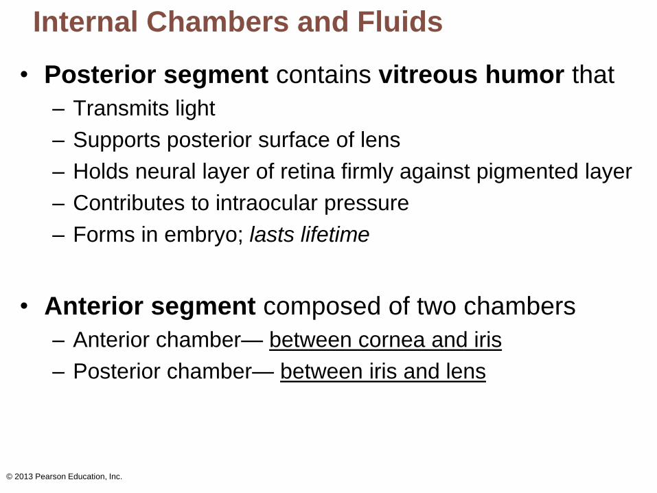
• Posterior segment contains vitreous humor that
– Transmits light
– Supports posterior surface of lens
– Holds neural layer of retina firmly against pigmented layer
– Contributes to intraocular pressure
– Forms in embryo; lasts lifetime
• Anterior segment composed of two chambers
– Anterior chamber— between cornea and iris
– Posterior chamber— between iris and lens
© 2013 Pearson Education, Inc.
Internal Chambers and Fluids
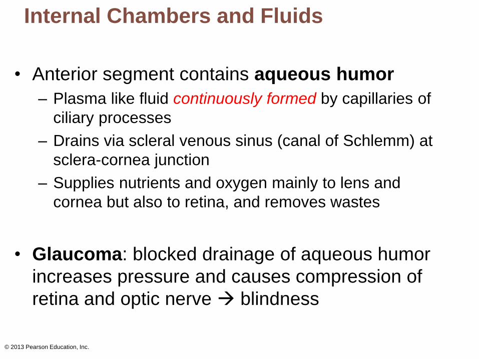
• Anterior segment contains aqueous humor
– Plasma like fluid continuously formed by capillaries of
ciliary processes
– Drains via scleral venous sinus (canal of Schlemm) at
sclera-cornea junction
– Supplies nutrients and oxygen mainly to lens and
cornea but also to retina, and removes wastes
• Glaucoma: blocked drainage of aqueous humor
increases pressure and causes compression of
retina and optic nerve blindness
© 2013 Pearson Education, Inc.
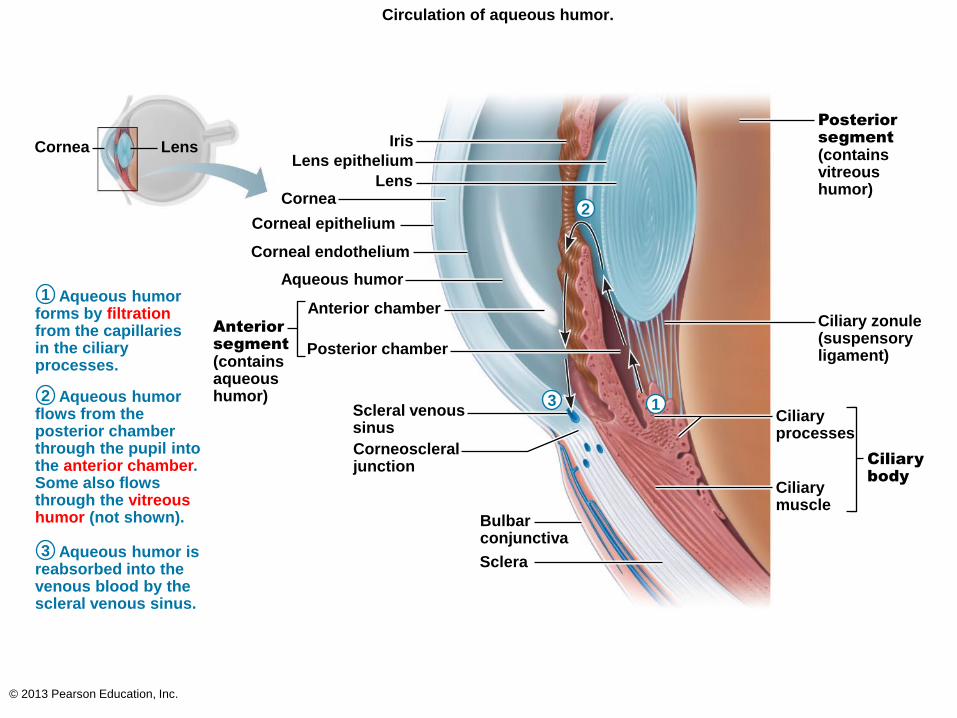
Circulation of aqueous humor.
Iris
Lens epithelium
Lens Cornea
Corneal epithelium
Corneal endothelium
Aqueous humor
Anterior chamber
Posterior chamber
Anterior
segment (contains aqueous humor)
Scleral venous sinus
Corneoscleral junction
Bulbar conjunctiva
Sclera
Ciliary
body
Ciliary zonule (suspensory ligament)
Posterior
segment
(contains vitreous humor)
Ciliary processes
Ciliary muscle
Aqueous humor forms by filtration from the capillaries in the ciliary processes.
Aqueous humor flows from the posterior chamber through the pupil into the anterior chamber. Some also flows through the vitreous humor (not shown).
Aqueous humor is reabsorbed into the venous blood by the scleral venous sinus.
Lens Cornea
1
2
3
1
2
3
© 2013 Pearson Education, Inc.
Lens
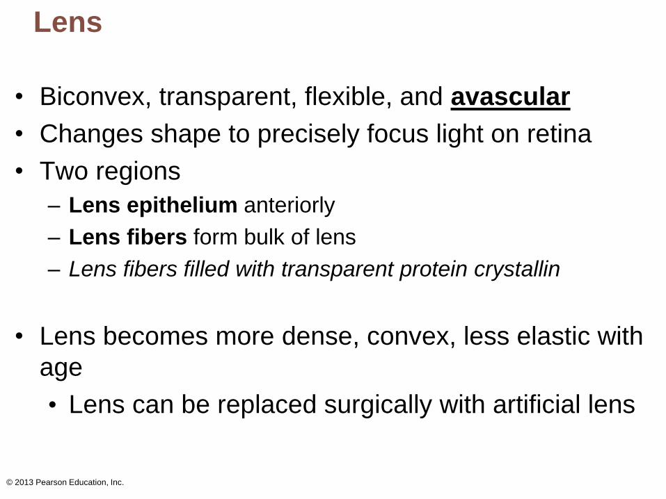
• Biconvex, transparent, flexible, and avascular
• Changes shape to precisely focus light on retina
• Two regions
– Lens epithelium anteriorly
– Lens fibers form bulk of lens
– Lens fibers filled with transparent protein crystallin
• Lens becomes more dense, convex, less elastic with
age
• Lens can be replaced surgically with artificial lens
© 2013 Pearson Education, Inc.
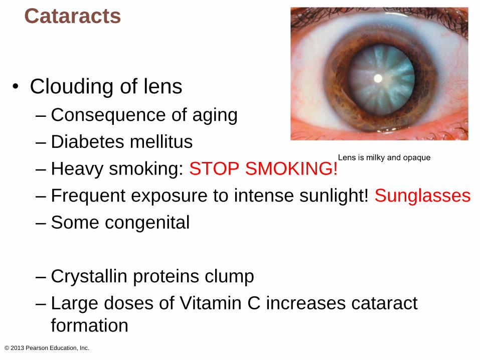
Cataracts
• Clouding of lens
– Consequence of aging
– Diabetes mellitus
– Heavy smoking: STOP SMOKING!
– Frequent exposure to intense sunlight! Sunglasses
– Some congenital
– Crystallin proteins clump
– Large doses of Vitamin C increases cataract
formation
BREAK
Functions of the Eye
1. Light/energy strikes the retina
2. Converts energy into action potentials
3. Relayed to brain for processing
© 2013 Pearson Education, Inc.
© 2013 Pearson Education, Inc.
Light And Optics: Wavelength And Color
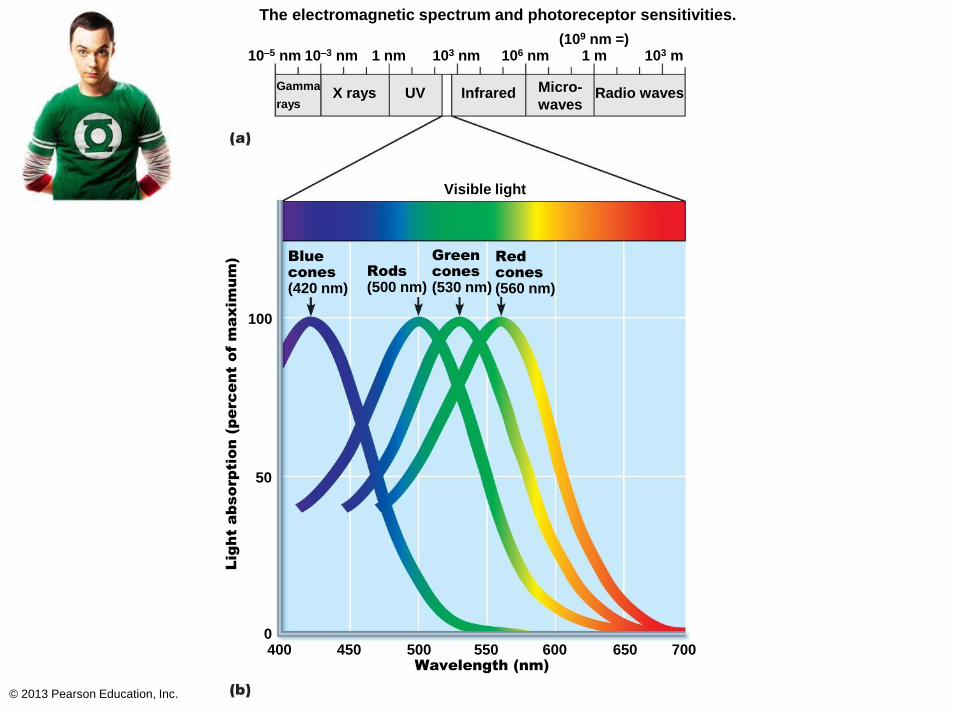
• Eyes respond to visible light
– Small portion of electromagnetic spectrum
– Wavelengths of 400-700 nm
• Light
– Packets of energy (photons or quanta) that travel
in wavelike fashion at high speeds
– Color of objects:
1. They absorb some wavelength
2. They reflect some wavelength
– Objects reflect determines color eye perceives
• Red apple reflects red, green grass reflects green
• Black: do not reflect anything, but absorbs everything
© 2013 Pearson Education, Inc.
The electromagnetic spectrum and photoreceptor sensitivities.
10–5 nm
Gamma
rays X rays UV Infrared Micro-
waves Radio waves
Visible light
Blue
cones (420 nm)
Rods (500 nm)
Green
cones (530 nm)
Red
cones (560 nm)
Light absorp
tion (p
erc
ent of m
axim
um
)
100
50
0 400
10–3 nm 1 nm 103 nm 106 nm 1 m 103 m (109 nm =)
Wavelength (nm)
450 500 550 600 650 700
© 2013 Pearson Education, Inc.
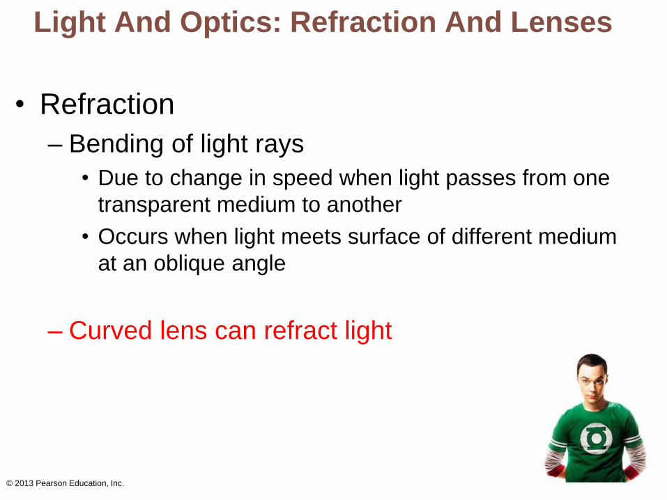
Light And Optics: Refraction And Lenses
• Refraction
– Bending of light rays
• Due to change in speed when light passes from one
transparent medium to another
• Occurs when light meets surface of different medium
at an oblique angle
– Curved lens can refract light
© 2013 Pearson Education, Inc.
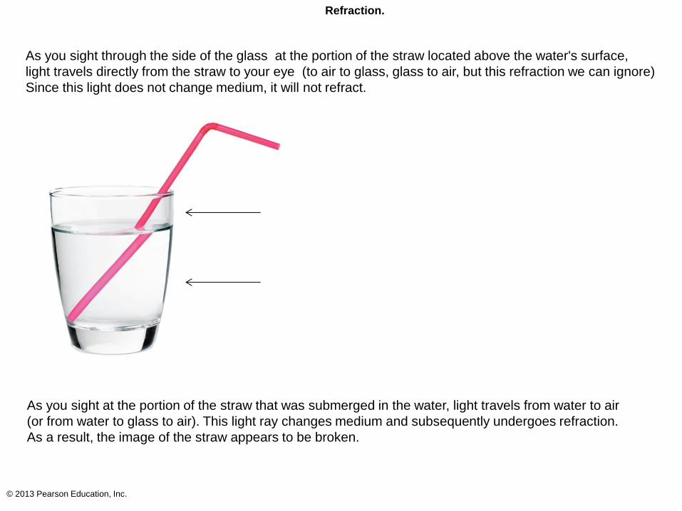
Refraction.
As you sight through the side of the glass at the portion of the straw located above the water's surface,
light travels directly from the straw to your eye (to air to glass, glass to air, but this refraction we can ignore)
Since this light does not change medium, it will not refract.
As you sight at the portion of the straw that was submerged in the water, light travels from water to air
(or from water to glass to air). This light ray changes medium and subsequently undergoes refraction.
As a result, the image of the straw appears to be broken.
© 2013 Pearson Education, Inc.
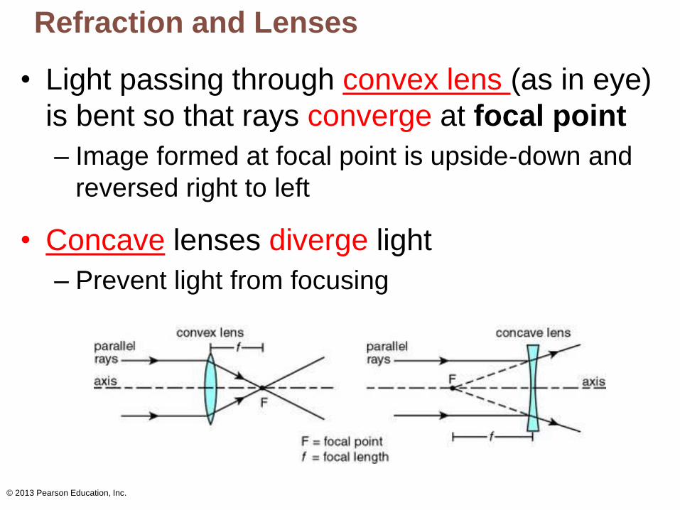
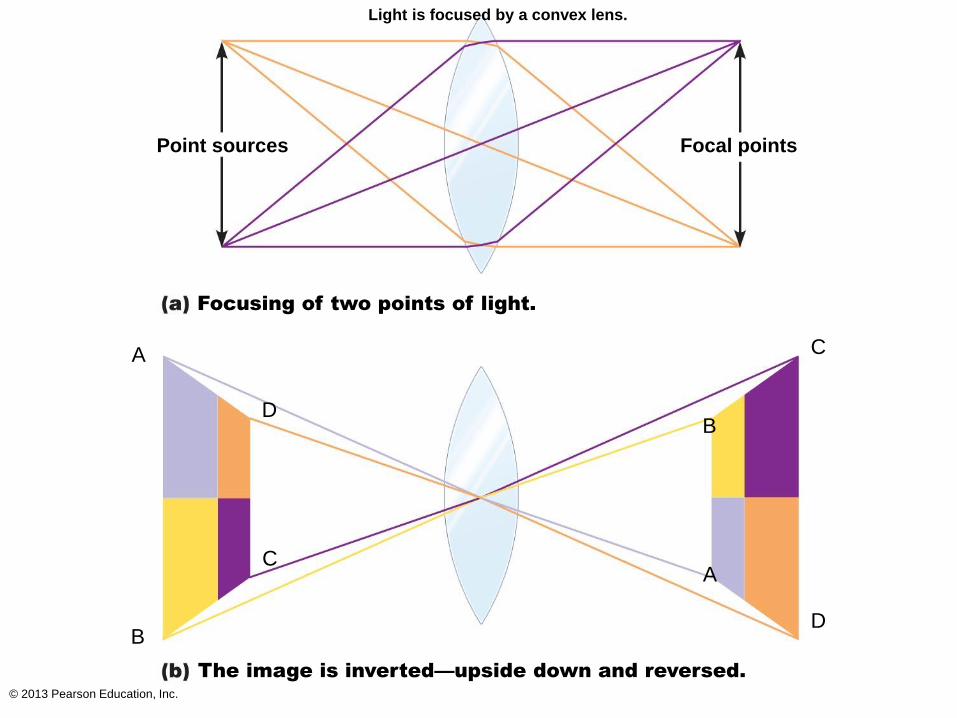
Refraction and Lenses
• Light passing through convex lens (as in eye)
is bent so that rays converge at focal point
– Image formed at focal point is upside-down and
reversed right to left
• Concave lenses diverge light
– Prevent light from focusing
© 2013 Pearson Education, Inc.
Light is focused by a convex lens.
Point sources Focal points
Focusing of two points of light.
The image is inverted—upside down and reversed.
A
B
C
D
C
D
A
B
© 2013 Pearson Education, Inc.
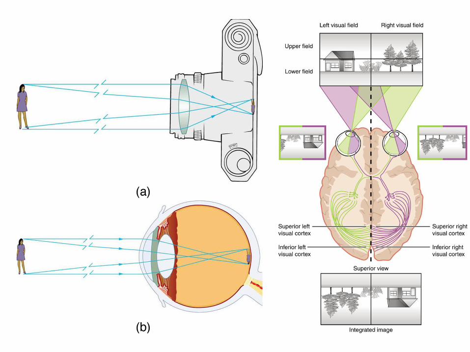
Focusing Light on The Retina
• Pathway of light entering eye:
cornea, aqueous humor, lens, vitreous humor, entire
neural layer of retina, photoreceptors
• Light refracted 3 (-4) times along pathway
1. Entering cornea
2. Entering lens
3. Leaving lens
• Majority of refractory power in cornea
• Change in lens curvature allows for fine focusing
© 2013 Pearson Education, Inc.
Focusing For Distant Vision
• Eyes best adapted for distant vision
• Far point of vision
– Distance beyond which no change in lens shape
needed for focusing
• 6m for emmetropic (normal) eye
• Cornea and lens focus light precisely on retina
• Ciliary muscles relaxed
• Lens stretched flat by tension in ciliary zonule
© 2013 Pearson Education, Inc.
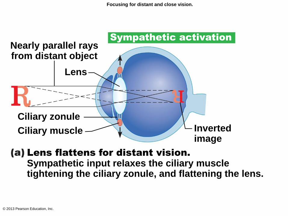
Focusing for distant and close vision.
Sympathetic activation
Nearly parallel rays from distant object
Lens
Ciliary zonule
Ciliary muscle Inverted image
Lens flattens for distant vision. Sympathetic input relaxes the ciliary muscle tightening the ciliary zonule, and flattening the lens.
© 2013 Pearson Education, Inc.
Focusing For Close Vision
• Light from close objects (<6 m) diverges as
approaches eye
– Requires eye to make active adjustments using 3
simultaneous processes
1. Accommodation of lenses
2. Constriction of pupils
3. Convergence of eyeballs
© 2013 Pearson Education, Inc.
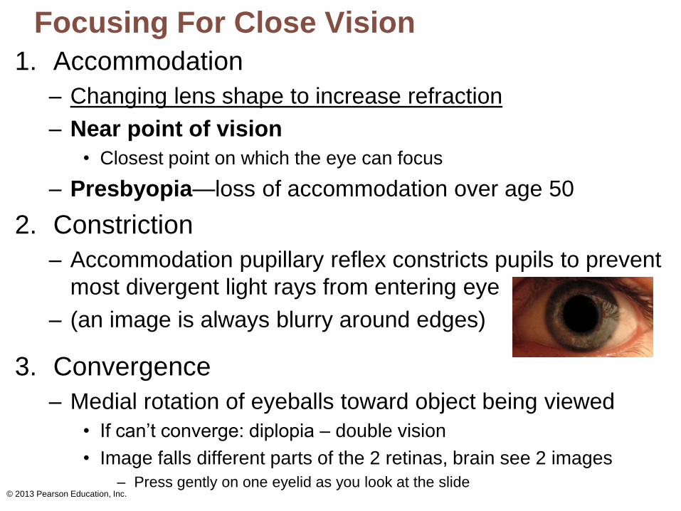
Focusing For Close Vision
1. Accommodation
– Changing lens shape to increase refraction
– Near point of vision
• Closest point on which the eye can focus
– Presbyopia—loss of accommodation over age 50
2. Constriction
– Accommodation pupillary reflex constricts pupils to prevent
most divergent light rays from entering eye
– (an image is always blurry around edges)
3. Convergence
– Medial rotation of eyeballs toward object being viewed
• If can’t converge: diplopia – double vision
• Image falls different parts of the 2 retinas, brain see 2 images
– Press gently on one eyelid as you look at the slide
© 2013 Pearson Education, Inc.
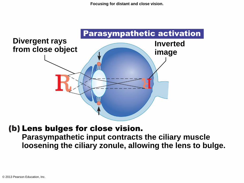
Focusing for distant and close vision.
Parasympathetic activation
Inverted image
Divergent rays from close object
Lens bulges for close vision. Parasympathetic input contracts the ciliary muscle loosening the ciliary zonule, allowing the lens to bulge.
© 2013 Pearson Education, Inc.
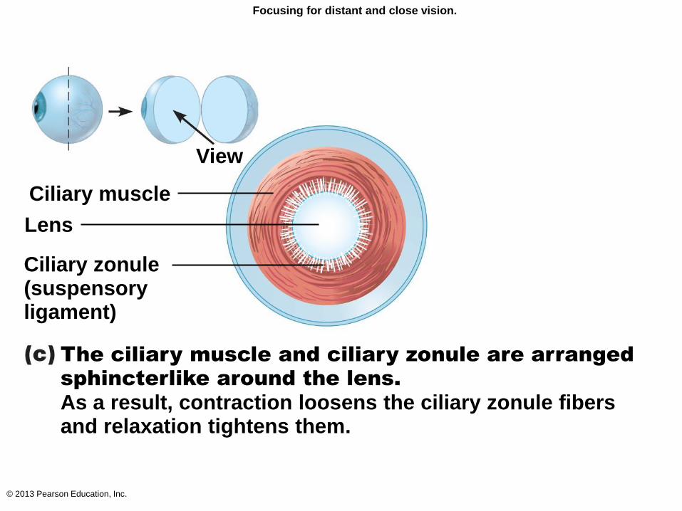
Focusing for distant and close vision.
Ciliary muscle
Lens
Ciliary zonule (suspensory ligament)
View
The ciliary muscle and ciliary zonule are arranged
sphincterlike around the lens. As a result, contraction loosens the ciliary zonule fibers and relaxation tightens them.
© 2013 Pearson Education, Inc.
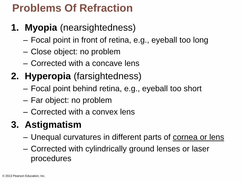
Problems Of Refraction
1. Myopia (nearsightedness)
– Focal point in front of retina, e.g., eyeball too long
– Close object: no problem
– Corrected with a concave lens
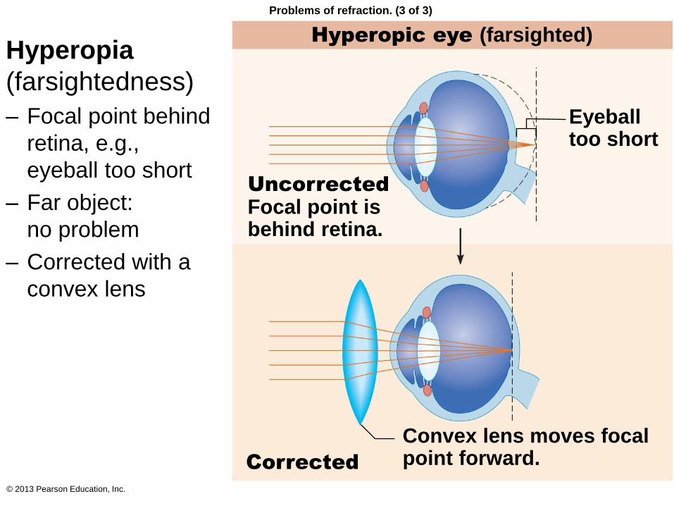
2. Hyperopia (farsightedness)
– Focal point behind retina, e.g., eyeball too short
– Far object: no problem
– Corrected with a convex lens
3. Astigmatism
– Unequal curvatures in different parts of cornea or lens
– Corrected with cylindrically ground lenses or laser
procedures
© 2013 Pearson Education, Inc.
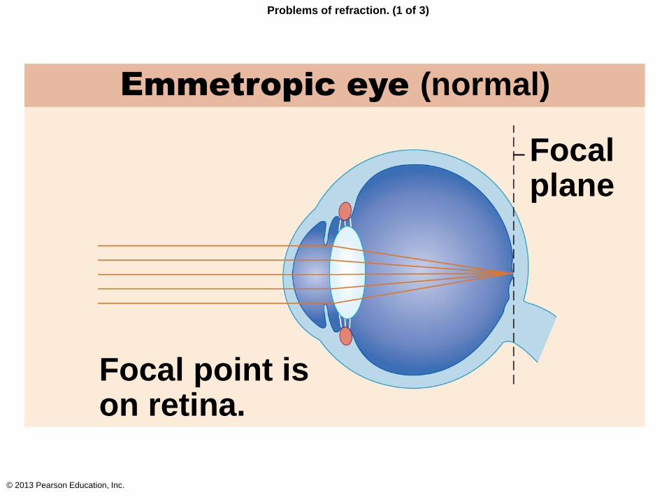
Problems of refraction. (1 of 3)
Emmetropic eye (normal)
Focal plane
Focal point is on retina.
© 2013 Pearson Education, Inc.
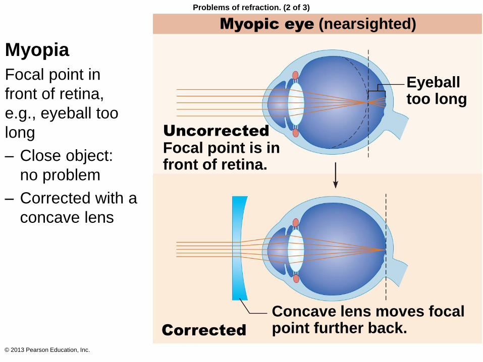
Myopic eye (nearsighted)
Uncorrected Focal point is in front of retina.
Concave lens moves focal point further back.
Eyeball too long
Corrected
Problems of refraction. (2 of 3)
Myopia
Focal point in
front of retina,
e.g., eyeball too
long
– Close object:
no problem
– Corrected with a
concave lens
© 2013 Pearson Education, Inc.
Hyperopic eye (farsighted)
Eyeball too short
Uncorrected Focal point is behind retina.
Corrected
Convex lens moves focal point forward.
Problems of refraction. (3 of 3)
Hyperopia
(farsightedness)
– Focal point behind
retina, e.g.,
eyeball too short
– Far object:
no problem
– Corrected with a
convex lens
© 2013 Pearson Education, Inc.
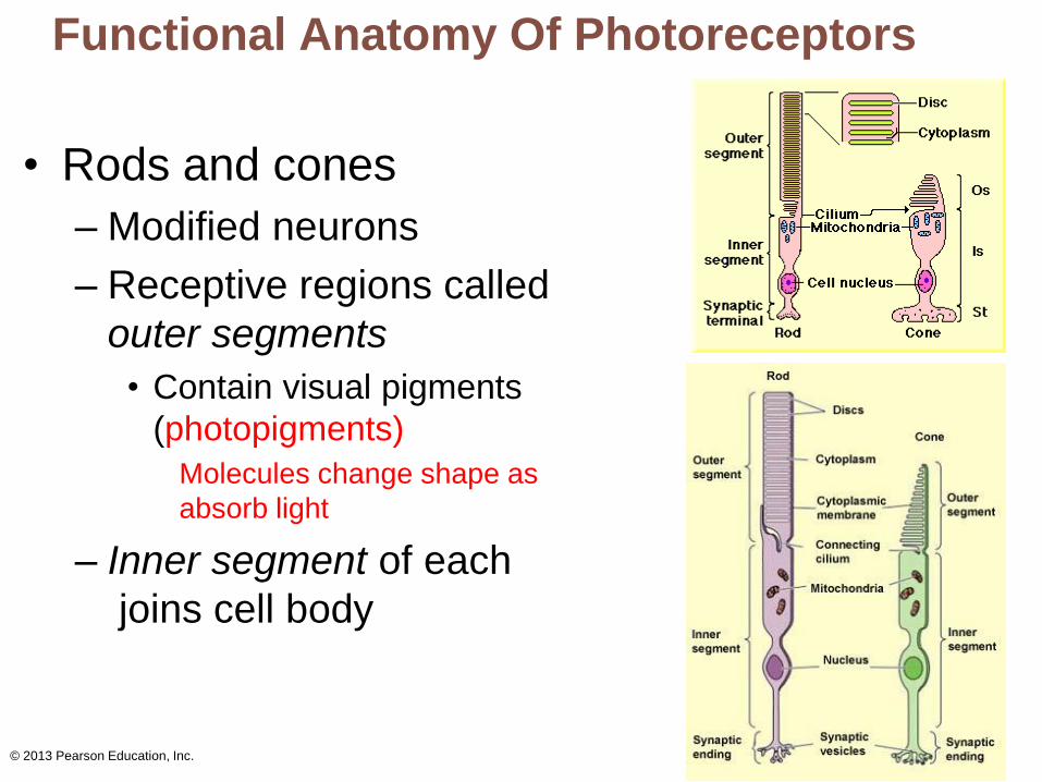
Functional Anatomy Of Photoreceptors
• Rods and cones
– Modified neurons
– Receptive regions called
outer segments
• Contain visual pigments
(photopigments)
Molecules change shape as
absorb light
– Inner segment of each
joins cell body
© 2013 Pearson Education, Inc.
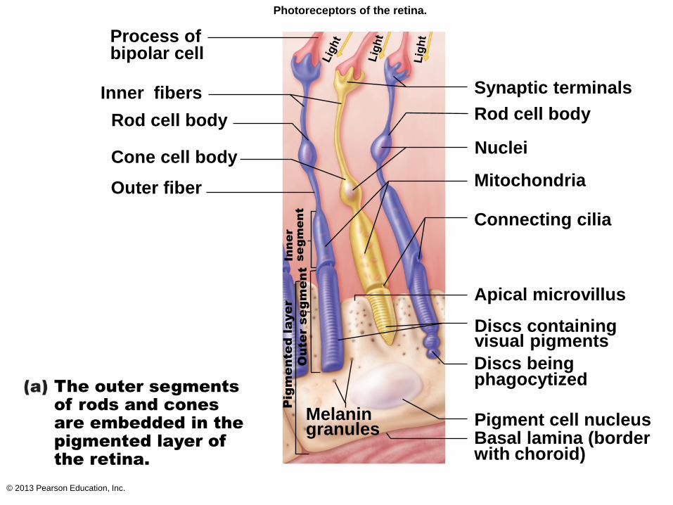
Photoreceptors of the retina.
Process of bipolar cell
Synaptic terminals
Rod cell body
Inner fibers
Nuclei Cone cell body
Mitochondria
Connecting cilia
Outer fiber
Apical microvillus
Discs containing visual pigments
Discs being phagocytized
Melanin granules
Pigment cell nucleus Basal lamina (border with choroid)
In
ne
r
se
gm
en
t
Pig
me
nte
d la
ye
r
Ou
te
r se
gm
en
t
The outer segments
of rods and cones
are embedded in the
pigmented layer of
the retina.
Rod cell body
© 2013 Pearson Education, Inc.
Photoreceptor Cells
• Vulnerable to damage
• Degenerate if retina detached
• Destroyed by intense light
• Outer segment renewed every 24 hours
– Tips fragment off and are phagocytized
© 2013 Pearson Education, Inc.
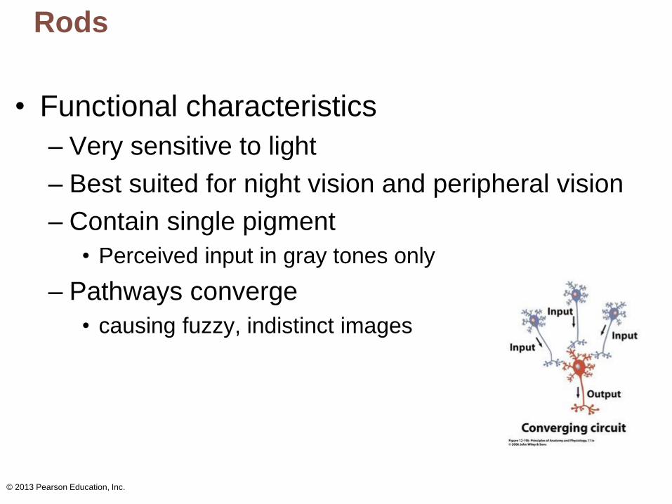
Rods
• Functional characteristics
– Very sensitive to light
– Best suited for night vision and peripheral vision
– Contain single pigment
• Perceived input in gray tones only
– Pathways converge
• causing fuzzy, indistinct images
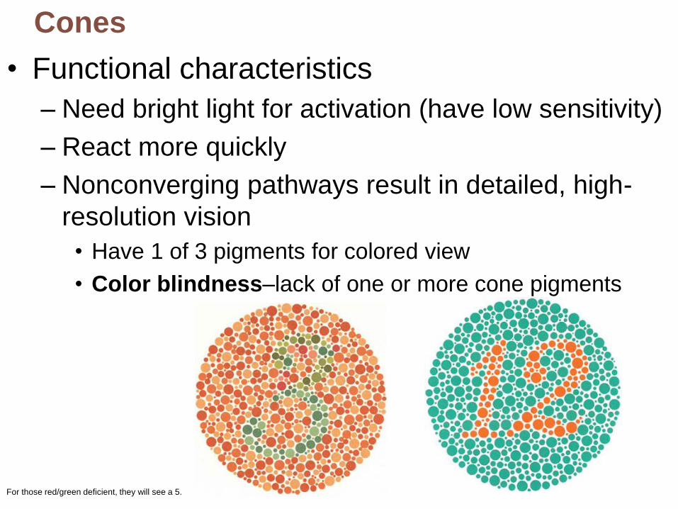
For those red/green deficient, they will see a 5.
Cones
• Functional characteristics
– Need bright light for activation (have low sensitivity)
– React more quickly
– Nonconverging pathways result in detailed, high-
resolution vision
• Have 1 of 3 pigments for colored view
• Color blindness–lack of one or more cone pigments
© 2013 Pearson Education, Inc.
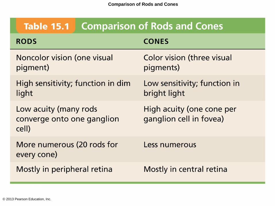
Comparison of Rods and Cones
© 2013 Pearson Education, Inc.
Chemistry Of Visual Pigments
• Retinal
– Light-absorbing molecule that combines with one of
four proteins (opsins) to form visual pigments
• Synthesized from vitamin A
– Retinal has 2 forms: bent form and straight form
• Bent form change to straight form when pigment absorbs
light
• Conversion of bent to straight initiates reactions
electrical impulses along optic nerve
© 2013 Pearson Education, Inc.
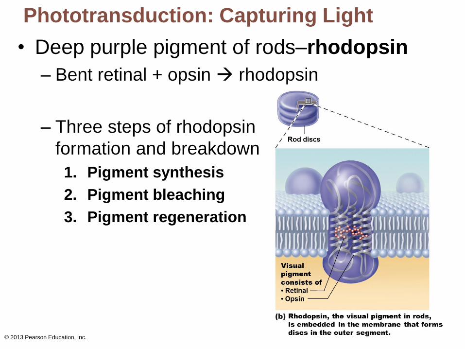
Phototransduction: Capturing Light
• Deep purple pigment of rods–rhodopsin
– Bent retinal + opsin rhodopsin
– Three steps of rhodopsin
formation and breakdown
1. Pigment synthesis
2. Pigment bleaching
3. Pigment regeneration
© 2013 Pearson Education, Inc.
Phototransduction: Capturing Light
1. Pigment synthesis
– Rhodopsin forms and accumulates in dark
2. Pigment bleaching
– When rhodopsin absorbs light, retinal changes to straight form
– Retinal and opsin separate (rhodopsin breakdown)
3. Pigment regeneration
– Straight retinal converted to bent
– Rhodopsin regenerated in outer segments
© 2013 Pearson Education, Inc.
Phototransduction In Cones
• Similar as process in rods
• Cones far less sensitive to light
– Takes higher-intensity light to activate cones
© 2013 Pearson Education, Inc.
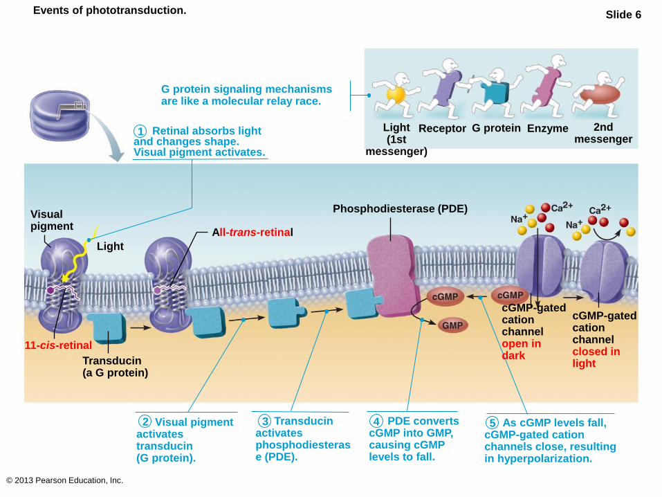
Events of phototransduction. Slide 6
G protein signaling mechanisms are like a molecular relay race.
Retinal absorbs light and changes shape. Visual pigment activates.
Receptor G protein Enzyme 2nd messenger
Visual pigment
1
Light
11-cis-retinal
Transducin (a G protein)
All-trans-retinal
2 3 Visual pigment activates transducin (G protein).
Transducin activates phosphodiesterase (PDE).
4 5 PDE converts cGMP into GMP, causing cGMP levels to fall.
As cGMP levels fall, cGMP-gated cation channels close, resulting in hyperpolarization.
cGMP-gated cation channel open in dark
cGMP-gated cation channel closed in light
Phosphodiesterase (PDE)
Light (1st
messenger)
© 2013 Pearson Education, Inc.
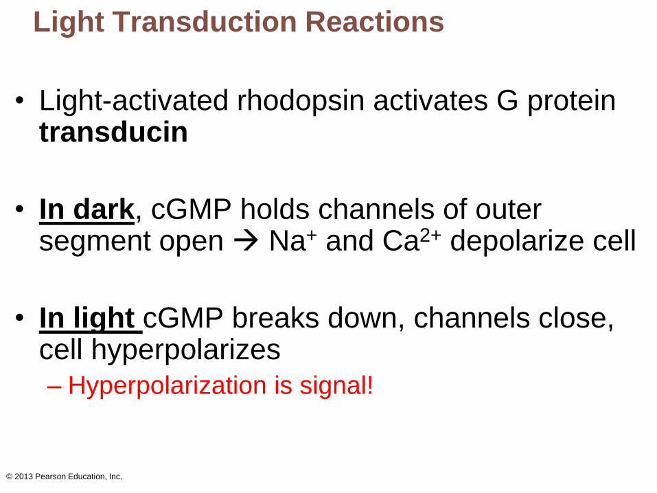
Light Transduction Reactions
• Light-activated rhodopsin activates G protein transducin
• In dark, cGMP holds channels of outer segment open Na+ and Ca2+ depolarize cell
• In light cGMP breaks down, channels close, cell hyperpolarizes
– Hyperpolarization is signal!
© 2013 Pearson Education, Inc.
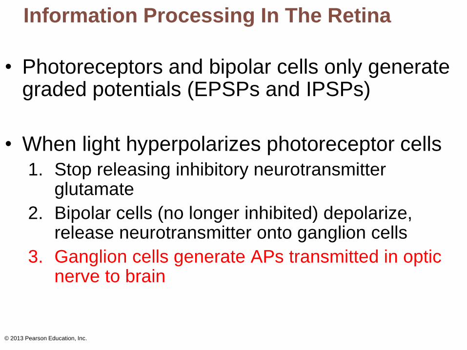
Information Processing In The Retina
• Photoreceptors and bipolar cells only generate graded potentials (EPSPs and IPSPs)
• When light hyperpolarizes photoreceptor cells
1. Stop releasing inhibitory neurotransmitter glutamate
2. Bipolar cells (no longer inhibited) depolarize, release neurotransmitter onto ganglion cells
3. Ganglion cells generate APs transmitted in optic nerve to brain

In the dark
© 2013 Pearson Education, Inc.
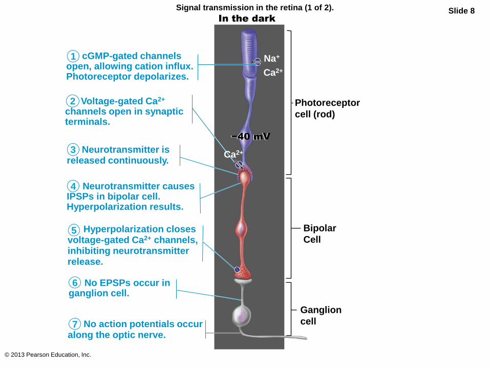
Signal transmission in the retina (1 of 2). Slide 8 In the dark
cGMP-gated channels open, allowing cation influx. Photoreceptor depolarizes.
1
Voltage-gated Ca2+ channels open in synaptic terminals.
Neurotransmitter is released continuously.
Neurotransmitter causes IPSPs in bipolar cell. Hyperpolarization results.
Hyperpolarization closes voltage-gated Ca2+ channels, inhibiting neurotransmitter release.
No EPSPs occur in ganglion cell.
No action potentials occur along the optic nerve.
Photoreceptor
cell (rod)
Bipolar
Cell
Ganglion
cell
Ca2+
−40 mV −40 mV
2
3
4
5
6
7
Ca2+
Na+
In the light
© 2013 Pearson Education, Inc.
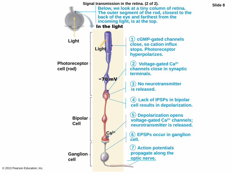
Signal transmission in the retina. (2 of 2). Slide 8
−70 mV
No neurotransmitter
is released.
Depolarization opens voltage-gated Ca2+ channels; neurotransmitter is released.
EPSPs occur in ganglion cell.
Action potentials propagate along the optic nerve.
cGMP-gated channels close, so cation influx stops. Photoreceptor hyperpolarizes.
Lack of IPSPs in bipolar
cell results in depolarization.
Voltage-gated Ca2+ channels close in synaptic terminals.
1
Photoreceptor
cell (rod)
Bipolar
Cell
Ganglion
cell
In the light
Light
Ca2+
−70 mV
2
3
4
5
6
7
Below, we look at a tiny column of retina. The outer segment of the rod, closest to the back of the eye and farthest from the incoming light, is at the top.
Light
© 2013 Pearson Education, Inc.
Light Adaptation
• Move from darkness into bright light
– Both rods and cones strongly stimulated
• Pupils constrict
– Large amounts of pigments broken down
instantaneously, producing glare
– Visual acuity improves over 5–10 minutes as:
• Rod system turns off
• Retinal sensitivity decreases
• Cones and neurons rapidly adapt
© 2013 Pearson Education, Inc.
Dark Adaptation
• Move from bright light into darkness
– Cones stop functioning in low-intensity light
– Rod pigments bleached; system turned off
– Rhodopsin accumulates in dark
– Transducin returns to outer segments
– Retinal sensitivity increases within 20–30 minutes
– Pupils dilate
© 2013 Pearson Education, Inc.
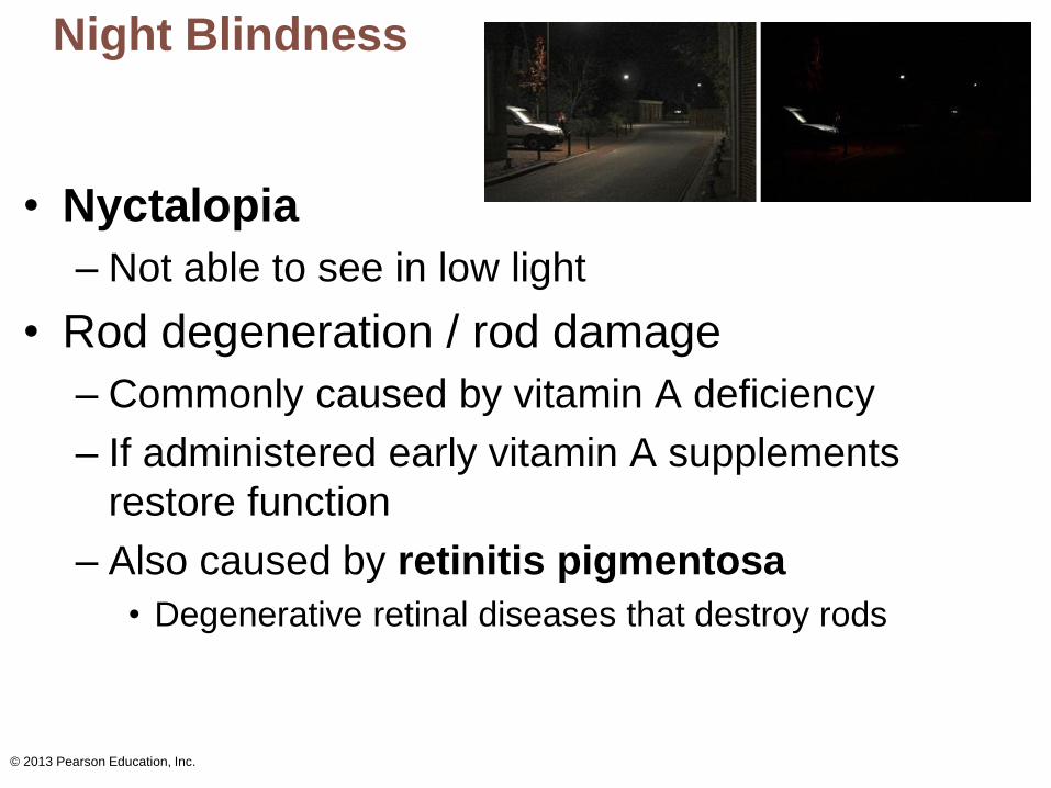
Night Blindness
• Nyctalopia
– Not able to see in low light
• Rod degeneration / rod damage
– Commonly caused by vitamin A deficiency
– If administered early vitamin A supplements
restore function
– Also caused by retinitis pigmentosa
• Degenerative retinal diseases that destroy rods
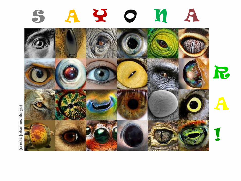
S A Y O N A
R
A
!


![[PPT]PowerPoint Presentation - DeannaRussler - Home · Web viewLacrimal apparatus Consists of lacrimal gland and several ducts Ducts drain lacrimal secretions into nasal cavity Gland](https://img.pdfslide.net/doc/110x75/5ae7f9f47f8b9acc268f6a96/pptpowerpoint-presentation-deannarussler-home-viewlacrimal-apparatus-consists.jpg)