Embed Size (px)
Citation preview
Muscular System: Muscle Tissue(Chapter 10)
Lecture Materials
for
Amy Warenda Czura, Ph.D.
Suffolk County Community College
Eastern Campus
Primary Sources for figures and content:
Marieb, E. N. Human Anatomy & Physiology 6th ed. San Francisco: Pearson BenjaminCummings, 2004.
Martini, F. H. Fundamentals of Anatomy & Physiology 6th ed. San Francisco: PearsonBenjamin Cummings, 2004.
Amy Warenda Czura, Ph.D. 1 SCCC BIO130 Chapter 10 Lecture Slides
Muscle Tissue Types:1. Skeletal muscle = voluntary striated muscle2. Cardiac muscle = involuntary striated
muscle3. Smooth muscle = involuntary nonstriated
muscle
Characteristics of all muscle tissues:1. Specialized cells: elongated, high density of
myofilaments = cytoplasmic filaments of actin and myosin
2. Excitability/irritability: receive and respondto stimulus
3. Contractility: shorten and produce force upon stimulation
4. Extensibility: can be stretched5. Elasticity: recoil after stretch
Amy Warenda Czura, Ph.D. 2 SCCC BIO130 Chapter 10 Lecture Slides
Skeletal Muscle Tissue-forms skeletal muscles (44% of body mass)A skeletal muscle = an organ: composed of
skeletal muscle cells (fibers), CT, nervesand blood vessels
Functions of skeletal muscles:1. Produce skeletal movement2. Maintain posture and upright position3. Stabilize joints4. Support soft tissues5. Guard entrances and exits6. Generate heat (maintain body temp)
Skeletal Muscle Anatomy-each muscle innervated by one nerve: must
branch and contact each skeletal muscle fiber (cell)
-also one artery, branches into extensive capillaries around each fiber to supply oxygen and remove wastesAmy Warenda Czura, Ph.D. 3 SCCC BIO130 Chapter 10 Lecture Slides
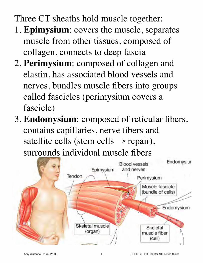
Three CT sheaths hold muscle together:1. Epimysium: covers the muscle, separates
muscle from other tissues, composed ofcollagen, connects to deep fascia
2. Perimysium: composed of collagen andelastin, has associated blood vessels andnerves, bundles muscle fibers into groupscalled fascicles (perimysium covers afascicle)
3. Endomysium: composed of reticular fibers,contains capillaries, nerve fibers andsatellite cells (stem cells → repair),surrounds individual muscle fibers
Amy Warenda Czura, Ph.D. 4 SCCC BIO130 Chapter 10 Lecture Slides
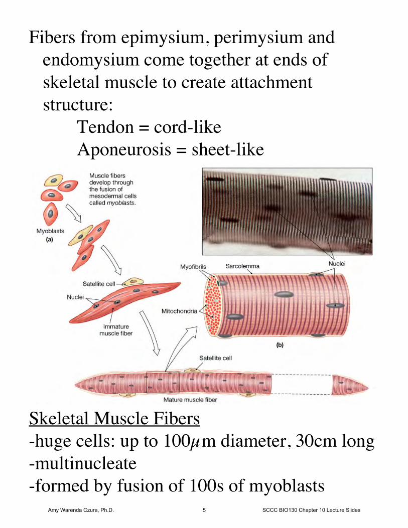
Fibers from epimysium, perimysium and endomysium come together at ends of skeletal muscle to create attachment structure:
Tendon = cord-likeAponeurosis = sheet-like
Skeletal Muscle Fibers-huge cells: up to 100µm diameter, 30cm long-multinucleate-formed by fusion of 100s of myoblasts
Amy Warenda Czura, Ph.D. 5 SCCC BIO130 Chapter 10 Lecture Slides
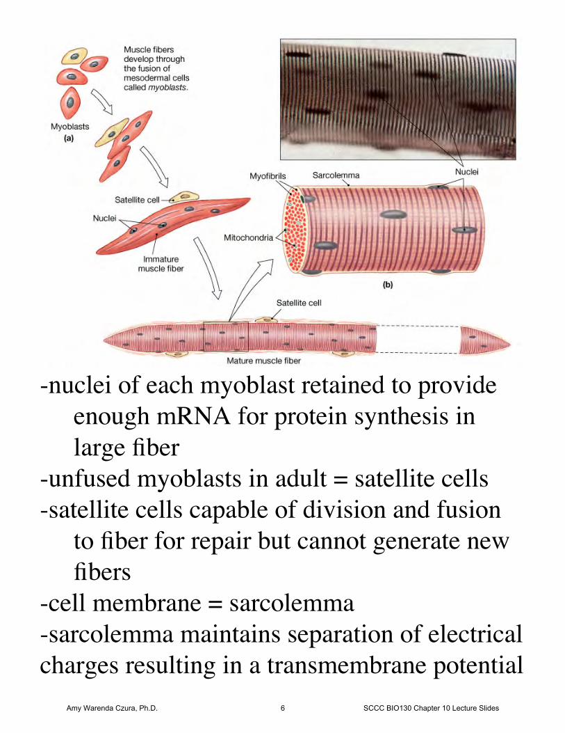
-nuclei of each myoblast retained to provide enough mRNA for protein synthesis in large fiber
-unfused myoblasts in adult = satellite cells-satellite cells capable of division and fusion
to fiber for repair but cannot generate newfibers
-cell membrane = sarcolemma-sarcolemma maintains separation of electricalcharges resulting in a transmembrane potential
Amy Warenda Czura, Ph.D. 6 SCCC BIO130 Chapter 10 Lecture Slides
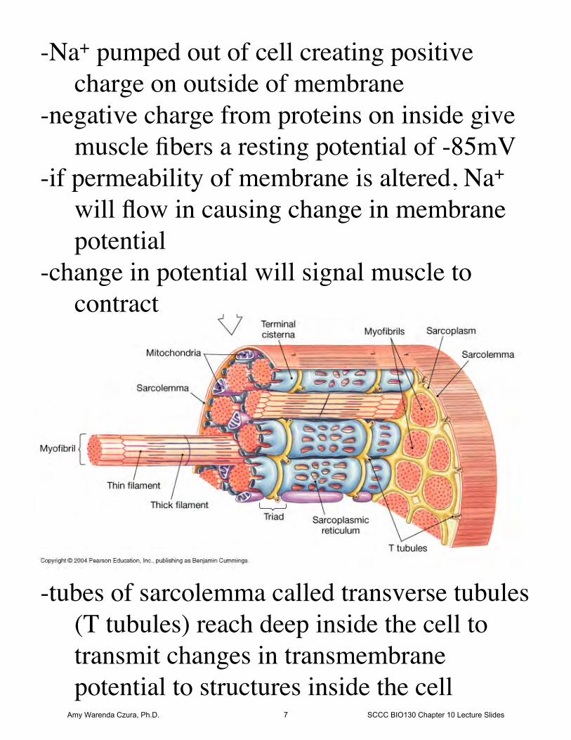
-Na+ pumped out of cell creating positive charge on outside of membrane
-negative charge from proteins on inside givemuscle fibers a resting potential of -85mV
-if permeability of membrane is altered, Na+ will flow in causing change in membranepotential
-change in potential will signal muscle to contract
-tubes of sarcolemma called transverse tubules(T tubules) reach deep inside the cell to transmit changes in transmembranepotential to structures inside the cell
Amy Warenda Czura, Ph.D. 7 SCCC BIO130 Chapter 10 Lecture Slides
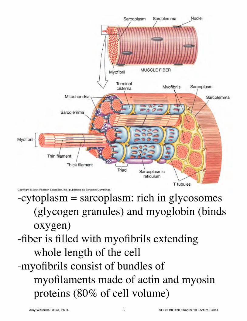
-cytoplasm = sarcoplasm: rich in glycosomes(glycogen granules) and myoglobin (bindsoxygen)
-fiber is filled with myofibrils extending whole length of the cell
-myofibrils consist of bundles of myofilaments made of actin and myosin proteins (80% of cell volume)
Amy Warenda Czura, Ph.D. 8 SCCC BIO130 Chapter 10 Lecture Slides
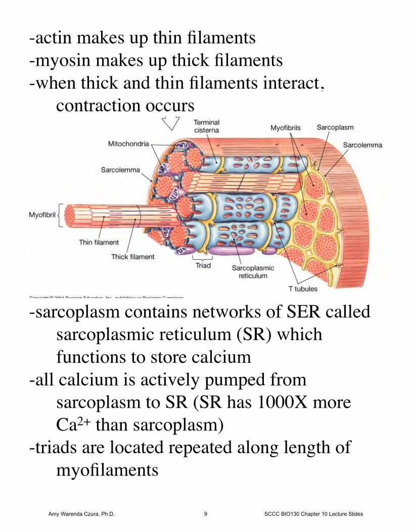
-sarcoplasm contains networks of SER calledsarcoplasmic reticulum (SR) which functions to store calcium
-all calcium is actively pumped from sarcoplasm to SR (SR has 1000X more Ca2+ than sarcoplasm)
-triads are located repeated along length of myofilaments
-actin makes up thin filaments-myosin makes up thick filaments-when thick and thin filaments interact,
contraction occurs
Amy Warenda Czura, Ph.D. 9 SCCC BIO130 Chapter 10 Lecture Slides
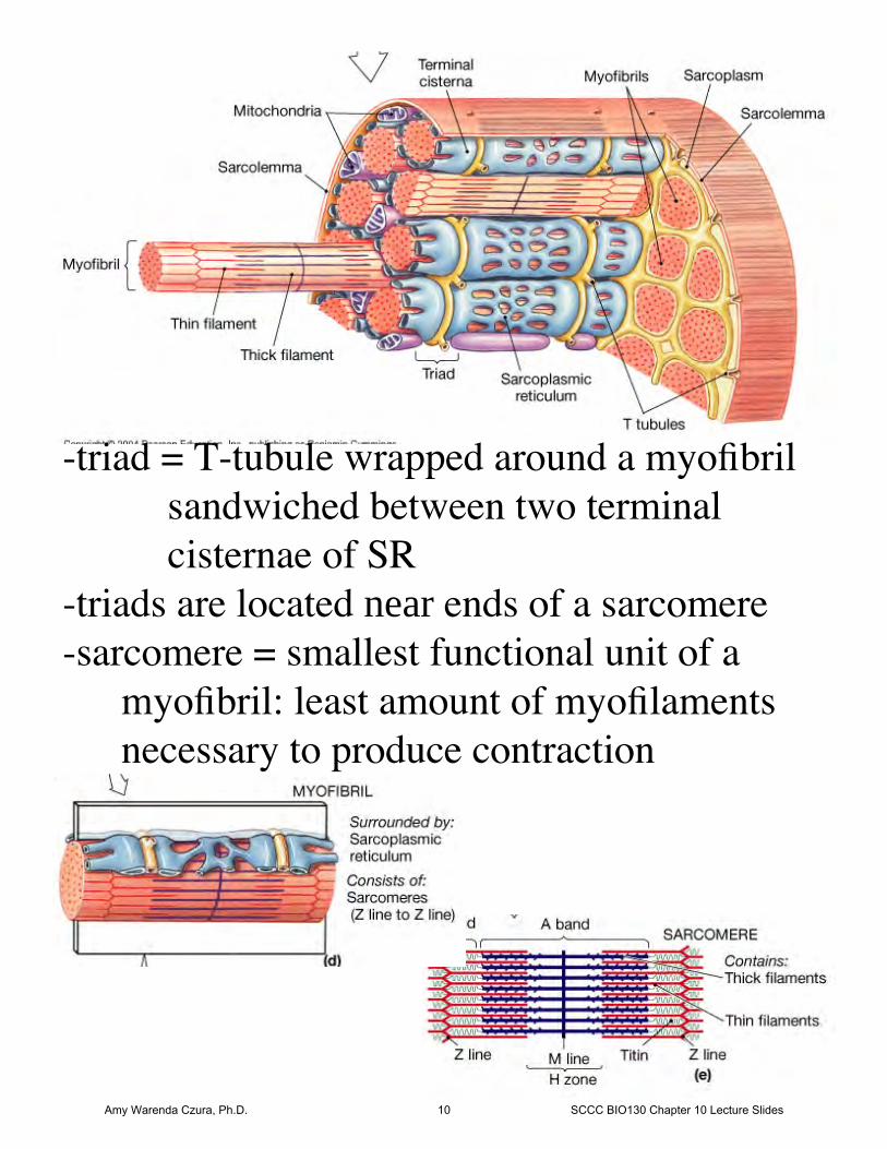
-triad = T-tubule wrapped around a myofibrilsandwiched between two terminal cisternae of SR
-triads are located near ends of a sarcomere-sarcomere = smallest functional unit of a
myofibril: least amount of myofilaments necessary to produce contraction
Amy Warenda Czura, Ph.D. 10 SCCC BIO130 Chapter 10 Lecture Slides
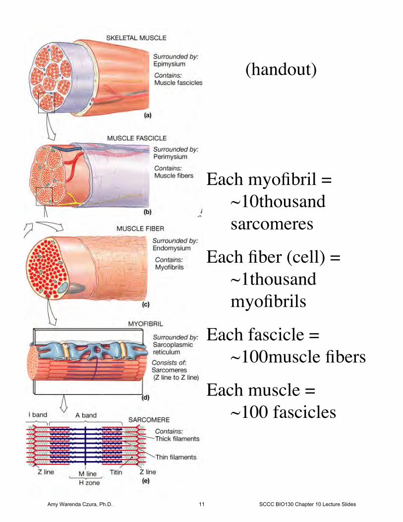
Each myofibril = ~10thousand sarcomeres
Each fiber (cell) = ~1thousand myofibrils
Each fascicle = ~100muscle fibers
Each muscle = ~100 fascicles
(handout)
Amy Warenda Czura, Ph.D. 11 SCCC BIO130 Chapter 10 Lecture Slides
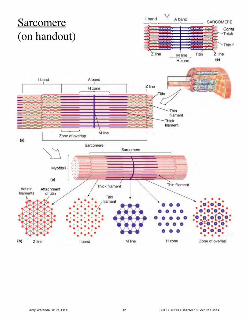
Sarcomere(on handout)
Amy Warenda Czura, Ph.D. 12 SCCC BIO130 Chapter 10 Lecture Slides
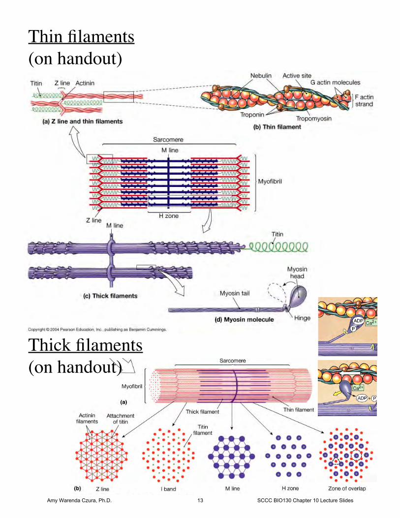
Thin filaments(on handout)
Thick filaments(on handout)
Amy Warenda Czura, Ph.D. 13 SCCC BIO130 Chapter 10 Lecture Slides
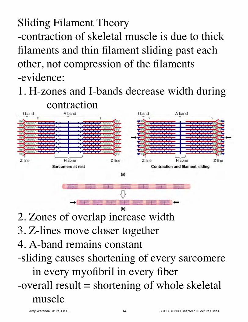
Sliding Filament Theory-contraction of skeletal muscle is due to thickfilaments and thin filament sliding past eachother, not compression of the filaments-evidence:1. H-zones and I-bands decrease width during
contraction
2. Zones of overlap increase width3. Z-lines move closer together4. A-band remains constant-sliding causes shortening of every sarcomere
in every myofibril in every fiber-overall result = shortening of whole skeletal
muscleAmy Warenda Czura, Ph.D. 14 SCCC BIO130 Chapter 10 Lecture Slides
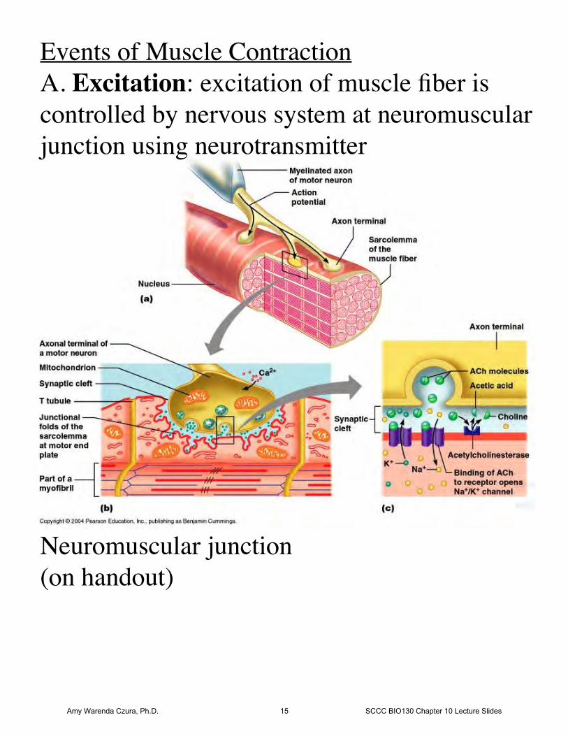
Events of Muscle ContractionA. Excitation: excitation of muscle fiber iscontrolled by nervous system at neuromuscularjunction using neurotransmitter
Neuromuscular junction(on handout)
Amy Warenda Czura, Ph.D. 15 SCCC BIO130 Chapter 10 Lecture Slides
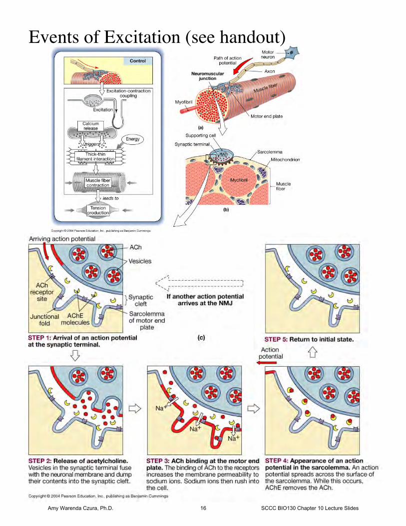
Events of Excitation (see handout)
Amy Warenda Czura, Ph.D. 16 SCCC BIO130 Chapter 10 Lecture Slides
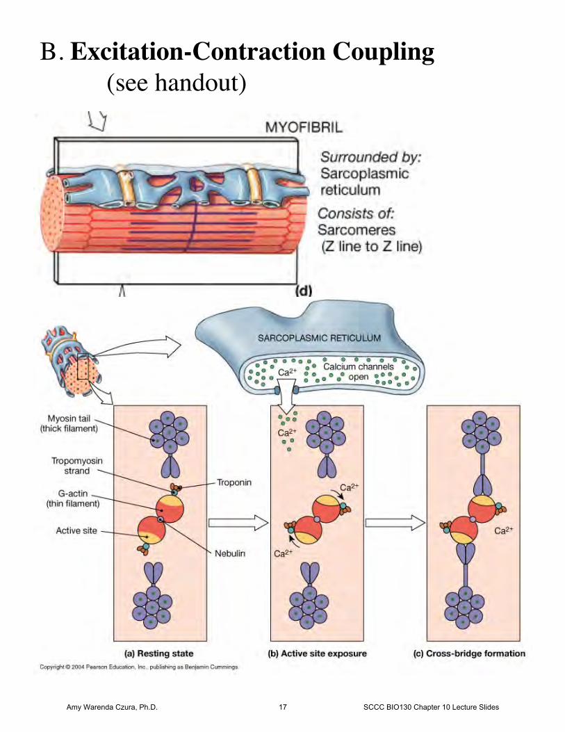
B. Excitation-Contraction Coupling(see handout)
Amy Warenda Czura, Ph.D. 17 SCCC BIO130 Chapter 10 Lecture Slides
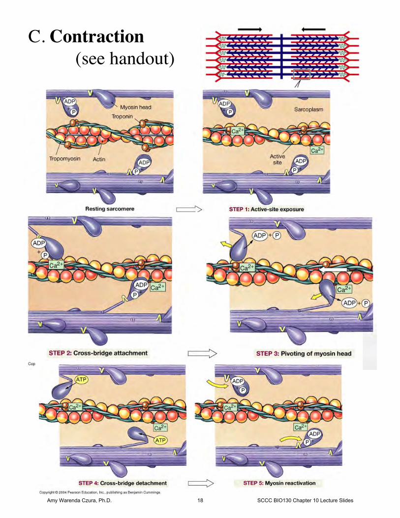
C. Contraction(see handout)
Amy Warenda Czura, Ph.D. 18 SCCC BIO130 Chapter 10 Lecture Slides
D. Relaxation 1. Ca2+ reabsorbed by sarcoplasmic reticulum 2. Ca2+ ions detach from troponin 3. Troponin, without Ca2+, pivots
tropomyosin back onto active sites onactin, no cross bridges can form
4. Sarcomeres stretch back out:-gravity-opposing muscle contractions-elastic recoil of titin protein
Muscle returns to resting lengthRigor Mortis:
Death, ATP used up,SR can not absorb Ca2+, Ca2+ binds troponin,
tropomyosin frees actin,cross bridges form,
no ATP to detach myosin head =fixed cross bridge(until necrosis releases lysosomal
enzymes which digest cross bridges)Amy Warenda Czura, Ph.D. 19 SCCC BIO130 Chapter 10 Lecture Slides
Diseases of muscle contraction:-Botulism/Botox: bacteria Clostridium
botulinum (grows in improperly cannedfoods) produces botulinum toxin: toxinprevents release of Ach atneuromuscular junction, results inflaccid paralysis
-Tetanus: Clostridium tetani (grows in soil)produces tetanus toxin: toxin causesover stimulation of motor neurons,results in spastic paralysis
-Myasthenia gravis: autoimmune disease,causes loss of Ach receptors, musclesbecome non-responsive
Amy Warenda Czura, Ph.D. 20 SCCC BIO130 Chapter 10 Lecture Slides
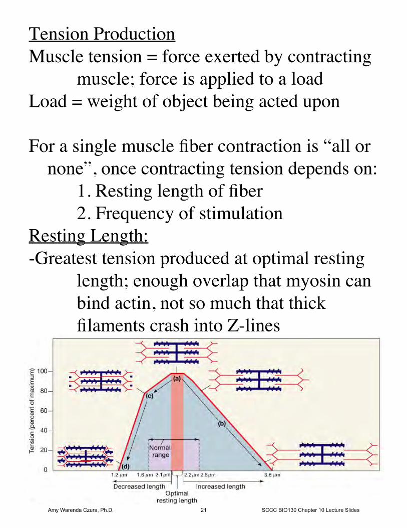
Tension ProductionMuscle tension = force exerted by contracting
muscle; force is applied to a loadLoad = weight of object being acted upon
For a single muscle fiber contraction is “all or none”, once contracting tension depends on:
1. Resting length of fiber2. Frequency of stimulation
Resting Length:-Greatest tension produced at optimal resting
length; enough overlap that myosin canbind actin, not so much that thickfilaments crash into Z-lines
Amy Warenda Czura, Ph.D. 21 SCCC BIO130 Chapter 10 Lecture Slides
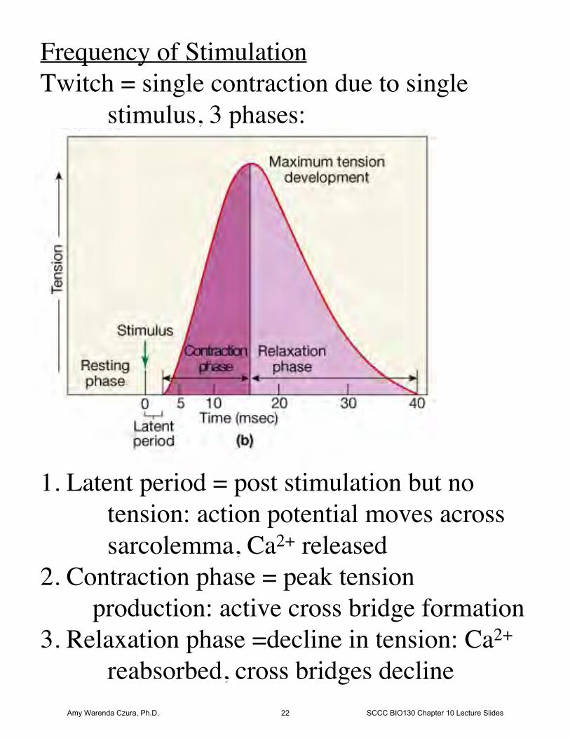
Frequency of StimulationTwitch = single contraction due to single
stimulus, 3 phases:
1. Latent period = post stimulation but notension: action potential moves acrosssarcolemma, Ca2+ released
2. Contraction phase = peak tension production: active cross bridge formation3. Relaxation phase =decline in tension: Ca2+
reabsorbed, cross bridges declineAmy Warenda Czura, Ph.D. 22 SCCC BIO130 Chapter 10 Lecture Slides
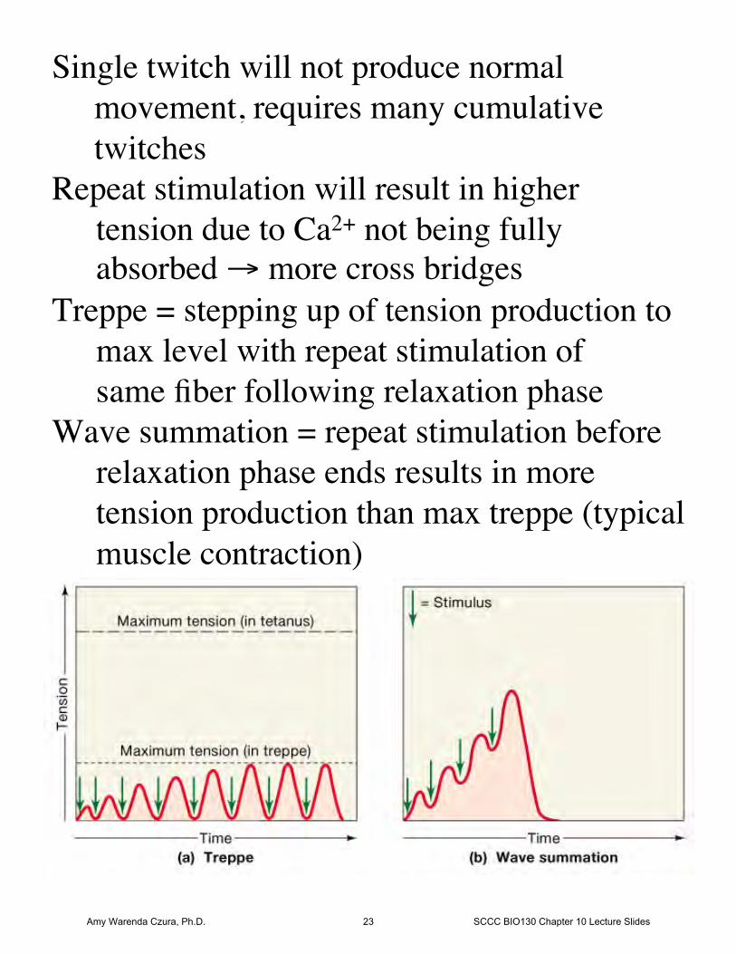
Single twitch will not produce normal movement, requires many cumulative twitchesRepeat stimulation will result in higher
tension due to Ca2+ not being fully absorbed → more cross bridges
Treppe = stepping up of tension production tomax level with repeat stimulation ofsame fiber following relaxation phase
Wave summation = repeat stimulation beforerelaxation phase ends results in more tension production than max treppe (typicalmuscle contraction)
Amy Warenda Czura, Ph.D. 23 SCCC BIO130 Chapter 10 Lecture Slides
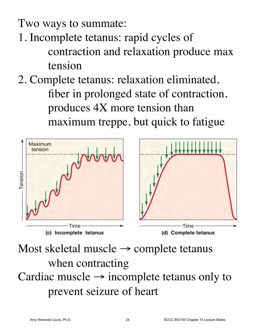
Two ways to summate:1. Incomplete tetanus: rapid cycles of
contraction and relaxation produce maxtension
2. Complete tetanus: relaxation eliminated,fiber in prolonged state of contraction,produces 4X more tension thanmaximum treppe, but quick to fatigue
Most skeletal muscle → complete tetanuswhen contracting
Cardiac muscle → incomplete tetanus only toprevent seizure of heart
Amy Warenda Czura, Ph.D. 24 SCCC BIO130 Chapter 10 Lecture Slides
Tension produced in whole muscle depends on:1. Internal vs. external tension Internal tension = produced by sarcomeres,
not all transferred to load, some lost dueto elasticity of muscle tissues
External tension = tension applied to load
2. Number of muscle fibers stimulated-each skeletal muscle has thousands of fibers
organized into motor units-Motor unit = all fibers controlled by single
motor neuron (axon branches to contacteach fiber)
-Number of fibers in motor unit depends onfunction:
-fine control: 4/unit e.g. eye muscles -gross control: 2000/unit e.g. legs-Fibers from different motor units intermingled
in muscle so activation of one unit willproduce equal tension across wholemuscle
Amy Warenda Czura, Ph.D. 25 SCCC BIO130 Chapter 10 Lecture Slides
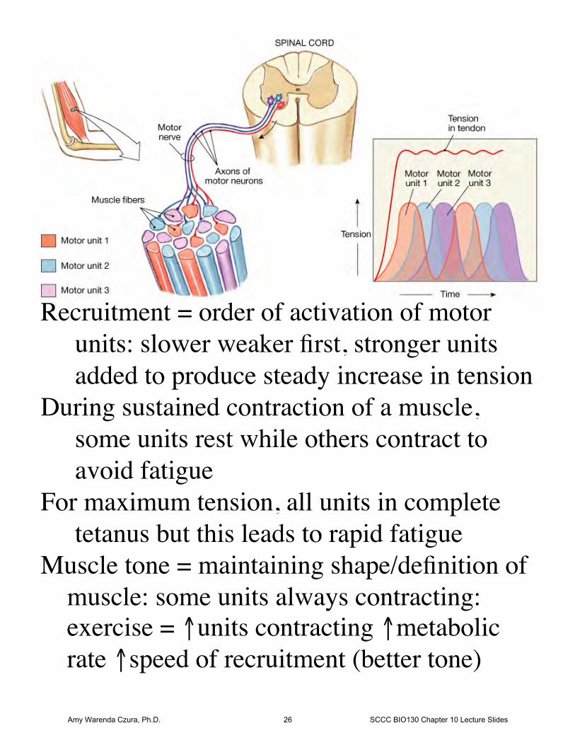
Recruitment = order of activation of motorunits: slower weaker first, stronger unitsadded to produce steady increase in tension
During sustained contraction of a muscle, some units rest while others contract toavoid fatigue
For maximum tension, all units in completetetanus but this leads to rapid fatigue
Muscle tone = maintaining shape/definition of muscle: some units always contracting: exercise = ↑units contracting ↑metabolic rate ↑speed of recruitment (better tone)
Amy Warenda Czura, Ph.D. 26 SCCC BIO130 Chapter 10 Lecture Slides
All contractions produce tension but notalways movement:
Isotonic contractions - muscle length changesresulting in movement
Isometric contractions -tension is producedwith no movement
Return to resting length: expansion via:1. elastic recoil after contraction2. opposing muscle contractions3. gravity
Muscle Metabolism- 1 fiber ~15 billion thick filaments- 1 thick filament ~ 2500 ATP/sec- 1 glucose (aerobic respiration) = 36 ATP- each fiber needs 1 X10¹² glucose/sec to
contract- ATP unstable, muscles store respiration
energy on creatine as CreatinePhosphate (CP)
Amy Warenda Czura, Ph.D. 27 SCCC BIO130 Chapter 10 Lecture Slides
-Creatine phosphokinase transfers P from CPto ADP when ATP needed to resetmyosin for next contraction
-Each cell has only ~20sec energy reserve
At Rest: use glucose and fatty acids with O2(from blood) →Aerobic respiration,resulting ATP used to build CP reserves, excess glucose stored as glycogen.
Moderate Activity: CP used up, glucose andfatty acids with O2 (from blood) used togenerate ATP (aerobic respiration).
High Activity: O2 not delivered adequately,glucose from glycogen reserves used forATP via fermentation (glycolysis only),pyruvic acid converted to lactic acid
Amy Warenda Czura, Ph.D. 28 SCCC BIO130 Chapter 10 Lecture Slides
Muscle Fatigue:-state where muscle can no longer contract
due to:1. Depletion of reserves (glycogen, ATP, CP)2. Decreased pH due to lactic acid
To restore function, cell needs:1. intracellular energy reserves (glycogen, CP)2. good circulation (nutrients in, wastes out)3. normal O2 levels4. normal pH
Lactic Acid Disposal-lactic acid diffuses into blood-filtered out by liver-converted back to glucose-returned to blood for use by cells-when O2 returns, remaining lactic acid in
muscle is converted to glucose and usedin aerobic cellular respiration
Amy Warenda Czura, Ph.D. 29 SCCC BIO130 Chapter 10 Lecture Slides
Muscle Performance-Depends on:
1. Types of fibers2. Physical conditioning
1. Fiber Types-types of fibers in a muscle are genetically
determined and mixedA. Fast glycolytic fibers (Fast twitch) -Myosin ATPases work quickly -anaerobic ATP production
(glycolysis only) -large diameter fibers -more myofilaments and glycogen -few mitochondria -fast to act, powerful, but quick to fatigue -catabolize glucose only
Amy Warenda Czura, Ph.D. 30 SCCC BIO130 Chapter 10 Lecture Slides
B. Slow oxidative fibers (slow twitch) -Myosin ATPases work slowly -specialized for aerobic respiration: -many mitochondria -extensive blood supply -myoglobin -smaller fibers for better diffusion -slow to contract, weaker tension, but resist
fatigue -catabolize glucose, lipids, and amino acids
C. Intermediate/ Fast oxidative fibers -qualities of both fast glycolytic and
slow oxidative fibers -fast acting but perform aerobic respiration
so resist fatigue -physical conditioning can convert some fast fibers into intermediate fibers for
stamina
Amy Warenda Czura, Ph.D. 31 SCCC BIO130 Chapter 10 Lecture Slides
2. Physical Conditioning A. Aerobic Exercise:
↑ capillary density↑ mitochondria and myoglobin
both then: ↑ efficiency of muscle metabolism ↑ strength and stamina ↓ fatigue
B. Resistance Exercise:-results in hypertrophy: fibers increase
in diameter but not number-↑glycogen, myofibrils, and myofilaments
results in ↑tension productionGrowth Hormone (pituitary) & testosterone
(male sex hormone) stimulate synthesis ofcontractile proteins = muscle enlargement
Epinephrine stimulates ↑muscles metabolism= ↑ force of contraction
Without stimulation muscles will atrophy:fibers shrink due to loss of myofilamentproteins. Loss: up to ~5%/day
Amy Warenda Czura, Ph.D. 32 SCCC BIO130 Chapter 10 Lecture Slides
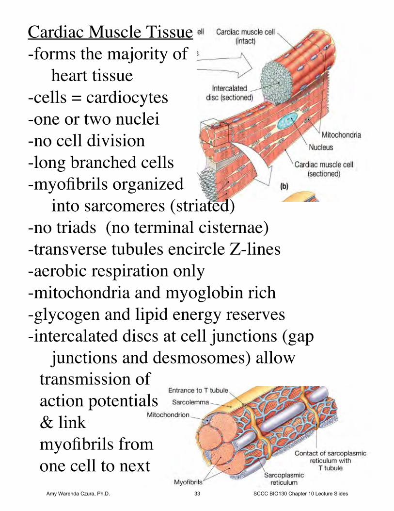
Cardiac Muscle Tissue-forms the majority of
heart tissue-cells = cardiocytes-one or two nuclei-no cell division-long branched cells-myofibrils organized
into sarcomeres (striated)-no triads (no terminal cisternae)-transverse tubules encircle Z-lines-aerobic respiration only-mitochondria and myoglobin rich-glycogen and lipid energy reserves-intercalated discs at cell junctions (gap
junctions and desmosomes) allowtransmission ofaction potentials& link myofibrils fromone cell to next
Amy Warenda Czura, Ph.D. 33 SCCC BIO130 Chapter 10 Lecture Slides
Features of cardiac muscle:1. Can contract without neural stimulation;
automaticity due to pacemaker cells thatgenerate action potentials spontaneously
2. Pace and amount of tension can be adjustedby nervous system
3. Contractions 10X longer than skeletalmuscle
4. Only twitches, no complete tetanus
Smooth Muscle Tissue-lines hollow organs: regulates blood flow and
movement of materials in organs-forms arrector pili muscles-usually organized into two layers:
-circular-longitudinal
Amy Warenda Czura, Ph.D. 34 SCCC BIO130 Chapter 10 Lecture Slides
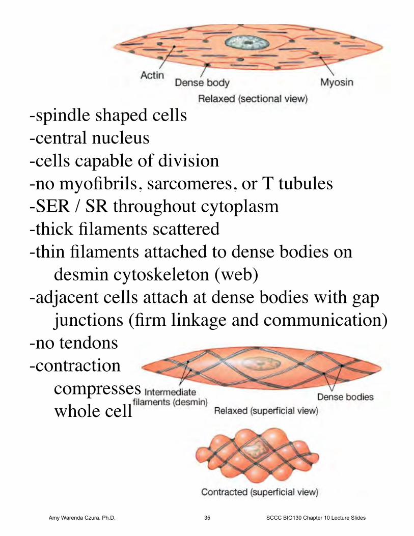
-spindle shaped cells-central nucleus-cells capable of division-no myofibrils, sarcomeres, or T tubules-SER / SR throughout cytoplasm-thick filaments scattered-thin filaments attached to dense bodies on
desmin cytoskeleton (web)-adjacent cells attach at dense bodies with gap
junctions (firm linkage and communication)-no tendons-contraction
compresseswhole cell
Amy Warenda Czura, Ph.D. 35 SCCC BIO130 Chapter 10 Lecture Slides
Smooth Muscle Excitation-Contraction-different than striated muscle: no troponin so
active sites on actin always exposedEvents:1. Stimulation causes Ca2+ release from SR
into cytoplasm2. Ca2+ binds calmodulin3. Calmodulin activates myosin light chain
kinase4. MLC Kinase converts ATP → ADP to cock
myosin head5. Cross bridges form → contraction, cells
pull toward centerStimulation is by involuntary control from:
-Autonomic Nervous System-hormones-other chemical factors
(Skeletal muscle = motor neurons)(Cardiac muscle = automaticity)
Amy Warenda Czura, Ph.D. 36 SCCC BIO130 Chapter 10 Lecture Slides
Effects of aging-skeletal muscle fibers become thinner
↓myofibrils, ↓energy reserves = ↓strength ↓endurance ↑fatigue
-↓cardiac and smooth muscle function = ↓cardiovascular performance
-↑fibrosis (CT)skeletal muscle less elastic
-↓ability to repair↓satellite cells↑scar formation
Amy Warenda Czura, Ph.D. 37 SCCC BIO130 Chapter 10 Lecture Slides