Embed Size (px)
Citation preview

© 2013 Pearson Education, Inc.
PowerPoint® Lecture Slides
prepared by
Meg Flemming
Austin Community College
C H A P T E R
The Urinary
System
18

© 2013 Pearson Education, Inc.
Chapter 18 Learning Outcomes
• 18-1
• Identify the components of the urinary system, and describe the
system's three primary functions.
• 18-2
• Describe the locations and structural features of the kidneys, trace
the path of blood flow to, within, and from a kidney, and describe
the structure of the nephron.
• 18-3
• Discuss the major functions of each portion of the nephron, and
outline the processes involved in urine formation.
• 18-4
• Describe the factors that influence glomerular filtration pressure
and the glomerular filtration rate (GFR).

© 2013 Pearson Education, Inc.
Chapter 18 Learning Outcomes
• 18-5
• Describe the structures and functions of the ureters, urinary
bladder, and urethra, discuss the control of urination, and describe
the micturition reflex.
• 18-6
• Define the terms fluid balance, electrolyte balance, and acid-base
balance, discuss their importance for homeostasis, and describe
how water and electrolytes are distributed within the body.
• 18-7
• Explain the basic mechanisms involved in maintaining fluid balance
and electrolyte balance.

© 2013 Pearson Education, Inc.
Chapter 18 Learning Outcomes
• 18-8
• Explain the buffering systems that balance the pH of the
intracellular and extracellular fluids, and identify the most common
threats to acid-base balance.
• 18-9
• Describe the effects of aging on the urinary system.
• 18-10
• Give examples of interactions between the urinary system
and other body systems.

© 2013 Pearson Education, Inc.
The Urinary System Structures (18-1)
• Two kidneys
• Produce urine that flows through:
• Urinary tract
• Two ureters
• Urinary bladder
• Urethra
• Elimination of urine is urination or micturition

© 2013 Pearson Education, Inc.
Figure 18-1 The Components of the Urinary System.
Kidney
Ureter
Urinary bladder
Urethra
Anterior view
Produces urine
Transports urinetoward the
urinary bladder
Temporarily storesurine prior
to elimination
Conducts urine toexterior; in males,transports semen
as well

© 2013 Pearson Education, Inc.
Three General Functions of the Urinary System
(18-1)
1. Excretion of organic wastes
2. Elimination of these wastes into the external
environment
3. Regulation of blood plasma volume and solute
concentration

© 2013 Pearson Education, Inc.
Homeostatic Functions of the Urinary System
(18-1)
• Regulate blood volume and blood pressure
• Regulate plasma ions, such as sodium,
potassium, chloride, and calcium
• Aid in stabilization of plasma pH
• Conserve valuable nutrients like glucose and
amino acids
• Eliminate wastes like urea and uric acid

© 2013 Pearson Education, Inc.
Checkpoint (18-1)
1. Name the three primary functions of the urinary
system.
2. Identify the components of the urinary system.
3. Define micturition.

© 2013 Pearson Education, Inc.
Location of the Kidneys (18-2)
• Either side of vertebral column
• Between last thoracic and 3rd lumbar vertebrae
• Right is often lower than left
• Retroperitoneal but supported by connective
tissue
• Fibrous capsule covers each kidney
• Capsule surrounded by adipose tissue

© 2013 Pearson Education, Inc.
Renal artery and vein
Adrenal gland
Left kidneyDiaphragm
11th and 12th ribs
Right kidney
Lumbar (L1) vertebra
UreterInferior vena cava
Iliac crest
Aorta
Urinary bladder
Urethra
This posterior view of the trunk showsthe positions of the kidneys and othercomponents of the urinary system.
Figure 18-2a The Position of the Kidneys.

© 2013 Pearson Education, Inc.
A superior view of a section at the level indicated inpart (a) shows the kidney’s retroperitoneal position.
Renalvein
Renalartery
Aorta
Hilumof kidney
Ureter
Kidney
Fibrouscapsule
Adiposetissue
L1
Inferiorvena cava
PancreasPancreas
eSpl enSpleen
Figure 18-2b The Position of the Kidneys.
Parietalperitoneum Stomach

© 2013 Pearson Education, Inc.
Superficial Anatomy of the Kidney (18-2)
• Kidneys are bean shaped
• Indentation on one side is the hilum
• Exit for ureter and renal vein
• Entrance for renal artery
• Fibrous capsule
• Covers outer surface
• Lines renal sinus, an internal cavity

© 2013 Pearson Education, Inc.
Sectional Anatomy of the Kidney (18-2)
• Renal cortex is outer layer
• Projects into medulla as renal columns
• Renal medulla is inner layer
• Contains 6–18 cone-shaped renal pyramids
• Tip is called renal papilla and projects into renal pelvis
• Renal lobe
• Contains pyramid, overlying cortex and renal columns

© 2013 Pearson Education, Inc.
Sectional Anatomy of the Kidney (18-2)
• Urine production occurs in nephrons of cortex
and pyramids of medulla
• Ducts drain urine into a cup-like structure called
the minor calyx
• 4–5 minor calyces empty into major calyx
• Major calyces combine to form the renal pelvis
• Nephron, functional unit of kidneys in cortex

© 2013 Pearson Education, Inc.
Figure 18-3 The Structure of the Kidney.
Proximal convoluted tubule
Distal convoluted tubule
Renal corpuscle
Collecting duct
Nephron loop
Renal
cortex
Renal
medulla
Renalsinus
Hilum
Renal
papillaUreter
A diagrammatic view of a frontal
section through the left kidney
An enlarged view showing
the location and general
structure of a nephron
Major calyx
Minor calyx
Renal pyramid
Renal lobe
Renal columns
Fibrous capsule
Hilum
Ureter
A frontal section through the left kidney
Renal
cortex
Renal
medulla
Renal
pyramids
Renal
sinus
Major
calyx
Renal
pelvis
Minor
calyx
Renal
papilla
Renal
pelvis

© 2013 Pearson Education, Inc.
Blood Supply to the Kidney (18-2)
• Kidneys receive 20–25 percent of cardiac output
• Blood flow starts with renal artery
• Interlobar artery
• Arcuate artery (along cortex-medulla boundary)
• Cortical radiate artery
• Afferent arterioles
• Glomerular capillaries
• Efferent arterioles
• Peritubular capillaries
• Cortical radiate vein
• Arcuate vein
• Interlobar vein
• Exits kidney renal vein
© 2013 Pearson Education, Inc.
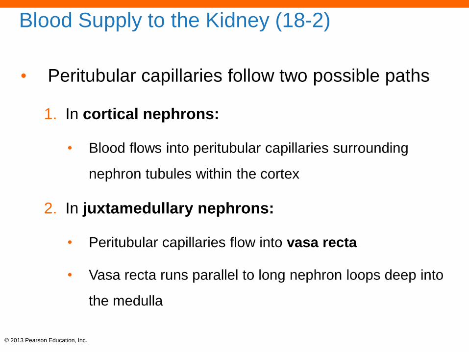
Blood Supply to the Kidney (18-2)
• Peritubular capillaries follow two possible paths
1. In cortical nephrons:
• Blood flows into peritubular capillaries surrounding
nephron tubules within the cortex
2. In juxtamedullary nephrons:
• Peritubular capillaries flow into vasa recta
• Vasa recta runs parallel to long nephron loops deep into
the medulla

© 2013 Pearson Education, Inc.
Cortical radiateveins
Cortical radiatearteries
Arcuateveins
Arcuatearteries
Adrenalartery
Renalartery
Renalvein
Interlobararteries
Interlobarveins
This sectional view of a kidney shows the major arteries and veins; compare with Figure 18-3a.
Figure 18-4a The Blood Supply to the Kidneys.

© 2013 Pearson Education, Inc.
Figure 18-4b The Blood Supply to the Kidneys.
Nephron
Cortex
Afferent
arterioles
Medulla
This enlargedview illustrates thecirculation in the cortex.

© 2013 Pearson Education, Inc.
Efferentarteriole
Afferentarteriole
Renalcorpuscle
Peritubularcapillaries
Nephronloop
Peritubularcapillaries Collecting
duct
Further enlargement shows the circulation to a cortical nephron.
Figure 18-4c The Blood Supply to the Kidneys.

© 2013 Pearson Education, Inc.
Peritubularcapillaries
Distalconvolutedtubule (DCT)
Vasa recta
Collectingduct
Nephronloop
Further enlargement shows the circulation to a juxtamedullary nephron.
Figure 18-4d The Blood Supply to the Kidneys.
Proximal convolutedtubule (PCT)
Efferentarteriole
Glomerulus
Afferentarteriole

© 2013 Pearson Education, Inc.
The Nephron (18-2)
• Renal corpuscle
• Glomerular (Bowman's) capsule surrounds glomerular capillaries
• Filtration occurs from capillaries to capsule
• Renal tubule
• Filtrate flows into proximal convoluted tubule (PCT)
• Nephron loop
• Distal convoluted tubule (DCT)
• Collecting duct
• Collecting system

© 2013 Pearson Education, Inc.
Functions of the Nephron (18-2)
• Corpuscle
• Filters plasma (minus proteins) into capsule
• Includes valuable nutrients, ions, and water
• Tubules
1. Reabsorb useful molecules and ions from filtrate back
into plasma
2. Reabsorb >90 percent of water back into plasma
3. Secrete any wastes missed by filtration

© 2013 Pearson Education, Inc.
NEPHRON
Proximal
convoluted
tubule
Distal
convoluted
tubule
COLLECTING SYSTEM
Collecting duct
Renal
corpuscle
Nephron loop
Papillary duct
KEY
Renal
tubule
GlomerulusEfferent arterioleAfferent arteriole
Collecting duct
Loop
begins
Loop
ends
Thin
descending
limb
Thick
ascending
limb
Descending
limb
Ascending
limb
FiltrateWater reabsorption
Variable waterreabsorptionSolute reabsorptionor secretionVariable solute reabsorptionor secretion
Minor
calyx
Figure 18-5 A Representative Nephron and the Collecting System.

© 2013 Pearson Education, Inc.
Renal Corpuscle Contains Filtration Membrane
(18-2)
• Glomerular capsule
• Outer layer forms wall of corpuscle
• Inner layer encloses glomerular capillaries
• Podocytes
• Epithelial cells have foot processes that sit on basement
membrane and wrap around capillaries
• Layers separated by capsular space
• Glomerular capillaries
• Highly fenestrated or leaky pores in endothelial cells

© 2013 Pearson Education, Inc.
Figure 18-6 The Renal Corpuscle.
Capsularspace
Glomerularcapillary
Capsularepithelium
Visceralepithelium(podocyte)
Glomerular capsule
Proximalconvoluted
tubule
Filtrationslits
Pedicels
Capsular space
Nucleus
Podocyte
Pores
SupportingcellCapillaryendothelialcell
Capsular
epithelium
This cross section through a segment of the glomerulus shows the components of the filtration membrane of the nephron.
Efferentarteriole
Distalconvoluted
tubule
Macula densa
Juxtaglomerularcells
Juxtaglomerularcomplex
Afferentarteriole
This sectional view illustrates the important
structural features of a renal corpuscle.
Pedicels
Podocyte
A podocyte SEM x 2,100
This colorized photomicrograph shows the glomerular surface, including individual podocytes and their processes.

© 2013 Pearson Education, Inc.
Capsularspace
Glomerularcapillary
Capsularepithelium
Visceralepithelium(podocyte)
Glomerular capsule
Proximalconvoluted
tubule
Efferentarteriole
Distalconvoluted
tubule
Macula densa
Juxtaglomerularcells
Juxtaglomerularcomplex
Afferentarteriole
This sectional view illustrates the important structural features of a renal corpuscle.
Figure 18-6a The Renal Corpuscle.

© 2013 Pearson Education, Inc.
Filtrationslits
Pedicels
Capsular space
Nucleus
Podocyte
Pores
Capillaryendothelialcell
Capsularepithelium
This cross section through a segment of the glomerulus shows the components of the filtration membrane of the nephron.
Figure 18-6b The Renal Corpuscle.
Supportingcell

© 2013 Pearson Education, Inc.
Figure 18-6c The Renal Corpuscle.
Pedicels
Podocyte
A podocyte SEM x 2,100
This colorized photomicrograph shows the glomerular surface, including individual podocytes and their processes.

© 2013 Pearson Education, Inc.
The Proximal Convoluted Tubule (18-2)
• First segment of renal tubule
• Majority of reabsorption occurs here
• Epithelium has transport mechanisms for nutrients and
ions
• Molecules are moved from tubule to IF
• Water follows osmotically

© 2013 Pearson Education, Inc.
The Nephron Loop (18-2)
• Fluid in descending limb flows toward renal pelvis
• Epithelium permeable to water, not solutes
• Tubule makes 180-degree turn
• Fluid in ascending limb flows toward renal cortex
• Epithelium permeable to solutes, not water
• IF of renal medulla has unusually high solute
concentration

© 2013 Pearson Education, Inc.
The Distal Convoluted Tubule (18-2)
• Has three vital processes
1. Active secretion of ions, acids, drugs, and toxins
2. Selective reabsorption of sodium
3. Selective reabsorption of water

© 2013 Pearson Education, Inc.
The Juxtaglomerular Complex (18-2)
• Macula densa
• Epithelial cells of DCT
• Sit next to afferent arterioles
• Juxtaglomerular cells
• Smooth muscle cells of afferent arterioles
• Together regulate blood volume and pressure
• Affected by secretion of erythropoietin and renin
© 2013 Pearson Education, Inc.
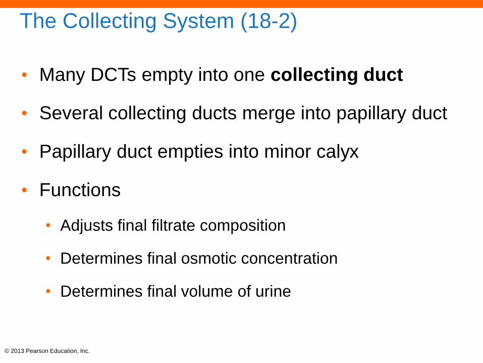
The Collecting System (18-2)
• Many DCTs empty into one collecting duct
• Several collecting ducts merge into papillary duct
• Papillary duct empties into minor calyx
• Functions
• Adjusts final filtrate composition
• Determines final osmotic concentration
• Determines final volume of urine

© 2013 Pearson Education, Inc.
Table 18-1 The Functions of the Nephron and Collecting System in the Kidney.

© 2013 Pearson Education, Inc.
Checkpoint (18-2)
4. How does the position of the kidneys differ from
that of most other organs in the abdominal
region?
5. Why don't plasma proteins pass into the capsular
space of the renal corpuscle under normal
circumstances?
6. Damage to which part of the nephron would
interfere with the control of blood pressure?

© 2013 Pearson Education, Inc.
Metabolic Wastes in Urine (18-3)
• Must be excreted to maintain homeostasis
1. Urea
• From amino acid breakdown
2. Creatinine
• From breakdown of high-energy compound: creatinine
phosphate in skeletal muscles
3. Uric acid
• From breakdown of RNA

© 2013 Pearson Education, Inc.
Three Nephron Processes (18-3)
1. Filtration
• Movement of water and solutes across filtration
membrane
• Occurs exclusively in renal corpuscle
2. Reabsorption
• Returning water and desirable solutes back to body
• Occurs primarily at PCT

© 2013 Pearson Education, Inc.
Three Nephron Processes (18-3)
3. Secretion
• Transport of undesirable material missed by filtration
• Occurs primarily at DCT
• Water, sodium, potassium
• Regulated by interaction between nephron loop and
collecting duct
ANIMATION Kidney Function: Reabsorption and SecretionPLAY

© 2013 Pearson Education, Inc.
Figure 18-7 Physiological Processes of the Nephron.
Blood
pressure
Glomerular
capillary
Filtration
membrane
Solute
Filtrate
Capsular
space
Filtration
Transport
proteins
Solute
Peritubular
fluidTubular
epithelium
Tubular
fluid
Reabsorption Secretion
Transport
proteins
Solute
Peritubular
fluidTubular
epitheliumTubular
fluid

© 2013 Pearson Education, Inc.
Table 18-2 Normal Laboratory Values for Solutes in Urine and Plasma

© 2013 Pearson Education, Inc.
Net Filtration Pressure (18-3)
• Filtration
• Result of outward blood pressure, inward blood osmotic
pressure, and inward fluid pressure from the capsule
• Glomerular BP
• Must stay high enough to overcome other two forces
• Serious loss of systemic BP can result in:
• Loss of filtration, reduction in kidney function
© 2013 Pearson Education, Inc.
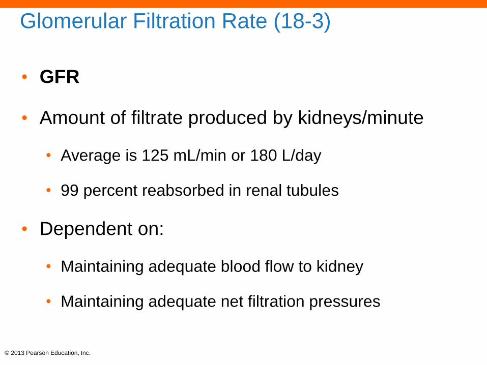
Glomerular Filtration Rate (18-3)
• GFR
• Amount of filtrate produced by kidneys/minute
• Average is 125 mL/min or 180 L/day
• 99 percent reabsorbed in renal tubules
• Dependent on:
• Maintaining adequate blood flow to kidney
• Maintaining adequate net filtration pressures

© 2013 Pearson Education, Inc.
Events at the Proximal Convoluted Tubule
(18-3)
• 60–70 percent of filtrate volume is reabsorbed at
PCT
• All glucose, amino acids, and other organic
nutrients are reabsorbed
• Ionic reabsorption of Na+, K+, Ca2+, Mg2+, HCO3–
• Some are hormonally regulated, like Ca2+ by PTH
• Some secretion occurs here, like H+
© 2013 Pearson Education, Inc.
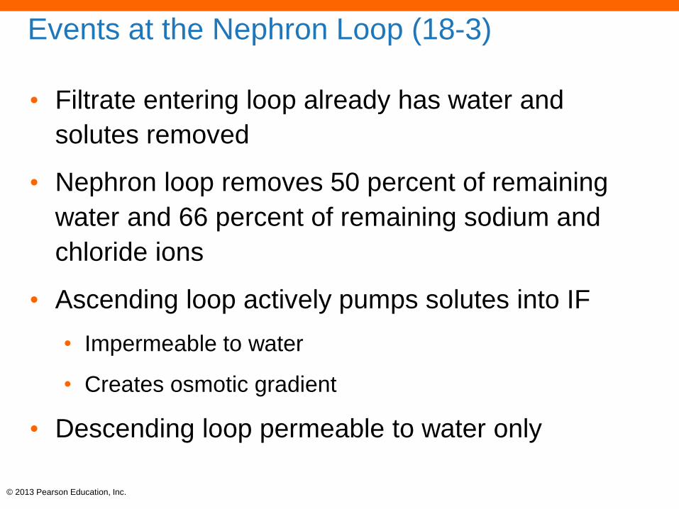
Events at the Nephron Loop (18-3)
• Filtrate entering loop already has water and
solutes removed
• Nephron loop removes 50 percent of remaining
water and 66 percent of remaining sodium and
chloride ions
• Ascending loop actively pumps solutes into IF
• Impermeable to water
• Creates osmotic gradient
• Descending loop permeable to water only

© 2013 Pearson Education, Inc.
Events at the Nephron Loop (18-3)
• As filtrate moves down the descending loop:
• Loses water to IF
• Becomes higher in osmotic concentration
• As filtrate moves up ascending loop:
• Pumps solutes to IF
• Becomes lower in osmotic concentration
• What remains are primarily waste products
ANIMATION Kidney function: Loop of HenlePLAY

© 2013 Pearson Education, Inc.
Events at the Distal Convoluted Tubule (18-3)
• 80 percent of water and 85 percent of solutes
have been reabsorbed
• Final adjustments to:
• Sodium reabsorption
• Increases in the presence of aldosterone
• Water reabsorption
• Increases in the presence of ADH
• Serves to concentrate filtrate as it passes through
collecting duct

© 2013 Pearson Education, Inc.
Figure 18-8 The Effects of ADH on the DCT and Collecting Duct.
Renal cortex
PCT DCT
Glomerulus
Solutes
Collectingduct
Renal medulla
Large volumeof dilute urine
The DCT and collecting duct
are impermeable to water in
the absence of ADH. The
result is the production of a
large volume of dilute urine.
Small volume of
concentrated urine
KEY
= Water
reabsorption
= Variable water
reabsorption
= Na+/Cl–
transport
= Antidiuretic
hormone
Renal cortex
Renal medulla
The DCT and collecting duct are
permeable to water in the presence
of ADH. The result is the production
of a small volume of concentrated
urine.

© 2013 Pearson Education, Inc.
Properties of Normal Urine (18-3)
• Once filtrate enters the minor calyx:
• No other secretion or reabsorption can occur
• Fluid is now called urine
• Concentration and composition vary based on metabolic
and hormonal activities
ANIMATION Kidney function: Urine formationPLAY

© 2013 Pearson Education, Inc.
Table 18-3 General Characteristics of Normal Urine

© 2013 Pearson Education, Inc.
Figure 18-9 A Summary of Kidney Function
The filtrate produced at
the renal corpuscle has the
same osmotic concentration
as plasma. It has the same
composition as blood plasma
but does not contain plasma
proteins.
In the proximal convoluted
tubule (PCT), the active
removal of ions and organic
nutrients produces a
continuous osmotic flow of
water out of the tubular fluid.
This reduces the volume of
filtrate but keeps the solute
concentration the same inside
and outside the tubule.
In the PCT and descending
limb of the nephron loop,
water moves into the
surrounding peritubular fluid
and then into the peritubular
capillaries and vasa recta. This
osmotic flow of water leaves a
small volume of highly
concentrated tubular fluid.
= Water
reabsorption
= Variable water
reabsorption
= Na+/Cl–
transport
= Aldosterone-
regulated pump
Nutrients
Electrolytes
Vasa
recta
Nephron
loopIn
cre
asin
g o
sm
ola
rity
Nephron
loop
Vasa
recta
Urine enters
renal pelvis
Tubular fluidfrom corticalnephrons
ADH-
regulated
permeability
The thick ascending limb is
impermeable to water and
solutes. The tubule cells actively
transport Na+ and Cl– out of the
tubule, thereby lowering the
solute concentration of the
tubular fluid. Because only Na+
and Cl– are removed, urea now
accounts for a higher proportion
of the total solute concentration
at the end of the nephron loop.1
Further alterations in the
composition of the tubular
fluid occur in the DCT and the
collecting system. The solute
concentration of the tubular
fluid can be adjusted through
active transport (reabsorption
or secretion).
The final adjustments in the
volume and solute
concentration of the tubular
fluid are made by controlling
the water permeabilities of the
distal portions of the DCT and
the collecting system. The
level of exposure to ADH
determines the final urine
volume and concentration.
The vasa recta absorbs
the solutes and water
reabsorbed by the nephron
loop and the collecting ducts.
By transporting these solutes
and water into the
bloodstream, the vasa recta
maintains the concentration
gradient of the renal medulla.
RENAL MEDULLA
KEY
RENAL CORTEX

© 2013 Pearson Education, Inc.
Checkpoint (18-3)
7. A decrease in blood pressure would have what
effect on the GFR?
8. If nephrons lacked a nephron loop, what would
be the effect on the volume and solute (osmotic)
concentration of the urine produced?
9. What effect would low circulating levels of
antidiuretic hormone (ADH) have on urine
production?

© 2013 Pearson Education, Inc.
GFR Is Key to Normal Kidney Function (18-4)
• Maintaining net filtration pressure and consistent
GFR is necessary for normal kidney function
• GFR is controlled by three mechanisms
1. Autoregulation (or local regulation)
2. Hormonal regulation
3. Autonomic regulation
• Through sympathetic division of ANS

© 2013 Pearson Education, Inc.
Local Regulation of Kidney Function (18-4)
• Autoregulation of diameters of afferent and
efferent arterioles
• Quick, short-term adjustment of GFR
• Decrease in GFR triggers:
• Dilation of afferent arteriole
• Constriction of efferent arteriole
• Brings glomerular blood pressure up
• Increases GFR back to normal
© 2013 Pearson Education, Inc.
Local Regulation of Kidney Function (18-4)
• Increase in BP causes increase in GFR
• Increase in GFR triggers:
• Stretch of wall of afferent arteriole
• Smooth muscle responds by contracting
• Reduces afferent arteriolar diameter
• Reduces flow into glomerulus
• Decreases GFR back to normal

© 2013 Pearson Education, Inc.
Hormonal Control of Kidney Function (18-4)
• Long-term adjustment of blood pressure and
volume stabilizes GFR
• Major hormones
• Angiotensin II
• ADH
• Aldosterone
• ANP

© 2013 Pearson Education, Inc.
Renin-Angiotensin System (18-4)
• Decrease in BP, blood volume causes:
• Release of renin from JGC, which converts
angiotensinogen into angiotensin I
• ACE converts angiotensin I into angiotensin II
• Causes peripheral vasoconstriction, increases BP
• Efferent arteriole constriction, increase in GFR
• Posterior pituitary releases ADH
• Adrenal glands secrete aldosterone, E, and NE
• Result is increase in BP and blood volume

© 2013 Pearson Education, Inc.
Figure 18-10 The Renin-Angiotensin System and Regulation of GFR.
Decreased filtrationpressure; decreased
filtrate and urine production
HOMEOSTASISDISTURBED
Decreased bloodflow to kidneys
HOMEOSTASIS
Normalglomerular
filtration rate
Endocrine
response
Juxtaglomerularcomplex increasesproduction of renin.
HOMEOSTASISRESTORED
Increasedglomerularpressure
Renin in thebloodstream triggers
formation ofangiotensin I, which is
then activated toangiotensin II by
angiotensin convertingenzyme (ACE) in the
capillaries of the lungs.
Angiotensin IIconstricts peripheralarterioles and furtherconstricts the efferent
arterioles.
Increasedsystemic
bloodpressure
Increasedblood
volume
Angiotensin II triggersincreased aldosterone
secretion by theadrenal glands.
Aldosteroneincreases
Na+ retention.
Increased fluidconsumption
Increased fluidretention
Constriction ofvenous reservoirs
Increasedcardiac output
Together, angiotensin IIand sympathetic activation
stimulate peripheralvasoconstriction.
Increasedstimulation ofthirst centers
Increased ADHproduction
Increasedsympathetic
activation
Angiotensin IItriggersneural
responses.
Renin–Angiotensin System

© 2013 Pearson Education, Inc.
Antidiuretic Hormone (18-4)
• ADH
• Release stimulated by:
• Angiotensin II
• Drop in BP
• Increase in plasma solute concentration
• Results in:
• Increased water permeability at DCT and collecting duct
• Stimulates thirst center in hypothalamus

© 2013 Pearson Education, Inc.
Aldosterone (18-4)
• Release stimulated by:
• Angiotensin II
• Increase in K+ concentration in plasma
• Results in:
• Reabsorption of Na+
• Secretion of K+
• Occurs at DCT and collecting ducts

© 2013 Pearson Education, Inc.
Atrial Natriuretic Peptide (18-4)
• ANP
• Release stimulated by:
• Rise in BP and blood volume in atria of heart
• Results in:
• Inhibition of renin, aldosterone, ADH
• Opposite of renin-angiotensin system
• Decrease in sodium reabsorption
• Dilation of glomerular capillaries, increasing GFR
© 2013 Pearson Education, Inc.
Sympathetic Activation (18-4)
• Direct, sudden effect of acute crisis
• Constriction of afferent arterioles
• Decreases GFR
• Can override local effects to stabilize GFR
• Indirect effect
• Shunts blood away from kidneys to perfuse tissues and
organs with higher needs
• Problematic in strenuous exercise

© 2013 Pearson Education, Inc.
Checkpoint (18-4)
10. List the factors that affect the glomerular filtration
rate (GFR).
11. What effect would increased aldosterone
secretion have on the K+ concentration in urine?
12. What is the effect of sympathetic activation on
kidney function?

© 2013 Pearson Education, Inc.
The Ureters (18-5)
• Paired muscular tubes conduct urine to bladder
• Begins at funnel-shaped renal pelvis
• Ends at posterior, slightly inferior bladder wall
• Slitlike ureteral openings prevent backflow
• Wall contains:
• Transitional epithelium
• Smooth muscle that undergoes peristalsis

© 2013 Pearson Education, Inc.
The Urinary Bladder (18-5)
• Hollow muscular organ that stores urine
• Base has triangular area called trigone, formed by:
• Two ureteral openings
• Urethral entrance
• Contains involuntary internal urethral sphincter
• Wall contains:
• Transitional epithelium
• Layers of smooth muscle called detrusor muscle

© 2013 Pearson Education, Inc.
The Urethra (18-5)
• Extends from neck of urinary bladder to exterior of
body through:
• External urethral orifice
• Passes through pelvic floor
• External urethral sphincter voluntary, skeletal muscle
• In males, passes through penis
• In females it is very short, anterior to vagina

© 2013 Pearson Education, Inc.
Figure 18-11a Organs for the Conduction and Storage of Urine.
Peritoneum
Urinary bladder
Pubic symphysis
Prostate gland
External urethralsphincter
Urethra
External urethralorifice
Left ureter Rectum
Male

© 2013 Pearson Education, Inc.
Figure 18-11b Organs for the Conduction and Storage of Urine.
RectumRight ureter
Uterus
Peritoneum
Urinary bladder
Pubic symphysis
Vagina
Urethra
External urethralsphincter
Female

© 2013 Pearson Education, Inc.
Figure 18-11c Organs for the Conduction and Storage of Urine.
Ureter
Ligaments
Detrusor muscle
Ureteral openings
Trigone
Internal urethralsphincter
External urethralsphincter
Neck
Prostate gland
Urinary bladder in male
Urethra

© 2013 Pearson Education, Inc.
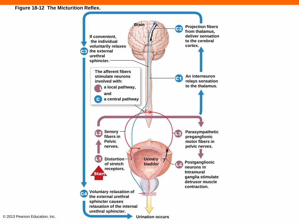
The Micturition Reflex and Urination (18-5)
• Reflex pathway
• Increased urine volume triggers stretch receptors in wall
• Activates afferent fibers to sacral spinal cord
• Activates parasympathetic pathway back to bladder wall
• Activates interneurons relaying information to CNS
• Contraction
• Elevates pressure, forces internal sphincter to open
© 2013 Pearson Education, Inc.
Figure 18-12 The Micturition Reflex.
If convenient,the individual
voluntarily relaxesthe external
urethralsphincter.
C3
The afferent fibersstimulate neuronsinvolved with:
a local pathway,
andL
C
Projection fibersfrom thalamus,deliver sensationto the cerebralcortex.
C2
An interneuron
relays sensation
to the thalamus.
Parasympatheticpreganglionicmotor fibers inpelvic nerves.
L3
Postganglionic
neurons in
Intramural
ganglia stimulate
detrusor musclecontraction.
Distortionof stretchreceptors.
Senory
fibers in
Pelvic
nerves.
Voluntary relaxation of
the external urethral
sphincter causes
relaxation of the internal
urethral sphincter.
Urination occurs
a central pathway
C1
L2
L1
Start
C4
L4
Brain
Urinary
bladder

© 2013 Pearson Education, Inc.
Checkpoint (18-5)
13. What is responsible for the movement of urine
from the kidneys to the urinary bladder?
14. An obstruction of a ureter by a kidney stone
would interfere with the flow of urine between
what two structures?
15. Control of the micturition reflex depends on the
ability to control which muscle?
© 2013 Pearson Education, Inc.
Fluid, Electrolyte, and pH Balance (18-6)
• Topic integrates information from multiple chapters
• Key to proper treatment of multiple diseases and
conditions
• Water balance is essential to maintain enzyme function
• Electrolyte balance is essential to maintain excitable
tissue function
• pH balance is essential to maintain normal chemical
reactions, cell structure, and function

© 2013 Pearson Education, Inc.
Three Interrelated Factors in Homeostasis
(18-6)
• For balance of all factors, intake must equal
output
1. Fluid balance
• Movement between ECF and ICF driven by osmosis
2. Electrolyte balance
• Creates osmotic gradients
3. Acid-base balance
• H+ ion balance establishes normal pH

© 2013 Pearson Education, Inc.
Basics of Body Fluid Compartments (18-6)
• Water is 50–60 percent of body weight
• Fluid compartments
• Intercellular fluid (ICF)
• Extracellular fluid (ECF)

© 2013 Pearson Education, Inc.
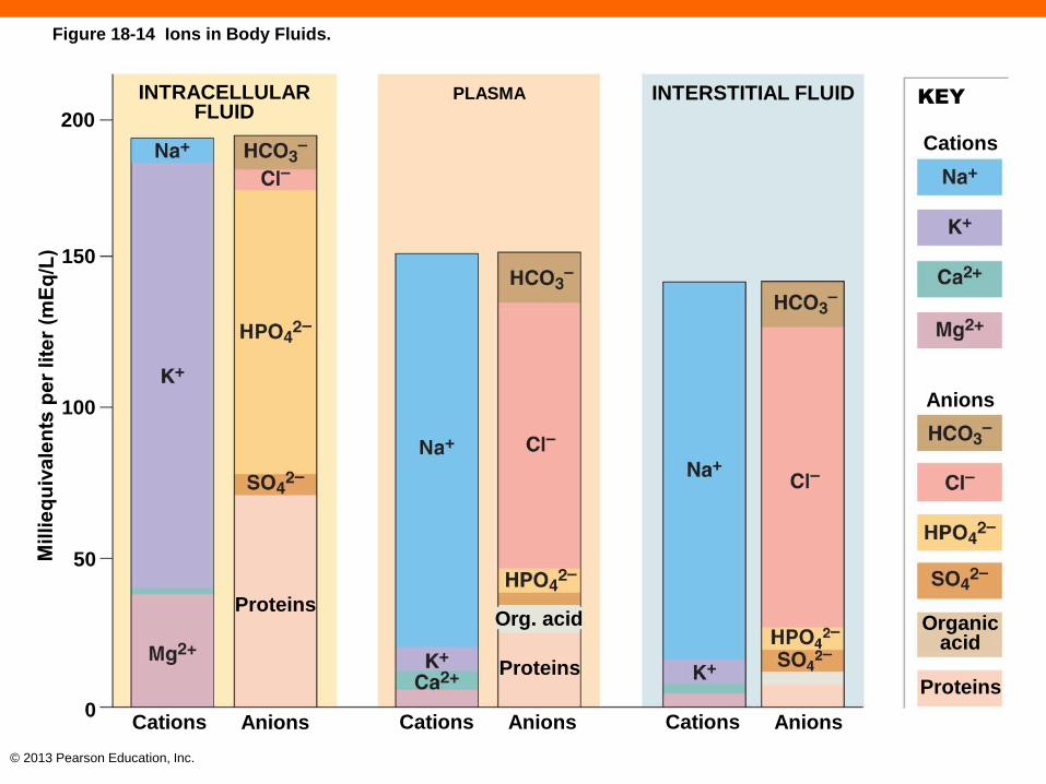
Intracellular Fluid (18-6)
• Is largest compartment
• 27 percent of total in adult females, 33 percent in males
• Contains high K+, Mg2+, HPO42–
• Also contains a lot of negatively charged proteins
• Exerts primary osmotic pressure

© 2013 Pearson Education, Inc.
Extracellular Fluid (18-6)
• Major subdivisions
• Plasma 4.5 percent of total in males and females
• Interstitial fluid (IF) 18 percent in females, 21.5 percent
in males
• Contains high Na+, Cl–, HCO3–
• Plasma has proteins
• Exert osmotic pressure
• IF has few proteins

© 2013 Pearson Education, Inc.
Figure 18-13a The Composition of the Human Body.
SOLID COMPONENTS
(31.5 kg; 69.3 lbs)
WATER (38.5 kg; 84.7 lbs)
15
10
Kg
5
0
20
15
Liters
10
Other
Plasma
Interstitialfluid
Proteins Lipids Minerals Carbohydrates Miscellaneous Intracellular fluid Extracellular fluid
The body composition (by weight, averaged for both sexes) and major body fluid compartments of a 70-kg individual. For technical reasons, it is extremely difficult to determine the precise size of any of these compartments; estimates of their relative sizes vary widely.
5
0

© 2013 Pearson Education, Inc.
Figure 18-13b The Composition of the Human Body.
ICFECF
Intracellularfluid 33%
Interstitialfluid 21.5%
Plasma 4.5%
Solids 40%(organic and inorganic materials)
Otherbodyfluids(≤1%)
Intracellularfluid 27%
Interstitialfluid 18%
Solids 50%(organic and inorganic materials)
Otherbodyfluids(≤1%)
Adult males Adult females
A comparison of the body compositions of adult males and females, ages 18–40 years.
ICF ECF
Plasma 4.5%
© 2013 Pearson Education, Inc.
Figure 18-14 Ions in Body Fluids.
INTRACELLULARFLUID200
150
100
50
0
Proteins
PLASMA
Org. acid
Proteins
INTERSTITIAL FLUID KEY
Cations
Anions
Organicacid
Proteins
Cations Anions Cations Anions Cations Anions

© 2013 Pearson Education, Inc.
Checkpoint (18-6)
16. Identify the three interrelated processes
essential to stabilizing body fluid volumes.
17. List the major component(s) of the intracellular
fluid (ICF) and the extracellular fluid (ECF).
© 2013 Pearson Education, Inc.
Fluid Balance (18-7)
• Water moves continuously
• Across capillary endothelium, serous membranes,
synovial membranes, CSF, eye humors, peri- and
endolymph of inner ear
• 2.5 L/day lost insensibly and sensibly
• Gains are 40 percent from food, 48 percent drink,
12 percent metabolic generation

© 2013 Pearson Education, Inc.
Table 18-4 Water Balance

© 2013 Pearson Education, Inc.
Fluid Shifts (18-7)
• Water movement between ECF and ICF
• Occurs due to changes in osmotic concentration
(osmolarity) of ECF
• If ECF osmolarity increases, water leaves cells
• If ECF osmolarity decreases, water moves into
cells
• Movement progresses until equilibrium reached
© 2013 Pearson Education, Inc.
Electrolyte Balance (18-7)
• Loss of balance results in:
• Loss of water balance
• Loss of cell function
• Ca2+ and K+ imbalances affect cardiac muscle tissue
• Na+ and K+ major contributors to osmolarity of ICF and
ECF

© 2013 Pearson Education, Inc.
Sodium Balance (18-7)
• Created between Na+ absorption from gut and
excretion by kidneys
• Regulated by aldosterone and ANP
• Excess dietary salt results in additional water
absorption from gut, raising blood volume and BP
• Imbalance of Na+ is most common cause of
electrolyte imbalances

© 2013 Pearson Education, Inc.
Potassium Balance (18-7)
• Most K+ is in cells
• ECF low K+ content a balance between absorption
from gut and excretion by kidneys
• Regulated by aldosterone
• Problems with K+ balance less common, but much
more dangerous
• Disrupts membrane potentials in excitable tissues

© 2013 Pearson Education, Inc.
Checkpoint (18-7)
18. Define a fluid shift.
19. How would eating a meal high in salt content
affect the amount of fluid in the intracellular fluid
compartment (ICF)?
20. What effect would being in the desert without
water for a day have on your blood osmotic
concentration?

© 2013 Pearson Education, Inc.
Acid-Base Balance (18-8)
• Normal ECF pH range 7.35–7.45
• Imbalances result in:
• Unstable cell membranes
• Denaturation of proteins, including enzymes
• CNS, cardiac tissue fail
• Vasodilation results in drop of BP
• Below 7.35 is acidosis
• Above 7.45 is alkalosis

© 2013 Pearson Education, Inc.
Acids in the Body (18-8)
• Carbonic acid (H2CO3) most abundant, but
dissociates in reversible reaction
•
• CO2 and H+ concentrations are proportional
• PCO2 and pH are inversely proportional
• Other metabolic sources of acids
• Lactic acid, ketoacids

© 2013 Pearson Education, Inc.
Figure 18-15 The Basic Relationship between Carbon Dioxide and Plasma pH.
P
40–45mm Hg
HOMEOSTASIS
pH
7.35–7.45
If P
When carbon dioxide levels rise, more carbonic acid forms, additional hydrogen ions and bicarbonate ions are released, and the pH goes down.
When carbon dioxide levels fall, carbonic acid dissociates into carbon dioxide and water. This removes H+ ions from solution and increases the pH.
co2
co2fallsIf Pco2
rises

© 2013 Pearson Education, Inc.
Buffers (18-8)
• Weak acids donate H+; weak bases absorb H+
• Buffer system
• Combination of H+ and an anion
• Three key buffer systems
1. Protein buffer systems
2. Carbonic acid–bicarbonate buffer system
3. Phosphate buffer system
© 2013 Pearson Education, Inc.
Protein Buffer Systems (18-8)
• Regulate pH in ICF and ECF
• If pH rises:
• —COOH group acts as weak acid
• Dissociates from amino acid and releases a H+
• If pH drops:
• —NH2 group acts as weak base
• Picks up a free H+

© 2013 Pearson Education, Inc.
Carbonic Acid–Bicarbonate Buffer System
(18-8)
• Regulates pH in ECF
•
• Metabolic acids release H+, lowering pH
• Is picked up by HCO3– and drives above formula to left
• Pco2 rises, respiratory system excretes CO2
• pH rises
• Formula is driven to the right

© 2013 Pearson Education, Inc.
Phosphate Buffer System (18-8)
• Primary buffer system of ICF
•

© 2013 Pearson Education, Inc.
Maintaining Acid-Base Balance (18-8)
• Buffer systems can tie up excess H+
• Temporary and limited
• Respiratory and renal mechanisms contribute
• Secrete or absorb H+
• Control excretion of acids and bases
• Generate additional buffers

© 2013 Pearson Education, Inc.
Respiratory Compensation of pH (18-8)
• Accomplished by altering respiration rate
• Low Pco2 results in rise in pH
• Respiration is suppressed
• Carbonic acid formula driven to right, conserving CO2
• Lowers pH
• High Pco2 results in lower pH
• Respiration is stimulated
• Carbonic acid formula driven to left, blowing off CO2
• Raises pH

© 2013 Pearson Education, Inc.
Renal Compensation of pH (18-8)
• Only way to truly eliminate H+
• Tubules can:
• Conserve H+ and eliminate HCO3– when pH is high
• Eliminate H+ and conserve HCO3– when pH is low

© 2013 Pearson Education, Inc.
Acid-Base Disorders (18-8)
• Respiratory disorders
• Caused by changes in CO2 in ECF
• Metabolic disorders
• Caused by overproduction of acids
• Alteration in HCO3– concentrations

© 2013 Pearson Education, Inc.
Table 18-5 Acid-Base Disorders

© 2013 Pearson Education, Inc.
Checkpoint (18-8)
21. Identify the body's three major buffer systems.
22. What effect would a decrease in the pH of body
fluids have on the respiratory rate?
23. How would a prolonged fast affect the body's
pH?

© 2013 Pearson Education, Inc.
Age-Related Changes in the Renal System
(18-9)
1. Decline in number of functional nephrons
2. Reduction in GFR
3. Reduction in sensitivity to ADH and aldosterone
4. Problems with micturition reflex
• Sphincters weaken
• CNS loss of control over sphincters
• Prostate enlargement in males restricts urine flow

© 2013 Pearson Education, Inc.
Age-Related Changes in the Renal System
(18-9)
5. Gradual decrease in total body water content
6. Net loss of body mineral content
7. Increase incidence of disorders in other systems
impacting fluid, electrolyte, and pH balance

© 2013 Pearson Education, Inc.
Checkpoint (18-9)
24. What effect does aging have on the GFR?
25. After age 40, does total body water content
increase or decrease?

© 2013 Pearson Education, Inc.
Coordinated Excretion of Wastes (18-10)
• The renal system excretes wastes but so do other
systems
• Integumentary system through sweat
• Respiratory system removes CO2
• Digestive system through excretion of bile
© 2013 Pearson Education, Inc.
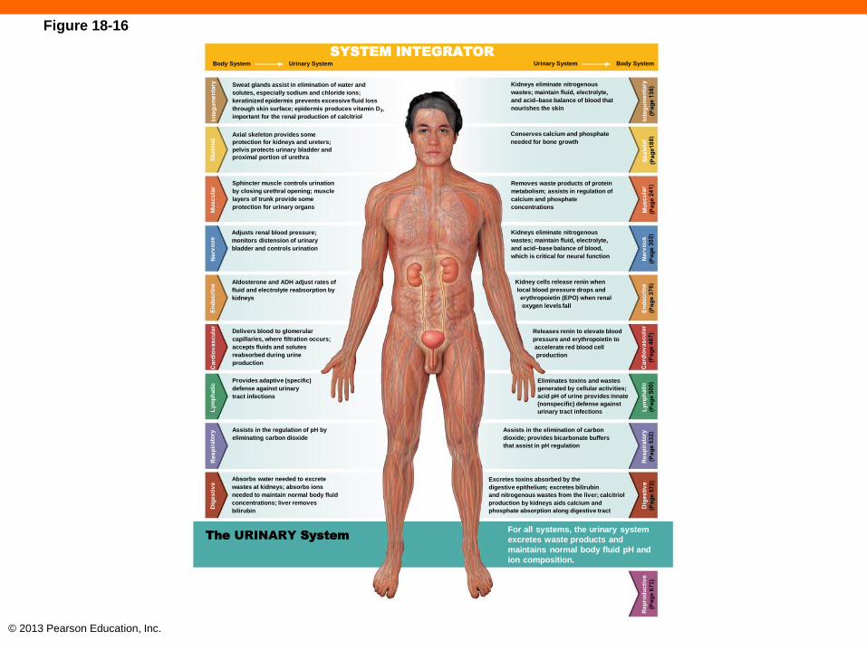
Figure 18-16
SYSTEM INTEGRATOR
Body System Urinary System
Sweat glands assist in elimination of water and
solutes, especially sodium and chloride ions;
keratinized epidermis prevents excessive fluid loss
through skin surface; epidermis produces vitamin D3,
important for the renal production of calcitriol
Axial skeleton provides some
protection for kidneys and ureters;
pelvis protects urinary bladder and
proximal portion of urethra
Sphincter muscle controls urination
by closing urethral opening; muscle
layers of trunk provide some
protection for urinary organs
Adjusts renal blood pressure;
monitors distension of urinary
bladder and controls urination
Aldosterone and ADH adjust rates of
fluid and electrolyte reabsorption by
kidneys
Delivers blood to glomerular
capillaries, where filtration occurs;
accepts fluids and solutes
reabsorbed during urine
production
Provides adaptive (specific)
defense against urinary
tract infections
Assists in the regulation of pH by
eliminating carbon dioxide
Absorbs water needed to excrete
wastes at kidneys; absorbs ions
needed to maintain normal body fluid
concentrations; liver removes
bilirubin
The URINARY System
Sk
ele
tal
Mu
sc
ula
rL
ym
ph
ati
cD
ige
sti
ve
For all systems, the urinary system
excretes waste products and
maintains normal body fluid pH and
ion composition.
Rep
rod
uc
tiv
e
(Pa
ge
67
1)
Body SystemUrinary System
Excretes toxins absorbed by the
digestive epithelium; excretes bilirubin
and nitrogenous wastes from the liver; calcitriol
production by kidneys aids calcium and
phosphate absorption along digestive tract
Assists in the elimination of carbon
dioxide; provides bicarbonate buffers
that assist in pH regulation
Eliminates toxins and wastes
generated by cellular activities;
acid pH of urine provides innate
(nonspecific) defense against
urinary tract infections
Releases renin to elevate blood
pressure and erythropoietin to
accelerate red blood cell
production
Kidney cells release renin when
local blood pressure drops and
erythropoietin (EPO) when renal
oxygen levels fall
Kidneys eliminate nitrogenous
wastes; maintain fluid, electrolyte,
and acid–base balance of blood,
which is critical for neural function
Removes waste products of protein
metabolism; assists in regulation of
calcium and phosphate
concentrations
Conserves calcium and phosphate
needed for bone growth
Kidneys eliminate nitrogenous
wastes; maintain fluid, electrolyte,
and acid–base balance of blood that
nourishes the skin

© 2013 Pearson Education, Inc.
Checkpoint (18-10)
26. What organ systems make up the body's
excretory system?
27. Identify the role the urinary system plays for all
other body systems.