Embed Size (px)
Citation preview

Microbiology Lab ( )............... Lecturer : kholoud A.H. ...................... First stage
Lectures of the Microbiology Laboratory
second stage
Lectures: 1- General introduction of Microbiology
2- Scope of Microbiology
3-Safety and Laboratory Guideline
4- Sterilization
5- Microscopes and the Study of Microbial Structure
6- The Structure and Shape of Bacterial cell
7- Media for Bacterial Growth and Methods of Isolation
8- Transfer Instruments & Methods of Isolation
9- Bacterial stain
10- Techniques of smear preparation
11-Simple stain
12-Negative stain
13-Gram stain
14- Acide fast stain
15- Physical and Chemical agents for the Control of Microbial Growth
-Teacher:-
Khuloud Abdulkareem Hussein
-Basrah univesity / Nursing college / Essential science.
-February 2020

Microbiology Lab ( )............... Lecturer : khulood A.H...............second stage
General introduction of Microbiology:-
a specialized area of biology is the study of living organisms are
not readily observed without magnification , which is to say they are
microscopic.* These microscopic organisms are collectively referred
to as(‘microorganisms’ or ‘microbes’), simple in structure, and
usually small in size, that are generally considered to be neither plants
nor animals; they include bacteria, algae, fungi, protozoa, and viruses..
Microorganisms were first seen and described by the Dutch lens-
maker Antonie van Leeuwenhoek (1632–1723).
Branches of Microbiology
1. Medical microbiology 2. Industrial microbiology 3. Food microbiology 4. Soil microbiology 5. Plant
microbiology.
Here we are concerned with medical microbiology. It is studied
under following headings:
a. Parasitology :- deals with the study of parasites causing diseases in
human being.
b. Mycoloy :- deals with the study of fungi causing diseases in human
beings.
c. Immunology :- is concerned with mechanism involved in the
development of resistance by body to infectious diseases.
d. Bacteriology :- deals with the study of bacteria.
e. Genetics :- is the study of heredity and variations.
f. Virology :- is the study of viruses.

Microbiology Lab ( )............... Lecturer : khulood A.H...............second stage
SO Medical microbiology mean (Gr. mikros-small, bios-life, logos-
science) is the study of causative agents of infectious disease of human
beings and their reactions to such infections. In other words it deals with
etiology, pathogenesis, laboratory diagnosis, treatment, epidemiology and control
of infection.
Scope of Microbiology :-
*1. Diagnostic, e.g. isolation and identification of causative organism
from the pathological lesions. We can also diagnose typhoid fever by
doing Widal’s test.
*2. Prognosis of disease, e.g. in Widal’s test rising titer signifies active
disease and ineffective treatment. Falling titer means effective treatment
and curing of disease.
*3. Guidance in treatment, e.g. by culturing the organism in pure form
and then performing drug sensitivity test we can suggest the effective
drug for the treatment of that particular infection.
*4. Source of infection, e.g. in sudden outbreak of infectious disease we
can find out the source of infection.
*5. Detection of new pathogens and then development of vaccines.

Microbiology Lab ( )............... Lecturer : khulood A.H...............second stage
Safety and Laboratory Guidelines:-
Because microorganisms present varying degrees of risk to laboratory
personnel (students, technicians, and faculty), people outside the
laboratory, and the environment, microbial cultures must be handled
safely, so the Basic Laboratory Safety are:-
1. Wear protective clothing (i.e., a lab coat) in the laboratory when handling microbes.
Remove the coat prior to leaving the lab ( Figure I ).
2. Do not wear sandals or open-toed shoes in the laboratory.
3. Wear eye protection whenever you are heating chemicals, even if you wear glasses or
contacts (Figure I ).
4. Turn off your Bunsen burner when it is not in use.
5. Tie back long hair, as it is a potential source of contamination as well
as a likely target for fire.
6. If you are feeling ill, go home. A microbiology laboratory is not a safe
place if you are ill.
7. If you are pregnant, immune compromised, or are taking
immunosuppressant drugs, please see the instructor.
8. wear disposable gloves If it is your lab’s practice while handling
microorganisms, staining microbes , handling blood products ( plasma,
serum, antiserum ) or whole blood , be sure to remove them each time
you leave the laboratory , and wash your hands ( Figure I ) .
9. Use an antiseptic (e.g., Betadine) on your skin if it is exposed to a spill
containing microorganisms.
10. Never pipette by mouth. Always use mechanical pipettors .
11. Dispose of broken glass or any other item in an appropriate “sharps” or broken-
glass container (Figure 2 ).

Microbiology Lab ( )............... Lecturer : khulood A.H...............second stage
Figure 1-wearing a protective lab coat, gloves, and goggles Figure 2-Sharps Container
Student Conduct in Laboratory :-
1. To reduce the risk of infection, do not smoke, eat, drink, or bring
food or drinks into the laboratory room—even if lab work is not
being done at the time.
2. Do not apply cosmetics or handle contact lenses in the laboratory.
3. Wash your hands thoroughly with soap and water before working in
the lab, after handling living microbes, and before leaving the
laboratory at any time , Also, wash your hands after removing
gloves.
4. Do not remove any organisms or chemicals from the laboratory.
5. Work carefully and methodically. Do not hurry through any
laboratory procedure.

Microbiology Lab ( )............... Lecturer : khulood A.H...............second stage
Sterilization:-
is the removal of all microbes, including endospores, and can be achieved by
Physical , chemicals, mechanical, or radiation methods.
Physical Methods
a) Dry heat
1. Incineration (fire) used for sterilize the slides , needles , loops, and
test tubes ).
2. ovens used for sterilize oils , powder, petridishes , pipettes , minerals
and surgical instruments , under 160-180°c for 1.5 -2h.
b) Moist heat
1. under pressure is provided by autoclaving is used ( under 121°c for
20 min and pressure 1-1.5 ) to sterilize culture media , liquids , waste
culture media , and all other substances which damaged by dry heat
2. without pressure by Fractional sterilization or tyndallization
sterilizer is used ( under 100°c for 15 min./3 days) to sterilize any
substance which damaged over 100°c like sugar solutions.
Chemical methods
a) Disinfection or disinfectants Chemical germicides are substances designed to
reduce the number of pathogens on a surface (floors, tables, sinks, countertops,
surgical instruments, etc.) or liquids such as 0.01%, 0.1%, and 1.0% household
bleach and 25%, 50%, and 100% Lysol® Brand II Disinfectant
b) Antiseptics Chemical Germicides are substances designed to reduce the
number of pathogens on or in living tissue such as 0.03%, 0.3%, and 3%
hydrogen peroxide (3% is full strength as purchased at the pharmacy) or 10%,
30%, and 50% isopropyl alcohol (70% is full strength as purchased at the
pharmacy)
Mechanical Methods such as : Filtration This Method is used for sterilization
of biological liquids , which are damage with high temperature degrees , such as
the solutions of serum , enzymes , vitamins , and antibiotics .

Microbiology Lab ( )............... Lecturer : khulood A.H...............second stage
Radiation such as : gamma ray , X-ray, and alpha ray. The most substances that
sterilized by radiation are plastic petridishes , plastic injections , plastic gloves
, plastic pipettes , operation rooms , packaging rooms of drugs , yoghurt
production .
Critical thanking:-
- Moist heat is more adequacy for killing a living cell, Why?

Microbiology Lab ( )............... Lecturer : khulood A.H...............second stage
Microscopes and the Study of Microbial Structure
-Microscope is the device for magnifying objects that are too small to be seen with the
naked eye
Microscopes are divided into three categories :
1- Microscopes use a beam of electrons as the source of illumination instead of light
- are subdivided into
a) Scanning electron microscope (SEM) Maximum effective magnification SEM =
650,000X
b) Transmission electron microscope (TEM) Maximum effective magnification TEM =
1,000,000X
2- Microscopes with ultraviolet or laser beam as the source of illumination instead
of light, Maximum effective magnification = 1,000. to 2,000 X
- are subdivided into
a) Fluorescent Microscope
b) Confocal Microscope
3- Microscopes with visible light as the source of illumination Maximum effective
magnification = 1,000. to 2,000.
- are subdivided into
a) Bright-field microscope
b) Dark-field microscope
c) Phase-contrast microscope
d) Differential interference contrast microscope
*As Figure 1-2-3 Respectively

Microbiology Lab ( )............... Lecturer : khulood A.H...............second stage
-1
2-
3-

Microbiology Lab ( )............... Lecturer : khulood A.H...............second stage
Bright –field microscope :
This microscope is used to study the size , shape , and arrangement of the microbial cells
, but it provides little information about the internal cell structure . There is two types of this
microscope :
* Simple microscope : It has a single lens magnifier.
* Compound microscope : It employs two or more lenses called ocular and objective
lenses.
The magnification of the microscope depends on the type of objective lens used with the
ocular lens . So , the total magnification is calculated by multiplying the objective lens by
ocular lens.

Microbiology Lab ( )............... Lecturer : khulood A.H...............second stage
Workings of an oil immersion lens :
To maximize its resolving power, an oil immersion lens (the one with highest
magnification) must have a drop of oil placed at its tip. This transmits a
continuous cone of light from the condenser to the objective, thereby increasing the amount
of light and, consequently, the numerical aperture. Without oil, some of the peripheral light
that passes through the specimen is scattered into the air or onto the glass slide; this
scattering decreases resolution ,as figure (1)
figure (1)
Microscope care
Microscope is a very important tool in microbiology and it must be used carefully and
correctly . Follow these guidelines every time you use a microscope :
1- carry the microscope with both hands , one hand beneath the base and the other on the arm
. Never slide a microscope a cross a bench surface .
2- Clean the microscope both before and after use . Use only lens paper and lens cleaner .
3-Only use oil when using the 100X oil immersion lens . Do not get oil on the other objective
lenses.
4-Do not wrap the cord around the microscope . Instead , fold the cord and place it between
the arm and the stage or beneath the stage.

Microbiology Lab ( )............... Lecturer : khulood A.H...............second stage
5- Observe the slide with both eyes open , to avoid eyestrain.
6- Always focus with low power first .
7- Keep the stage clean and free of oil .
8- wipe oil off the oil immersion lens before putting your microscope away . Do not touch
the lenses with your hands .
9- Clean the ocular lens carefully with lens paper if dust is present .
10- Replace the dust cover before putting the microscope away .
11- Make sure that the specimen is on the top –side of the slide when using the oil immersion
lens .
12- Increase the amount of light when using the oil immersion lens .
Citical thinking:-
1- For what purpose each of the following microscope components?
a. Coarse-adjustment knob
b. Fine-adjustment knob
c. Condenser
d. Mechanical stage control
2- How can you calculated the total magnificent for the microscope?

Microbiology Lab ( )............... Lecturer : khulood A.H...............second stage
The Structure and Shape of Bacterial cell
In general the organisms are divided into two groups (As table 1) :-
1- Prokaryotes : Include two kingdoms appear similar in morphology but they have
major molecular and biochemical differences :
a- Kingdom (1) : Bacteria ( Eubacteria ) : They are common in the environment
and include all bacteria that infect human . They all possess peptidoglycan ( murein or cell
wall skeleton ) which contains a unique sugar and muramic acid, not found elsewhere in
nature.
d- Kingdom (2) : Archaea ( Archaebacteria ) : These organisms do not infect
human and they contain a pseudomurein ( pseudopeptidoglycan ) that is different in
structure to eubacterial murein .
2- Eukaryotes : Include all other forms of life ( plants, animals, human, algae, fungi and
parasite).
Table (1): Comparison of Prokaryotic and Eukaryotic Cell Organization:-
Characteristic Prokaryote Eukaryote
- organelles
Absent present
- nuclear membrane Absent present
- Chromosomal DNA Circular; complexed with RNA Linear; complexed with histones
and other proteins basic
- Sterols in cytoplasmic membrane Absent present
- Ribosomes: site of protein Present smaller in size (70S) Present, larger in size (80S)
synthesis
- cell division faster slower

Microbiology Lab ( )............... Lecturer : khulood A.H...............second stage
Prokaryote Cell
Eukaryote Cell

Microbiology Lab ( )............... Lecturer : khulood A.H...............second stage
Shapes of Bacteria:-
Microscopic Shapes:-
Bacteria vary in size from 0.4 to 2 μm. They occur in three basic shapes:
• Cocci (spherical)
• Bacilli (rod-shaped)
• Spirochetes (spiral) or ( Helical and curved bacteria).
Individual bacteria may form characteristic groupings.
Cocci:- (plural of coccus) may occur singly
in pairs (diplococci),
in chains (streptococci),
in four cells grouped together (tetrads)
in eight cells grouped together (sarcinae form)
or in clusters (staphylococci). As figure (1)
Figure (1):- Arrangement of Cocci.

Microbiology Lab ( )............... Lecturer : khulood A.H...............second stage
Staphylococcus aureus
Bacilli:-
(plural of bacillus) Bacilli may occur as single rods, The cells of this group vary in
length .
in chains
short coccobacilli to long filamentous rods. As figure (2)
Figure (2):- Arrangement of Bacilli Bacillus subtilis
Spirochetes (spiral) or ( Helical and curved bacteria):-
The cells in this group are either helical ( spirillum and slender spirochaetes)
or curved (comma shape) .As figure(3)

Microbiology Lab ( )............... Lecturer : khulood A.H...............second stage
Figure(3):-Arrangement of Spirochetes
Curved bactera:-Vibrio cholera Spirochetes:- Treponema pallidum

Microbiology Lab ( )............... Lecturer : khulood A.H...............second stage
Media for Bacterial Growth and Methods of Isolation:-
Culture media gives artificial environment simulating natural conditions necessary
for growth of bacteria. The basic requirement of culture media are:
1. Energy source.
2. Carbon source.
3. Nitrogen source.
4. Salts like sulphates, phosphates, chlorides and carbonates of sodium, potassium,
magnesium, ferric, calcium and trace elements like copper, etc.
5. Satisfactory pH 7.2 to 7.6.
6. Adequate oxidation-reduction potential.
7. Growth factor.
Media used for obtaining the growth of bacteria are:
FLUID MEDIA
Bacteria grow very well in fluid media in 3 to 4 hours. Hence, they are used as enriched
media before plating on solid media. They are not suitable for the isolation of organism in
pure culture. We cannot study colony characters as well Examples of fluid media are
nutrient broth, peptone water, etc.
SOLID MEDIA
They are used to study colonies of individual bacteria. They are essential for isolation of
organism in pure form. Agar it is important constituent of solid media such as blood and
nutrient agar ,etc.
Culture Techniques
In clinical laboratory indications for culture are:
a. Isolation of bacteria in pure culture.
b. To demonstrate their properties.
c. To obtain sufficient pure growth for preparation of antigen and for other tests.
e. To determine sensitivity to antibiotics.
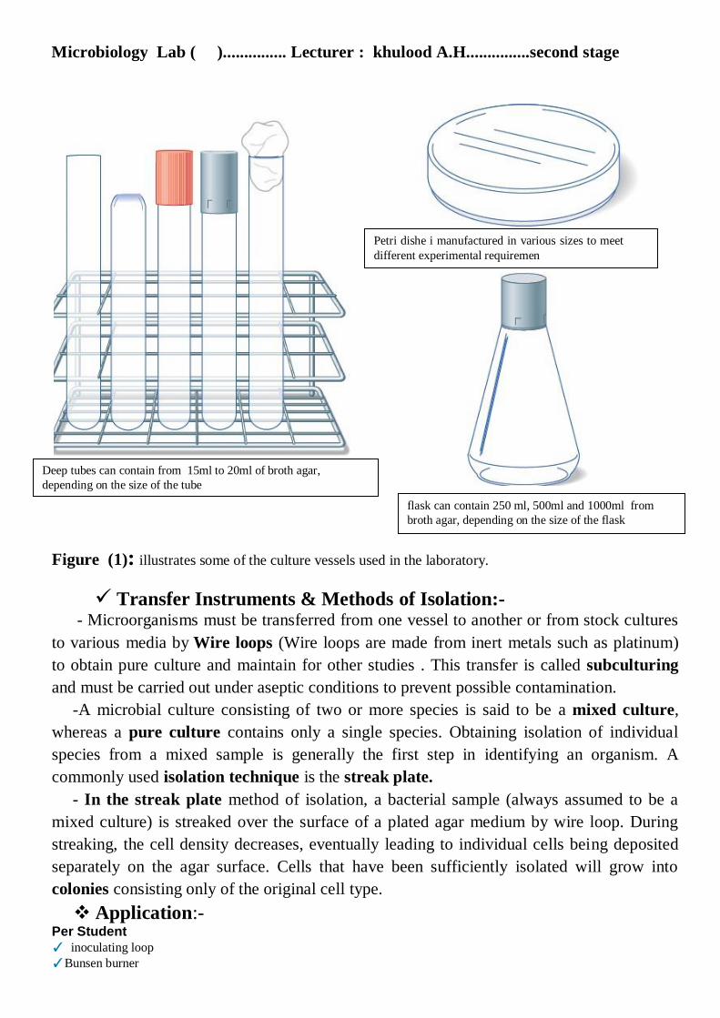
Culture Tubes and Petri Dishes:-Glass test tubes and glass or plastic Petri dishes are used to cultivate microorganisms.
A suitable nutrient medium in the form of broth or agar may be added to the tubes, while
only a solid medium is used in Petri dishes. A sterile environment is maintained in culture
tubes by various types of closures. Figure (1) below illustrates some of the culture vessels
used in the laboratory.
Microbiology Lab ( )............... Lecturer : khulood A.H...............second stage
Deep tubes can contain from 15ml to 20ml of broth agar,
depending on the size of the tube
Petri dishe i manufactured in various sizes to meet
different experimental requiremen
Figure (1): illustrates some of the culture vessels used in the laboratory.
Transfer Instruments & Methods of Isolation:-- Microorganisms must be transferred from one vessel to another or from stock cultures
to various media by Wire loops (Wire loops are made from inert metals such as platinum)
to obtain pure culture and maintain for other studies . This transfer is called subculturing
and must be carried out under aseptic conditions to prevent possible contamination.
-A microbial culture consisting of two or more species is said to be a mixed culture,
whereas a pure culture contains only a single species. Obtaining isolation of individual
species from a mixed sample is generally the first step in identifying an organism. A
commonly used isolation technique is the streak plate.
- In the streak plate method of isolation, a bacterial sample (always assumed to be a
mixed culture) is streaked over the surface of a plated agar medium by wire loop. During
streaking, the cell density decreases, eventually leading to individual cells being deposited
separately on the agar surface. Cells that have been sufficiently isolated will grow into
colonies consisting only of the original cell type.
Application:- Per Student
✓ inoculating loop
✓Bunsen burner
flask can contain 250 ml, 500ml and 1000ml from
broth agar, depending on the size of the flask

Microbiology Lab ( )............... Lecturer : khulood A.H...............second stage
✓ sterile fluid media such as Nutrient Broth tubes
✓ sterile solid media such as Nutrient and manitol Agar media.
✓marking pens
procedure:-
1-Flaming Loop 2- Mixing Broth by Hand
3- Removing the Tube Cap 4- Holding the Tube at an Angle
5- 1 Remove the loop, Flaming the Tube
and replace the lid. Wire loop
handl
shaft
Loop

Microbiology Lab ( )............... Lecturer : khulood A.H...............second stage
Note:-Removing the Loop from Broth notice the film of broth in the loop (see inset). Be
careful not to catch the loop on the lip of the tube when removing it.
7- Obtain the sample of mixed culture from broth media with a sterile loop on to solid media
and Beginning the Streak Pattern. such as figure below.
Figure illustrate streak pattern.
A- Beginning the Streak Pattern: Label the plate's base. Then, streak the mixed culture back and forth in
one quadrant of the agar plate. Do not cut the agar with the loop. Flame the loop, then proceed.
B- Streaking Again: Rotate the plate nearly 90° and touch the agar in an uninoculated region to cool the
loop. Streak again using the same wrist motion. Flame the loop.
C- Streaking Yet Again: Rotate the plate nearly 90° and streak again using the same wrist motion. Be sure
to cool the loop prior to streaking. Flame again.
D-Streaking Into the Center: After cooling the loop, streak one last time into the center of the plate.
Flame the loop. Then
8- Incubate the plate in an inverted position for then assigned time at the appropriate
temperature at 37°C at for 24 to 48 hours ( incubate temperature depended on type of
isolation from soil, water, plants, and animals including humans ). after incubation , for
isolation and identification of bacteria depended on
1- examine colony morphology on the solid media plate, this including : colony shape,
margin (edge), elevation, texture, and color. such as figure below.
2- Examine shape and arrangement of bacterial cell under microscope after smear
preparation and staining, explain in the next lab .

Microbiology Lab ( )............... Lecturer : khulood A.H...............second stage
Critical thinking:- 1- Any method to sterilize Wire loop and any portion in this method (discuses the practical
methods )?
2- Petri dishes (after inoculation) are incubated in an inverted position (top down)Why?

Microbiology Lab ( )............... Lecturer : khulood A.H...............second stage
Bacterial staining :-
Visualization of microorganisms in the living state is quite difficult, not only because
they are minute, but also because they are transparent and practically colorless when
suspended in an aqueous medium . To study their properties and to divide microorganisms
into specific groups for diagnostic purposes, biological stains and staining procedures in
conjunction with light microscopy have become major tools in microbiology.
- Chemical Basis:-
a stain (dye) may be defined as an organic compound containing a benzene ring
plus a chromophore and an auxochrome group(Figure 1) , The ability of a stain to bind to
macromolecular cellular components such as proteins or nucleic acids depends on the
electrical charge found on the chromogen portion, as well as on the cellular component to
be stained.
Bellow, a summary of acidic and basic stains is outlined in Figure 2 .
Acidic stains are anionic, which means that, on ionization of the stain, the
chromogen portion exhibits a negative charge and therefore has a strong affinity for the
positive constituents of the cell. Proteins, positively charged cellular components, will
readily bind to and accept the color of the negatively charged, anionic chromogen of an
acidic stain.
- Structurally, like Eosin stain is an example of an acidic stain that produces Eosinate- an
anionic chromogen,
Eosin stain: Eosin
Sodium+
+ Eosinate-
( Anionic chromogen)
Basic stains are cationic, because on ionization the chromogen portion exhibits a
positive charge and therefore has a strong affinity for the negative constituents of the cell.
Figure 1 :- Chemical composition of a stain

Microbiology Lab ( )............... Lecturer : khulood A.H...............second stage
Nucleic acids, negatively charged cellular components, will readily bind to and accept the
color of the positively charged, cationic chromogen of a basic stain.
-Structurally, methylene blue is a basic stain that produces methylene+ a cationic
chromogen
Methylene blue methylene+ + Chloride -
cationic chromogen
Basic stains are more commonly used for bacterial staining. The presence of a
negative charge on the bacterial surface acts to repel most acidic stains and thus prevent
their penetration into the cell.
Numerous staining techniques are available for :-
1. visualization
2. differentiation, and
3. separation of bacteria in terms of morphological characteristics
and cellular structures.
A summary of commonly used procedures and their purposes is outlined in Figure3
Figure3: Staining techniques
Figure 2:- Acidic and basic stains

Microbiology Lab ( )............... Lecturer : khulood A.H...............second stage
Techniques of smear preparation (preparation of bacteial smear):-
Materials
- Cultures
Twenty-four–hour nutrient agar slant culture and a 24-hour nutrient broth culture of
any bacteial growth
- Equipment
1. Glass microscope slides
2. Bunsen burner,
3. inoculating loop, and
4. glassware marking pencil.
Smears from a Broth Medium
-Procedure
Label three clean slides with the initials of the organism, and number them 1, 2, and 3.
Resuspend the sedimented cells in the broth culture by tapping the culture tube with your
finger. The next four steps of this procedure are illustrated
1. With a sterile loop, place one loopful of culture on Slide 1, two loopfuls on Slide 2, and
three loopfuls on Slide 3, respectively.
2. With a circular movement of the loop, spread the cell suspension into an area
approximately the size of a dime.
3. Allow the slide to air-dry completely. This may be done by placing the slide on a drying
tray attached to a microincinerator or by placing the slide on the bench.
4. Heat fix the preparation. Note: Pass the airdried slide in front of the entrance to the
microincinerator or pass the slide through the outer portion of the Bunsen flame to prevent
overheating, which can distort the morphology through plasmolysis of the cell wall.
-Examine each slide under microscope and record your results in the Lab Report.
Smears from a Solid Medium
-Procedure
Label four clean slides with the initials of the organism. Label Slides 1 and 2 with an L for
loop, and Slides 3 and 4 with an N for needle. The next four steps of this procedure are
illustrated in
1. Using a loop, place one to two loops of water on each slide.

Microbiology Lab ( )............... Lecturer : khulood A.H...............second stage
2. With a sterile loop, touch the entire loop to the culture and emulsify the cells in water on
Slide, Then, with a sterile loop, just touch the tip of the loop to the culture and emulsify it in
the water on Slide 2. Repeat Steps 1 and 2 using a sterile inoculating needle on Slides 3 and
4.
3. Allow all slides to air-dry completely. This may be done by placing the slide on a drying
tray attached to a microincinerator or by placing the slide on the bench.
4. Heat fix the preparation , pass the slide through the outer portion of the Bunsen flame
to prevent overheating, which can distort the morphology through plasmolysis of the cell
wall.
-Examine each slide under microscope and record your results in the Lab Report.
Heat fixing:- kills the bacteria and the coagulated proteins from the cells will cause
cells to stick to the slide. Fixing denatures bacterial enzymes , preventing them from
digesting cell parts , which causes the cell to break , a process called autolysis.
Critical thinking:-
1. How does the heaviness of a bacterial smear affect its microscopic analysis?
2. Why should you be careful not to underheat a smear during the heat-fixing
process?
3. What is heat fixation? How is it carried out?

Microbiology Lab ( )............... Lecturer : khulood A.H...............second stage
Type of Bacterial Staining:-
1- Simple staining :-
Principle
In simple staining, the bacterial smear is stained with a single reagent, which produces a
distinctive contrast between the organism and its background. Basic stains with a positively
charged chromogen are preferred because bacterial nucleic acids and certain cell wall
components carry a negative charge that strongly attracts and binds to the cationic
chromogen.
Clinical Application :-
Simple stains are relatively quick and useful methods of testing for the presence of,
determining the shape of, or determining the numbers of bacteria present in a sample.
Generally involving a single staining step, simple staining methods are not considered
differential or diagnostic and will have limited uses. However, this is a quick procedure for
determining whether a clinical sample has the presence of a foreign bacterial pathogen.
The purpose of simple staining is to elucidate the morphology and arrangement of
bacterial cells .
The most commonly used basic stains are
methylene blue,
crystal violet, and
carbol fuchsin.
reagents
Methylene blue, crystal violet, and carbol fuchsin.
Equipment
Bunsen burner, loop, staining tray, microscope, lens paper, and glass slides.
Procedure
Prepare separate bacterial smears of the organisms and following the procedure described
in bellow.
Note: All smears must be heat fixed prior to staining.
The following steps are illustrated in Figure 4a.
1. Place a slide on the staining tray and flood the smear with one of the indicated stains,
using the appropriate exposure time for each: carbol fuchsin, 15 to 30 seconds; crystal
violet, 20 to 60 seconds; methylene blue (shown in Figure 4 a), 1 to 2 minutes.

Microbiology Lab ( )............... Lecturer : khulood A.H...............second stage
2. Gently wash the smear with tap water to remove excess stain. During this step, hold the
slide parallel to the stream of water; in this way you can reduce the loss of organisms from
the preparation.
3. Using bibulous paper, blot dry, but do not wipe the slide.
4. Repeat this procedure with the remaining two organisms, using a different stain for each.
5. Examine all stained slides under oil immersion.
6. In the chart provided in the Lab Report 1, complete the following:
a. Draw a representative field for each organism. Refer to page 16 for proper drawing
procedure.
b. Describe the morphology of the organisms with reference to their shapes (bacilli, cocci,
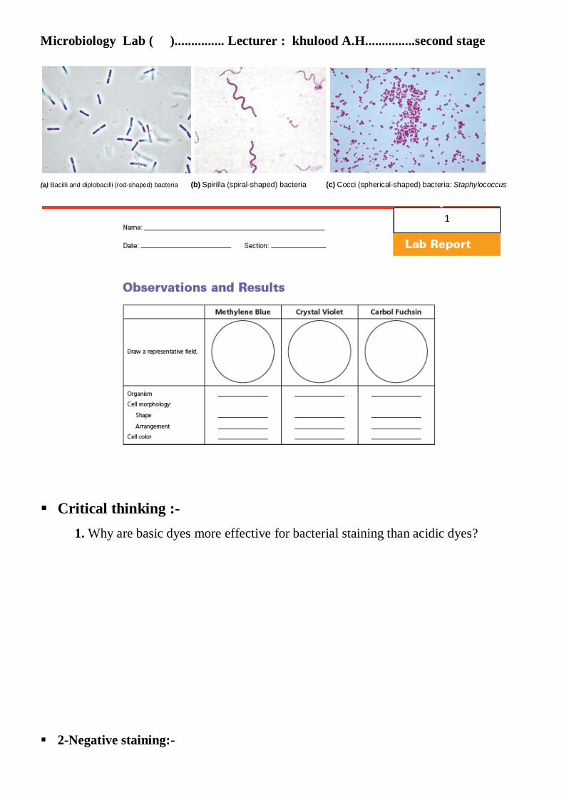
spirilla) and arrangements (chains, clusters, pairs) as Figure 4b.
Figure 4 b :-
Figure 4a:- Simple staining procedure
Microbiology Lab ( )............... Lecturer : khulood A.H...............second stage
(a) Bacilli and diplobacilli (rod-shaped) bacteria (b) Spirilla (spiral-shaped) bacteria (c) Cocci (spherical-shaped) bacteria: Staphylococcus
Critical thinking :-
1. Why are basic dyes more effective for bacterial staining than acidic dyes?
2-Negative staining:-
1

Microbiology Lab ( )............... Lecturer : khulood A.H...............second stage
Principle:-
Negative staining requires the use of an acidic stain such as India ink or nigrosin. The
acidic stain,with its negatively charged chromogen, will not penetrate the cells because of
the negative charge on the surface of bacteria. Therefore, the unstained cells are easily
discernible against the colored background.
The practical application of negative staining is twofold:-
-First, since heat fixation is not required and the cells are not subjected to the distorting
effects of chemicals and heat, their natural size and shape can be seen.
-Second, it is possible to observe bacteria that are difficult to stain, such as some spirilla.
Because heat fixation is not done during the staining process, keep in mind that the
organisms are not killed and slides should be handled with care. Figure 5b shows a
negative stain of bacilli.
Figure 5b: negative staining: Bacilli (1000×)
Clinical Application :-
Detecting Encapsulated invaders
The principle application of negative staining is to determine if an organism possesses a
capsule (agelatinous outer layer that makes the microorganism more virulent), although it
can also be used to demonstrate spore formation. The technique is frequently used in the
identification of fungi such as Cryptococcus neoformans, an important infectious agent
found in bird dropping that is linked to meningeal and lung infections in humans.

Microbiology Lab ( )............... Lecturer : khulood A.H...............second stage
Materials
Cultures
Twenty-four–hour agar slant cultures of Micrococcus luteu or Bacillus cereus, and other
alternate bacterial cultures.
reagent
Nigrosin. or indian ink
Equipment
Bunsen burner, loop, staining tray, glass slides, lens paper, and microscope.
Procedure
Steps 1–4 are illustrated in Figure 5a.
1. Place a small drop of nigrosin close to one end of a clean slide.
2. Using aseptic technique, place a loopful of inoculum from the bacteial culture in the
drop of nigrosin and mix.
3. Place a slide against the drop of suspended organisms at a 45° angle and allow the drop
to spread along the edge of the applied slide.
4. Push the slide away from the drop of suspended organisms to form a thin smear. Air-dry.
Note: Do not heat fix the slide.
5. Repeat Steps 1–4 for slide preparations of the remaining cultures.
6. Examine the slides under oil immersion.
.
Critical thinking:-
1. Why can’t methylene blue be used in place of nigrosin for negative staining? Explain.
2. What is the principle application of negative staining?
3. Why must slides be carefully handled during the negative staining process?
Figure 5a: negative staining procedure

Microbiology Lab ( )............... Lecturer : khulood A.H...............second stage
3- Gram stain (Differential staining)
Principle:
Differential staining requires the use of at least four chemical reagents ( discus bellow)
that are applied sequentially to a heat-fixed smear.
-The first reagent is called the primary stain (Crystal Violet (hucker’s)) This violet stain
is used first and stains all cells purple. Its function is to impart its color to all cells.
-The second stain is a mordant(Gram’s iodine)This reagent serves not only as a killing
agent but also as a mordant, a substance that increases the cells’ affinity for a stain) used to
intensify the color of the primary stain. In order to establish a color contrast.
-The third reagent used is the decolorizing agent ( Ethyl Alcohol, 95%) This reagent serves
a dual function as a protein-dehydrating agent and as a lipid solvent. Its action is determined
by two factors, the concentration of lipids and the thickness of the peptidoglycan layer in
bacterial cell walls.
-The final reagent, the counterstain ( Safranin ) This is the final reagent, used to stain
pink those cells that have been previously decolorized. Since only gram-negative cells
undergo decolorization, they may now absorb the counterstain.
- The most important differential stain used in bacteriology is the Gram stain, named
after Dr. Hans Christian Gram. It divides bacterial cells into two major groups, gram
positive and gram negative, which makes it an essential tool for classification and
differentiation of microorganisms. Figure 6B shows gram-positive and gram-negative cells
- The Gram stain reaction is based on the difference in the chemical composition of
bacterial cell walls. Gram-positive cells have a thick peptidoglycan layer, whereas the
peptidoglycan layer in gram-negative cells is much thinner and surrounded by outer lipid
containing layers.

Microbiology Lab ( )............... Lecturer : khulood A.H...............second stage
Figure 6B: Gram-stained cells
(a) Gram-positive stain of streptococci (b) Gram-negative stain of E. coli
Materials:-
Cultures:
Twenty-four–hour nutrient agar slant cultures of Escherichia coli, Staphylococcus aureus
and Bacillus cereus.
reagents:
Crystal violet, Gram’s iodine, 95% ethyl alcohol,and safranin.
Equipment:
Bunsen burner, inoculating loop, staining tray, glass slides, lens paper, and microscope.
Procedure:
-The following steps are shown in Figure 6a:
1. Smear Preparation and allow smears to air-dry and then heat fix in the usual manner
2. Gently flood smears with crystal violet and let stand for 1 minute.
Figure 6C:- Microscopic observation of cells following steps in the Gram staining procedure

Microbiology Lab ( )............... Lecturer : khulood A.H...............second stage
3. Gently wash with tap water.
4. Gently flood smears with the Gram’s iodine mordant and let stand for 1 minute.
5. Gently wash with tap water.
6. Decolorize with 95% ethyl alcohol. Note: Do not over-decolorize. Add reagent drop by
drop until the alcohol runs almost clear, showing only a blue tinge.
7. Gently wash with tap water.
8. Counterstain with safranin for 45 seconds.
9. Gently wash with tap water.
10. Blot dry with bibulous paper and examine under oil immersion.
11. As you observe each slide under oil immersion, complete the chart provided in the Lab
Report.
Figure 6a: Gram staining procedure
Microbiology Lab ( )............... Lecturer : khulood A.H...............second stage
Critical thining:-
1. Explain why only gram-negative cells undergo decolorization during the Gram staining
procedure.
2. Cite the purpose of each of the following reagents in a differential staining procedure.
-a. Primary stain:
-b. Mordant:
-c. Decolorizing agent:
-d. Counterstain:
4. What might happen if the Gram staining procedure is performed on a culture incubated
for a little over a day?
2

Microbiology Lab ( )............... Lecturer : khulood A.H...............second stage
4- Acid fast stain:-
Principle:-
While the majority of bacterial organisms are stainable by either simple or Gram staining
procedures, a few genera, particularly the members of the genus Mycobacterium, are
visualized more clearly by the acid-fast method. Since M. tuberculosis and M. leprae
represent bacteria that are pathogenic to humans, the stain is of diagnostic value in
identifying these organisms. The characteristic difference between mycobacteria and other
microorganisms is the presence of a thick, waxy (lipoidal) wall that makes penetration by
stains extremely difficult.
Mycobacteria tend to clump together, and it is difficult to identify individual cells in
stained preparations if this clumping effect occurs. The acid-fast stain uses three different
reagents.
-Primary Stain
Carbol Fuchsin Unlike cells that are easily stained by ordinary aqueous stains, most
species of mycobacteria are not stainable with common dyes such as methylene blue and
crystal violet. Carbol fuchsin, a dark red stain in 5% phenol that is soluble in the lipoidal
materials that constitute most of the mycobacterial cell wall, does penetrate these bacteria
and is retained. Penetration is further enhanced by the application of heat, which drives the
carbol fuchsin through the lipoidal wall and into the cytoplasm. This application of heat is
used in the Ziehl-Neelsen method. The Kinyoun method, a modification of the Ziehl-
Neelsen method, circumvents the use of heat by addition of a wetting agent (Tergitol®) to
this stain, which reduces surface tension between the cell wall of the mycobacteria and the
stain. Following application of the primary stain, all cells will appear red.
- Decolorizing Agent
Acid-Alcohol (3% hCl + 95% Ethanol) Prior to decolorization, the smear is cooled, which
allows the waxy cell substances to harden. On application of acid-alcohol, acid-fast cells
will be resistant to decolorization since the primary stain is more soluble in the cellular
waxes than in the decolorizing agent. In this event, the primary stain is retained

Microbiology Lab ( )............... Lecturer : khulood A.H...............second stage
and the mycobacteria will stay red. This is not the case with non–acid-fast organisms, which
lack cellular waxes. The primary stain is more easily removed during decolorization,
leaving these cells colorless or unstained.
- Counterstain
Methylene Blue This is used as the final reagent to stain previously decolorized cells. As
only non–acid-fast cells undergo decolorization, they may now absorb the counterstain and
take on its blue color, while acid-fast cells retain the red of the primary stain.
At the bench:-
Materials
Cultures:-
Seventy-two– to 96-hour Trypticase™ soy broth culture of Mycobacterium smegmatis and
18- to 24-hour culture of Staphylococcus aureus BSL -2 .
reagents:
Carbol fuchsin, acid-alcohol, and methylene blue.
Equipment:
Bunsen burner, hot plate, 250-ml beaker, inoculating loop, glass slides, lens paper, staining
tray, and microscope.
Procedure:-
Steps 1–7 are pictured in Figure 7a
1- Smear Preparation and allow smears to air-dry and then heat fix in the
usual manner.
2. a. Flood smears with carbol fuchsin and place over a beaker of water on a warm hot
plate, allowing the preparation to steam for 5 minutes. Note: Do not allow stain to
evaporate; replenish stain as needed. Also, prevent stain from boiling by adjusting
the hot-plate temperature.
b. For a heatless method, flood the smear with carbol fuchsin containing Tergitol® for 5 to
10 minutes.
2. Wash with tap water. Heated slides must be cooled prior to washing.
3. Decolorize with acid-alcohol, adding the reagent drop by drop until the alcohol runs
almost clear with a slight red tinge.
4. Wash with tap water.
5. Counterstain with methylene blue for 2 minutes.
6. Wash smear with tap water.
7. Blot dry with bibulous paper and examine under oil immersion.
8. In the chart provided in the Lab Report, complete the following:
a. Draw a representative microscopic field for each preparation.
b. Describe the cells according to their shapes and arrangements.
c. Describe the color of the stained cells.

Microbiology Lab ( )............... Lecturer : khulood A.H...............second stage
d. Classify the organisms as to reaction: acidfast or non–acid-fast.
Refer to Figure 7b for a photograph of an acid-fast stain.
Figure 7b:- Acid-fast stain of mycobacteria.
Figure 7a: acid fast stain procedure
3

Microbiology Lab ( )............... Lecturer : khulood A.H...............second stage
Critical thinking:-
1. Why must heat or a surface-active agent be used with application of the primary stain
during acid-fast staining?
2. Explain the importance of using methylene blue as the counterstain in the acid-fast
staining method
3.Why is the application of heat or a surface-active agent not required during the
application of the counterstain in acid-fast staining?

Microbiology Lab ( )............... Lecturer : khulood A.H...............second stage
Physical and Chemical agents for the Control of Microbial Growth:-
Physical Methods for Control of Microbial Growth:
The modes of action of the different physical agents of control vary, although they all
produce damaging effects to one or more essential cellular structures or molecules in order
to cause cell death or inhibition of growth such as heat (Explain beforehand in lab. 2 ).
Sites of damage that can result in malfunction are :-
1. Cell-wall injury: Failure to synthesize a missing segment of the cell wall results
in an unprotected protoplast.
2. Cell-membrane damage: This may be the result of lysis of the membrane, which will
cause immediate cell death
3. Alteration of the colloidal state of cytoplasm:
Certain agents cause denaturing of cytoplasmic proteins. Denaturing processes are
responsible for enzyme inactivation and cellular death
4. Inactivation of cellular enzymes
5. Interference with the structure and function of the DNA molecule
Chemical Methods for Control of Microbial Growth such as :-
1. Antiseptics
2. Disinfectants
3. Chemotherapeutic agents:
Chemotherapeutic agents are chemical substances used in the treatment of infectious
diseases ( that destroy or inhibit the growth of microorganisms in living tissues). Their
mode of action is to interfere with microbial metabolism, thereby producing a
bacteriostatic or bactericidal effect on the microorganisms,without producing a like effect
in host cells.Chemotherapeutic agents act on a number of cellular targets.
Their mechanisms of action include
1-inhibition of cell-wall synthesis
2- inhibition of protein synthesis
3- inhibition of nucleic acid synthesis
4-disruption of the cell membrane, and
5- inhibition of folic acid synthesis.
Explain beforehand in lab. 2

Microbiology Lab ( )............... Lecturer : khulood A.H...............second stage
These drugs can be separated into two categories:
1. Antibiotics are synthesized and secreted by some true bacteria, actinomycetes, and fungi
that destroy or inhibit the growth of other microorganisms. Today, some antibiotics are
laboratory synthesized or modified; however, their origins are living cells.
2. Synthetic drugs are synthesized in the laboratory. To determine a therapeutic drug of
choice, it is important to determine its mode of action, possible adverse side effects in the
host, and the scope of its antimicrobial activity. The specific mechanism of action varies
among different drugs, and the short-term or long-term use of many drugs can produce
systemic side effects in the host. These vary in severity from mild and temporary upsets to
permanent tissue damage (Table 2).
Principle:-
A standardized diffusion procedure with filterpaper discs on agar, known as the Kirby-
Bauer method, is frequently used to determine the drug susceptibility of microorganisms
isolated from infectious processes.
2

Microbiology Lab ( )............... Lecturer : khulood A.H...............second stage
Media
Per designated student group: seven Mueller- Hinton agar plates.
Antimicrobial-Sensitivity Discs
Penicillin G, 10 μg; streptomycin, 10 μg; tetracycline, 30 μg; chloramphenicol, 30 μg;
gentamicin, 10 μg; vancomycin, 30 μg; and sulfanilamide, 300 μg.
Equipment
Sensi-Disc™ dispensers or forceps, microincinerator or Bunsen burner, sterile cotton
swabs, glassware marking pencil, 70% ethyl alcohol, and millimeter ruler.
Procedure:-
1. Place agar plates right side up in an incubator heated to 37°C for 10 to 20 minutes with
the covers adjusted so that the plates are slightly opened, allowing the plates to warm up
and the surface to dry.
2. Label the bottom of each of the agar plates with the name of the test organism to be
inoculated.
3. Using aseptic technique, inoculate all agar plates with their respective test organisms as
follows:
a. Dip a sterile cotton swab into a well-mixed saline test culture and remove excess
inoculum by pressing the saturated swab against the inner wall of the culture tube.
b. Using the swab, streak the entire agar surface horizontally, vertically, and around the
outer edge of the plate to ensure a heavy growth over the entire surface.
4. Allow all culture plates to dry for about 5 minutes.
5. Using the Sensi-Disc dispenser, apply the antibiotic discs by placing the dispenser over
the agar surface and pressing the plunger, depositing the discs simultaneously onto the agar
surface (Figure 8, Step 1a). Or, if dispensers are not available, distribute the individual
discs at equal distances with forceps dipped in alcohol and flamed (Figure 8, Step 1b).
6. Gently press each disc down with the wooden end of a cotton swab or with sterile forceps
to ensure that the discs adhere to the surface of the agar (Figure 8, Step 2). Note: Do not
press the discs into the agar.
7. Incubate all plate cultures in an inverted position for 24 to 48 hours at 37°C.

Microbiology Lab ( )............... Lecturer : khulood A.H...............second stage
Figure 8:- Kirby-Bauer antibiotic sensitivity procedure
3

Microbiology Lab ( )............... Lecturer : khulood A.H...............second stage
Figure 8: Kirby-Bauer antibiotic sensitivity test.
Reference:-
1- Cappuccino J.G.; and Welsh C. (2018). Microbiology a laboratory manual, Pearson
Education Limited.
2- Mahon C.R.;and Lehman D.C.(2015) .Textbook of Diagnostic Microbiology.
SAUNDERS ELSEVER.
3- Satish G.M.(2010).Medical Microbiology (Including Parasitology). Jitendar P Vij.

Parasitology................second stage............... lec. Khulood Abdulkareem Hussein
Medical Parasitology: Medical parasitology is the study of the parasites which cause disease in
man. Here, as a matter of fact we study host/parasite relationship,
geographical distribution, habitat, morphology, lifecycle, mode of
infection, disease manifestations, host response, laboratory diagnosis,
treatment.
parasite a living organism which gets nourishment from another living
organism (Host) where it lives is called parasite, may be:
Ectoparasite (living on the surface of other organisms lice, ticks, mites,
etc.)
Endoparasites (lives inside the body of other organism, e.g. Entamoeba
histolytica, Ascaris lumbricoides, etc.)
Obligate parasite (who must spend some part of their life cycle in or on
host, e.g. plasmodium).
Facultative parasite (may be free living but can obtain the nutrition from
hosts too).
Host An organism which harbors the parasite. Host may be of
following types:
Definitive host (when it harbors parasite in adult form or where parasite
utilizes sexual method of reproduction).
Intermediate host (harbor’s larval stages of parasite).
Natural host (which is naturally infected with certain species of parasite).
Accidental host (which is by and large under normal circumstances not
infected with parasite).
The parasites divided into two subkingdom are Protozoa and
Helminths.
Protozoa:-
Protozoa are:-

Parasitology................second stage............... lec. Khulood Abdulkareem Hussein
1- unicellular
2-consist of tow membrane bound nucleus and cytoplasm
protozoa are subdivided into four groups :-
1-Amoebae
2-Flagellates
3- Sporozoa , and
4- Ciliates
Helminthes:-
Helminthes are :-
1- multicellular
2- Elongated , bilaterally symmetrical covered with thick cuticle
and vary in length.
Helminthes are subdivided into four groups :-
1-Trematodes
2-Cestodes
3- Nematodes
.......................................................................................................................
Subkingdom: protozoa
Genus: Entamoeba
Species: Entamoeba histolytica
Cause disease (amebiasis )
Geographical Distribution
Entamoeba histolytica has been found in all populations throughout the world
where search has been conducted. Predominantly infecting humans and other
Mammals such as dogs and cats
Habitats
Trophozoites of Entamoeba histolytica live in the mucous and submucous layers
of large intestine.

Parasitology................second stage............... lec. Khulood Abdulkareem Hussein
Morphology
Three stages are encountered:-
(a) active ameba trophozoite
(b) inactive cyst and
— Transmision: Transmitted by fecal-oral , swallowing cysts in contaminated
water or food.
Pathology
• Man is the reservoir of infection. Infections occur by cysts.
• Entamoeba histolytica produces dysentery with frequent passing of stools
mixed with mucus and blood.
Life cycle of Entamoeba histolytica
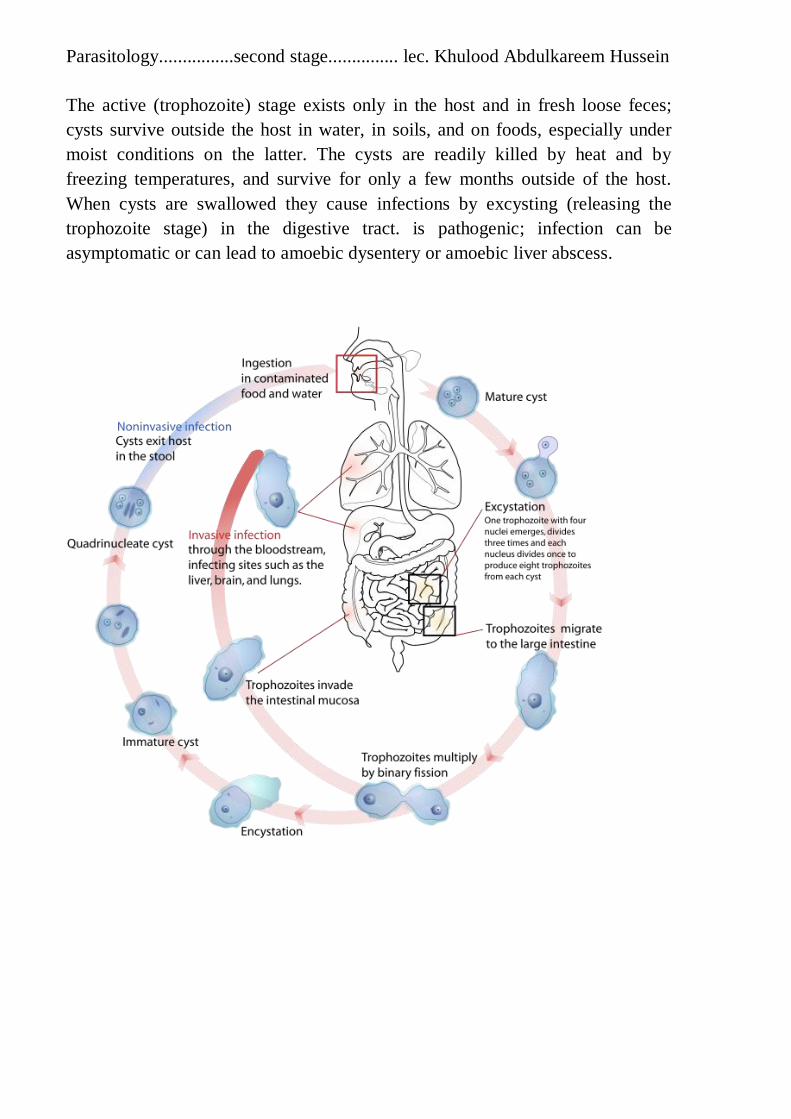
Parasitology................second stage............... lec. Khulood Abdulkareem Hussein
The active (trophozoite) stage exists only in the host and in fresh loose feces;
cysts survive outside the host in water, in soils, and on foods, especially under
moist conditions on the latter. The cysts are readily killed by heat and by
freezing temperatures, and survive for only a few months outside of the host.
When cysts are swallowed they cause infections by excysting (releasing the
trophozoite stage) in the digestive tract. is pathogenic; infection can be
asymptomatic or can lead to amoebic dysentery or amoebic liver abscess.
Parasitology................second stage............... lec. Khulood Abdulkareem Hussein
Laboratory Diagnosis
• Macroscopic examination of stool (dark red stool mixed with blood and
mucus).
• Microscopic examination of stool for demonstration of trophozoite or cyst of
Entamoeba histolytica,
• Proctosigmoidoscopy, scraping and biopsy samples collected under direct
vision by endoscopy
• Culture techniques can be done
• Serological techniques
• DNA examination techniques
.......................................................................................................................
Subkingdom: protozoa
Genus: Giardia
Species: Giardia lamblia
Cause disease (Giardiasis )
Geographical Distribution:-
It occurs all over the world. It is prevalent in 2to 25 percent population.
Habitat:-
is an aerobes flagellated protozoan parasite that colonizes and reproduces in
the Duodenum and upper part of small intestine.
Morphology:-
It is found in the following two forms:-
1-Trophozoite: It resembles longitudinally-cut pears, The dorsal surfaceis
convex and ventral surface is concave.There are a pair of axostyles, two nuclei
and 4 pairs of flagellae. It multiplies by binary fission.
Parasitology................second stage............... lec. Khulood Abdulkareem Hussein
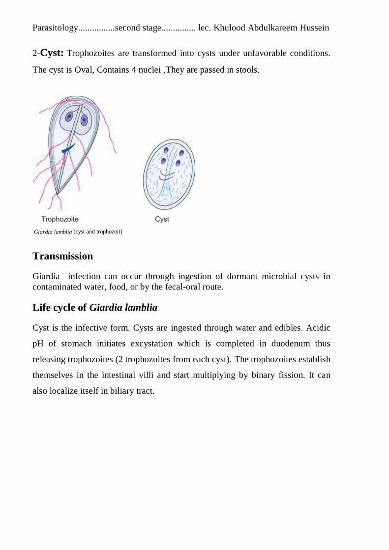
2-Cyst: Trophozoites are transformed into cysts under unfavorable conditions.
The cyst is Oval, Contains 4 nuclei ,They are passed in stools.
Giardia lamblia (cyst and trophozoit)
Transmission
Giardia infection can occur through ingestion of dormant microbial cysts in
contaminated water, food, or by the fecal-oral route.
Life cycle of Giardia lamblia
Cyst is the infective form. Cysts are ingested through water and edibles. Acidic
pH of stomach initiates excystation which is completed in duodenum thus
releasing trophozoites (2 trophozoites from each cyst). The trophozoites establish
themselves in the intestinal villi and start multiplying by binary fission. It can
also localize itself in biliary tract.

Parasitology................second stage............... lec. Khulood Abdulkareem Hussein
Laboratory Diagnosis
• Demonstration of cysts in the stool microscopically.
• Demonstration of trophozoites in duodenalaspirate.
• Intestinal biopsy.
• Immunological techniques like ELISA
......................................................................................................................
Subkingdom: protozoa
Genus: Leishmania
Species: Leishmani donovani
Leishmani tropica
Leishmani brasiliensis
Cause disease (Leishmaniasis)

Parasitology................second stage............... lec. Khulood Abdulkareem Hussein
Leishmania: is a disease caused by protozoan flagellates parasites and spread by
the bite of certain types of sandflies. The disease can present in three main ways
as:
Leishmani donovani :
Habitat :
The natural habitat of Leishmania donovani in man is reticuloendothelial system
especially spleen, liver, bone marrow, intestinal mucosa, and also in the
macrophages of intestinal wall. causes visceral leishmaniasis (kala azar).
Geographical Distribution
Visceral leishmaniasis is widely distributed,It is endemic in many places in
America, Africa, China, South Europe, Europe and India.
Leishmani tropica:
Geographical Distribution
Central and Western India. The infection does not coexist with kala-azar.
Habitat :
Amastigote in reticuloendothelial cells of skin (clasmatocyte). Promastigote
form in sandfly causes cautaneous leishmaniasis (oriental sore,Baghdad boil).
Leishmani brasiliensis:
Geographical Distribution
Central and South America.
Habitat :
Amastigote form occurs in the macrophages of skin and mucous membrane of
the nose and buccal cavity. causes mucocutaneous leishmaniasis.

Parasitology................second stage............... lec. Khulood Abdulkareem Hussein
Morphology
Leishmania exists in two forms:
(a)- amastigote form also called aflagellar form , and
(b)- Promastigote form also called flagellar form .
Life cycle:
The female sandfly after sucking leishmania along with blood of the
patient,female sandfly is small hairy fly (1.5 to 3.5 mm). Its usual biting
time is at dusk or night. Leishmania spp. undergoes development inside the
body of female sandfly. The promastigote forms after multiplication ascend to
pharynx and reach the proboscis. It takes 9 days to complete the cycle in sandfly.
Ultimately the buccal cavity of sandfly is blocked by promastigote form. For
taking second meal the sandfly has to release the promastigote form from its
mouth into the bite wound caused by its proboscis. The promastigotes thus enter
the circulation are mainly destroyed by vertebrate host (man). Still some
promastigotes take shelter inside cells of reticuloendothelial system where
promastigote form is transformed into amastigote one. They undergo
multiplication

Parasitology................second stage............... lec. Khulood Abdulkareem Hussein
there at a slow rate. When the infected cells of reticuloendothelial system
rupture, the free amastigote forms attack other cells. Sometimes they may be
phagocytosed.
Laboratory Diagnosis
Direct smear, culture and serological techniques
Biopsy or aspirate from these specimens is smeared on clean glass slide
fixed with methyl alcohol and stained with Giemsa stain.
Immunological tests include tests to detect antigen ,e.g. ELISA

Parasitology................second stage............... lec. Khulood Abdulkareem Hussein
Subkingdom: protozoa
Genus: Trichomonas
Species: Trichomonas vaginalis
Cause disease (Trichomoniasis).
-Is an anaerobic , flagellated protozoan.
Geographical Distribution
world wide distribution
Habitat
In female it is found mainly in vagina and in male it is in urethia.
Morphology
It is found only in trophozoitic form which bears

Parasitology................second stage............... lec. Khulood Abdulkareem Hussein
Mode of Transmission
It is primarily a venereal disease in which transmission can also be from person-
toperson contact. However, newborns may get infected during birth. Fomities
also form another way of transmission of infection.
Incubation Time
It varies from 4 to 30 days.
Laboratory Diagnosis
In female patient, Trichomonas vaginalis may be demonstrated in sedimented
urine, vaginal secretion
In male patient Trichomonas vaginalis may be found in the centrifuged urine
and prostatic secretions
Culture: It is quite sensitive technique.
Life cycle

Parasitology................second stage............... lec. Khulood Abdulkareem Hussein
Subkingdom: protozoa
Genus: Plasmodium (Malarial parasite)
Species: Plasmodium falciparum
Plasmodium vivax
Plasmodium ovale and
Plasmodium malariae.
GEOGRAPHICAL DISTRIBUTION
It is sporozoa parasite, occurs in all countries in the tropics and subtropics.
Transmission: The infection is initiated when sporozoites are injected with the
saliva of a feeding mosquito
Habitat :
It is found in parenchymal cells of liver, erythrocytes and other organs.
Life cycle: All species complete life cycle in man and female anopheles
mosquito. The life cycle of Plasmodium involves several distinct stages in
the insect and
vertebrate hosts.
In infected mosquitoes, parasites in the salivary gland are called sporozoites.
When the mosquito bites a vertebrate host, sporozoites are injected into the host

Parasitology................second stage............... lec. Khulood Abdulkareem Hussein
with the saliva. From there, the sporozoites enter the bloodstream and are
transported to the liver, where they invade and replicate within hepatocytes. At
this point, some species of Plasmodium can form a long-lived dormant stage
called a hypnozoite which can remain in the liver for many years. The parasites
that emerge from infected hepatocytes are called merozoites, and these return to
the blood to infect red blood cells. Within the red blood cells, the merozoites
grow first to a ring-shaped form and then to a larger form called a trophozoite.
Trophozoites then mature to schizonts which divide several times to produce
new merozoites. The infected red blood cell eventually bursts, allowing the new
merozoites to travel within the bloodstream to infect new red blood cells. Most
merozoites continue this replicative cycle, however some merozoites upon
infecting red blood cells differentiate into male or female sexual forms called
gametocytes. These gametocytes circulate in the blood until they are taken up
when a mosquito feeds on the infected vertebrate host, taking up blood which
includes the gametocytes.
In the mosquito, the gametocytes move along with the blood meal to the
mosquito's midgut. Here the gametocytes develop into male and female gametes
which fertilize each other, forming a zygote. Zygotes then develop into a motile
form called an ookinete, which penetrates the wall of the midgut. Upon
traversing the midgut wall, the ookinete embeds into the gut's exterior membrane
and develops into an oocyst. Oocysts divide many times to produce large
numbers of small elongated sporozoites. These sporozoites migrate to the
salivary glands of the mosquito where they can be injected into the blood of the
next host the mosquito bites, repeating the cycle.

Parasitology................second stage............... lec. Khulood Abdulkareem Hussein
Laboratory Diagnosis
• Peripheral blood film for parasites (thick and thin smear) is studied
microscopically after staining
• Serological techniques like ELISA
............................................................................................................................. .....
-2-class: NEMATODES
species: Trichuris trichiura
Geographical Distribution :Worldwide.
Habitat :Adult worm lives in large intestine of man.
Disease: causes Trichuriasis
Transmission: A soil transmitted swallowing infective eggs in
contaminated soil, food or water.

Parasitology................second stage............... lec. Khulood Abdulkareem Hussein
Life cycle: No intermediate host is required. Eggs are passed in stools of
infected patient. A rhabditiform larva develops from egg and infection to
healthy person occurs by ingestion of embryonated eggs in food and water.
The egg shell is dissolved in the stomach and larvae liberated pass down the
cecum which grow into adult worms and embed their anterior parts in the
intestinal mucosa. They grow in adult form. The life-cycle is completed in
one host, i.e. man.
Laboratory Diagnosis :It is established by detecting characteristic
eggs
in stool. Sometimes adult worm may be detected in stools but rarely.
....................................................................................................................
2-species :Ascaris lumbricoides
Geographical Distribution :It is cosmopolitan.
Habitat :Adult worm lives in the lumen of the small intestine
(jejunum) of man.

Parasitology................second stage............... lec. Khulood Abdulkareem Hussein
Transmission: swallowing infective eggs in contaminated soil, food or
water.
Disease: Ascariasis
Life Cycle
Infects humans when an ingested fertilised egg becomes a larval worm that
penetrates the wall of the duodenum and enters the blood stream. From
there, it is carried to the liver and heart, and enters pulmonary circulation to
break free in the alveoli, where it grows and molts. In three weeks, the
larva passes from the respiratory system to be coughed up, swallowed, and
thus returned to the small intestine, where it matures to an adult male or
female worm. Fertilization can now occur and the female produces as
many as 200,000 eggs per day for a year. These fertilized eggs become
infectious after two weeks in soil; they can persist in soil for 10 years or
more
Laboratory Diagnosis
Detection of adult worms in stool.
Microscopic detection of eggs in feces or bile obtained by duodenal
intubation.

Parasitology................second stage............... lec. Khulood Abdulkareem Hussein
3-Specise:Enterobius vermicularis
Geographical Distribution :It is cosmopolitan.
Habitat :Adult worm (female resides in cecum and appendix of man).
Disease: cause Enterobiasis
Transmission: Do not need to rely on a vector for transmission. infection
usually occurs via ingestion of infectious eggs by direct anus-to-mouth
transfer by fingers.
Life Cycle
The female worm when fully gravid passes down to migrate several inches
outside the anus to deposit eggs. These eggs are transferred by fingers
(autoinfection) and by contaminated food or fomites to the mouth and they are
swallowed. On reaching the intestine, outer shell is dissolved by digestive
enzyme thus liberating the larvae. In the presence of oxygen, larvae become
infective.
Laboratory Diagnosis
• Detection of adult worm in the stools.
• Demonstration of eggs in stool and finger nails.
3-Class :Cestoda (Tapeworms) :- live in the digestive tracts of vertebrates
as adults.
Transmission: Humans are subject to parasitism by several species of
tapeworms if they eat undercooked meat such as pork (Taenia solium), beef
(T. saginata), and fish (Diphyllobothrium spp.), or if they live in, or eat food
prepared in, conditions of poor hygiene (Hymenolepis or Echinococcus
species).
Laboratory Diagnosis

Parasitology................second stage............... lec. Khulood Abdulkareem Hussein
Demonstration of proglottids or eggs.
Serodiagnosis is done with the help of tests like indirect hemagglutination,
and ELISA.
Taenia solium Taenia saginata Echinococcus granulosus