Embed Size (px)
Citation preview
1
LEEDS TEACHING HOSPITALS TRUST
GUIDELINES FOR THE CARE AND MANAGEMENT OF ADULT PATIENTS WITH A TRACHEOSTOMY at LTHT
Guideline Detail Publication date: original publication Sept 2004, updated Nov 2011, Updated July 2015 Next Review date: September 2018 Status: Current Contents
Section Page number
Summary 2
Aims and Scope 2
Objectives 2
Background 3
Consent 3
Treatment / Management 3
1 1.1 1.2
Tracheostomy Standard surgical technique Percutaneous dilatation technique
3 4 4
2 Laryngectomy 4
3 Psychological implications 4
4 Infection control 5
5 5.1 5.2 5.3 5.4 5.5 5.6 5.7
Tracheostomy tube and stoma care Type of tubes Cleaning the tracheostomy stoma Securing the tube Cleaning inner tube Changing tubes Monitoring Cuff pressures
5 5 6 6 6 7 7 7
6 Suctioning 8
7 Humidification 8
8 Swallow assessment 9
9 Passy Muir Speaking Valves 9
10 10.1 10.2 10.3 10.4 10.5 10,6 10.7 10.8
Safety and emergency care Experienced personnel Immediate complications Delayed complications Late complications Blocked tracheostomy Displaced tracheostomy Haemorrhage RED FLAGS
10 10 11 11 11 11 12 12 13
2
10.9 10.10 10.11
Other complications: local infection, mucosal ischaemia Emergency airway management Other considerations
14 15 15
11 Weaning 16
12 Decannulation 16
13 Discharge to community 17
Audit 17
Provenance 17
Evidence 17
Appendix
1 Tracheostomy Insertion record 20
2 Nursing care standard 21
3 Types of tubes 23
4 SOP for cleaning tubes 26
5 Assessment of competency for decannulation and tube change 27
6 SOP Passy-Muir tube 34
7 Emergency algorithm 39
8 Equipment at bedside 43
9 Audit tool 44
Summary of Guideline
These guidelines have been developed and updated to provide guidance for staff caring for patients with a tracheostomy within the Leeds Teaching Hospitals NHS Trust These guidelines are intended to support and inform practitioners caring for adults with tracheostomies within the acute in-patient setting to improve care and safety. There are supporting teaching packages, competencies, care plan and algorithms in addition to the guidelines. These guidelines include information about procedures, consent, management, emergency care, decannulation and tube changes. Patients with tracheostomies should only be cared for in clinical areas where staff are competent in caring for them.
Aims That patients with a tracheostomy receive up to date efficient care based on their assessed needs and delivered by trained staff who have been assessed as competent in this skill.
Scope These guidelines relate to all adult patients who have a tracheostomy tube in situ cared for within the Leeds Teaching Hospitals NHS Trust.
They are aimed at all practitioners caring for the patient with a tracheostomy tube.
Objectives That all patients or relatives receive information about the needs for a Tracheostomy. That all patients within the LTH with a tracheostomy tube will have their care needs
assessed and delivered as per agreed plan of care.
3
Care will be performed in a safe and timely manner according to the patient’s individual
plan with any deviations to planned care accurately documented. Care will be documented and evaluated. The guidelines and competencies will be available to all staff to aid their safe practice. Background
Following the NCEPOD study and report (2014) and review of the Intensive Care society’s standards for the care of adult patients with a temporary Tracheostomy (2014) , these guidelines have been reviewed and updated to reflect best practice in caring for patients with a tracheostomy.
Consent Most patients will be able to give informed consent for an elective procedure, for those that are critically ill attempts should be made to seek their understanding and approval if this is possible. The role of the next of kin in healthcare decision making is increasingly formalised under the Mental Capacity Act (England and Wales) and the Adults with Incapacity Act (Scotland). It is considered good practice to inform relatives of the clinical decision and involve them in the planned care. Current directives from the GMC and Department of Health specify their involvement using Consent Form 4; ‘Form for Adults who are Unable to Consent to Investigation or Treatment’. This process requires provision of information on the nature of the procedure, proposed benefits, potential hazards and alternatives, ideally written and with visual aids in the first instance. Nevertheless it is the responsibility of medical staff to act in the best interests of patients lacking capacity, rather than pass the responsibility for consent to the next of kin. It should be emphasised that, like all surgical interventions, this procedure is not undertaken lightly and is not risk-free, however nor is prolonged translaryngeal intubation.
Treatment / Management 1. Tracheostomy There are two techniques for creating a tracheostomy.
Regardless of which technique is chosen, clinicians must be prepared for the possibility of losing the airway during insertion and difficulties in re-securing it. A comprehensive dedicated ‘difficult airway’ trolley must be available on the ICU. The contents of the trolley should include a range of tracheal tubes, laryngoscopes, bougies, airway exchange catheters, laryngeal mask airways, paediatric facemasks, and cricothyroid needles/cannulae for emergency oxygen insufflation.
4
1.1. The Standard Surgical Technique (performed in theatres) This surgical technique involves the dissection and transfixion of the thyroid isthmus and formation of an opening in the trachea at the level of the second and third tracheal rings. After this procedure a tract forms between the trachea and the skin over a period of 5-6 days (Webb et al 1999).
1.2. Percutaneous Dilatational Tracheostomy (PDT)(usually performed within
critical care)
The procedure is performed by the introduction of a guide wire percutaneously into the trachea, followed by dilation of the tract formed to enable the insertion of a tracheostomy tube (Carrillo et al, 1997). After this procedure a tract forms between the trachea and the skin over a period of 5-6 days (Webb et al 1999). For further details please refer to Intensive Care society’s standards for the care of adult patients with a temporary Tracheostomy (2014). See Appendix 1 for insertion form documentation.
2. Laryngectomy A laryngectomy creates a permanent stoma. The larynx is resected and the trachea is
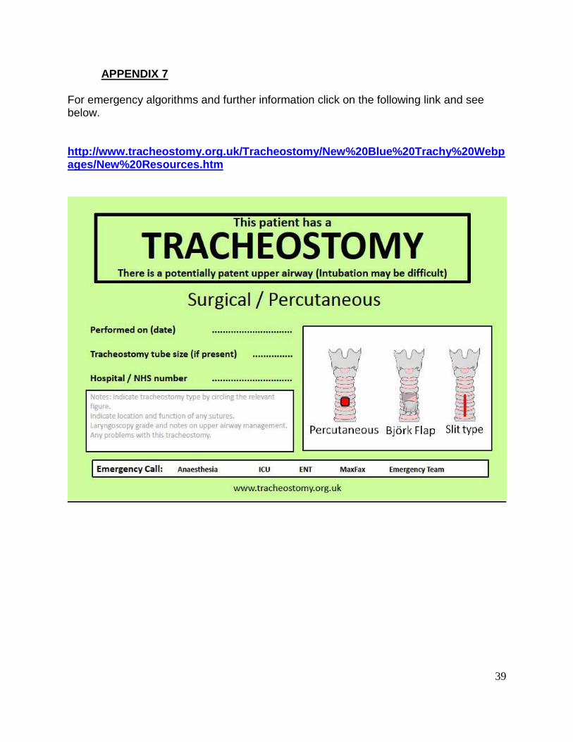
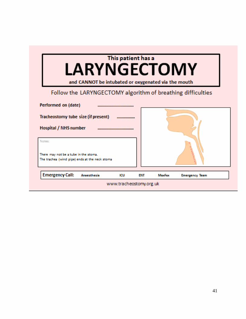
brought to the surface of the skin and sutured to the neck wall to form a stoma. Thus the lungs are completely disconnected from the nose and mouth. The rigid rings of cartilage of which the trachea is comprised prevent the stoma from closing. Staff must be aware of the differences between tracheostomy and laryngectomy especially in emergency situations (NPSA 2005). In emergency situations, patients with a laryngectomy have an end stoma and cannot be intubated or ventilated via the mouth. Call for expert help - Anaesthetics/ITU and ENT/Max Fax. If bleeding from the laryngectomy, await expert advice before deflating the cuff.
3. Psychological considerations Patients with a tracheostomy will have no voice whilst the cuff is inflated, this can limit
their ability to communicate. In the absence of an appropriate speaking valve, aids such as pen and paper or picture cards can facilitate communication. “Closed” questions should be used where possible.
Psychological support for patients with a tracheostomy is important both pre and post
procedure. In some circumstances the procedure is elective and patients can be prepared in some way for the potential difficulties. Unconscious patients however, will not receive preparation and may need extra psychological support when they begin to recover.
Due to an altered body image the patient should have careful pre-operative explanation. If this is not possible the patient must receive explanation and support post-operatively and an information leaflet provided.
Clear explanations of what is going to happen, and why, should be given to the patient
5
before any procedures such as suction or changing of tubes takes place. 4. Infection control
The presence of a tracheostomy tube, the resultant secretions and stoma site in an already debilitated and possibly immuno-compromised patient all increase the risk of infection. Hand washing is essential before and after all procedures. Gloves must be worn and contaminated gloves changed between procedures. Aprons must be worn for all procedures. Eye protection should be worn where there is a risk a patient may cough secretions towards the carer. Closed suction should be used whilst patient is ventilated. Any infections must be clearly documented and communicated and Trust isolation procedures adhered to. Hand Hygiene Policy -LTHT: http://nww.lhp.leedsth.nhs.uk/common/guidelines/detail.aspx?ID=673 Standard Infection Prevention and Control Precautions Policy http://nww.lhp.leedsth.nhs.uk/common/guidelines/detail.aspx?ID=671 Source Isolation Policy http://nww.lhp.leedsth.nhs.uk/common/guidelines/detail.aspx?ID=678
5. Tracheostomy tube and stoma care
All nurses caring for patients with tracheostomies should adhere to the nursing care standard (see Appendix 2)
5.1. Types of tracheostomy tubes
The type of tracheostomy tube used within The Leeds Teaching Hospitals NHS Trust will vary according to the needs of the individual patient (see Appendix 3). Documentation and communication of the type and size of tube is essential, including a record of the date of insertion. The tubes may be cuffed or uncuffed, these may have an inner liner, and in addition they may be fenestrated or non-fenestrated. Fenestrated tracheostomy tubes may help the patient breath with a tracheostomy tube in situ. Provided the inner cannula (if not fenestrated) is removed, the work of breathing may be reduced. Obese patients, or those with abnormal anatomy, or a low tracheostomy stoma may require a longer adjustable flange tube or a tube with a fixed extended distal or proximal portion. Patients must not be discharged from critical care unless a tracheostomy tube with
6
an inner liner tube is in situ. Tracheostomy tubes may also have supraglottic drainage ports to help reduce the incidence of Ventilator acquired pneumonia (VAP). The type of tube that is in placed should be clearly documented. An emergency pack containing a tube of the same size must be available at the bedside.
5.2. Cleaning a tracheostomy stoma
Until the stoma site is established, the site and surrounding skin should be cleaned as often as required with sterile sodium chloride 0.9%, using an aseptic non touch technique. A clean dry stoma site will facilitate wound healing. The condition of the skin around the stoma should be assessed regularly for signs of infection and delayed healing. This should be documented in the critical care nursing plan or LTH tracheostomy care plan. The routine use of dressings around the stoma site is unnecessary and should only be used if clinically indicated. Once the stoma site has healed, responsibility for cleaning it can be negotiated between the patient and the team caring for them. It is then possible to care for the stoma adopting a clean procedure using sterile water or cooled previously boiled water. A skin barrier cream can be applied if indicated.
5.3. Securing the tube
Ties should be changed when soiled. The tube should be secure without impeding venous return (Docherty and Bench 2002). Velcro tracheostomy ties with a soft backing should be used as these are more comfortable for the patient. It is recommended that this procedure is done with 2 people, particularly with restless or confused patients.
5.4. Cleaning tracheostomy inner tubes (see Appendix 4).
Where a tracheostomy tube is in situ the inner cannula should be removed and cleaned at a maximum interval of 4 hourly in a patient with a productive chest, and at least 8 hourly in all cases being conscious of the patient’s need for rest and sleep. Repeated ventilator disconnection to change and clean the inner tube may result in derecruitment, especially in patients receiving high levels of PEEP and / or inspiratory pressures, and increase the risk of VAP/infection. The risks and benefits of using inner cannulae should be considered. It is recommended that inner tubes are cleaned with sterile sodium chloride 0.9% or sterile water using cleaning swabs where required. However, some areas with longer term tracheostomies may use tap water. Tracheostomy cleaning solution, which is a non-foaming biological detergent which breaks down sputum and cleans tubes well, may also be used (available from NHS supplies). Bedside sinks should not be used
7
for cleaning tracheostomies; these are only for hand washing (DoH 2010).
5.5. Changing tracheostomy tubes
After the initial formation of a tracheostomy, the tube should ideally remain in place for at least five to six days to allow a tract to form between the trachea and skin. Tubes should be changed according to patient need and/or as per manufacturers’ guidelines, at least every 30 days. Tracheostomy tubes without an inner liner should only remain in situ for up to seven days. Should a tracheostomy be required for a longer period, then a tube with an inner liner should be considered. All practitioners must hold an appropriate professional qualification and the procedure must be within their scope of practice. Where this is an extended scope of practice the practitioner must have undertaken training appropriate to their profession and be deemed competent. (See Appendix 5). All patients should be pre-oxygenated prior to tracheostomy tube change. Consider changing the tube in theatre with capnography and endoscopy available if any difficulty is anticipated. It is essential that if the new tube cannot be inserted or is misplaced, there is an agreed procedure for managing the situation, with the appropriate equipment and drugs available.
5.6. Monitoring
Waveform capnography is mandatory for any acutely ventilator dependent tracheostomy patient.
5.7. Cuff pressures Cuff manometers should be used to measure cuff pressures and they should be recorded on the observation chart at least 4 hourly, and maintained ideally between 20-25cm H2O. Maintaining an appropriate inflation pressure of the tracheal cuff is important, since under inflation (< 20cm H2O) of the tracheal cuff is associated with ventilator acquired pneumonia (VAP). Over inflation (>30cm H2O) is also harmful and contributes to long term tracheal damage (DoH 2011). Pressures above 30cm H2O have been shown to cause ischaemia, tracheal wall necrosis and cartilage destruction (Bernhard et al, 1985). Anaesthetic gases can increase cuff pressures (Stanley and Liu 1975). If an air leak occurs with the cuff pressure at maximum recommended, the tracheostomy tube may have become displaced or may require changing: medical or other professionals who are competent in tracheostomy management should review the patient (ICS, 2014). Cuff pressure should not exceed 25 cm H2O. Cuff deflation should be a 2 person technique; as one deflates the cuff the second performs tracheal suction to prevent aspiration. Following cuff deflation, expiratory noises may be heard, the patient should be reassured that this is normal.
8
6. Suctioning Please refer to LTHT suctioning guidelines (2012) http://nww.lhp.leedsth.nhs.uk/common/guidelines/detail.aspx?ID=3152 Always encourage patients to expectorate their secretions where possible. An upright position will facilitate clearance of secretions. Tracheal suction should be performed following assessment of the patient and only performed if needed. Following suction further assessment should be made. Results of the procedure and any adverse effects should be documented. When receiving oxygen, pre-oxygenation with high flow or a higher percentage oxygen may help to reduce hypoxia during the procedure. The oxygen should be prescribed on the prescription chart with a dose and target oxygen specified if being cared for out of the critical care environment.
Tracheal suction is performed using an aseptic non touch technique. An appropriate sized, multi-eyed suction catheter should be used. The suction pressure used should not exceed 120mmHg. The pressure should only be applied during removal of the suction catheter. The procedure should last no longer than 10 seconds. It is advised that a visor be worn if closed suction is not being used.
7. Humidification
Additional humidification is necessary to maintain the relative humidity of the inspired gases and prevent secretions drying and blocking the tracheostomy tube. It is essential that patients with a tracheostomy receive adequate humidification in order to prevent tracheal secretions becoming hard and crusty thus preventing consolidation, obstruction and atelectasis. Where oxygen is not required, humidification via a heat & moisture exchanger (HME) (often known as Swedish nose), or bibs should be considered. HME’s can be dislodged by strong coughing (Shelley 1996) and become clogged with secretions, leading to infection risk. They should never be used overnight and are not suitable for patients with copious secretions. HME’s should be changed at least every 24 hours but more often if required due to secretions. Buchanan/ Hydrolux bibs/protectors may be more comfortable, and are appropriate to be used overnight. For hospital patients, these should be changed every 24 hours. If secretions increase in viscosity then alternative methods of humidification should be used. Regular (4-6 hourly) nebulised sodium chloride 0.9% 5mls (Prescribed) can be considered to augment clearance of thick secretions (Conway 1992, Sutton 1998) but should be used in addition to and not in place of other methods of humidification. Bolus Sodium chloride 0.9% up to 2-3ml instillation into the tube may be useful to loosen and remove thick secretions in certain patients but should never be a routine procedure
9
(Hess 1999). Assessment of hydration, humidification and the nature of secretions should be carried out by an experienced practitioner who should make the decision to use sodium chloride 0.9% instillation (Hudak 1996). The most important method of maintaining humidification is to ensure the patient has adequate hydration. In patients with thick, difficult to clear secretions, a mucolytic may be considered. Physiotherapy or outreach teams can give advice on the most appropriate method of humidification for specific patients
Monitoring & Review: Humidification, observations and NEWS score are monitored from the graded response chart. Fluid balance is monitored on a fluid balance chart
8. Swallowing assessment Any patient with a tracheostomy tube should be considered at risk of aspiration (Dikeman
& Kazandjian 1995, Tippett 2000). Before commencing eating and drinking, all patients should be assessed, a referral to speech and language therapy (SALT) should be considered and ongoing assessment for signs of aspiration should continue. A blue dye test is no longer recommended by SALT. Laryngectomy patients will have consultant led input around the need for contrast swallow test.
Risk factors for swallowing problems in patients with a tracheostomy (ICS, 2014)
Neurological injury e.g. bulbar palsy
Disuse atrophy
Head & neck surgery
Evidence of aspiration of enteral feed or oral secretions on tracheal suctioning
Increased secretion load, or persistent wet / weak voice, when cuff is deflated
Coughing and / or desaturation following oral intake
Patient anxiety or distress during oral intake
High FiO2 9. Passy Muir Speaking Valves (see Appendix 6)
Passy Muir Speaking valves (PMV) are a closed diaphragm speaking valve, designed for use with tracheostomies, which diverts all air to pass through the upper airway on expiration in comparison to previous speaking valves which only partially close on expiration. Therefore the PMV allows, amongst other things, improved laryngeal excursion, improved airway protection and restoration of subglottic pressure. They have been shown to improve weaning and therefore decannulation. If you are considering the use of a PMV with your patient please refer to Physiotherapy for assessment and guidance. If a speaking valve is in use the cuff must be fully deflated at all times with an adequate leak past the tube. Preferably a non-cuffed tube or one with a fenestration should be used to allow easier expiration of air.
10
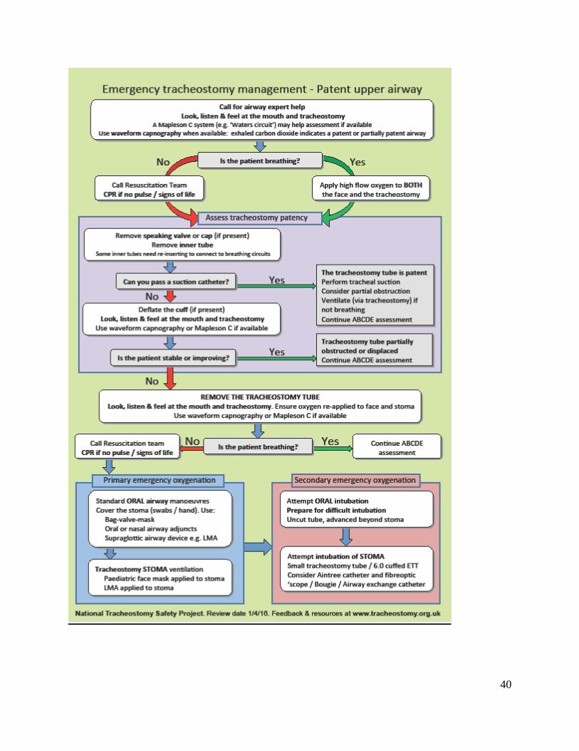
10. Safety and emergency care See link below for algorithms from National Tracheostomy Safety Project, NTSP, for detailed guidance http://www.tracheostomy.org.uk/Templates/Algorithms.html Patients with tracheostomies need access to appropriate equipment (see Appendix 8) and to staff with the skills capable of caring for and managing them. This equipment should be checked daily as part of the tracheostomy standard (See Appendix 2). Respiratory difficulty should lead to urgent assessment of the airway, and to consider whether the tube has become displaced (inside or outside the trachea), or blocked. Every ward looking after patients with a tracheostomy must have a clear procedure for emergencies.
10.1 EXPERIENCED PERSONNEL Acute Anaesthetist:- SJUH: Bleep 80 5011 LGI: Bleep (ICU general) 80 3451, (ICU neuro) 80 2752 ICU: SJUH 69154/64584 ICU: LGI 27403/25768 Outreach:- SJUH: 66367, Bleep 80 6425 BEXLEY WING: 67637, Bleep 80 5781 LGI: 28179, Bleep 80 1450 Physiotherapists:- SJUH: 80 4334/6250/1088 LGI: 80 2981/2844 Emergencies, Common Complications and their Management This section is taken from section 6 common complications and emergency management in “Standards for the care of adult patients with a temporary tracheostomy (ICS, 2014). The main life threatening complications associated with a tracheostomy are blockage, dislodgement and bleeding. All staff working in clinical locations where tracheostomy patients are managed must be competent to assess and initiate management in the event of an airway emergency occurring. These complications may occur at any time or location. They may be life threatening and may be challenging for patients and staff. Complications associated with a tracheostomy Complications can be divided into those associated with insertion of the tracheostomy
11
(surgical or percutaneous), those which arise following the procedure (usually blocked or displaced tracheostomy tubes) or later complications. These can be serious and sometimes fatal. These complications may coexist and are usually grouped as follows:
10.2. Immediate Complications (peri-procedural period)
Haemorrhage (this is usually minor, but can be severe if thyroid or major paratracheal blood vessels are damaged).
Primary misplacement of tube - within tissues around trachea or to main bronchus, or secondary displacement e.g. after severe coughing on emergence.
Pneumothorax.
Tube occlusion.
Surgical emphysema.
Loss of the upper airway.
10.3. Delayed Complications (post-operative period < 7 days)
Tube blockage with secretions or blood. May be sudden or gradual.
Partial or complete tube displacement.
Infection of the stoma site.
Infection of the bronchial tree (pneumonia).
Ulceration, and/or necrosis of trachea.
Mucosal ulceration by tube migration (due to loose tapes or patient intervention).
Risk of occlusion of the tracheostomy tube in obese or fatigued patients who have difficulty extending their neck.
Tracheo-oesphageal fistula formation
Haemorrhage (local tissue trauma or erosion through blood vessels).
10.4. Late Complications (late post-operative period >7 days)
Granulomata of the trachea may cause respiratory difficulty when the tracheostomy tube is removed.
Tracheal dilation, stenosis, persistent sinus or collapse (tracheomalacia).
Scar formation-requiring revision.
Blocked tubes may occur at any time, especially if secretions become thick, the secretions are not managed appropriately (suction) and humidification is not used.
Haemorrhage.
10.5 Blocked Tracheostomy
Inserting a tracheostomy tube bypasses the natural warming and humidification mechanisms of the upper airway. Patients may have reduced ability to cough or clear secretions, which may be increased or purulent if infected, or thickened if the patient
12
is dehydrated. The risks of blocked tracheostomy tubes can be minimised by careful humidification, tracheal suction and inner tube care. Prevention is better than cure. Staff treating patients with a tracheostomy in an emergency situation need to be aware: that ‘bag and mask’ ventilation via the mouth and nose is not possible with a cuffed tracheostomy tube in situ.
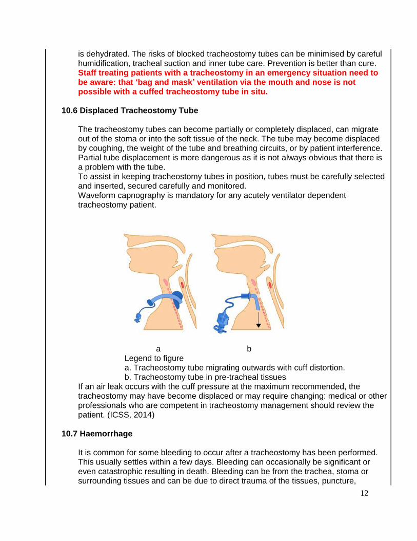
10.6 Displaced Tracheostomy Tube
The tracheostomy tubes can become partially or completely displaced, can migrate out of the stoma or into the soft tissue of the neck. The tube may become displaced by coughing, the weight of the tube and breathing circuits, or by patient interference. Partial tube displacement is more dangerous as it is not always obvious that there is a problem with the tube. To assist in keeping tracheostomy tubes in position, tubes must be carefully selected and inserted, secured carefully and monitored. Waveform capnography is mandatory for any acutely ventilator dependent tracheostomy patient.
a b
Legend to figure a. Tracheostomy tube migrating outwards with cuff distortion. b. Tracheostomy tube in pre-tracheal tissues
If an air leak occurs with the cuff pressure at the maximum recommended, the tracheostomy may have become displaced or may require changing: medical or other professionals who are competent in tracheostomy management should review the patient. (ICSS, 2014)
10.7 Haemorrhage
It is common for some bleeding to occur after a tracheostomy has been performed. This usually settles within a few days. Bleeding can occasionally be significant or even catastrophic resulting in death. Bleeding can be from the trachea, stoma or surrounding tissues and can be due to direct trauma of the tissues, puncture,
13
infection or injury to adjacent blood vessels or the tube or cuff eroding into surrounding tissues or vessels over time. Bleeding can also come from the lungs themselves and become evident through tracheal suction. These problems are compounded in the presence of a coagulopathy. If airway obstruction occurs due to blood clot in the airway (identify if possible with an endoscope) then direct suction on the tracheal tube with suction tubing may be required to remove it. Large clots will not pass through a suction catheter or a fibre optic scope suction channel. Suction may need to be applied directly to the tracheal tube and/or the tube removed with suction applied. Do not make repeated attempts at suctioning with conventional catheters, as delay in removing a blocked tracheostomy tube may cause hypoxia, which may lead to significant morbidity or may even be fatal. At this stage, the tracheostomy tube must be removed. The stoma can be suctioned directly once the tube is removed. Direct endobronchial intubation, (under fibre optic guidance) with an uncut endotracheal tube and with suction then applied directly to the endotracheal tube may be needed to try to remove obstructing clots. It may be necessary to repeat this procedure and it may be necessary to apply suction in this manner to both main bronchi. A tracheo-arterial fistula can occur if the tube erodes into the brachiocephalic artery. This is a rare complication but is associated with lower placement of the tube in the trachea. There may be a warning or ‘sentinel’ bleed. Any haemorrhage should prompt a fibre optic inspection of the trachea. If an arterial bleed is suspected, this examination should occur immediately with an experienced surgeon and resuscitating measures available. Arterial haemorrhage can become rapidly fatal. Hyperinflation of the tracheostomy tube cuff or an endotracheal tube cuff, or digital pressure in the stoma may help to tamponade the bleeding point, prior to definitive surgical management.
10.8 Tracheostomy Red Flags
Tracheostomy ‘Red Flags’ are used as a warning signal that a problem has, or is about to occur and need to be acted upon. Prompt assessment by someone competent to do so is required and a fibre optic inspection of the position of the tracheostomy tube to confirm correct placement within the trachea may be indicated. All staff caring for patients with tracheostomies should be familiar with these warning signs. As with all assessments of the acutely unwell patient, an ABCDE assessment includes ensuring the airway is patent, including assessment of the tracheostomy tube if present.
Red flags include: 1. Airway
a. A suction catheter not passing easily into the trachea b. A changing, inadequate or absent capnograph trace
14
c. The patient with a cuffed tracheostomy tube suddenly being able to talk or noises or bubbles coming from the upper airways (implying gas escaping proximally and the cuff no longer ‘sealing’ the trachea) d. Frequent requirement for (excessive) inflation of the cuff to prevent air leak e. Pain at the tracheostomy site f. Visibly displaced tracheostomy tube. If this is an adjustable flange tube, check to see where (at what length?) it was last positioned g. Active bleeding from the tube or stoma
2. Breathing
a. Increasing ventilator support or increasing oxygen requirements b. Respiratory distress c. Surgical (subcutaneous) emphysema (gas in the soft tissues) d. The patient complaining that they cannot breathe or have difficulties in breathing e. Suspicion of aspiration (feed aspirated on tracheal toilet – suggests that the cuff is not functioning adequately)
3. Circulation or any other general clinical deterioration
a. An airway emergency may lead to cardiovascular collapse. b. Anxiety, restlessness, agitation and confusion may also be due to an airway problem.
10.9 Other complications following tracheostomy
Local Infection The stoma is an open surgical wound and routine wound care is covered above. Colonisation with respiratory flora is inevitable and occasionally more serious local site infection may develop which can progress to spreading cellulitis or mediastinitis. This is more common with open surgical procedures due to the greater surgical exposure and dissection. Systemic antibiotics and occasional surgical debridement are required. Tracheal Damage/Mucosal Ischaemia Cuff pressure above 20cm H2O risks ischaemic damage to the tracheal mucosa. Modern tracheostomy tubes have low-pressure cuffs, which extend over a greater surface area; however over-inflation should still be avoided. The pressure in the cuff should be just adequate to prevent air leakage and seal the airway against aspiration
Direct mucosal damage can also occur by poor tracheal suctioning techniques, ill-fitting tubes, or excessive movement of the tube within the trachea. Such problems may be manifest by the development of granuloma and stenosis above or below the tracheostomy stoma.
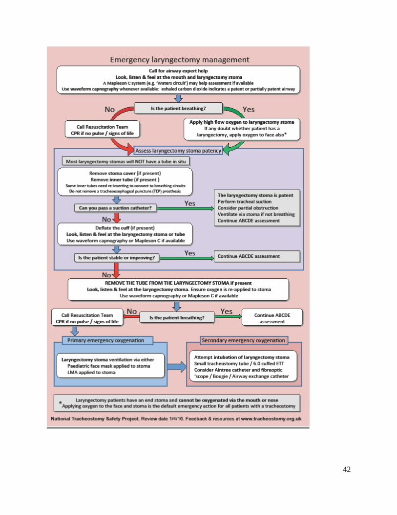
15
10.10 Emergency airway management of the patient with a Tracheostomy or Laryngectomy. (See Appendix 7 and links below for algorithms) The management of tracheostomy (and laryngectomy) related emergencies is summarised in Multidisciplinary guidelines for the management of tracheostomy and laryngectomy airway emergencies. Anaesthesia 2012; 67:1025-41. The algorithms are coupled with bed-head signs, allowing essential information to be clearly displayed and immediately available to responders in an emergency, including key details regarding the nature and date of the tracheostomy, method of forming the stoma and the function of any ‘stay sutures.’ Separate, colour-coded algorithms and bed-head signs are available for patients with a potentially patent upper airway and those with a laryngectomy. Two key principles
Oxygenation of the patient takes priority (not necessarily securing the airway immediately and definitively, unless required for oxygenation)
Skilled assistance should be sought early. The generic algorithms cover the majority of common and easily reversible clinical situations that arise. Even in complicated scenarios, key airway management principles should be followed. The primary responder (typically a nurse, junior doctor or allied health professional) needs to detect airway problems, to assess tracheostomy and airway patency and to provide basic emergency oxygenation. The secondary responder (typically an anaesthetist, intensivist, head and neck surgeon or specialist practitioner) will have skills in conventional airway management and will also be guided to use skills in managing the tracheostomy or stoma. These skills would include oro-nasal intubation techniques (including difficult intubations), ability to use fibre optic endoscope to assess or replace tracheostomy tubes and the ability to perform and manage an emergency surgical airway or tracheostomy. The algorithms are applicable for any urgent or emergency situation that develops in a patient with a tracheostomy or laryngectomy. Interactive algorithms with video links to each of the key steps are available from the website www.tracheostomy.org.uk, in Smart Phone applications (NTSP) and explained in the associated e-learning resources. The website also includes details of in-house training for staff, links to training courses and the freely available NTSP Manual 2013. Sample algorithms and bedhead signs see below; Reproduced from McGrath BA, Bates L, Atkinson D, Moore JA. Multidisciplinary guidelines for the management of tracheostomy and laryngectomy airway emergencies. Anaesthesia. 2012 Jun 26. doi: 10.1111/j.1365-2044.2012.07217, with permission from the Association of Anaesthetists of Great Britain & Ireland/Blackwell Publishing Ltd.
10.11 Special considerations
Tracheostomy emergencies in ICU patients usually involve displacement of a tube that is likely to have been placed percutaneously. Percutaneous stomas do not usually become established tracts for 7-10 days after
16
initial insertion. This may make early reinsertion of a displaced tube difficult or impossible. Bedside details of the nature and date of tracheostomy should guide the responders. Conventional upper airway management is likely to be safer and easier when managing a recently formed, percutaneous, displaced tracheostomy tube. For patients with a known, difficult upper airway (including the absent upper airway of the laryngectomy patient) emergency management plans should be made in advance, communicated and recorded where they are readily accessible
11.0 Weaning
The decision to start weaning a patient is done on an individualised basis following assessment by the MDT and making a clear plan prior to the process. This will be continuously re-evaluated and updated based on the individual needs of the patient. The patient must be involved in the process and given full explanations and reassurance about the procedures. A tracheostomy tube should be removed as soon as it is no longer required. It must be clear which person or team is responsible for management of the tracheostomy, especially if it is not the speciality with primary responsibility for the patient’s care. Reviewing the need for a tracheostomy and planning weaning should be part of the daily assessment. Some patients may tolerate rapid decannulation, especially if their ventilation period has been short or if they do not suffer significant lung or airway pathology or neuromuscular problems. Others, particularly those with underlying cardiopulmonary disease, muscle weakness, neurological deficits, upper airway oedema or problems managing airway secretions, will take much longer to wean and it is important that the process is both planned and sequential. A checklist to use prior to commencing weaning should ascertain: • Is the upper airway patent? (may require endoscopic assessment) • Can the patient maintain and protect their airway spontaneously? • Are they free from ventilatory support?* • Are they haemodynamically stable? • Are they absent of fever or active infection? • Is the patient consistently alert? • Do they have a strong consistent cough (able to cough out of tube or into mouth)? • Do they have control of saliva with or without a competent swallow? • Are there any planned procedures requiring anaesthesia within next 7-10 days? • Is this patient causing concern to any healthcare practitioner? • Can we safely support the weaning process in the patient’s current clinical environment? Under specialist supervision, it is possible to decannulate some patients who will need on-going non-invasive respiratory support via a face or nasal mask.” (ICS 2014)
12.0 Decannulation
Care should be taken when deflating a tracheostomy tube cuff as secretions may have
17
pooled above the cuff. Prior to performing the procedure explanations should be given to the patient. If there is a subglottal drainage port, secretions should be aspirated prior to cuff deflation. Cuff deflation should be a 2 person technique; as one deflates the cuff the second performs tracheal suction to prevent aspiration. Following cuff deflation, expiratory noises may be heard, the patient should be reassured that this is normal. The decision to attempt decannulation should be made with the MDT caring for the patient. Each area should follow their protocol for decannulation. The decannulation process should be commenced in the morning to allow a full period of assessment when the MDT is available.
13.0 Discharge to community with tracheostomy tube Effective discharge planning based on individual assessment of the patient needs will help facilitate a smooth transition from hospital into the community. The patient and any additional carers should be trained and confident in performing all aspects of tracheostomy tube care prior to discharge.
Audit and Monitoring Compliance
This guideline will be audited with the audit tool in appendix 9 Audit results will be presented to the critical care clinical governance audit meeting, which will agree actions arising from the recommendations, and monitor the progress of the actions.
Provenance:
Author name: Sheelah Ainsworth Senior Sister Adult Critical Care / including major sections from ICS guidelines Clinical condition: Patients requiring tracheostomy for airway management Target patient group: Patients requiring tracheostomy for airway management Target professional group (clinical competence): All healthcare professionals managing the care of adult patients with tracheostomies including: Nursing staff, physiotherapists, speech and language therapists, anaesthetists, respiratory physicians, ENT surgeons.
Evidence Base: References : Standards for the care of adult patients with a temporary Tracheostomy; Standards and Guidelines, Intensive Care Society Standards 2014;(ICSS, 2014) http://www.ics.ac.uk/ics-homepage/guidelines-and-standards/ On the right trach? A review of the care received by patients who underwent a tracheostomy 2014. http://www.ncepod.org.uk/index.htm UK National Tracheostomy Safety Project. http://www.tracheostomy.org.uk/ Algorithm Summary in: Multidisciplinary guidelines for the management of tracheostomy and laryngectomy airway emergencies. Anaesthesia 2012; 67:1025-41. The Global tracheostomy collaborative. http://globaltrach.org/ 4th National Audit Project (NAP4): Major complications of airway management in the United Kingdom Report and Findings – March 2011 http://www.rcoa.ac.uk/nap4
18
Tracheostomy guidelines. St Georges Healthcare NHS Trust. https://www.stgeorges.nhs.uk/gps-and-clinicians/clinical-resources/tracheostomy-guidelines/ NHS quality improvement Scotland. Best Practice Statement ~ March 2007 Caring for the patient with a tracheostomy. http://www.healthcareimprovementscotland.org/previous_resources/best_practice_statement/ tracheostomy care.aspx Bernhard et al, (1985) Intracuff Pressures in Endotracheal Tracheostomy Tubes, Chest. 87 (6) 720 – 725.
Carrillo EH, Spain DA and Bumpous JM (1997) Percutaneous Dilational Tracheostomy for Airway control, The American Journal of Surgery. 174, 469 – 473. Conway JH (1992) The Effects of Humidification for Patients with Chronic Airway Disease, Physiotherapy, 78 (2) 97 – 100 DoH (2010) High Impact Intervention No 5 (draft) 24/03/2010 Deppe SA, Kellly JW, Thoi LL (1993) Incidence of Colonisation Noscomial Pneumonia and Mortality in Critically Ill patients using a Trach Care Closed system versus an Open suction system: Prospective Randomized Controlled Study, Critical Care Medicine. 18 (12) 1389 – 1393 Dezfulian C et al (2005) Subglottic secretion drainage for the prevention of ventilator-associated pneumonia. The American Journal of Medicine 118: 11-18 Diaz E, Rodriguez AH, Rello J (2005) Ventilator associated pneumonia: issues related to the artificial airway. Respiratory Care 50: 139-43 Dikeman KJ and Kazandjian MS (1995) Communication and Swallowing Management of Trachoestomised and Ventilator Dependant Adults, Singular Publishing Group Inc. San Diego & London. Dixon L and Wasson D (1998) Comparing use and cost effectiveness of Tracheostomy tube securing devices, MEDSURG Nursing. 7 (5) 270 – 274. Docherty B and Bench S (2002) Trachoestomy management for patients in general ward settings, Professional Nurse. 18 (2) 100 – 104. Doerksen K, Ladyshewsky A and Stansfield K (1994) A comparative study of Systemised versus Random Tracheostomy Weaning, AXON. September Edition. DoH (2010) Alert warning hospital wash basins identified as a source of gram negative bacteria (DoH Sept 2010) DoH (2011) Reducing healthcare associated infections http://hcai.dh.gov.uk/whatdoido/high-impact-interventions/ Hess D R, Branson RD (1999) Humidification - Respiratory Care Equipment 2a Philadelphia: Lippincott, Williams & Wilkins: 101-132 Hudak M (1996) Post operative head and neck cancer patients with artificial airways: the effect of sodium chloride 0.9% lavage on tracheal mucus evacuation and oxygen saturations. ORL - head and neck nursing. Jackson C. Humidification in the Upper Respiratory Tract: a physiological overview Intensive and Critical Care, 12: 1
19
NPSA Protecting patients who are neck breathers Patient safety information (7th March 2005)
Peterson B D. Heated humidifiers structure & function In: Branson RD, Peterson BD, Karson KD Humidification: Current therapy and controversy. Respiratory Care Clinics of North America pgs 243-259 Russell C & Matta B. Tracheostomy: A Multi-Professional Handbook. London, Greenwich Medical Media Ltd Sheppard M & Wright M (2000) Principles & Practice of High Dependency Nursing Harcourt Publishers Ltd Stanley TH and Liu WS (1975) Tracheostomy and Endotracheal cuff volume and pressure changes during Thoracic operations, Annals of Thoracic Surgery. 20 (2) 144 – 151. Thomacot L, Vialet R, Viguier JM et al (1998) Efficacy of heat and moisture exchangers after changing every 48 hours rather than 24 hours. Critical Care Medicine: 26; 477-481 Tippett DC (2000) Tracheostomy and Ventilator Dependency, Thieme Medical Publishers, Inc. Webb AR, Shapiro M, Singer M and Suter PM(1999) Oxford Textbook of Critical Care, 1
st Edition, Oxford
university Press, Oxford.
20
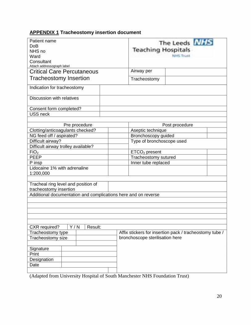
APPENDIX 1 Tracheostomy insertion document
Patient name DoB NHS no Ward Consultant Attach addressograph label
Critical Care Percutaneous Tracheostomy Insertion
Airway per
Tracheostomy
Indication for tracheostomy
Discussion with relatives
Consent form completed?
USS neck
Pre procedure Post procedure
Clotting/anticoagulants checked? Aseptic technique
NG feed off / aspirated? Bronchoscopy guided
Difficult airway? Type of bronchoscope used Difficult airway trolley available?
FiO2 ETCO2 present
PEEP Tracheostomy sutured
P insp Inner tube replaced
Lidocaine 1% with adrenaline 1:200,000
Tracheal ring level and position of tracheostomy insertion
Additional documentation and complications here and on reverse
CXR required? Y / N Result:
Tracheostomy type Affix stickers for insertion pack / tracheostomy tube / bronchoscope sterilisation here Tracheostomy size
Signature
Designation
Date
(Adapted from University Hospital of South Manchester NHS Foundation Trust)
21
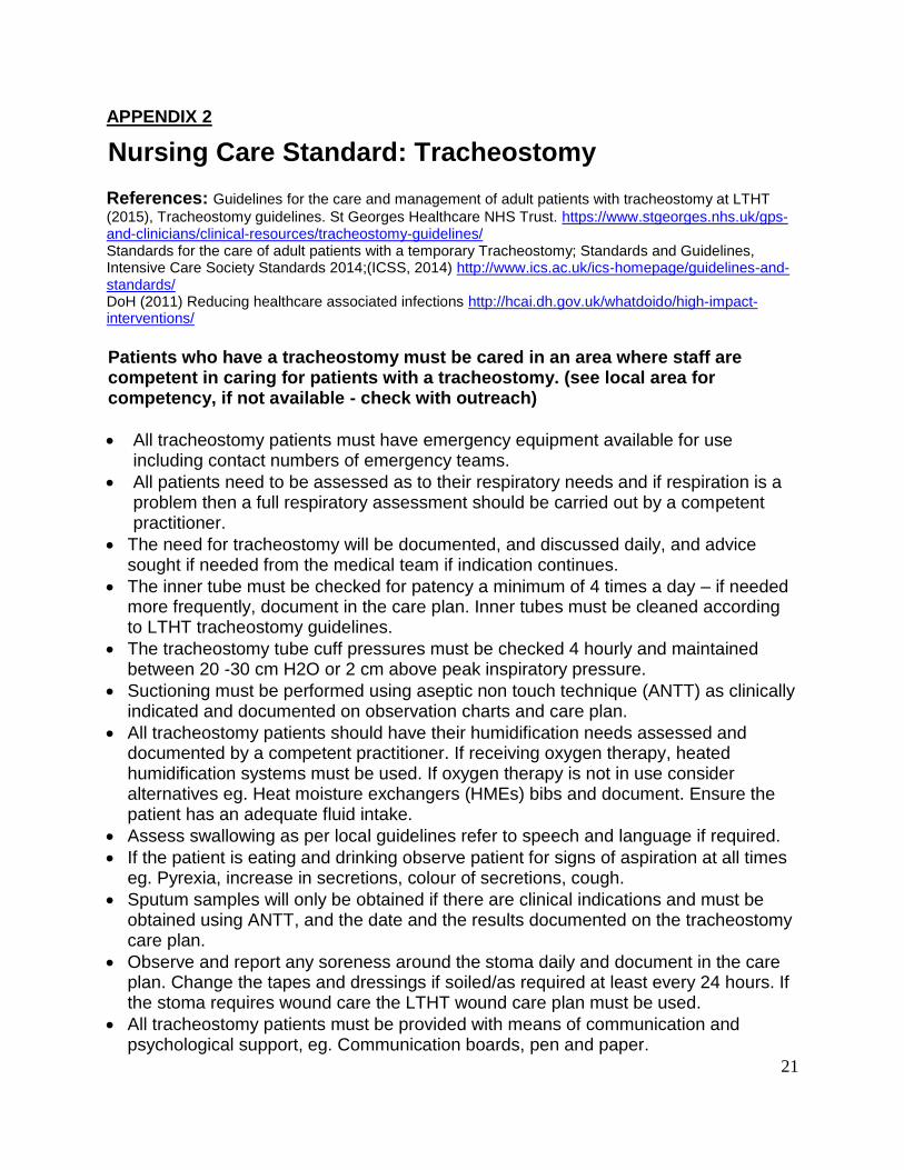
APPENDIX 2
Nursing Care Standard: Tracheostomy References: Guidelines for the care and management of adult patients with tracheostomy at LTHT
(2015), Tracheostomy guidelines. St Georges Healthcare NHS Trust. https://www.stgeorges.nhs.uk/gps-and-clinicians/clinical-resources/tracheostomy-guidelines/ Standards for the care of adult patients with a temporary Tracheostomy; Standards and Guidelines, Intensive Care Society Standards 2014;(ICSS, 2014) http://www.ics.ac.uk/ics-homepage/guidelines-and-standards/ DoH (2011) Reducing healthcare associated infections http://hcai.dh.gov.uk/whatdoido/high-impact-interventions/
Patients who have a tracheostomy must be cared in an area where staff are competent in caring for patients with a tracheostomy. (see local area for competency, if not available - check with outreach)
All tracheostomy patients must have emergency equipment available for use including contact numbers of emergency teams.
All patients need to be assessed as to their respiratory needs and if respiration is a problem then a full respiratory assessment should be carried out by a competent practitioner.
The need for tracheostomy will be documented, and discussed daily, and advice sought if needed from the medical team if indication continues.
The inner tube must be checked for patency a minimum of 4 times a day – if needed more frequently, document in the care plan. Inner tubes must be cleaned according to LTHT tracheostomy guidelines.
The tracheostomy tube cuff pressures must be checked 4 hourly and maintained between 20 -30 cm H2O or 2 cm above peak inspiratory pressure.
Suctioning must be performed using aseptic non touch technique (ANTT) as clinically indicated and documented on observation charts and care plan.
All tracheostomy patients should have their humidification needs assessed and documented by a competent practitioner. If receiving oxygen therapy, heated humidification systems must be used. If oxygen therapy is not in use consider alternatives eg. Heat moisture exchangers (HMEs) bibs and document. Ensure the patient has an adequate fluid intake.
Assess swallowing as per local guidelines refer to speech and language if required.
If the patient is eating and drinking observe patient for signs of aspiration at all times eg. Pyrexia, increase in secretions, colour of secretions, cough.
Sputum samples will only be obtained if there are clinical indications and must be obtained using ANTT, and the date and the results documented on the tracheostomy care plan.
Observe and report any soreness around the stoma daily and document in the care plan. Change the tapes and dressings if soiled/as required at least every 24 hours. If the stoma requires wound care the LTHT wound care plan must be used.
All tracheostomy patients must be provided with means of communication and psychological support, eg. Communication boards, pen and paper.

22
Tracheostomy tubes should not be changed as routine within the first week of stoma formation. Tracheostomy tubes must be changed as per manufacturer instructions. They must be changed by a competent practitioner.
For emergency care see LTHT tracheostomy guidelines and algorithms on www.tracheostomy.org.uk

23
APPENDIX 3
Anatomy of a tracheostomy tube
Fig 1: A Twist 301 cuffed double lumen tube © Tracoe.
A) Outer cannula or tube: This is the main part of the tracheostomy tube, which sits in the trachea. It remains in situ for the lifespan of the tracheostomy tube. The outer diameter (OD) measurement refers to the diameter of the outside of this cannula in mm, usually at the widest point but not including the cuff. The inner diameter (ID) refers to the internal diameter of this cannula in mm; usually the size of a tracheostomy tube refers to the ID but not always. Outer cannulae may end proximally with a standard 15mm anaesthetic connector, a flush fitting or with a twist lock (as in Fig 1) to enable placement of an inner cannula. B) Cuff: This is a balloon at the distal end of the outer cannula. It can be inflated to create an airtight seal in the trachea to allow ventilation. Modern cuffs are low pressure and high volume with a large contact area to reduce pressure on the wall of the trachea; this prevents tracheal dilatation, mucosal necrosis and tracheal stenosis. Ideally cuffs should only be inflated to a pressure of between 15 & 25cm H2O using a cuff pressure manometer [2, 3].
Fig 2: A cuff pressure manometer © Teleflex Medical.
D
E
G
1.0. G
A
B
C
F
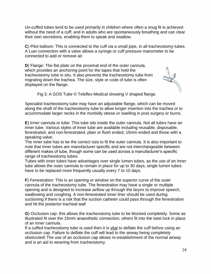
24
Un-cuffed tubes tend to be used primarily in children where often a snug fit is achieved without the need of a cuff; and in adults who are spontaneously breathing and can clear their own secretions, enabling them to speak and swallow. C) Pilot balloon: This is connected to the cuff via a small pipe, in all tracheostomy tubes. A Luer connection with a valve allows a syringe or cuff pressure manometer to be connected to add or remove air. D) Flange: The flat plate on the proximal end of the outer cannula, which provides an anchoring point for the tapes that hold the tracheostomy tube in situ. It also prevents the tracheostomy tube from migrating down the trachea. The size, style or code of tube is often displayed on the flange.
Fig 3: A GOS Tube © Teleflex Medical showing V shaped flange. Specialist tracheostomy tube may have an adjustable flange, which can be moved along the shaft of the tracheostomy tube to allow longer insertion into the trachea or to accommodate larger necks in the morbidly obese or swelling in post surgery or burns. E) Inner cannula or tube: This tube sits inside the outer cannula. Not all tubes have an inner tube. Various styles of inner tube are available including reusable, disposable, fenestrated, and non-fenestrated, plain or flush ended, 15mm ended and those with a speaking valve. The inner tube has to be the correct size to fit the outer cannula. It is also important to note that inner tubes are manufacturer specific and are not interchangeable between different makes of tube, though some can be used across a manufacturer’s specific range of tracheostomy tubes. Tubes with inner tubes have advantages over single lumen tubes, as the use of an inner tube allows the outer cannula to remain in place for up to 30 days, single lumen tubes have to be replaced more frequently usually every 7 to 10 days. F) Fenestration: This is an opening or window on the superior curve of the outer cannula of the tracheostomy tube. The fenestration may have a single or multiple opening and is designed to increase airflow up through the larynx to improve speech, swallowing and coughing. A non-fenestrated inner liner should be used during suctioning if there is a risk that the suction catheter could pass through the fenestration and hit the posterior tracheal wall G) Occlusion cap: this allows the tracheostomy tube to be blocked completely. Some as illustrated fit over the 15mm anaesthetic connection, others fit into the twist lock in place of an inner cannula. If a cuffed tracheostomy tube is used then it is vital to deflate the cuff before using an occlusion cap. Failure to deflate the cuff will lead to the airway being completely obstructed! The use of an occlusion cap allows re-establishment of the normal airway and is an aid to weaning from tracheostomy.
25
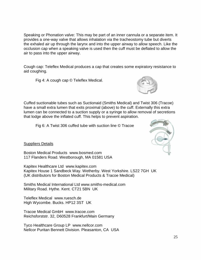
Speaking or Phonation valve: This may be part of an inner cannula or a separate item. It provides a one-way valve that allows inhalation via the tracheostomy tube but diverts the exhaled air up through the larynx and into the upper airway to allow speech. Like the occlusion cap when a speaking valve is used then the cuff must be deflated to allow the air to pass into the upper airway. Cough cap: Teleflex Medical produces a cap that creates some expiratory resistance to aid coughing.
Fig 4: A cough cap © Teleflex Medical.
Cuffed suctionable tubes such as Suctionaid (Smiths Medical) and Twist 306 (Tracoe) have a small extra lumen that exits proximal (above) to the cuff. Externally this extra lumen can be connected to a suction supply or a syringe to allow removal of secretions that lodge above the inflated cuff. This helps to prevent aspiration.
Fig 6: A Twist 306 cuffed tube with suction line © Tracoe
Suppliers Details Boston Medical Products www.bosmed.com 117 Flanders Road. Westborough, MA 01581 USA Kapitex Healthcare Ltd www.kapitex.com Kapitex House 1 Sandbeck Way. Wetherby. West Yorkshire. LS22 7GH UK (UK distributors for Boston Medical Products & Tracoe Medical) Smiths Medical International Ltd www.smiths-medical.com Military Road. Hythe. Kent. CT21 5BN UK Teleflex Medical www.ruesch.de High Wycombe. Bucks. HP12 3ST UK Tracoe Medical GmbH www.tracoe.com Reichsforststr. 32, D60528 Frankfurt/Main Germany Tyco Healthcare Group LP www.nellcor.com Nellcor Puritan Bennett Division. Pleasanton, CA USA
26
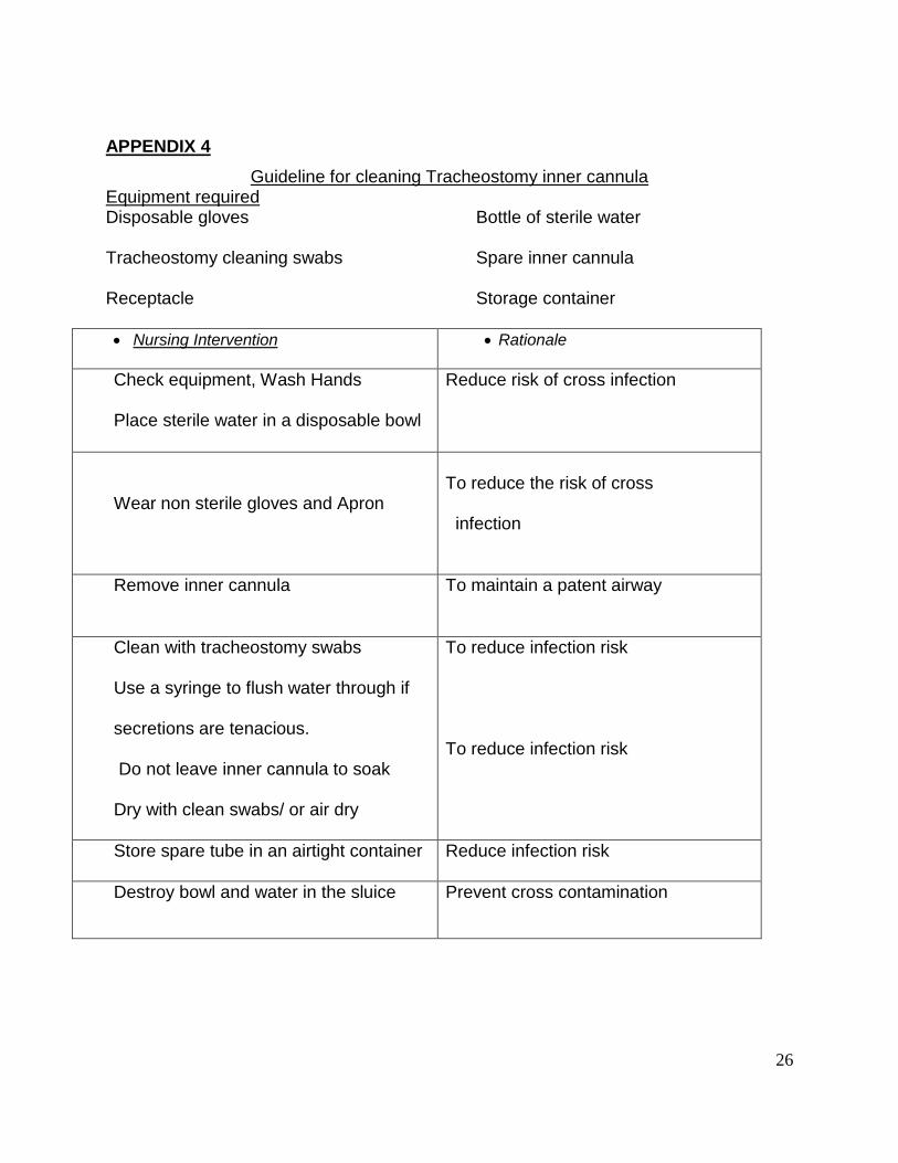
APPENDIX 4
Guideline for cleaning Tracheostomy inner cannula Equipment required Disposable gloves Bottle of sterile water
Tracheostomy cleaning swabs Spare inner cannula
Receptacle Storage container
Nursing Intervention Rationale
Check equipment, Wash Hands
Place sterile water in a disposable bowl
Reduce risk of cross infection
Wear non sterile gloves and Apron
To reduce the risk of cross
infection
Remove inner cannula To maintain a patent airway
Clean with tracheostomy swabs
Use a syringe to flush water through if
secretions are tenacious.
Do not leave inner cannula to soak
Dry with clean swabs/ or air dry
To reduce infection risk
To reduce infection risk
Store spare tube in an airtight container Reduce infection risk
Destroy bowl and water in the sluice Prevent cross contamination
27
APPENDIX 5
Competency Assessment Tool for
Tracheostomy Decannulation and Changes Introduction This competency package includes:-
Tracheostomy competencies - Theory
Tracheostomy competencies - Practical
The ICS (Intensive Care Society) Standards for care of adult patients with temporary tracheostomies (2007)
Anatomy and Physiology teaching pack
Anaesthetic Equipment - visual aid pack
Airway management and Ventilatory support - Skills Identified
Algorithm for tracheostomy weaning It has been designed for use for practitioners who have the appropriate knowledge and skills to undertake this practice It is up to the individual to maintain their own competence. If for any reason the practitioner feels they are no longer competent (e.g. post maternity leave or a period of longer than 6 months without practice) it is essential that they are re-assessed. As part of the assessment process the criteria laid down in the tracheostomy competencies section must be achieved and signed off by the appropriate assessor before unsupervised practice takes place. It is recommended that there are at least two operators to perform the procedure one of whom MUST be a competent practitioner. Precautionary Note Ensure that resuscitation and suction equipment is functional and available prior to the start of the procedure Discussion must have taken place with an appropriate anaesthetist prior to the procedure and must be informed when the procedure is going to take place
28
Adapted from St Georges Healthcare NHS trust - Guidelines for the care of Patients with Tracheostomy Tubes
Tracheostomy Competencies - Theory
Criteria Objectives Date
Achieved Assessed by
Anatomy/ Physiology
To accurately discuss upper airway anatomy
To be able to identify appropriate surface anatomy
To show an understanding of the physiological effects of having a tracheostomy in situ
(see late complications following decannulation))
Tracheostomy change
Can accurately state indications for tracheostomy changes
Be able to accurately produce equipment required for tracheostomy change
Can accurately identify risk factors
Be aware of medico-legal guidance for change
Tracheostomy change complications
Can state what procedures should be activated if the following occur: -Tube inserted incorrectly - Tube cannot be reinserted - Excessive bleeding - Airway collapse - Excessive coughing
- Laryngeal spasm -Tracheal stenosis -Tracheoesophageal fistula -Tracheocutaneous fistula -Tracheo-innominate artery fistula -Cellulitis -Stoma Site infection
Decannulation
Can accurately state indications for decannulation
Can accurately gather equipment for decannulation
Can identify when decannulation is not appropriate
29
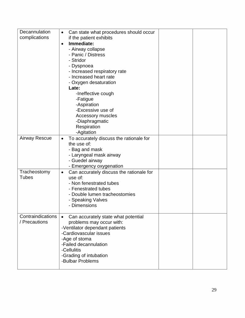
Decannulation complications
Can state what procedures should occur if the patient exhibits
Immediate: - Airway collapse - Panic / Distress - Stridor - Dyspnoea - Increased respiratory rate - Increased heart rate - Oxygen desaturation Late:
-Ineffective cough -Fatigue -Aspiration
-Excessive use of Accessory muscles -Diaphragmatic Respiration -Agitation
Airway Rescue
To accurately discuss the rationale for the use of: - Bag and mask - Laryngeal mask airway - Guedel airway - Emergency oxygenation
Tracheostomy Tubes
Can accurately discuss the rationale for use of: - Non fenestrated tubes - Fenestrated tubes - Double lumen tracheostomies - Speaking Valves - Dimensions
Contraindications/ Precautions
Can accurately state what potential problems may occur with:
-Ventilator dependant patients -Cardiovascular issues -Age of stoma -Failed decannulation -Cellulitis -Grading of intubation -Bulbar Problems

30
Tracheostomy Competencies - Practical
Decannulation Assessment Date
Observed by Competent (Y/N)
Date
1.Mandatory
2. Mandatory
3. Mandatory
Tracheostomy Change Assessment Date
Observed by Competent (Y/N)
Date
1.Mandatory
2. Mandatory
3. Mandatory
Airway Rescue Observed by Competent (Y/N)
Date
a. Bag and Mask b. Laryngeal mask airway c. Guedel Airway d. Emergency oxygenation
31
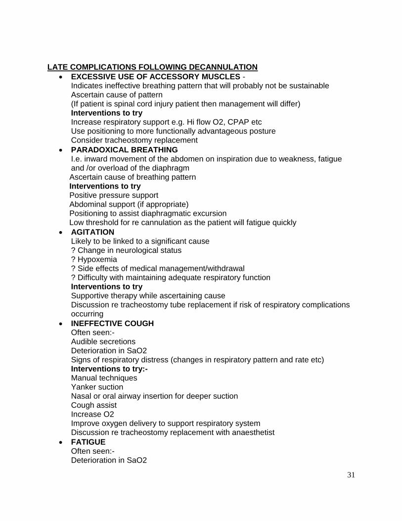
LATE COMPLICATIONS FOLLOWING DECANNULATION
EXCESSIVE USE OF ACCESSORY MUSCLES - Indicates ineffective breathing pattern that will probably not be sustainable Ascertain cause of pattern (If patient is spinal cord injury patient then management will differ) Interventions to try Increase respiratory support e.g. Hi flow O2, CPAP etc Use positioning to more functionally advantageous posture Consider tracheostomy replacement
PARADOXICAL BREATHING I.e. inward movement of the abdomen on inspiration due to weakness, fatigue and /or overload of the diaphragm
Ascertain cause of breathing pattern Interventions to try Positive pressure support Abdominal support (if appropriate) Positioning to assist diaphragmatic excursion Low threshold for re cannulation as the patient will fatigue quickly
AGITATION Likely to be linked to a significant cause ? Change in neurological status ? Hypoxemia ? Side effects of medical management/withdrawal ? Difficulty with maintaining adequate respiratory function Interventions to try Supportive therapy while ascertaining cause Discussion re tracheostomy tube replacement if risk of respiratory complications occurring
INEFFECTIVE COUGH Often seen:-
Audible secretions Deterioration in SaO2 Signs of respiratory distress (changes in respiratory pattern and rate etc) Interventions to try:- Manual techniques Yanker suction Nasal or oral airway insertion for deeper suction Cough assist Increase O2 Improve oxygen delivery to support respiratory system Discussion re tracheostomy replacement with anaesthetist
FATIGUE Often seen:- Deterioration in SaO2
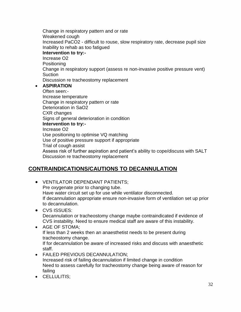
32
Change in respiratory pattern and or rate Weakened cough Increased PaCO2 - difficult to rouse, slow respiratory rate, decrease pupil size Inability to rehab as too fatigued Intervention to try:- Increase O2 Positioning Change in respiratory support (assess re non-invasive positive pressure vent) Suction Discussion re tracheostomy replacement
ASPIRATION Often seen:- Increase temperature Change in respiratory pattern or rate Deterioration in SaO2 CXR changes Signs of general deterioration in condition Intervention to try:- Increase O2 Use positioning to optimise VQ matching Use of positive pressure support if appropriate Trial of cough assist Assess risk of further aspiration and patient’s ability to cope/discuss with SALT Discussion re tracheostomy replacement
CONTRAINDICATIONS/CAUTIONS TO DECANNULATION
VENTILATOR DEPENDANT PATIENTS; Pre oxygenate prior to changing tube. Have water circuit set up for use while ventilator disconnected. If decannulation appropriate ensure non-invasive form of ventilation set up prior to decannulation.
CVS ISSUES: Decannulation or tracheostomy change maybe contraindicated if evidence of CVS instability. Need to ensure medical staff are aware of this instability.
AGE OF STOMA; If less than 2 weeks then an anaesthetist needs to be present during tracheostomy change. If for decannulation be aware of increased risks and discuss with anaesthetic staff.
FAILED PREVIOUS DECANNULATION; Increased risk of failing decannulation if limited change in condition Need to assess carefully for tracheostomy change being aware of reason for failing
CELLULITIS;
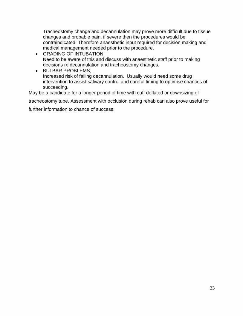
33
Tracheostomy change and decannulation may prove more difficult due to tissue changes and probable pain, if severe then the procedures would be contraindicated. Therefore anaesthetic input required for decision making and medical management needed prior to the procedure.
GRADING OF INTUBATION; Need to be aware of this and discuss with anaesthetic staff prior to making decisions re decannulation and tracheostomy changes.
BULBAR PROBLEMS; Increased risk of failing decannulation. Usually would need some drug intervention to assist salivary control and careful timing to optimise chances of succeeding.
May be a candidate for a longer period of time with cuff deflated or downsizing of
tracheostomy tube. Assessment with occlusion during rehab can also prove useful for
further information to chance of success.
34
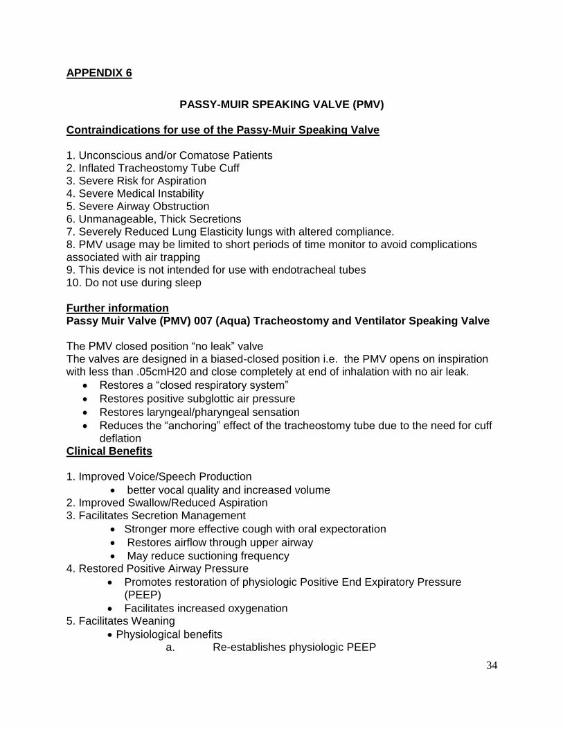
APPENDIX 6
PASSY-MUIR SPEAKING VALVE (PMV)
Contraindications for use of the Passy-Muir Speaking Valve 1. Unconscious and/or Comatose Patients 2. Inflated Tracheostomy Tube Cuff 3. Severe Risk for Aspiration 4. Severe Medical Instability 5. Severe Airway Obstruction 6. Unmanageable, Thick Secretions 7. Severely Reduced Lung Elasticity lungs with altered compliance. 8. PMV usage may be limited to short periods of time monitor to avoid complications associated with air trapping 9. This device is not intended for use with endotracheal tubes 10. Do not use during sleep Further information Passy Muir Valve (PMV) 007 (Aqua) Tracheostomy and Ventilator Speaking Valve The PMV closed position “no leak” valve The valves are designed in a biased-closed position i.e. the PMV opens on inspiration with less than .05cmH20 and close completely at end of inhalation with no air leak.
Restores a “closed respiratory system”
Restores positive subglottic air pressure
Restores laryngeal/pharyngeal sensation
Reduces the “anchoring” effect of the tracheostomy tube due to the need for cuff deflation
Clinical Benefits 1. Improved Voice/Speech Production
better vocal quality and increased volume 2. Improved Swallow/Reduced Aspiration 3. Facilitates Secretion Management
Stronger more effective cough with oral expectoration
Restores airflow through upper airway
May reduce suctioning frequency 4. Restored Positive Airway Pressure
Promotes restoration of physiologic Positive End Expiratory Pressure (PEEP)
Facilitates increased oxygenation 5. Facilitates Weaning
Physiological benefits a. Re-establishes physiologic PEEP
35
b. More effective cough c. Restores a more normal breathing pattern d. Improved assessment capabilities e. Improved secretion management
Psychological Benefits a. Patient confidence b. Communication and socialization c. Motivation and independence
6. Reduces Decannulation Time
Interim step in the decannulation process
Adjusting to a more normal breathing pattern
Improved ability to assess the airway
Builds patient confidence
Vocal cord stimulation due to airflow through the oronasopharynx 7. Improved Olfaction
Improved sense of smell by re-establishing airflow through the oral/nasal cavities which may stimulate appetite.
8. Quality of Life
Improved communication
More normal voice/speech production
Facilitates more active interaction. Assessment
Discuss with MDT
The PMV may be placed 48 to 72 hours after the tracheostomy is performed if tracheal oedema and/or secretions from the procedure have decreased. If the tracheostomy tube has been changed, PMV placement may need to be delayed 48-72 hours.
Patient Assessment The patient should be assessed before, during and after PMV placement 1. Cognitive status
Awake, responsive, attempting to communicate 2. Medical/Pulmonary Status
The patient should be medically stable. 3. Positioning
position comfortably at a 45°angle or sitting position to facilitate proper diaphragmatic movement and maintenance of a patent airway. 4. Cuff Deflation
Cuff deflation is mandatory when utilizing the PMV in order to allow expired air to pass around the tracheostomy tube and through the oronasopharynx.
Assess for secretions, tracheal and oral suction if required Slow deflation of the cuff to facilitates adjustment to the feeling of airflow in the
upper airway and secretions being dislodged in trachea. 5. Airway patency
36
Patient should be able to exhale efficiently and completely around the tracheostomy tube and through the upper airway
Check history for diagnosis of airway obstruction (i.e. severe stenosis, traumatic intubation, etc.)
Tracheostomy tube cuff (even when deflated) may create bulk in the airway causing obstruction to exhaled airflow.
Assessment for airway patency 1. Check tracheostomy tube cuff deflated 2. Instruct the patient to inhale through open tracheostomy tube 3. Manually finger occlude during exhalation 4. Encourage oral exhalation and voice Vocalisation may be heard.
6. During placement of PMV
Observe the PMV to ensure that the diaphragm of the PMV opens during inspiration and remains closed during expiration.
Monitor to ensure adequate airflow around the tracheostomy tube a. Monitor baseline measurements. b. If signs of respiratory distress, remove the PMV immediately and reassess 7. Use of Warning Labels
Attach warning labels provided with PMV to the pilot line of tracheostomy tube 8. Secretion Management. In-line Suction Systems The PMV should be connected to the side port to prevent the suction catheter from obstructing or damaging the PMV. 9. Swallowing Status
PMV can improve efficiency of swallow and may reduce aspiration, asses the presence and/or risk of aspiration to determine appropriate use of the PMV for swallow. 10. Lung Compliance
a. Altered compliance due to loss of lung elasticity requires careful assessment to prevent air trapping
b. Prolonged exhalation potentially causing complications associated with air trapping
c. PMV use may be limited to short periods of time 11. Oxygen:
a. Oxygen administration with PMV via tracheostomy mask, T-piece along with appropriate humidification.
b. Heat moisture exchange filter (HME) performance may be reduced with the PMV as no expiration through the filter to enable moisture collection. If an HME is used, it is important that it be placed between the PMV and the patient so that some moisture may be gained. 12. Facilitating Voice/Speech
If PMV tolerated encourage vocalization. Speech may be immediately possible. 13. Nebulisers Remove PMV prior to nebulizer treatments.
37
If the PMV is inadvertently used during nebulizer treatment it should be removed immediately and cleaned to remove medication residue as some medications may adversely affect the PMV diaphragm. Duration of use Dependant on toleration and can vary from few minutes to full day. Constant reassessment required to prevent respiratory distress/tiring due to fluctuating respiratory status (e.g. presence of infection, thick secretions, etc) tolerance of the PMV can vary. Care and lifetime of the PMV The PMVs are packaged in single units and are single patient use. The PMV should be cleaned daily after wearing. 1. Cleaning Procedure • Swish PMV in soapy, warm water (not hot water.) Rinse thoroughly with warm water. Allow PMV to air dry thoroughly before placing in storage container. Do not apply heat to dry PMV. • DO NOT use hot water 2. Lifetime of the PMV Each PMV is guaranteed to last for a minimum of two months. Lifetime cannot be guaranteed if cleaned or used improperly. The PMV can continue to be used as long as it does not exhibit stickiness, noise, vibration, increased resistance on inspiration or any other difficulties.
38
PMV NOT INDICATED
ASSESSMENT FOR PASSY-MUIR VALVE (PMV) *****ONLY FOR USE ON PATIENTS WITH A TRACHEOSTOMY*****
BY A COMPETENT PRACTITIONER
Is your patient
Awake & co-operative?
Able to tolerate cuff deflation
Cardiovascularly stable
Does your patient have:
Upper airway obstruction
Unmanageable thick secretions
An endotracheal tube
High aspiration risk
Is your patient on:
High flow/CPAP
FiO2 ≤ 50%
PEEP ≤ 7.5cmH2O
Are all members of MDT in agreement
with PMV placement?
Sit patient up to at least 45⁰
Perform oral and tracheal suction
DEFLATE THE TRACHEOSTOMY CUFF
MONITOR FOR:
Resp Rate & pattern
Heart Rate & BP
SaO2 level
Assess for airflow around
tracheostomy (see blue
box)
If patient stable & comfortable
IF AIRFLOW THROUGH MOUTH IS
ADEQUATE
Insert PMV into oxygen circuit
YES
NO
Points to remember: Do not use with severe airway obstruction/stenosis Cuff MUST be deflated Do not use with a nebuliser Remove valve for overnight sleeping Clean valve with sterile water
For those who are not needing respiratory support and a cuff is not needed, then it is safer and easier for the patient to have a non-cuffed tube or one with a fenestration, which will make it easier to vocalise with a PMV
APPLY WARNING LABEL TO PILOT
TUBE OF TRACHEOSTOMY
NO YES
NO
YES
ASSESSMENT FOR AIRFLOW
Ask patient to inhale
Occlude Tracheostomy with finger
Assess ability to vocalise
NO
YES
39
APPENDIX 7 For emergency algorithms and further information click on the following link and see below. http://www.tracheostomy.org.uk/Tracheostomy/New%20Blue%20Trachy%20Webpages/New%20Resources.htm
40
41
42
43
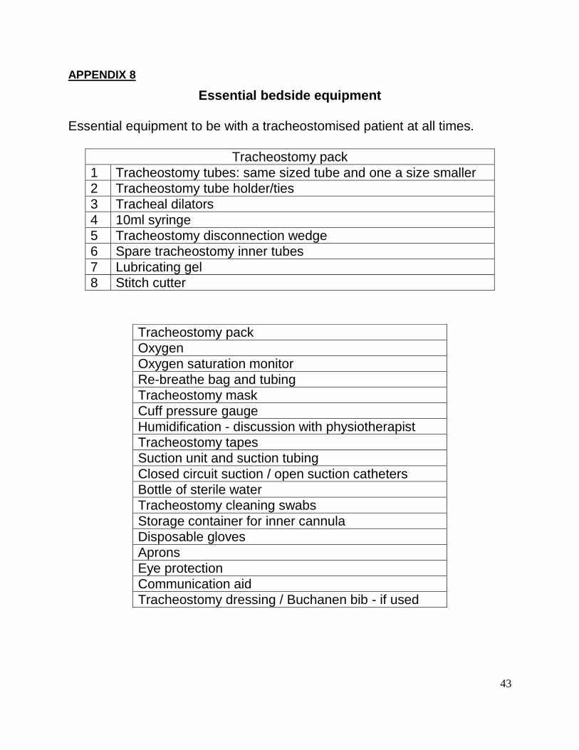
APPENDIX 8
Essential bedside equipment
Essential equipment to be with a tracheostomised patient at all times.
Tracheostomy pack
1 Tracheostomy tubes: same sized tube and one a size smaller
2 Tracheostomy tube holder/ties
3 Tracheal dilators
4 10ml syringe
5 Tracheostomy disconnection wedge
6 Spare tracheostomy inner tubes
7 Lubricating gel
8 Stitch cutter
Tracheostomy pack
Oxygen
Oxygen saturation monitor
Re-breathe bag and tubing
Tracheostomy mask
Cuff pressure gauge
Humidification - discussion with physiotherapist
Tracheostomy tapes
Suction unit and suction tubing
Closed circuit suction / open suction catheters
Bottle of sterile water
Tracheostomy cleaning swabs
Storage container for inner cannula
Disposable gloves
Aprons
Eye protection
Communication aid
Tracheostomy dressing / Buchanen bib - if used

44
APPENDIX 9 Audit tool
Tracheostomy guidelines audit (standard of achievement 100%)
Yes No N/A
Consent was obtained for tracheostomy procedure
The need for tracheostomy is documented in the medical
notes
Emergency equipment is available
Suctioning is recorded on the observation chart
Cuff pressures are recorded on the observation chart 4
hourly and maintained between 20-30cm H2O
Appropriate humidification is provided
Swallow assessment has been performed (if applicable)
Inner tube has been checked for patency 4 times daily
and recorded on the observation chart
Care of tracheostomy site is documented in the care plan
Communication aid is available