Embed Size (px)
Citation preview
IMAGES IN CARDIOTHORACIC SURGERY
Left Atrial Myxoma With Tumor VascularityArising From the Left and Right Coronary ArteriesMatthias Bauer, MD, and Roland Hetzer, MD, PhDDeutsches Herzzentrum Berlin, Berlin, Germany
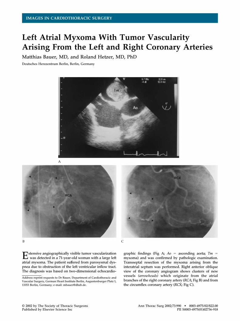
Extensive angiographically visible tumor vascularizationwas detected in a 71-year-old woman with a large left
atrial myxoma. The patient suffered from paroxysmal dys-pnea due to obstruction of the left ventricular inflow tract.The diagnosis was based on two-dimensional echocardio-
graphic findings (Fig A; Ao � ascending aorta; Tm �myxoma) and was confirmed by pathologic examination.Transseptal resection of the myxoma arising from theinteratrial septum was performed. Right anterior obliqueview of the coronary angiogram shows clusters of newvessels (arrowheads) which originate from the atrialbranches of the right coronary artery (RCA; Fig B) and fromthe circumflex coronary artery (RCX; Fig C).
Address reprint requests to Dr Bauer, Department of Cardiothoracic andVascular Surgery, German Heart Institute Berlin, Augustenburger Platz 1,13353 Berlin, Germany; e-mail: [email protected].
© 2002 by The Society of Thoracic Surgeons Ann Thorac Surg 2002;73:990 • 0003-4975/02/$22.00Published by Elsevier Science Inc PII S0003-4975(01)02736-918


![Mobile left atrial mass-clot or left atrial myxoma....mass includes thrombus, myxoma, lipoma and non-myxomatous neoplasm [7,8]. Among them, cardiac myxoma is the most common benign](https://img.pdfslide.net/doc/110x75/60fedab34ecd6d6c000feba7/mobile-left-atrial-mass-clot-or-left-atrial-mass-includes-thrombus-myxoma.jpg)