Embed Size (px)
Citation preview
Leo Semes, OD, FAAO
UAB Optometry
Safe; inert
Has long track record - over 45 years
Mixes with plasma and highlights blood
vessel compromise
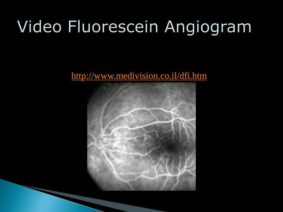
Using specific exciting (490 nm)and absorption (510 nm) filters…
B/W film documents vascular changes
Eliminated through liver and kidneys in 24-36 hours
Stains skin and mucous membranes (24-36)
May contaminate results of urine tests for reducing sugars (up to 4-5 days)
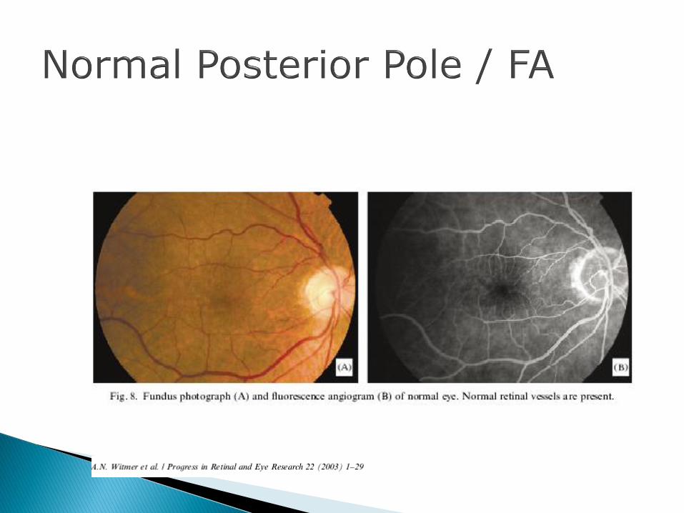
Does not diffuse through NORMAL retinal blood vessels
Normally leaky choroidal vessels show uniform “choroidal flush”

Mild◦ Nausea, sneezing
◦ Pruritis
◦ Vomiting
◦ Extravasation at injection site
Moderate
◦ Urticaria (< 2%)◦ Syncope (< 0.2%)◦ Other (< 0.006%) Skin eruptions
Thrombophelibitis
Pyrexia
Focal tissue necrosis
Nerve palsy
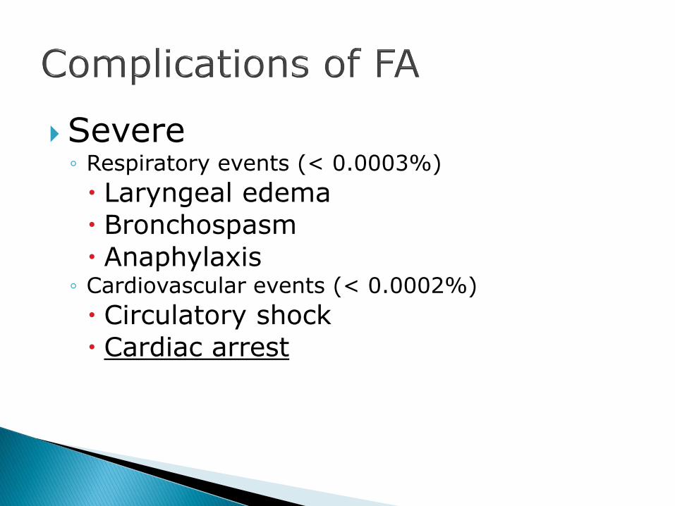
Severe◦ Respiratory events (< 0.0003%)
Laryngeal edema
Bronchospasm
Anaphylaxis ◦ Cardiovascular events (< 0.0002%)
Circulatory shock
Cardiac arrest
Previous anaphylactic reaction
Other previous reactions-weigh risk:benefit
Kidney disease (relative
contraindication)
Pregnancy ◦ Avoid,especially during the first trimester
Inner
◦ Vascular endothelium (integrity)
Outer
◦ Retinal Pigment Epithelium
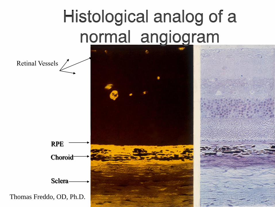
RPE
Choroid
Sclera
Retinal Vessels
Histological analog of a
normal angiogram
Thomas Freddo, OD, Ph.D.
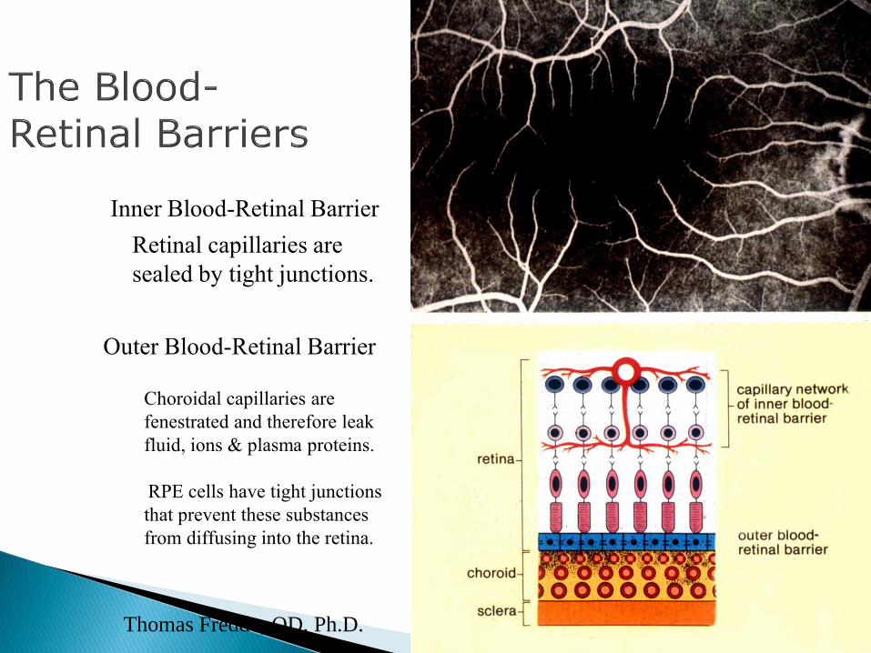
Inner Blood-Retinal Barrier
Retinal capillaries are
sealed by tight junctions.
Outer Blood-Retinal Barrier
Choroidal capillaries are
fenestrated and therefore leak
fluid, ions & plasma proteins.
RPE cells have tight junctions
that prevent these substances
from diffusing into the retina.
Thomas Freddo, OD, Ph.D.
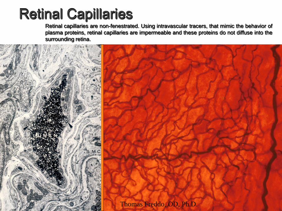
Retinal CapillariesRetinal capillaries are non-fenestrated. Using intravascular tracers, that mimic the behavior of
plasma proteins, retinal capillaries are impermeable and these proteins do not diffuse into the
surrounding retina.
Thomas Freddo, OD, Ph.D.
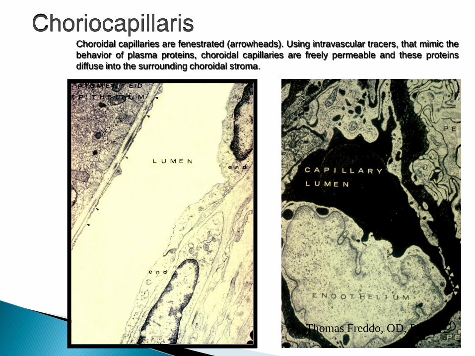
ChoriocapillarisChoroidal capillaries are fenestrated (arrowheads). Using intravascular tracers, that mimic the
behavior of plasma proteins, choroidal capillaries are freely permeable and these proteins
diffuse into the surrounding choroidal stroma.
Thomas Freddo, OD, Ph.D.
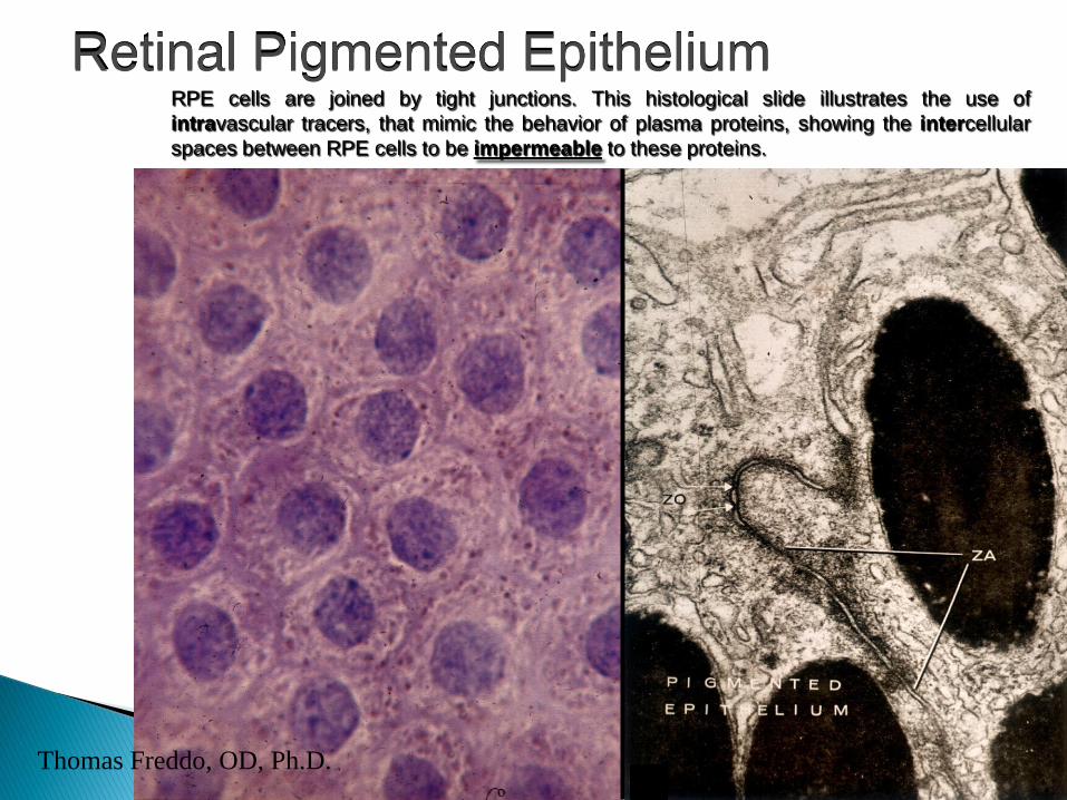
Retinal Pigmented EpitheliumRPE cells are joined by tight junctions. This histological slide illustrates the use of
intravascular tracers, that mimic the behavior of plasma proteins, showing the intercellular
spaces between RPE cells to be impermeable to these proteins.
Thomas Freddo, OD, Ph.D.
Tight junctions between retinal vascular endothelial cells prevent plasma proteins from diffusing into the retina.
Tight junctions between RPE cells prevent plasma proteins that have leaked from the fenestrated choroidal capillaries from diffusing into the retina.
Oral (shows only late leakage by angioscopy) May be applicable in children or where IV route in
impossible
Shows only late stage; not circulation phases (angioscopy)
Intravenous (IV) - shows transit of fluorescein as blood circulates and re-circulates)
FA is employed to support diagnoses and direct treatment
Examples of conditions that may be amenable to treatment (e.g., photocoagulation)
◦ Diabetic macular edema
◦ Age-related macular degeneration
◦ Other diseases that have neovascularization or macular edema as a consequence
Arm to retina 10-15 [7.5-25] sec (via???)
Arterial retinal 0.9-1.1
Venous retinal 1.7-2.2
Total retinal 2.6-3.3
*total transit time can be up to ~ 30 sec
Reduced in youth and anemia
Prolonged (slow/impaired blood flow)
◦ Carotid stenosis
◦ Sludging disorders

Color fundus photo
Red-free photo (reference)
Camera ready (SLO, also)
Patient prep
◦ Injection site
◦ Syringe w/5 ml (10% ); injected into the antecubital vein (3-7 sec as bolus)
Photographic sequence◦ 2-3 frames/10-12 sec
◦ Then every 2 – 3 sec for next 10 sec
◦ Late frames @ 5 to 20 minutes
Choroidal flush (prearterial) Early Phase (arterial)
Mid Phase (arteriovenous,venous,laminar)
Late Phase (recirculatory)
Choroidal flush (prearterial) The initial arm-to retina time is recorded as
the first appearance of fluorescein in the choroidal vasculature system (10-20 sec)
Uniform glow from permeable choroidal circulation
Cilioretinal artery fills
Early Phase (arterial)
The arteries alone in the retinal vascular tree are visibly filled with fluorescein against the choroidal flush
Note: a cilioretinal artery fills with the choroidalcirculation
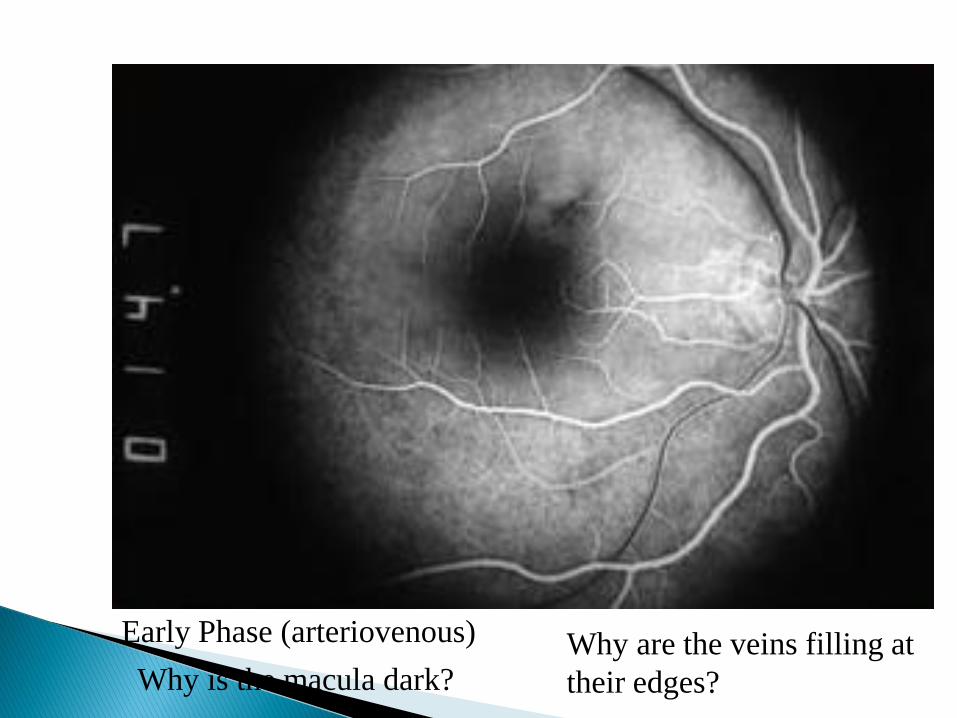
Early Phase (arteriovenous)
Why is the macula dark?Why are the veins filling at
their edges?
Early / Mid Phase (arteriovenous,venous; 3-5 min) Early filling of the veins,laminar flow, or border
filling of the veins (looks like railroad tracks), followed by…
Arteries and veins fully filled with fluorescein
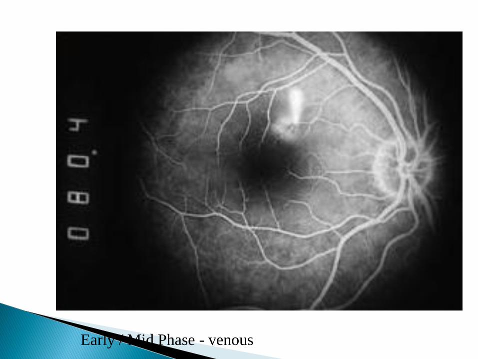
Early / Mid Phase - venous
The Late Phase This phase is documented 5-20 min following injection
In normals, no staining or leakage is seen
Abnormal results include late leakage not contained by the circulation, or staining
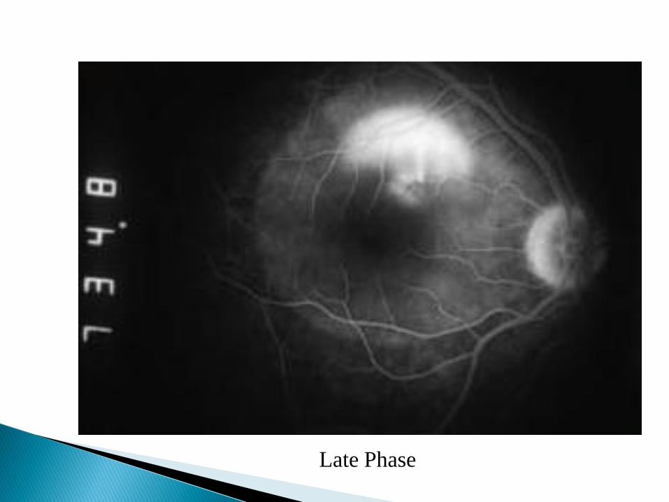
Late Phase
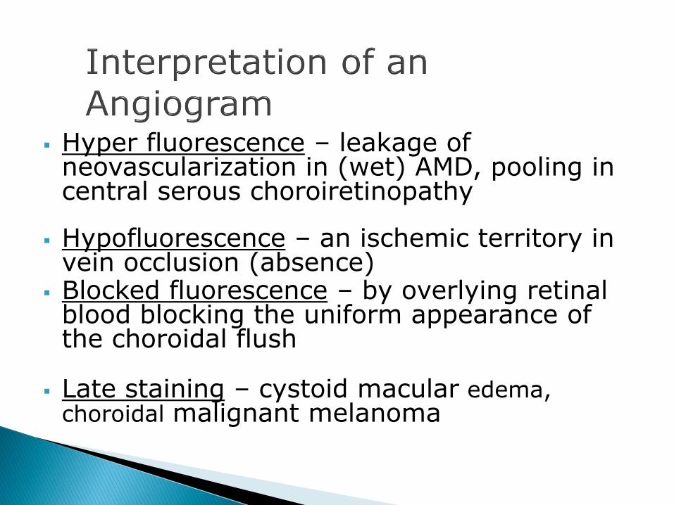
Hyper fluorescence – leakage of neovascularization in (wet) AMD, pooling in central serous choroiretinopathy
Hypofluorescence – an ischemic territory in vein occlusion (absence)
Blocked fluorescence – by overlying retinal blood blocking the uniform appearance of the choroidal flush
Late staining – cystoid macular edema, choroidal malignant melanoma
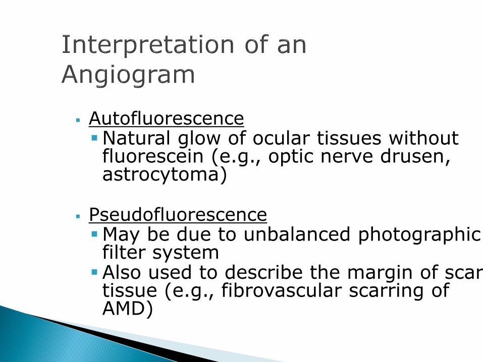
AutofluorescenceNatural glow of ocular tissues without fluorescein (e.g., optic nerve drusen, astrocytoma)
PseudofluorescenceMay be due to unbalanced photographic filter systemAlso used to describe the margin of scar tissue (e.g., fibrovascular scarring of AMD)
FIRST - compare the color and R/F photos
NEXT - identify the stage of the angiogram
Look for one of the 4 characteristic abnormalities (hyper-, hypo-, blocked or late-phase staining/leakage)
Determine if the abnormal fluorescence is changing or remaining the same throughout the angiogram
Correlate the anatomical (clinical) and angiographic sites; again refer to the color and R/F photos
!!! FA is to confirm diagnoses and guide treatment; not to make a diagnosis
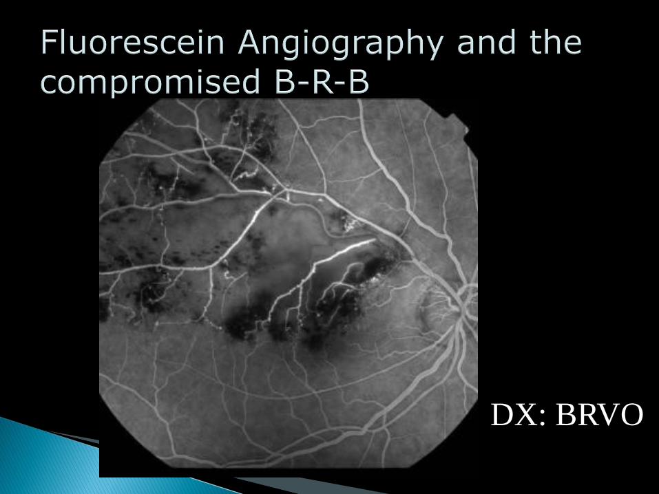
DX: BRVO

Abnormal examples
A: 1. CWS in papillomacular
bundle
2. Intraretinal hemorrhage
B: FA
1. capillary nonperfusion in
the area corresponding to
the cotton-wool patch; 2.
blocked fluorescence
caused by the intraretinal
hemorrhage
Yanoff and Duker in MD
consult
Link to
http://www.mdconsult.com/das/bo
ok/body/189639736-
13/969371360/1869/I4-u1.0-B978-
0-323-04332-8..00092-5--
f0030.fig?tocnode=56523153
Healthy 37 YO M
• 3-hour history of visual loss (VA 20/60).
A: Retinal whitening is very subtle and
the retinal vessels appear normal.
(B) FA reveals abnormal arterial filling.
(confirming CRAO)
http://www.mdconsult.c
om/das/book/body/189
639736-
17/969373663/1869/I4-
u1.0-B978-0-323-
04332-8..00089-5--
f0010.fig?tocnode=565
23082
FA is employed to support diagnoses and direct treatment
Examples of conditions that may be amenable to treatment (e.g., photocoagulation)◦ Diabetic macular edema
◦ Age-related macular degeneration
◦ Other diseases that have neovascularization or macular edema as a consequence
ICG (tricarbocyanine)
Near IR (780/810 nm) absorption/emission
Similar injection protocol / safety spectrum to fluorescein, ex: ◦ metabolized in liver◦ contains iodine
Advantages◦ Defines CNV better than FA◦ Visualization improved through media opacities,
pigment, blood, and scar tissue

Most useful for choroidal filling assessment
application in AMD
Example
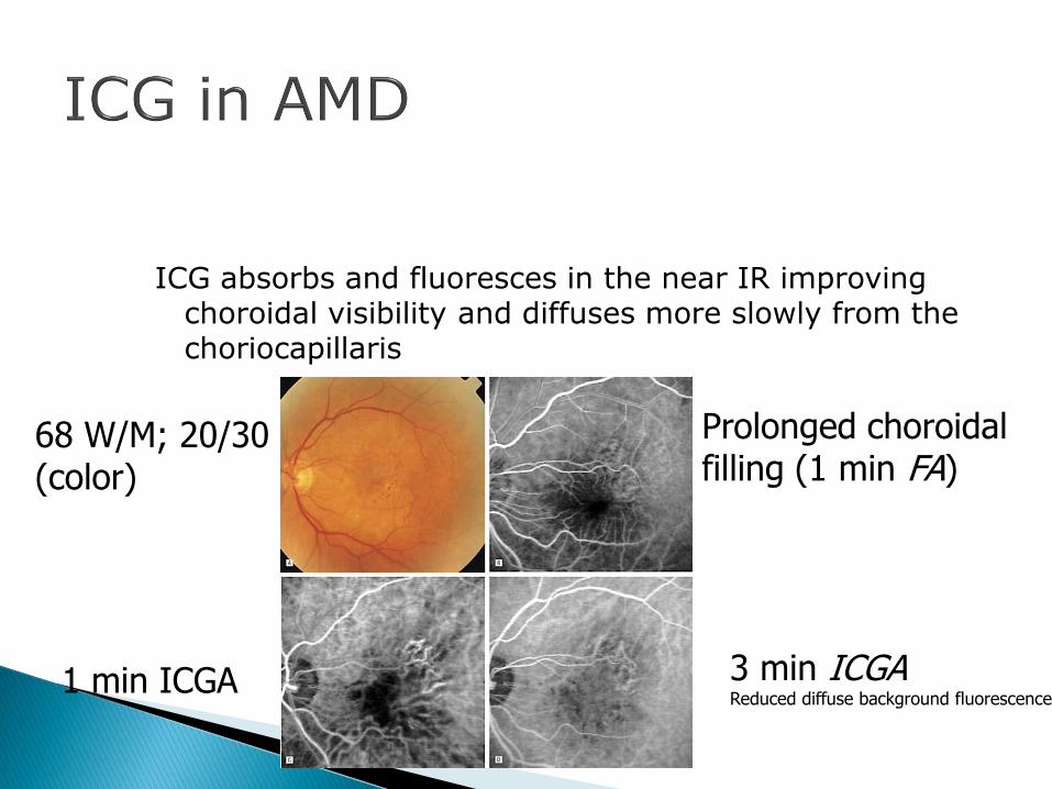
ICG absorbs and fluoresces in the near IR improving choroidal visibility and diffuses more slowly from the choriocapillaris
68 W/M; 20/30(color)
Prolonged choroidalfilling (1 min FA)
3 min ICGAReduced diffuse background fluorescence
1 min ICGA
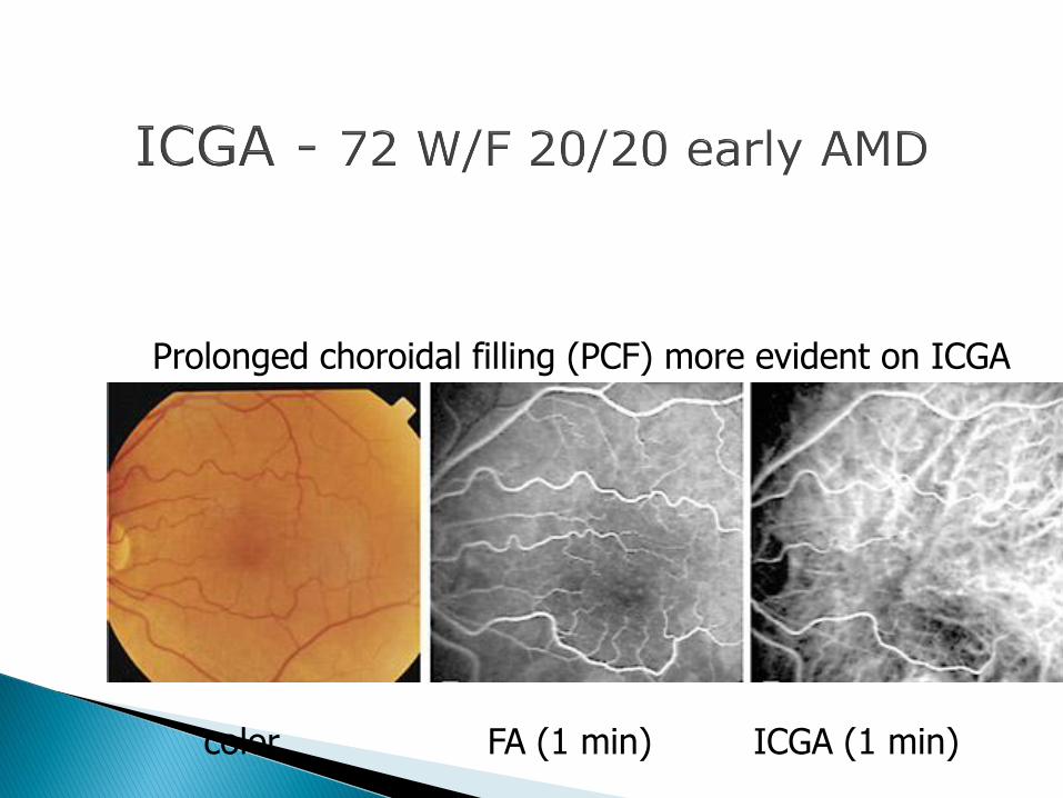
color FA (1 min) ICGA (1 min)
Prolonged choroidal filling (PCF) more evident on ICGA
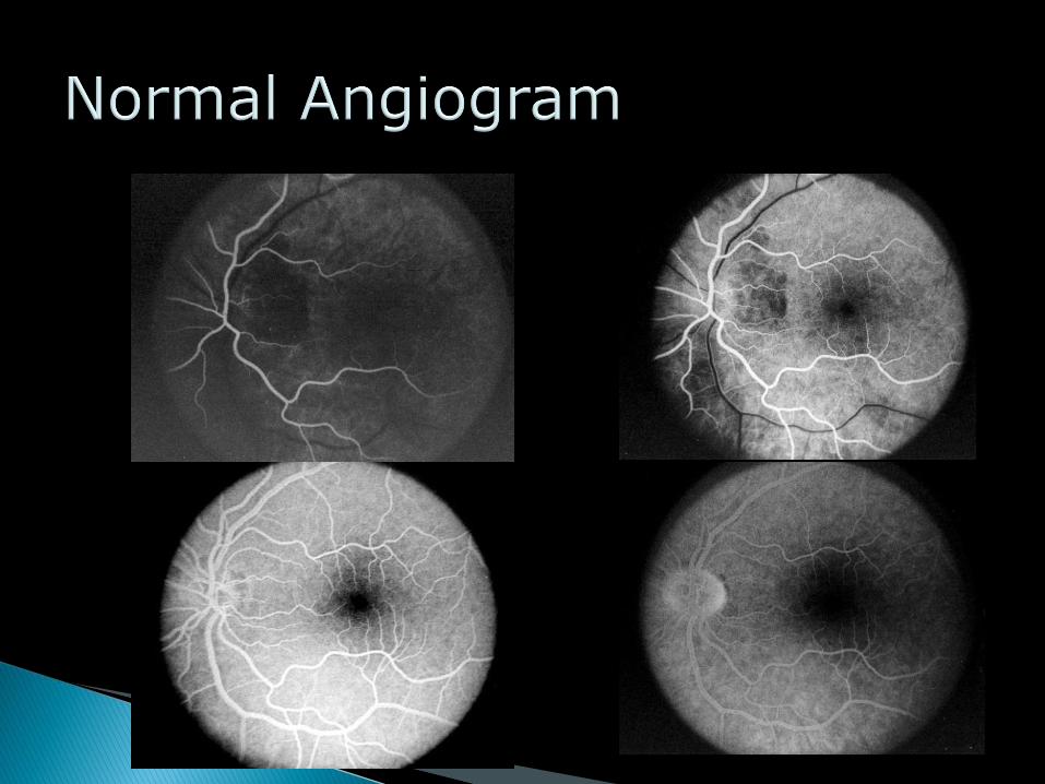
Normal Angiogram
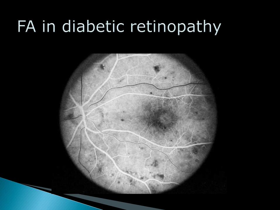
Diabetic Retinopathy (NPDR)
Proliferative Diabetic Retinopathy (PDR)
Age-related Macular Degeneration (AMD)
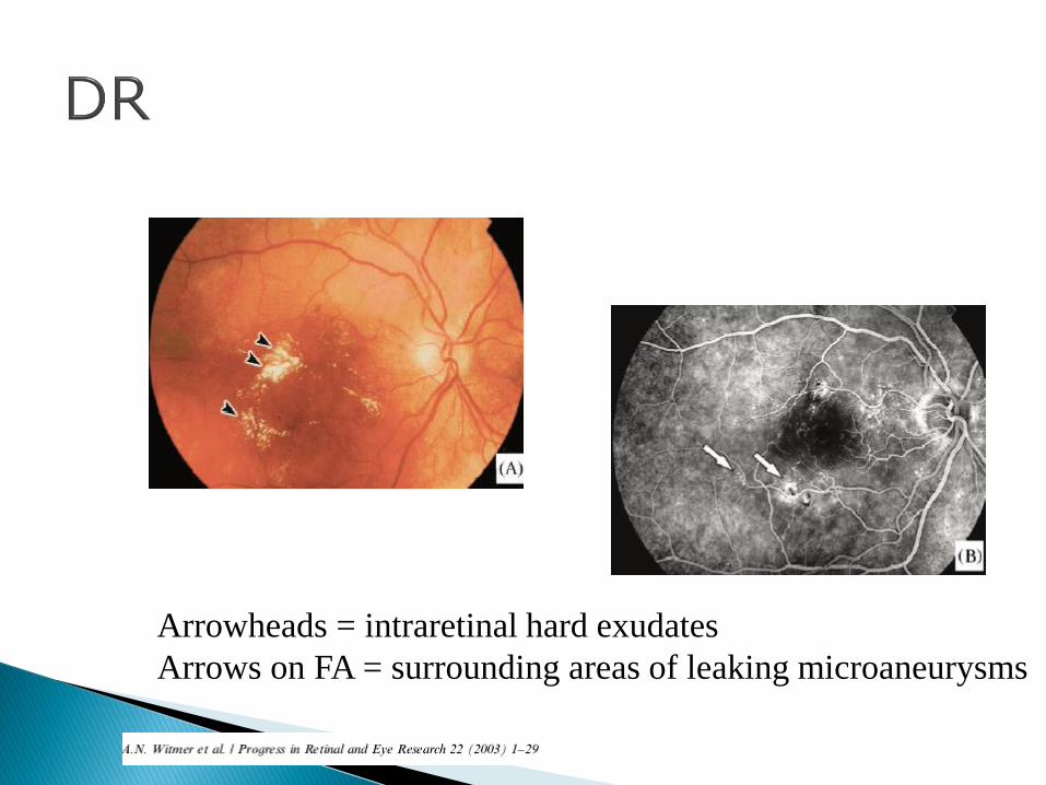
Arrowheads = intraretinal hard exudates
Arrows on FA = surrounding areas of leaking microaneurysms

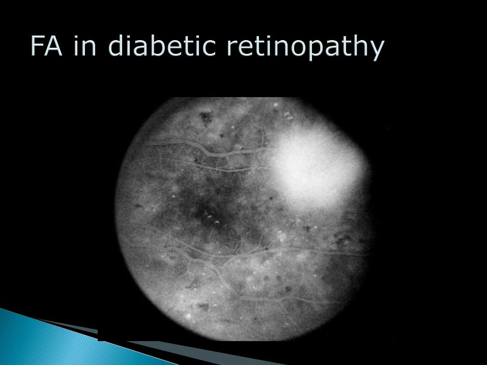
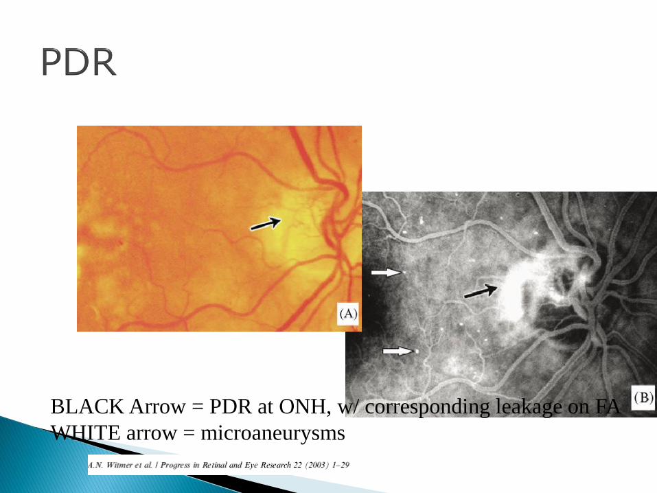
BLACK Arrow = PDR at ONH, w/ corresponding leakage on FA
WHITE arrow = microaneurysms
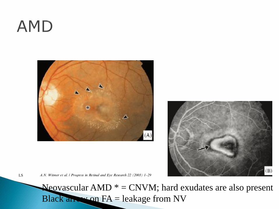
Neovascular AMD * = CNVM; hard exudates are also present
Black arrow on FA = leakage from NV
LS