Embed Size (px)
Citation preview

Objectives
• To be able to localize 3 symptoms, and their respective locations in the brain
• Name and understand the primary prognostic indicator for patients with brain metastasis and leptomeningeal disease
• Understand what differentiates the treatment goals for a patient with good prognosis vs a poor prognosis patient
• Name approaches to each form of treatment‐Surgery, Radiation, and chemotherapy

Introduction
• Brain metastasis and leptomeningeal disease are lethal
• Untreated brain metastasis from solid tumors has a prognosis of 1‐2 months
• Once diagnosed leptomeningeal disease has a prognosis of 2 weeks–2 months

The Lobes of the Brain

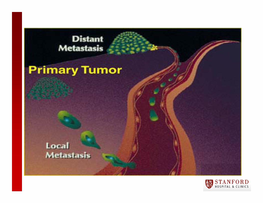
Brain Metastasis
• Brain metastasis is the most common intracranial tumor in adults
• In systemic malignancy brain metastasis occurs in 10‐30% of adults, and 6‐10% of children
• Incidences increasing – Improved imaging with MRI– Improve control of extracranial disease


Common Malignancies Responsible for Brain Metastasis
• Adults– Lung– Breast– Kidney– Colorectal– Melanoma
• Children– Sarcomas– Germ Cell tumors– Neuroblastoma

Clinical Manifestations• Headache
– 40‐50% of patients – Early morning headache
• Focal neurologic dysfunction– 20‐40% pf patients– Hemiparesis most common
• Cognitive dysfunction– 30‐35% of patients
• Seizures• Stroke• Others

Imaging
• Magnetic resonance imaging with contrast• More sensitive than non contrast MRI or CT scan

FRONT
LEFTRIGHT
BACK


Prognostic Indicators
• Performance status• Age younger than 65• Control of extracranial disease• Underlying cancer histology and genealogy

Karnofsky Performance Scale

Treatments
• Surgery• Radiation• Chemotherapy

Surgery
• There is a single metastatic lesion• Large symptom producing tumors• Or if there is a uncertain diagnosis, excisional biopsy is considered

Eloquent Areas of the Brain

Radiation Therapy
• Many who undergo surgery get local radiation therapy to the surgical bed
• For those with a limited number of small brain metastasis, they may have stereotactic radiosurgery alone
• Whole brain radiation therapy

Chemotherapy
• Chemotherapy is based on the primary site of cancer– Breast– Lung–Melanoma

Surveillance
• Imaging– 1 month after initial therapy, and then every 2‐3 months after
– Up to 50% progress within the first 6 months to one year
Recurrence
• Pseudo‐progression• Recurrence
– Additional surgery a possible option– Additional radiation therapy is unlikely– Chemotherapy
Leptomeningeal Disease
• Malignant cancer cells in the CSF• Rare and devastating complication of advanced cancer• Diagnosed in approximately 5% of patients with metastatic cancer
• Most common cancers to result in leptomeningeal disease–breast, lung, melanoma, GI cancers
• Primary brain tumors may also lead to the leptomeningeal disease‐ high‐grade astrocytomas, oligodendroglioma, medulloblastoma, , pineoblastoma
• The development may be influenced by treatment
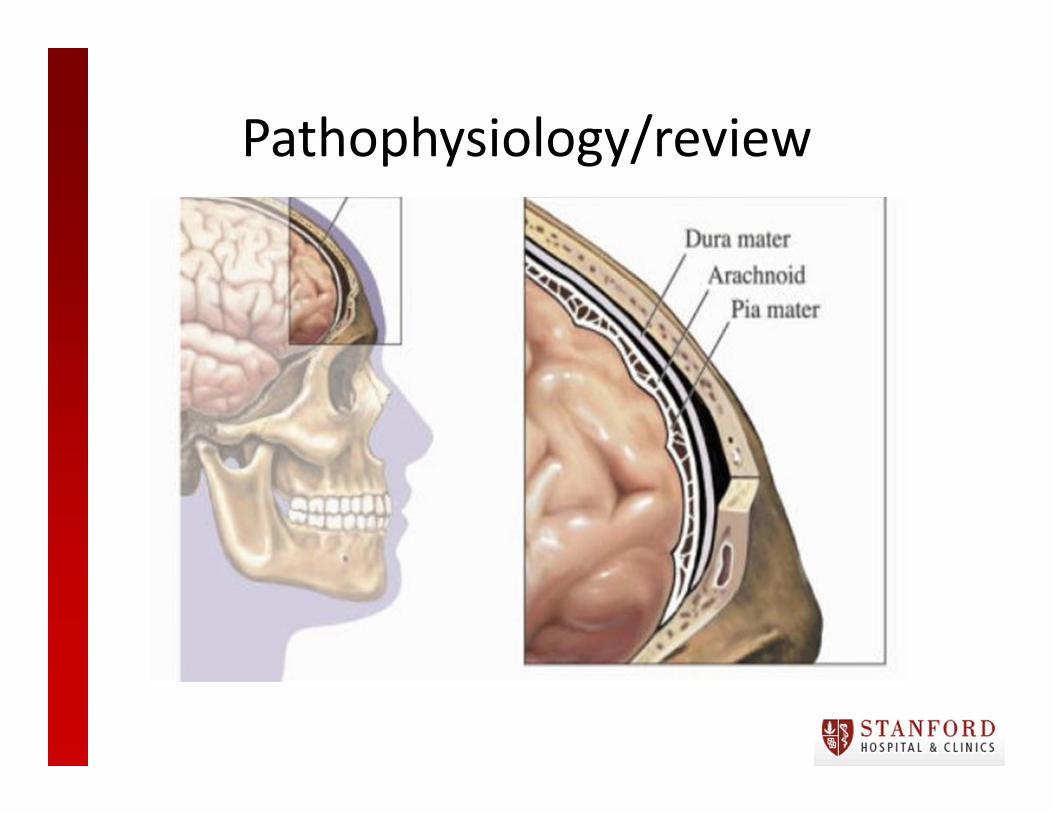
Pathophysiology/review

Cerebrospinal fluid
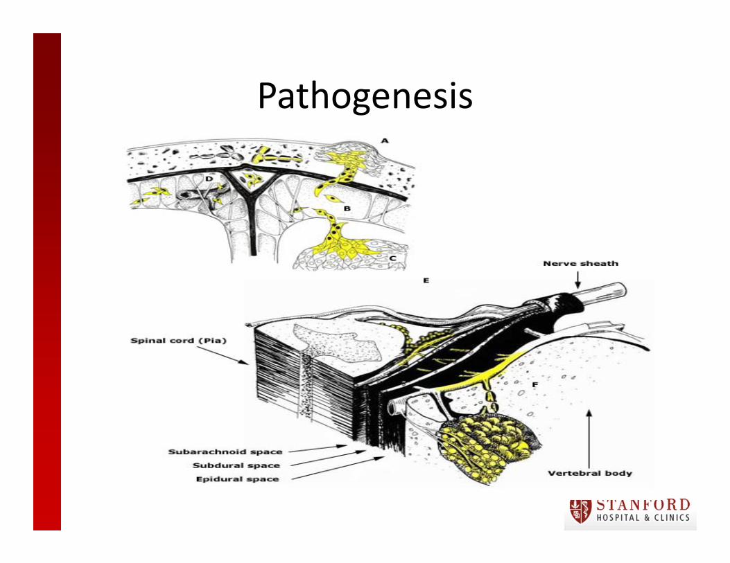
Pathogenesis
Clinical Manifestations
• Mass effect• Cranial nerves and spinal root dysfunction• Invasion of the brain parenchyma• Disruption of the blood brain barrier

Signs and symptoms
• Any neurological symptom may be related to LM
• Symptoms present acutely and progress within days to weeks
• Multifocal neurological signs and symptoms• Be aware of those that present with a single symptom

Diagnostics
• Brain MRI• CSF analysis through lumbar puncture

Leptomeningeal contrast enhancement

Leptomeningeal Contrast Enhancement

Treatment Goals
• Stabilizing or improving neurological function• Prolonging survival• Palliation of symptoms

Karnofsky Performance Scale

Poor Risk Patients
• Patients with multiple serious/fixed neurological deficits
• Extensive systemic disease even with active treatment
• Focus is largely on palliation of symptoms

Treatments
• Radiation therapy• Analgesics for pain• Corticosteroids• Anticonvulsants• VP shunting• SSRIs
Good Risk Patients
• Those without fixed neurological deficits• Minimal systemic disease• Cancer with reasonable treatment options• Goal is direct tumor control

Treatments
• Surgery• Radiation• Chemotherapy
Surgery
• Treatment of increased intracranial pressure– For signs of increased intracranial pressure initially treat with steroids
– VP shunting
Radiation Therapy
• Used to treat bulkier symptomatic areas of disease
• Appears to be more effective at relieving symptoms when compared to chemotherapy
• Standard radiation dose for leptomeningeal disease includes 30‐36 Gy, in 3 Gy daily fractions
• Major adverse effects during or after focal radiation therapy unusual
• With large extension radiation fields common adverse effects include myelosuppression, mucositis, esophagitis, leukoencephalopathy
Intrathecal chemotherapy
• Mainstay of treatment with leptomeningeal metastasis
• It may be delivered via lumbar puncture versus Ommaya reservoir
• Methotrexate is the chemotherapy most often used for the leptomeningeal disease

Systemic Chemotherapy
• There are several therapeutic chemotherapy agents provide therapeutic concentration within the CSF when given at appropriate doses
• Advantages– Surgery risks– Obstruction normal CSF flow– Increased availability of cytotoxic agents– Uniform drug distribution

Common Systemic Chemotherapy Agents
• High‐dose methotrexate with leucovorinrescue
• High‐dose cytarabine• Capecitabine• Tyrosine kinase inhibitors such as erlotinib• Anaplastic lymphoma kinase inhibitors such as Crizotinib

Investigational Therapies
• IT etoposide• Intrathecal trastuzumab• Intrathecal rituximab

Prognosis
• Despite aggressive therapy even good risk patients with leptomeningeal disease have limited survival
• Average survival with aggressive treatment is 3‐4 months
• Tumor histology and molecular subtype may influence prognosis
• Performance status and control of systemic disease are important factors

QUIZ
• Questions to ask yourself–What lobe of the brain is this lesion in?–Would you resect the tumor?–What part of the brain would receive radiation?– Name 2 symptoms the patient may experience with a metastatic lesion in this area.


ReferencesBindal RK, Sawaya R, Leavens ME, et al. Reoperation for recurrent metastatic brain tumors. J Neurosurg 1995; 83:600.
Burch PA, Grossman SA, Reinhard CS. Spinal cord penetration of intrathecally administered cytarabine and methotrexate: a quantitative autoradiographicstudy. J Natl Cancer Inst 1988; 80:1211.
Chamberlain MC. Combined‐modality treatment of leptomeningeal gliomatosis. Neurosurgery 2003; 52:324.Chamberlain MC, Tsao‐Wei D, Groshen S. Neoplastic meningitis‐related encephalopathy: prognostic significance. Neurology 2004; 63:2159.
Chang EL, Maor MH. Standard and novel radiotherapeutic approaches to neoplastic meningitis. Curr Oncol Rep 2003; 5:24
Delattre JY, Krol G, Thaler HT, Posner JB. Distribution of brain metastases. Arch Neurol 1988; 45:741.
Grossman SA, Reinhard CS, Loats HL. The intracerebral penetration of intraventricularly administered methotrexate: a quantitative autoradiographic study. J Neurooncol 1989; 7:319.
Grossman SA, Krabak MJ. Leptomeningeal carcinomatosis. Cancer Treat Rev 1999; 25:103.
Hitchins RN, Bell DR, Woods RL, Levi JA. A prospective randomized trial of single‐agent versus combination chemotherapy in meningeal carcinomatosis. J Clin Oncol 1987; 5:1655.
Kaplan JG, DeSouza TG, Farkash A, et al. Leptomeningeal metastases: comparison of clinical features and laboratory data of solid tumors, lymphomas and leukemias. J Neurooncol 1990; 9:225.
Lamovec J, Zidar A. Association of leptomeningeal carcinomatosis in carcinoma of the breast with infiltrating lobular carcinoma. An autopsy study. Arch Pathol Lab Med 1991; 115:507.

References Cont.Laufman LR, Forsthoefel KF. Use of intrathecal trastuzumab in a patient with carcinomatous meningitis. Clin Breast Cancer 2001; 2:235.
Mahajan A, Ahmed S, McAleer MF, et al. Post‐operative stereotactic radiosurgery versus observation for completely resected brain metastases: a single‐centre, randomised, controlled, phase 3 trial. Lancet Oncol 2017; 18:1040.
Mehta MP, Rodrigus P, Terhaard CH, et al. Survival and neurologic outcomes in a randomized trial of motexafin gadolinium and whole‐brain radiation therapy in brain metastases. J Clin Oncol 2003; 21:2529.
NCCN guidelines available at http://www.nccn.org.laneproxy.stanford.edu/professionals/physician_gls/f_guidelines.asp (Accessed on November 05, 2014).
Norris LK, Grossman SA, Olivi A. Neoplastic meningitis following surgical resection of isolated cerebellar metastasis: a potentially preventable complication. J Neurooncol 1997; 32:215
Patchell RA, Tibbs PA, Walsh JW, et al. A randomized trial of surgery in the treatment of single metastases to the brain. N Engl J Med 1990; 322:494.
Posner JB. Management of brain metastases. Rev Neurol (Paris) 1992; 148:477.
Saito R, Kumabe T, Jokura H, et al. Symptomatic spinal dissemination of malignant astrocytoma. J Neurooncol 2003; 61:227.
Shapiro WR, Young DF, Mehta BM. Methotrexate: distribution in cerebrospinal fluid after intravenous, ventricular and lumbar injections. N Engl J Med 1975; 293:161Siegal T, Lossos A, Pfeffer MR. Leptomeningeal metastases: analysis of 31 patients with sustained off‐therapy response following combined‐modality therapy. Neurology 1994; 44:1463.
Vecht CJ, Haaxma‐Reiche H, Noordijk EM, et al. Treatment of single brain metastasis: radiotherapy alone or combined with neurosurgery? Ann Neurol 1993; 33:583.