Embed Size (px)
Citation preview

LESSON PLAN Week Total Hours Contents
1 2 Introduction to biochemistry I and II
2,3 2+1 Carbohydrates
3 1 Lipid
4
5
6
7 2
8, 9 2+1
9,10 1+1 Enzymes: Biological catalyst
10 1 Test 2 – amino acid and protein
11 1
12
13,14 2+1
14 1 Test 3 – enzymes and nucleic acid
15, 16 2+1Final examination (26/10/09 – 15/11/09)
Test 1 – carbohydrate and lipid
Proteins: Amino acids
1+1 Lipid
Semester break
1
Proteins
Enzymes: Biological catalyst
Nucleic Acid
Hari Raya break (19 – 27 Sept)
Overview of metabolism

Protein: Protein: MONOMER – MONOMER – AMINO ACIDAMINO ACID
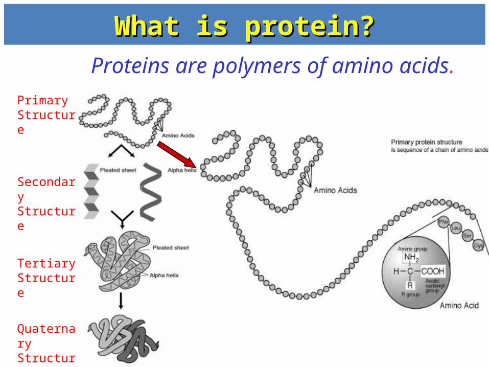
What is protein?What is protein? Proteins are polymers of amino acids.
Primary Structure
Secondary Structure
Tertiary Structure
Quaternary Structure

What is amino acid?What is amino acid?
Amino acid:Amino acid: a compound that contains both an amino group and a carboxyl group attach to -carbon
• -carbon also bound to side chain group, R
• R gives identity to amino acid

TerminologyTerminology• - carbon = the carbon that attach next to the carboxyl group
• - amino group = amino group that attach to -carbon
• Other type of amino group – eg. in Lysine, has
-amino group
Lysine

Amino acidAmino acid
1. All 20 are -amino acids
2. For 19 of the 20, the -amino group is primary; for proline, it is secondary amino acid
-Amino acid has an amino group attached to the carbon (-carbon) adjacent to the carboxyl group

Generic amino acid at physiological pH amino acids exist as dipolar ionic species (have positive and negative charge on the same molecule) - zwitterion form
Amino acid is an amphoteric molecule – act either as an acid or a base
- carboxyl group carboxylate ion
- amino group protonated amino acid
Physiological pH
Amino acids as dipolar ions

• The amino acids can exist in two enantiomeric forms
(nonsuperimposable mirror image) forms – exceptional for glycine
C
C
R1
HNH3
+
OO
C
C
R1
H NH3
+
O O
Mirror plane
carbon
EnantiomerEnantiomer
• Two steroisomers of amino acids are designated L- or D-. L – amino acid: abundant in nature, found in proteins, amino group on the left

Amino acidAmino acid
• Only the L - form of amino acids is commonly found in proteins.
• Depending on the nature of the R group, amino acids are classified into four groups.
1. nonpolar2. polar – neutral/uncharged side chain3. acidic4. basic
Polar, charged
Vs monosaccharide : D - form

Classification of amino acidClassification of amino acid
• Nonpolar (9 amino acids)
• Polar neutral/uncharged (6 amino acids)
charged basic (3 amino acids)
acidic (2 amino acids)

Classification of amino acids
Simplest amino acid due to the R group = H
No stereoisomer because the is achiral
Nonpolar
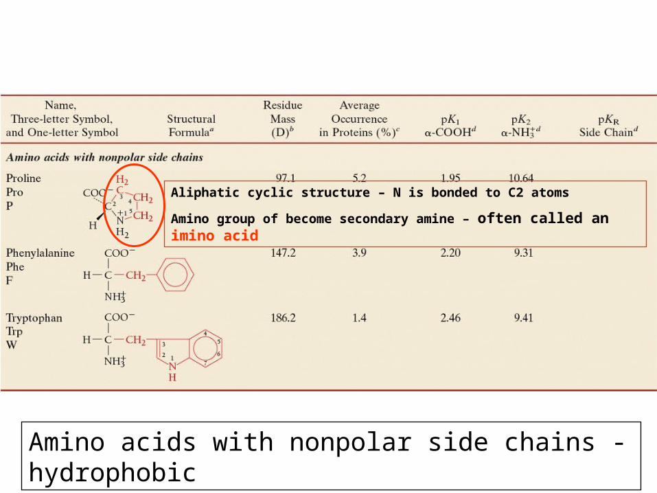
Aliphatic cyclic structure – N is bonded to C2 atoms
Amino group of become secondary amine – often called an imino acid
Amino acids with nonpolar side chains - hydrophobic

Polar unchargedPolar uncharged
Amide bond – highly polar
Thiol / sulfhydryl group – polar
– under oxidizing condition, with other thiol groups to form disulfide bridges (-S-S-) – important in 3o structure
Phenol

Polar chargedPolar charged
Basic
Acidic
Aspartate
Glutamate

Essential Amino acid Essential Amino acid • An essential amino acid or
indispensable amino acid is an amino acid that cannot be synthesized de novo by the organism (usually referring to humans), and therefore must be supplied in the diet.
• vs non-essential amino acid

Ionization of Amino AcidsIonization of Amino Acids
• Remember, amino acids without charged groups on side chain exist in neutral solution as zwitterions with no net charge
In acidic solution – as base (protonation)
In basic solution – as acid (deprotonation)

Ionization of amino acidsIonization of amino acids
• At physiological pH, the carboxyl group of the amino acid is negatively charged and the amino group is positively charged.
• Amino acids without charged side chains (Group 1 and 2) are zwitterions and have no net charge. (H3
+N-HCR-COO- ).
• A titration curve shows how the amine and carboxyl groups react with hydrogen ion.

Titration of amino acidTitration of amino acid• At low pH a nonacidic/nonbasic amino acid is protonated and has the structure
H3N+HCRCOOH (amino acid in cationic form)
• Increase of pH, dissociation of proton (H+) from –COOH group form H3N+HCRCOO-
(amino acid in zwitterionic form)
• At pK1, amount of cationic form = amount of zwitterionic form
• Beyond pK1, additional base ions will results in all amino acids in cationic forms deprotonated to zwitterionic forms – all amino acids have no net charge
pI = isoelectric point = pH at which the amino acid has no net charge/all amino acids are in zwitterionic form
• Increase of pH beyond pI, will cause the dissociation of H+ / deprotonation
from H3N+ resulting in formation of H2NHCRCOO- (anionic form)
• Increase of pH, more dissociation of proton (H+) from –H3N+group, more amino acids in anionic form
• At pK2, amount of zwitterionic form = amount of anionic form

Titration of AlanineTitration of Alanine
• When an amino acid is titrated, the titration curve represents the reaction of each functional group with the hydroxide ion
Cationic form All amino acids are in the zwitterion form – at isoelectric point (pI)
Anionic form
pI (isoelectric point) = pH at which the amino acid has no net charge/ all amino acids are in zwitterionic form

Titration of amino acidTitration of amino acid
• pK1 and pK2 are proton dissociation constant from carboxyl group and amino group
• From titration of amino acid, the pI can be calculated
• The charge behavior of acidic and basic amino acids is more complex. – Group Polar/charged amino acid

TerminologyTerminology• • peptidepeptide: the name given to a short polymer of
amino acids joined by peptide bonds; they are classified by the number of amino acids in the chain
• • dipeptidedipeptide: a molecule containing two amino acids joined by a peptide bond
• • tripeptidetripeptide: a molecule containing three amino acids joined by peptide bonds
• • polypeptidepolypeptide: a macromolecule containing many amino acids joined by peptide bonds
• • proteinprotein: a biological macromolecule of molecular weight 5000 g/mol or greater, consisting of one or more polypeptide chainsPrimary structure = one polypeptide
Protein: Protein: 11oo , 2 , 2o o and 3and 3oo structure structure

PeptidePeptide
* * * * *
Amino acid residue: a monomeric unit of amino acids

PROTEIN PROTEIN STRUCTURSTRUCTURE :OVERVIE :OVERVI
EWEW

Primary structurePrimary structure
Primary (1o) Structure = sequence of a chain of amino acids. Determines the final structure, eventually the properties of
proteins
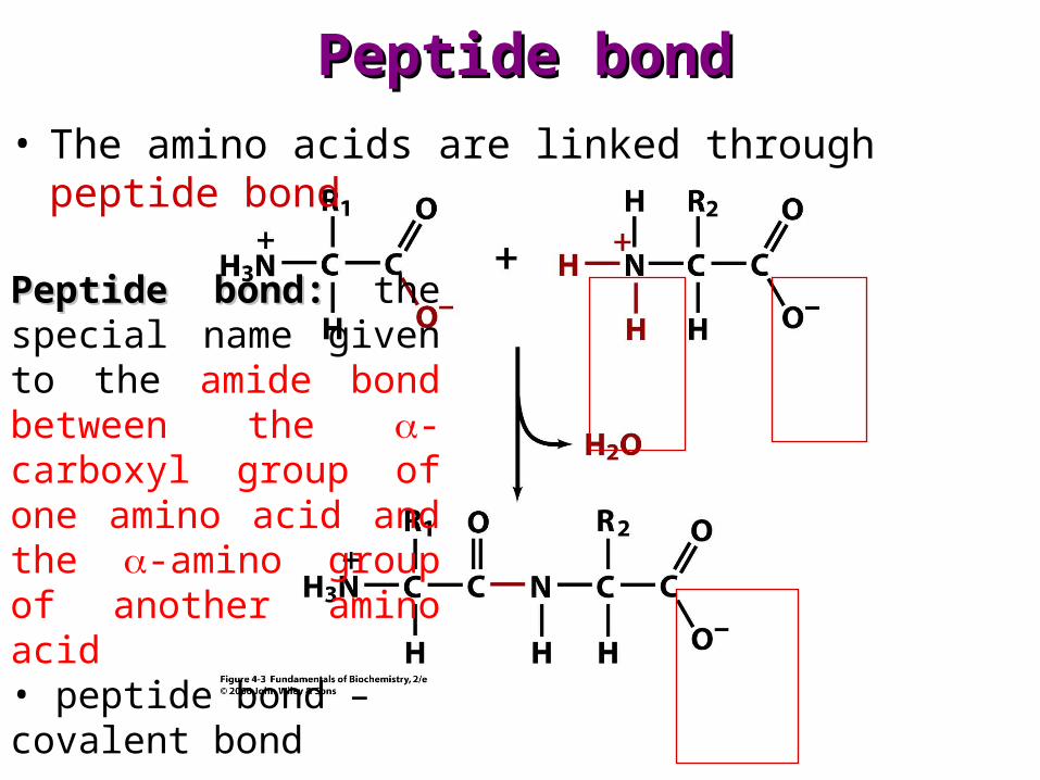
Peptide bondPeptide bond• The amino acids are linked through peptide bond
Peptide bond:Peptide bond: the special name given to the amide bond between the -carboxyl group of one amino acid and the -amino group of another amino acid • peptide bond – covalent bond

Peptide bond: FeaturePeptide bond: Feature
Peptide bond – in trans configuration, acts as a rigid and planar unit. Has limited rotation around the peptide bond (C-N).
COO-NH3
+
1 2 3 4 5Free
rotation
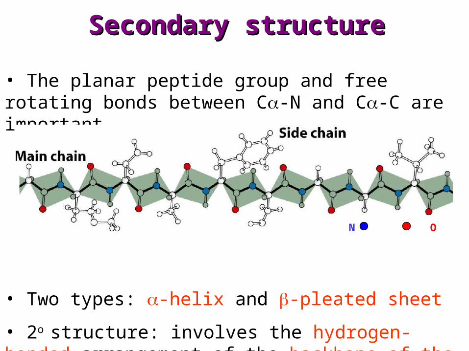
Secondary structureSecondary structure
• The planar peptide group and free rotating bonds between C-N and C-C are important
• Two types: -helix and -pleated sheet
• 2o structure: involves the hydrogen-bonded arrangement of the backbone of the protein
N O

Secondary structure: Secondary structure: -helix-helix
Structural features:
1. One polypeptide chain
2. Hydrogen bonds between the -CO and the –NH in the same polypeptide chain (intrachain)
3. The hydrogen bonds are parallel to the helix axis
4. Winding can be right- or left- handed (L- amino acid favor right-handed)
N O
H bond

Secondary structure: Secondary structure: -pleated sheet-pleated sheet
Structural features:
1. More than one polypeptide chain
2. Two types: antiparallel and parallel pleated sheet
3. Hydrogen bonds between the -CO and the –NH in the same polypeptide chain or with other polypeptide chain (interchain)
4. The hydrogen bonds are perpendicular to the direction of chain
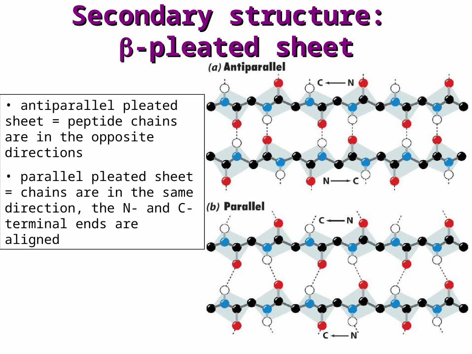
Secondary structure: Secondary structure: -pleated sheet-pleated sheet
• antiparallel pleated sheet = peptide chains are in the opposite directions
• parallel pleated sheet = chains are in the same direction, the N- and C- terminal ends are aligned
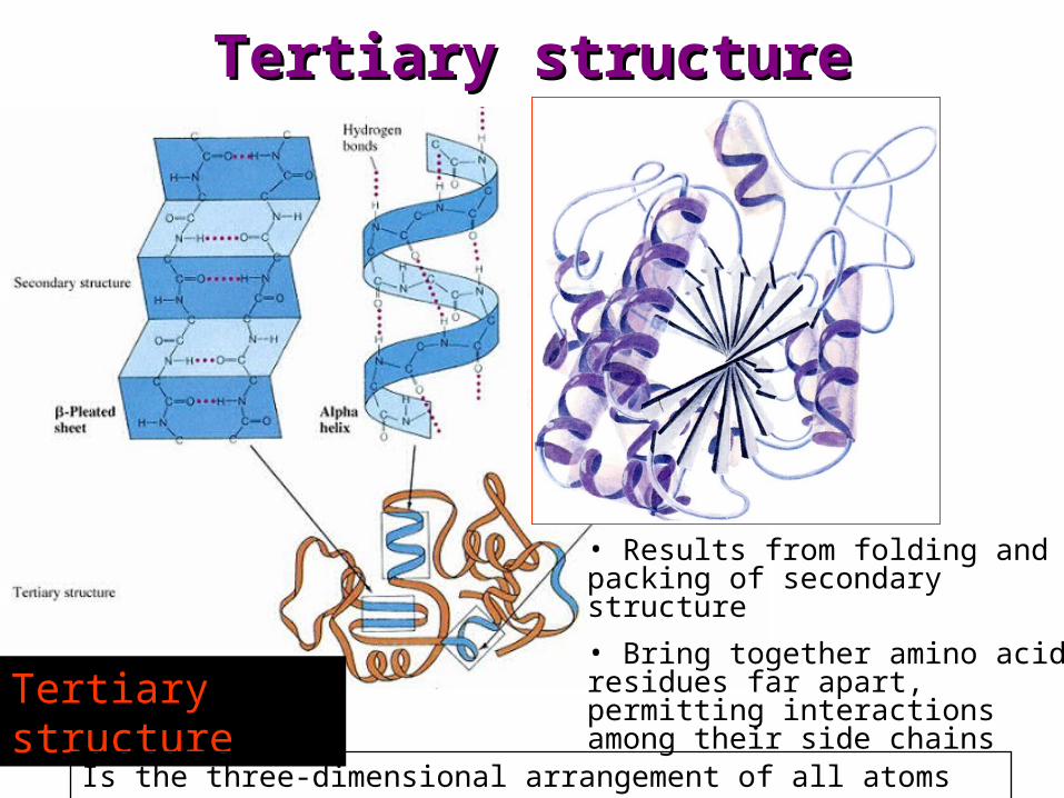
Tertiary structureTertiary structure
Tertiary structure
Is the three-dimensional arrangement of all atoms in protein molecule
• Results from folding and packing of secondary structure
• Bring together amino acid residues far apart, permitting interactions among their side chains

Tertiary structureTertiary structure
• Is the three-dimensional arrangement of all atoms in protein molecule
• Involves non-covalent interaction and covalent bonds
1. Hydrogen bonds between the side chain
2. Hydrophobic interaction
3. Electrostatic interactions/attractions
4. Disulfide bonds – between the R group
5. Complexation with metal ions

Forces in 3˚ StructureForces in 3˚ Structure
• Noncovalent interactions, including– hydrogen bonding between polar side chains, e.g., Ser
and Thr– hydrophobic interaction between nonpolar side
chains, e.g., Val and Ile– electrostatic attraction between side chains of
opposite charge, e.g., Lys and Glu– electrostatic repulsion between side chains of like
charge, e.g., Lys and Arg, Glu and Asp
• Covalent interactions: Disulfide (-S-S-) bonds between side chains of cysteines

• Native conformation: three-dimensional shape of a protein with biological activity
• Tertiary or quaternary structures

Quaternary structureQuaternary structure
• Final level of protein structure
• Association of more than one polypeptide chain to form a complex
• Subunit = individual parts of a large protein molecule = polypeptide chain
• Quaternary structure is the result of noncovalent interactions between two or more protein chains.
• Noncovalent interactions electrostatics, hydrogen bonds, hydrophobic
1
2 3
4

Quaternary StructureQuaternary Structure
• Oligomers are multisubunit proteins with all or some identical subunits.
• The subunits are called protomers.1. two subunits are called dimers2. four subunits are called tetramers
Quaternary structureQuaternary structure
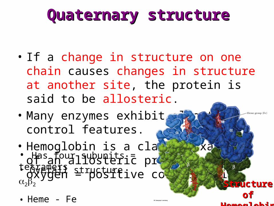
• If a change in structure on one chain causes changes in structure at another site, the protein is said to be allosteric.
• Many enzymes exhibit allosteric control features.
• Hemoglobin is a classic example of an allosteric protein. – oxygen = positive cooperativity
• Has four subunits = tetramers
Structure of Structure of HemoglobinHemoglobin
• Overall structure 22
• Heme - Fe

Classification of proteinClassification of protein
• Proteins are classified in two ways:
1. Shape
2. Composition

Fibrous ProteinsFibrous Proteins
• Fibrous proteins: contain polypeptide chains organized approximately parallel along a single axis. They– consist of long fibers or large sheets– tend to be mechanically strong– are insoluble in water and dilute salt solutions– play important structural roles in nature

Globular ProteinsGlobular Proteins
• Globular proteins: proteins which are folded to a more or less spherical shape
– they tend to be soluble in water and salt solutions– most of their polar side chains are on the outside and
interact with the aqueous environment by hydrogen bonding and ion-dipole interactions
– most of their nonpolar side chains are buried inside– nearly all have substantial sections of -helix and -
sheet

Comparison of Shapes of Fibrous and Comparison of Shapes of Fibrous and Globular ProteinsGlobular Proteins
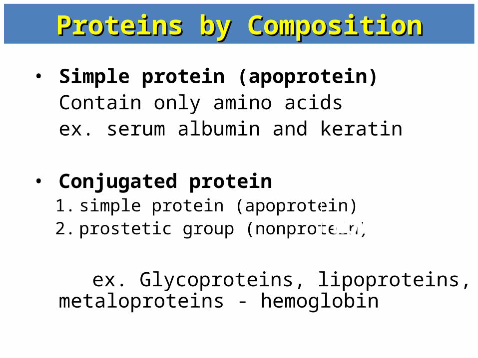
Proteins by CompositionProteins by Composition
• Simple protein (apoprotein) Contain only amino acidsex. serum albumin and keratin
• Conjugated protein1. simple protein (apoprotein)2. prostetic group (nonprotein)
ex. Glycoproteins, lipoproteins, metaloproteins
- hemoglobin
Holo-protein

DenaturationDenaturation Definition – complete loss of organized
structure in a protein, destroys the physiological function of the protein.
Definition – The unfolding of protein Eg. During cooking of egg
– Albumin (white egg) – denatured by heat and changes from a clear, colorless solution to a white coagulum
– Often irreversible – denatured protein cannot returned to its native biological form – lost of biological function – why microbes die when boiling

Due to loss of 2o 4o of protein structure, but not 1o , the amide bond (peptide bond) is intact

DenaturationDenaturationSeveral ways to denature proteins
• Heat – in temp, vibrations within the molecule, the energy of these vibrations can disrupt the 3o
• pH – or pH, affect the charges of protein, the electrostatic interactions that normally stabilize the native conformation is reduced.
• Detergents (eg. SDS) - disrupt hydrophobic interactions, if the detergent is charged, this can also disrupt electrostatic interactions
• Reducing agents(eg. Urea) – will form stronger H bonds, stronger than within the protein. Also disrupt the hydrophobic interaction
• Heavy metal ions
• Mechanical stress

DenaturationDenaturation
Reversible denaturation – organic solvents (ethyl alcohol or acetone), urea, detergents and acid or base
Denaturants disrupt only noncovalent interactions not the covalent linkages of the primary structure– If removed, possible protein to unwound to native
structure– eg. pH – addition of picric acid, protein (casein)
precipitate addition of NaOH, the solution clear
DenaturationDenaturation
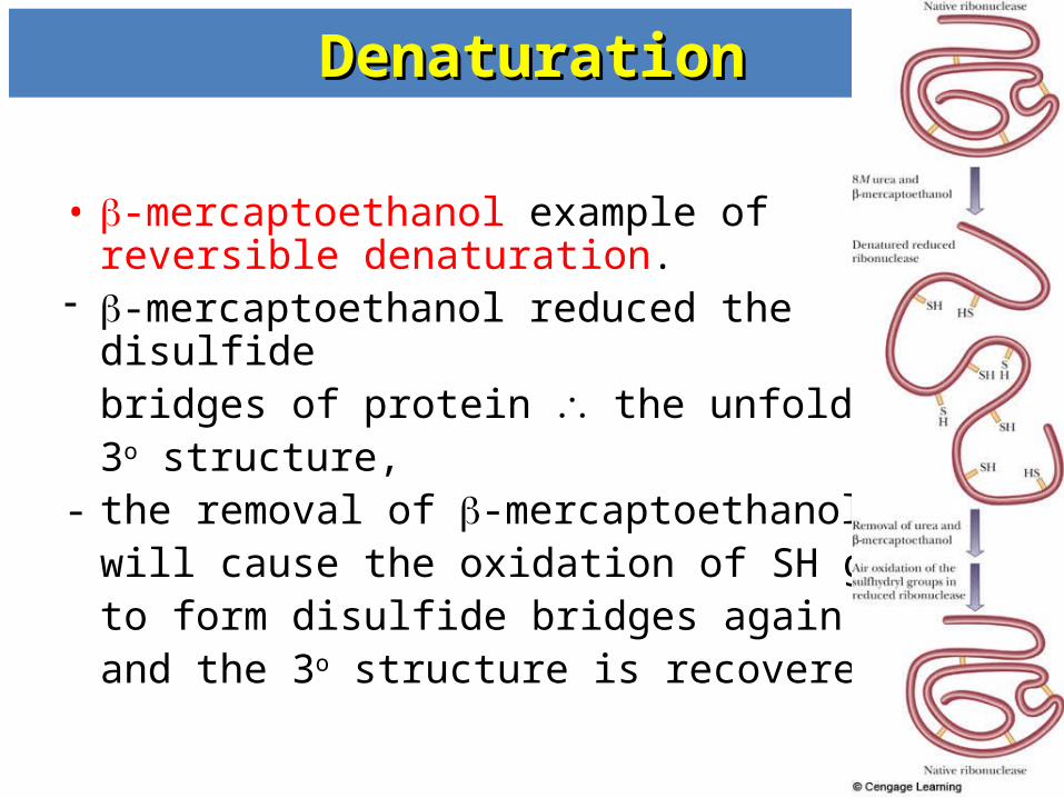
• -mercaptoethanol example of reversible denaturation.
-mercaptoethanol reduced the disulfide bridges of protein the unfolding of 3o structure,
- the removal of -mercaptoethanol will cause the oxidation of SH group to form disulfide bridges again and the 3o structure is recovered.

PPrrootteeiinn
FFuunnccttiioonnss


![Soldo 1969 [2,32 MiB]](https://img.pdfslide.net/doc/110x75/5852f3e51a28abfa398e4d84/soldo-1969-232-mib.jpg)
