Embed Size (px)
Citation preview
161
LEUCINE UROLITHIASIS IN A 3 WEEKS OLD MIXED GERMAN
SHEPHERD PUPPY
Laeish JUNKEE, Marian A. TAULESCU
Pathology Department, Faculty of Veterinary Medicine, University of Agricultural Sciences and Veterinary Medicine, 3-5 Calea Manastur, 400372, Cluj-Napoca (Romania)
Phone: +40 (0) 264 59 63 84 / 172; +40 (0) 753287436 Fax: +40 (0) 364816130; +40 (0) 264 59 37 92
[email protected], [email protected]
[email protected] Abstract
Canine urolithiasis is a common disorder of the urinary tract, characterized by stones located anywhere within the urinary tract, which is mostly encountered in middle-aged to older dogs. Urolithiasis is influenced by familial, congenital and pathophysiological factors including urinary pH, dehydration, urinary infection, anatomical abnormalities and drug administration. A 3 weeks old mixed German Shepherd male puppy with no antecedent clinical signs (sudden death) was submitted to the Pathology Department for necropsy. The animal was suspected of bronchopneumonia ab ingestis due to milk aspiration. Necropsy, cytological and histological exams were performed. Grossly, a large amount of urine was found within the peritoneal cavity (uroperitoneum) secondary to urinary bladder rupture, severe bilateral hydronephrosis and hydroureter, and urethral obstruction with numerous large white to gray calculi varying in size from 2-10mm were identified. The cytological exam showed several large, white to yellow spheroids with radial concentric laminations consistent with leucine crystals. Histologically, the renal tubules were diffusely dilated and contained pale eosinophilic hyaline casts, sloughed necrotic epithelial cells and lamellated concretions of amphophilic radiating structures. A diagnosis of urethral obstruction due to leucine urolithiasis was made, and it was associated with hydronephrosis, hydroureter and urinary bladder rupture. To the best of the authors’ knowledge this is the first report of leucine urolithiasis in a dog in Romania.
Key words: Canine, congenital, leucine, urolithiasis.
INTRODUCTION
Urolithiasis represents the formation of crystals and uroliths in the urinary system. In dogs, struvite uroliths are the most commonly reported uroliths in many studies worldwide (Ling, 1998). Urolithiasis (urinary calculi) can be located anywhere within the urinary tract, from the kidney, ureter, bladder, to the urethra and are referred to as nephroliths, ureteroliths, urocystoliths and urethroliths, respectively
(Zachary and McGavin, 2012) . Several dog breeds predisposed to urolithiasis include Dalmatians Cocker Spaniels, Bichon Frise (Bichons), and Miniature Schnauzers (Kruger et al., 2009) Morphologically, calculi vary in colour and composition; they can be white to gray (e.g., struvite and oxalate, leucine) and yellow (e.g.,
urate, cysteine, benzocoumarin, and xanthine) (Zachary and McGavin, 2012). The etiopathogenesis of the leucine urolithiasis is still unknown. The aim of the study was to describe the pathological findings of a juvenile leucine urolithiasis in a dog. MATERIALS AND METHODS Biological material
A 3 weeks old mixed German Shepherd male puppy with a clinical history of sudden death, and a suspicion of acute bronchopneumonia ab ingestis due to milk aspiration was submitted to the Pathology Department (Faculty of Veterinary Medicine of Cluj-Napoca) for necropsy. Necropsy, cytological and histological exams were performed on the same day.
Scientific Works. Series C. Veterinary Medicine. Vol. LXI (2)ISSN 2065-1295; ISSN 2343-9394 (CD-ROM); ISSN 2067-3663 (Online); ISSN-L 2065-1295
162
Necropsy
The kidneys were opened on the large curvature, while the urinary bladder, urethra and ureters were opened longitudinally. During the procedure, several samples from the kidneys, urethra, ureters and urinary bladder were collected and fixed in 10% buffered formalin and paraffin embedded.
Cytological analysis
Several specimens from the urethral sediments and calculi were examined by direct method.
Histological Analysis
Serial consecutive sections of 3µm-thick were stained with Hematoxylin and Eosin. The slides were analyzed with an Olympus BX51 microscope with an Olympus SP 350 digital camera.
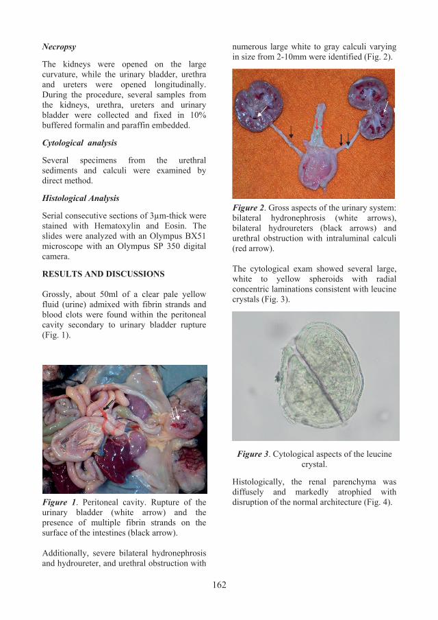
RESULTS AND DISCUSSIONS Grossly, about 50ml of a clear pale yellow fluid (urine) admixed with fibrin strands and blood clots were found within the peritoneal cavity secondary to urinary bladder rupture (Fig. 1).
Figure 1. Peritoneal cavity. Rupture of the urinary bladder (white arrow) and the presence of multiple fibrin strands on the surface of the intestines (black arrow). Additionally, severe bilateral hydronephrosis and hydroureter, and urethral obstruction with
numerous large white to gray calculi varying in size from 2-10mm were identified (Fig. 2).
Figure 2. Gross aspects of the urinary system: bilateral hydronephrosis (white arrows), bilateral hydroureters (black arrows) and urethral obstruction with intraluminal calculi (red arrow). The cytological exam showed several large, white to yellow spheroids with radial concentric laminations consistent with leucine crystals (Fig. 3).
Figure 3. Cytological aspects of the leucine crystal.
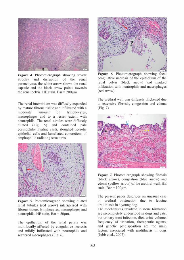
Histologically, the renal parenchyma was diffusely and markedly atrophied with disruption of the normal architecture (Fig. 4).
163
Figure 4. Photomicrograph showing severe atrophy and disruption of the renal parenchyma; the white arrow shows the renal capsule and the black arrow points towards the renal pelvis. HE stain. Bar = 200µm.
The renal interstitium was diffusely expanded by mature fibrous tissue and infiltrated with a moderate amount of lymphocytes, macrophages and to a lesser extent with neutrophils. The renal tubules were diffusely dilated (Fig. 5) and contained pale eosinophilic hyaline casts, sloughed necrotic epithelial cells and lamellated concretions of amphophilic radiating structures.
Figure 5. Photomicrograph showing dilated renal tubules (red arrow) interspersed with fibrous tissue, lymphocytes, macrophages and neutrophils. HE stain. Bar = 50µm. The epithelium of the renal pelvis was multifocally affected by coagulative necrosis and mildly infiltrated with neutrophils and scattered macrophages (Fig. 6).
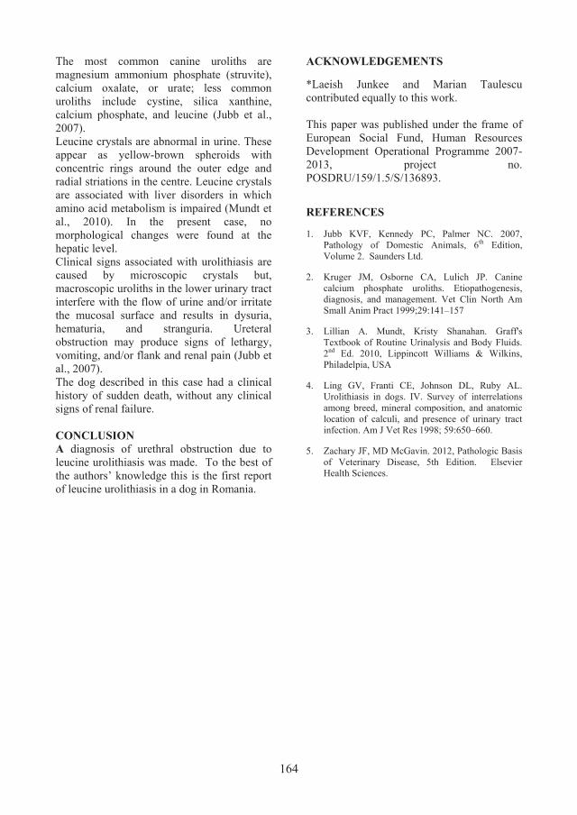
Figure 6. Photomicrograph showing focal coagulative necrosis of the epithelium of the renal pelvis (black arrow) and marked infiltration with neutrophils and macrophages (red arrow). The urethral wall was diffusely thickened due to extensive fibrosis, congestion and edema (Fig. 7).
Figure 7. Photomicrograph showing fibrosis (black arrow), congestion (blue arrow) and edema (yellow arrow) of the urethral wall. HE stain. Bar = 100µm. The present paper describes an unusual case of urethral obstruction due to leucine urolithiasis in a young dog. The mechanisms involved in stone formation are incompletely understood in dogs and cats, but urinary tract infection, diet, urine volume, frequency of urination, therapeutic agents, and genetic predisposition are the main factors associated with urolithiasis in dogs (Jubb et al., 2007).
164
The most common canine uroliths are magnesium ammonium phosphate (struvite), calcium oxalate, or urate; less common uroliths include cystine, silica xanthine, calcium phosphate, and leucine (Jubb et al., 2007). Leucine crystals are abnormal in urine. These appear as yellow-brown spheroids with concentric rings around the outer edge and radial striations in the centre. Leucine crystals are associated with liver disorders in which amino acid metabolism is impaired (Mundt et al., 2010). In the present case, no morphological changes were found at the hepatic level. Clinical signs associated with urolithiasis are caused by microscopic crystals but, macroscopic uroliths in the lower urinary tract interfere with the flow of urine and/or irritate the mucosal surface and results in dysuria, hematuria, and stranguria. Ureteral obstruction may produce signs of lethargy, vomiting, and/or flank and renal pain (Jubb et al., 2007). The dog described in this case had a clinical history of sudden death, without any clinical signs of renal failure. CONCLUSION A diagnosis of urethral obstruction due to leucine urolithiasis was made. To the best of the authors’ knowledge this is the first report of leucine urolithiasis in a dog in Romania.
ACKNOWLEDGEMENTS
*Laeish Junkee and Marian Taulescu contributed equally to this work. This paper was published under the frame of European Social Fund, Human Resources Development Operational Programme 2007-2013, project no. POSDRU/159/1.5/S/136893.
REFERENCES
1. Jubb KVF, Kennedy PC, Palmer NC. 2007, Pathology of Domestic Animals, 6th Edition, Volume 2. Saunders Ltd.
2. Kruger JM, Osborne CA, Lulich JP. Canine calcium phosphate uroliths. Etiopathogenesis, diagnosis, and management. Vet Clin North Am Small Anim Pract 1999;29:141–157
3. Lillian A. Mundt, Kristy Shanahan. Graff's Textbook of Routine Urinalysis and Body Fluids. 2nd Ed. 2010, Lippincott Williams & Wilkins, Philadelpia, USA
4. Ling GV, Franti CE, Johnson DL, Ruby AL. Urolithiasis in dogs. IV. Survey of interrelations among breed, mineral composition, and anatomic location of calculi, and presence of urinary tract infection. Am J Vet Res 1998; 59:650–660.
5. Zachary JF, MD McGavin. 2012, Pathologic Basis of Veterinary Disease, 5th Edition. Elsevier Health Sciences.