Embed Size (px)
Citation preview
DOI 10.1515/hf-2012-0120 Holzforschung 2013; 67(4): 463–471
Li-Ting Ma , Sheng-Yang Wang , Yen-Hsueh Tseng , Yi-Ru Lee and Fang-Hua Chu *
Cloning and characterization of a 2,3-oxidosqualene cyclase from Eleutherococcus trifoliatus Abstract: The 2,3-oxidosqualene cyclases (OSCs) are a
family of enzymes that have an important role in plant trit-
erpene biosynthesis. In this study, an OSC gene designed
Et LUS from Eleutherococcus trifoliatus has been cloned.
Et LUS includes a 2292-bp open reading frame and encodes
a 763-amino acid protein. Et LUS has an MLCYCR motif,
which is conserved in lupeol synthases. Comparison of
active-site residues and gene expression in yeast showed
that Et LUS synthesizes lupeol. However, Et LUS has the
highest sequence identity with β -amyrin synthases from
Araliaceae rather than lupeol synthases, adding new
perspective to the evolution of the OSCs of Araliaceae.
Furthermore, Et LUS is upregulated in leaf tissues under
methyl jasmonate treatment, which can be interpreted
that lupeol and its derivatives play an ecological and
physiological role in plant defense against pathogens and
insect herbivores.
Keywords: Araliaceae, Eleutherococcus trifoliatus , lupeol,
methyl jasmonate, 2,3-oxidosqualene cyclase
*Corresponding author: Fang-Hua Chu, School of Forestry and
Resource Conservation , National Taiwan University, Taipei 10617 ,
Taiwan , e-mail: [email protected]
Li-Ting Ma and Yi-Ru Lee: School of Forestry and Resource
Conservation , National Taiwan University, Taipei 10617 , Taiwan
Sheng-Yang Wang and Yen-Hsueh Tseng: Department of Forestry ,
National Chung-Hsing University, Taichung 40227 , Taiwan
Introduction Eleutherococcus trifoliatus (L.) S.Y. Hu var. trifoliatus
( = Acanthopanax trifoliatus , Araliaceae) is a shrub with
ternately compound leaves. It is used in folk medicine
in Vietnam, Taiwan, and China for bruising, neuralgia,
impotence, and gout because of its ginseng-like activity
(Lischewski et al. 1985 ; Ohashi 1993 ; Yook et al. 1999 ).
Many studies have reported the secondary metabolic bio-
activities of compounds from the Araliaceae. Triterpenes
such as ginsenoside and ciwujianosides isolated from
Panax species and Eleutherococcus senticosus , respec-
tively, are among the most commonly reported. Some
compounds from this family have been found to possess
important pharmacological activities, including anti-
inflammatory, anti-cancer, anti-amnestic, and anti-aging
effect, which led to an increased interest in this group
of plants ( Kenarova et al. 1990 ; Mochizuki et al. 1995 ;
Shinkai et al. 1996 ; Jung et al. 2003 ; Cheng et al. 2005 ; Seo
et al. 2005 ).
Terpene compounds have a plenty of bioactivi-
ties. Trapp and Croteau (2001) summarized the genomic
organization of plant terpene synthases, and in many
cases, typical terpene synthases were identified and char-
acterized, such an α -pinene synthase (Chu et al. 2009 ),
β -cadinene synthase (Kuo et al. 2012 ), or a sesquiterpene
synthase (Wen et al. 2012 ), to mention a few. Triter-
penes are a large subgroup of the terpene superfam-
ily (Xu et al. 2004 ), which are biosynthesized directly
from 2,3-oxidosqualene by an enzyme family named the
2,3-oxidosqualene cyclases (OSCs) (Segura et al. 2003 ).
Different OSCs have different product specificities that
can be divided into sterol precursors and triterpene alco-
hols; the OSCs can thus be considered a critical branch
point between primary and secondary metabolism and a
critical point in the generation of triterpene skeletal diver-
sity (Phillips et al. 2006 ).
In recent years, a number of triterpene synthases
have been cloned and characterized from various plant
species; those articles contributed to a better understand-
ing the biosynthesis of plant triterpenes (Basyuni et al.
2006 ; Guhling et al. 2006 ). Some triterpene compounds
are thought to participate in plant defense systems
because of the anti-microbial, fungicidal, and insecti-
cidal activities (Mylona et al. 2008 ). Multiple pharmaco-
logical activities have also been observed. Glycyrrhizin is
a triterpene compound that exists in the roots and stolons
of Glycyrrhiza plants and may be suitable for the treat-
ment of liver diseases and allergic diseases (Hayashi and
Sudo 2007). Panax ginseng contains unique triterpene
compounds named ginsenosides. Each ginsenoside has
been shown to have different pharmacological effects,
supporting its traditional use as a medical plant (Liang
and Zhao 2008 ).
To date, five triterpenoid carboxylic acids and three
lupane-glycosides have been reported in E. trifoliatus
(Ty et al. 1984, 1985 ; Lischewski et al. 1985 ; Yook et al.
Brought to you by | National Taiwan UniversityAuthenticated | 140.112.82.231
Download Date | 5/14/13 6:57 AM
464 L.-T. Ma et al.: Lupeol synthase from E. trifoliatus
1999 ). However, the biosynthesis pathways of triterpene
compounds in this plant remain unknown. In the present
study, a gene involved in triterpene biosynthesis in
E. trifoliatus should be identified and characterized. To
understand the physiology functions and properties
of this gene, its expression will be analyzed in different
tissues and under treatment conditions with an elicitor,
namely with methyl jasmonate (MeJA).
Materials and methods
Plant materials and culture conditions Plants were collected from a fi eld on the Chiufenershan Mountain
in Nantou County of Taiwan. The plants were incubated under
greenhouse conditions at 25 ° C with natural light. The plant tissue
was harvested and frozen instantaneously in liquid N 2 . The tissues
were stored at -80 ° C in advance for RNA preparation. Yeast strain
GIL77 ( gal2 hem3-6 erg7 ura3167 ) was selected as the host for gene
expression, maintained on YPD medium (1 % yeast extract, 2 % pep-
tone, 2 % dextrose) supplemented with hemin (13 μ g ml -1 ), ergosterol
(20 μ g ml -1 ), and Tween 80 (5 mg ml -1 ).
Cloning of OSC cDNA Fresh leaf tissue was weighed and ground to a fi ne powder under liq-
uid N 2 , and total RNA isolation was performed by Plant Total RNA
Miniprep Purifi cation Kit (Hopegen, Taichung, Taiwan). Total RNA
(2 μ g) was reverse-transcribed with SuperScript ™ III reverse tran-
scriptase (Invitrogen, Carlsbad, CA, USA) according to the manu-
facturer ’ s protocol. The core sequence was amplifi ed by degenerate
primers triTPS1F (CAATGGA(G/T)CA(C/T)(A/G)TTCATTATGA) and
triTPS4R (TCCT(C/G)(C/T)TGAGGAAA(A/G)TCACC), which was based
on highly conserved region on the alignment of published sequences
of OSC. The 3 ′ - and 5 ′ -end amplifi cation of cDNA were carried out
with 3 ′ - and 5 ′ -RACE System (Invitrogen, Carlsbad, CA, USA), respec-
tively. All specifi c primers were based on the sequence of core frag-
ment. Protein structure was predicted by Swiss-Model Repository
( http://swissmodel.expasy.org ) and visualized by the Chimera pro-
gram (Resource for Biocomputing, Visualization, and Informatics at
the University of California, San Francisco, CA, USA) (Pettersen et al.
2004 ).
Sequences alignment and phylogenetic analyses were based on
the neighbor-joining method and using the ClustalW website and
the MEGA5.0 soft ware (http://www.megasoft ware.net/). The phylo-
genetic tree was built based on 1000 bootstrap replications. All the
sequences of this analysis are des cribed in Table 1 .
Functional expression of OSC cDNA in yeast and analysis of triterpene products The full-length cDNA of Et LUS was obtained by a forward primer
with a Sac I site (5 ′ -CGAGCTC-ATG TGG AAG CTG AAG ATA GCC GAA
GGA G-3 ′ ) and a reverse primer with a Not I site (5 ′ -ATAGTTTAGCG-
GCCG-TTA GGC ACC TAG GGT CGG TAA TGG TAC-3 ′ ). PCR was per-
formed by Phusion ® High-Fidelity DNA Polymerase (Finnzyme,
Espo, Finland). The PCR amplifi cation program was designed as
follows: one cycle of initial denaturation at 98 ° C for 30 s, 30 cycles
at 98 ° C for 17 s, 60 ° C for 15 s, and 72 ° C for 60 s, and fi nally, 72 ° C for
10 min.
The full-length PCR product was digested with Sac I and Not I restriction enzymes (New Rngland Biolabs, Hitchin, UK), and
ligated into yeast expression vector pYES2 (Invitrogen, Carls-
bad, CA, USA) derived from GAL1 promoter with T4 DNA ligase
(Promega, Heidelberg, Germany). The plasmid was transformed
into yeast mutant GIL77 by electroporation. The transformed yeast
was inoculated in 30 ml synthetic complete medium without uracil
(SC-Ura) and containing hemin (13 μ g ml -1 ), ergosterol (20 μ g ml -1 ),
and Tween 80 (5 mg ml -1 ), incubated at 30 ° C for 2 days, and then
transferred to a new SC-Ura medium supplemented with 2 % galac-
tose for glucose. Aft er induction of galactose and the stabilization
of incubation for 24 h in 0.1 M potassium phosphate buff er (pH 7.0)
supplemented with 3 % glucose and hemin, cells were collected
by centrifugation at 1750 g for 5 min and refl uxed with 5 ml 20 %
KOH/50 % ethanol at 90 ° C for 10 min and extracted twice with 5
ml hexane. The solvent was dried out by a gentle stream of N 2 and
redissolved in 1 ml hexane.
The fi nal extract was analyzed by TLC that was developed with
hexane/ethyl acetate (9:1, v/v) and then visualized by 5 % phospho-
molybdic acid in ethanol. To identify the chemical structures, the
extracts were directly derivatized with bis-N,N-(trimethylsilyl)- trif-
luoroacetamide (BSTFA) in pyridine (30 min at 70 ° C). The pro duct
analysis was observed using an ITQ 900TM GC/MS instrument
(Thermo Scientifi c, Dreieich, Germany) and a DB-5 capillary column
(30 × 0.25 mm i.d., 0.25 μ m fi lm thickness; J&W Scientifi c, Folsom, CA,
USA). For the temperature program, injection was done at 110 ° C and
held constant for 1 min (He at 1 ml min -1 ), then increased to 260 ° C
(40 ° C min -1 ) and held constant for 5 min, and then further increased
to 290 ° C (5 ° C min -1 ) and held for 10 min.
Expression analysis of Et LUS in E. trifoliatus organs and MeJA treatment Unexpanded leaves, mature leaves, shoots, stems, roots, and fruits
were collected from plants. Total RNA was extracted from powder
with TRIzol ® reagent (Invitrogen, Carlsbad, CA, USA), then cDNA
was synthesized as described previously. The nucleotide sequences
of the gene specifi c primers were as follows: 2osc3R2 (5 ′ -GTT ATA
TCC TGG GCA CCG AAA GAA GGA G-3 ′ ) and 2oscR (5 ′ -ACC GAC CCT
AGG TGC CTA AAT AGC-3 ′ ). Actin as internal control was amplifi ed
using the primer sets: actin-F (5 ′ -GCC ATC CTT CGT CTT GAC CTT
GCT G-3 ′ ) and actin-R (5 ′ -CTG TTG GAA GGT GCT GAG GGA TGC-3 ′ ).
To eliminate the amplifi cation of homologous transcripts, two prim-
ers sets were designed in the coding region and the 3 ′ -UTR, respec-
tively. PCR amplifi cation was carried out with initial denaturation at
94 ° C for 3 min, 25 cycles each at 94 ° C for 30 s, 60 ° C for 30 s, and 72 ° C
for 30 s, and fi nally, 72 ° C for 60 s.
For MeJA treatment, plants were sprayed with 10 mM MeJA
solution until totally wet and placed alone in the plant growth
incubator. Leaves were harvested as samples aft er 6, 12, 24, and
48 h, and RNA extraction and RT-PCR were carried out as described
previously.
Brought to you by | National Taiwan UniversityAuthenticated | 140.112.82.231
Download Date | 5/14/13 6:57 AM
L.-T. Ma et al.: Lupeol synthase from E. trifoliatus 465
Results
Characterization of the encoding OSC in E. trifoliatus In previous studies of E. trifoliatus , triterpene com-
pounds were isolated mainly from leaves; in the present
study, cDNA from leaves was used to clone OSCs. A full-
length sequence named Et LUS (GenBank accession no.
JQ087376) was obtained using PCR amplification with
degenerated primers and the RACE method. The open
reading frame of Et LUS is 2292 bp and encodes a 763-
amino acid protein with a molecular weight of 87 kDa.
It contains four QW motifs that are thought to contribute
to the fold stability of the protein (Wendt et al. 1997 ) and
the OSC family-conserved substrate-binding site DCTAE
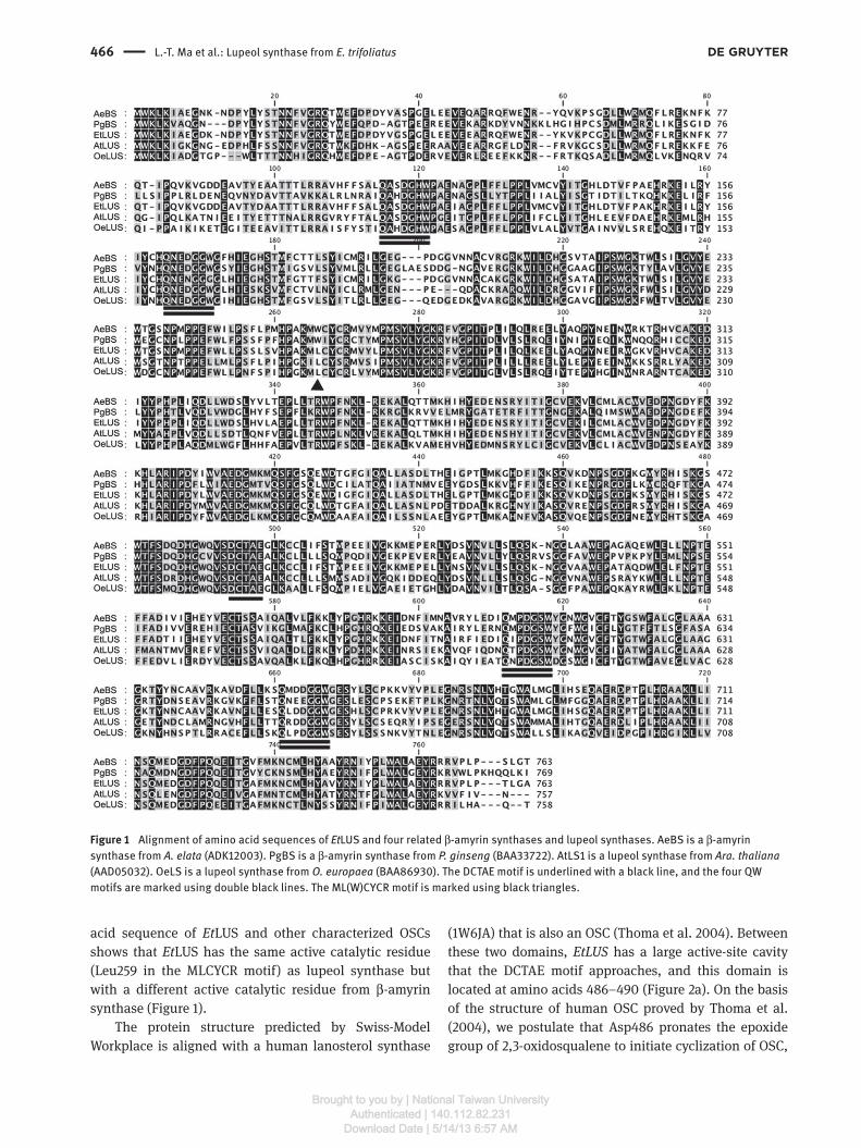
(Figure 1 ).
Active catalytic residue identification has been the
focus of study of OSCs, and it has been proved that the
MLCYCR and MWCYCR motifs are critical in the product
specificities of lupeol and β -amyrin synthase, respec-
tively (Kushiro et al. 2000 ). The alignment of the amino
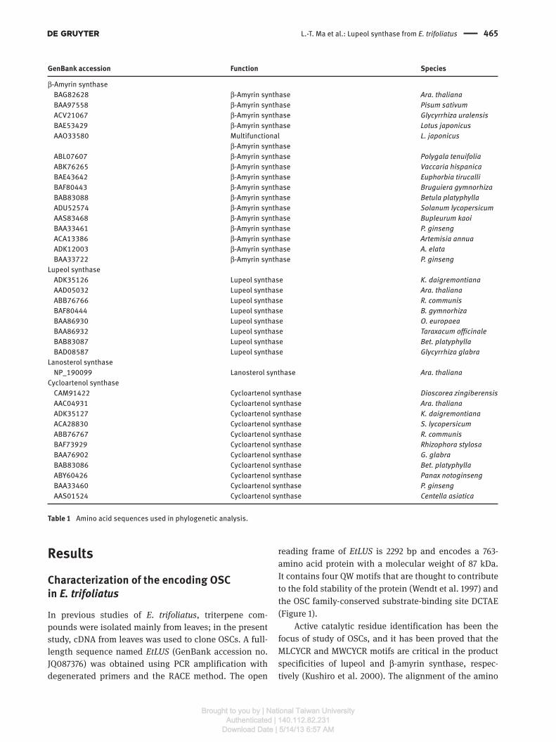
GenBank accession Function Species
β -Amyrin synthase
BAG82628 β -Amyrin synthase Ara. thaliana
BAA97558 β -Amyrin synthase Pisum sativum
ACV21067 β -Amyrin synthase Glycyrrhiza uralensis
BAE53429 β -Amyrin synthase Lotus japonicus
AAO33580 Multifunctional L. japonicus
β -Amyrin synthase
ABL07607 β -Amyrin synthase Polygala tenuifolia
ABK76265 β -Amyrin synthase Vaccaria hispanica
BAE43642 β -Amyrin synthase Euphorbia tirucalli BAF80443 β -Amyrin synthase Bruguiera gymnorhiza
BAB83088 β -Amyrin synthase Betula platyphylla
ADU52574 β -Amyrin synthase Solanum lycopersicum
AAS83468 β -Amyrin synthase Bupleurum kaoi BAA33461 β -Amyrin synthase P. ginseng
ACA13386 β -Amyrin synthase Artemisia annua
ADK12003 β -Amyrin synthase A. elata
BAA33722 β -Amyrin synthase P. ginseng
Lupeol synthase
ADK35126 Lupeol synthase K. daigremontiana
AAD05032 Lupeol synthase Ara. thaliana
ABB76766 Lupeol synthase R. communis
BAF80444 Lupeol synthase B. gymnorhiza
BAA86930 Lupeol synthase O. europaea
BAA86932 Lupeol synthase Taraxacum officinale
BAB83087 Lupeol synthase Bet. platyphylla
BAD08587 Lupeol synthase Glycyrrhiza glabra
Lanosterol synthase
NP_190099 Lanosterol synthase Ara. thaliana
Cycloartenol synthase
CAM91422 Cycloartenol synthase Dioscorea zingiberensis
AAC04931 Cycloartenol synthase Ara. thaliana
ADK35127 Cycloartenol synthase K. daigremontiana
ACA28830 Cycloartenol synthase S. lycopersicum
ABB76767 Cycloartenol synthase R. communis
BAF73929 Cycloartenol synthase Rhizophora stylosa
BAA76902 Cycloartenol synthase G. glabra
BAB83086 Cycloartenol synthase Bet. platyphylla
ABY60426 Cycloartenol synthase Panax notoginseng
BAA33460 Cycloartenol synthase P. ginseng
AAS01524 Cycloartenol synthase Centella asiatica
Table 1 Amino acid sequences used in phylogenetic analysis.
Brought to you by | National Taiwan UniversityAuthenticated | 140.112.82.231
Download Date | 5/14/13 6:57 AM
466 L.-T. Ma et al.: Lupeol synthase from E. trifoliatus
Figure 1 Alignment of amino acid sequences of Et LUS and four related β -amyrin synthases and lupeol synthases. AeBS is a β -amyrin
synthase from A. elata (ADK12003). PgBS is a β -amyrin synthase from P. ginseng (BAA33722). AtLS1 is a lupeol synthase from Ara. thaliana
(AAD05032). OeLS is a lupeol synthase from O. europaea (BAA86930). The DCTAE motif is underlined with a black line, and the four QW
motifs are marked using double black lines. The ML(W)CYCR motif is marked using black triangles.
acid sequence of Et LUS and other characterized OSCs
shows that Et LUS has the same active catalytic residue
(Leu259 in the MLCYCR motif) as lupeol synthase but
with a different active catalytic residue from β -amyrin
synthase (Figure 1).
The protein structure predicted by Swiss-Model
Workplace is aligned with a human lanosterol synthase
(1W6JA) that is also an OSC (Thoma et al. 2004 ). Between
these two domains, Et LUS has a large active-site cavity
that the DCTAE motif approaches, and this domain is
located at amino acids 486 – 490 (Figure 2 a). On the basis
of the structure of human OSC proved by Thoma et al.
(2004) , we postulate that Asp486 pronates the epoxide
group of 2,3-oxidosqualene to initiate cyclization of OSC,
Brought to you by | National Taiwan UniversityAuthenticated | 140.112.82.231
Download Date | 5/14/13 6:57 AM

L.-T. Ma et al.: Lupeol synthase from E. trifoliatus 467
and hydrogen-bonding partners with Asp486 were also
found in Cys487 and Cys565 (Figure 2b).
Functional expression of OSC in yeast
To further identify the function of Et LUS, functional
expression of the cloned gene was attempted. Et LUS cDNA
was ligated into the vector pYES2 and expressed under
the control of the GAL1 promoter in yeast mutant GIL77,
which lacks lanosterol synthase activity, to decrease the
competition for the substrate of OSC, 2,3-oxidosqualene.
Because of this deficiency, exogenous ergosterol, which is
derived from lanosterol, is essential for cell growth. The
empty vector was also transformed into GIL77 as a nega-
tive control.
Lipid extract and authentic standards were applied
to TLC plates. The gene expression of Et LUS in GIL77
resulted in a product with the same mobility as lupeol
(Figure 3 a) and β -amyrin (data not shown). Both trans-
formants (pYES2 and pYES2- Et LUS) had the same
signal, which was possibly ergosterol. To identify the
chemical structure of each signal in the TLC plates,
the extracts were analyzed by GC/MS. Comparison of
the MS fragmentation patterns of authentic triterpene
compounds such as β -amyrin, lupeol, cycloartenol, and
ergosterol showed that two transformants contained
ergosterol, in agreement with the results of TLC. In
addition, the extracts from yeast harboring Et LUS cDNA
comprised one major compound, which showed at 21.7
min and was consistent with the retention time of the
lupeol standard (Figure 3b). To elucidate the struc-
ture, MS characteristics were compared with the lupeol
standard, revealing fraction peaks at m / z 189, 218, and
ba
Figure 2 Protein structure prediction and active site of Et LUS. (a) The whole Et LUS structure with the active site in the center (red frame)
in which the substrate, 2,3-oxidosqualene (white) is catalyzed. (b) The relative positions of substrate-binding residue Asp486 (green), the
active catalytic residue Leu259 (blue), hydrogen-bonding partners Cys487 and Cys565 (red), and 2,3-oxidosqualene (white).
a
b 100
50
0100
50
0100
50
018 19 20 21 22
Time (min)
Rel
ativ
e ab
unda
nce
EtOSC2 product
Authentic lupeol
2
1
EtLUS product
Lupeol
Vector control
Vector control
23 24 25
Figure 3 Chromatographic analysis of the hexane extract from
yeast and authentic lupeol as standard. (a) Comparison patterns of
TLC patterns of yeast transformed with pYES2 (vector control) and
pYES2- Et LUS ( Et LUS product). (b) GC analysis of three extracts: com-
pound 1 (ergosterol) and compound 2 (lupeol). The mass spectrum
of compound 2 is shown in Figure 4.
498, which were also identical to the lupeol standard
(Figure 4 ).
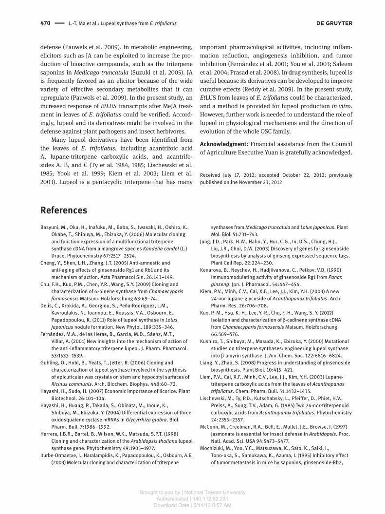
Expression of Et LUS in E. trifoliatus organs and MeJA treatment
RT-PCR was performed to examine the expression of
Et LUS in different tissues of E. trifoliatus. The accumula-
tion of Et LUS transcripts was observed in all leaf tissues
Brought to you by | National Taiwan UniversityAuthenticated | 140.112.82.231
Download Date | 5/14/13 6:57 AM
468 L.-T. Ma et al.: Lupeol synthase from E. trifoliatus
Discussion and conclusions
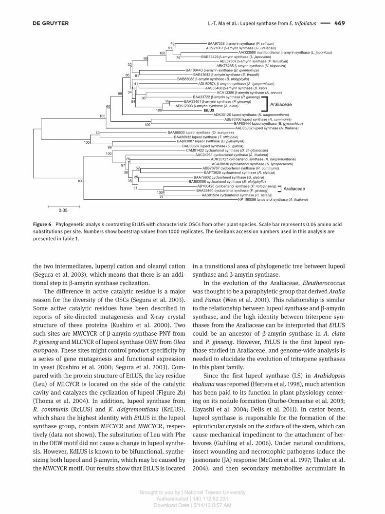
According to the phylogenetic analysis of Et LUS and 41
other previously characterized plant OSCs (Table 1), they
are separated into three groups: cycloartenol synthases,
β -amyrin synthases, and lupeol synthases (Figure 6 ). The
amino acid sequence of Et LUS exhibits 90 – 92 % sequence
identity with β -amyrin synthase from P. ginseng (GenBank
accession no. BAA33461) and Aralia elata (GenBank acces-
sion no. ADK12003), which belong to the same family
as E. trifoliatus and has a 72 – 76 % identity with lupeol
synthase from Ricinus communis (GenBank accession
no. ABB76766) and Kalanchoe daigremontiana (GenBank
accession no. ADK35126).
Alhough Et LUS shares the highest degree of sequence
identity with β -amyrin synthase (92 % ) based on phy-
logenetic tree analysis, GC/MS analysis revealed that it
encodes a lupeol synthase. In fact, β -amyrin and lupeol
synthases fold the substrate into the same pre- chair - chair -
chair conformation. Continuously, lupeol synthase turns
the dammarenyl cation into lupeol through an interme-
diate named lupenyl cation; β -amyrin synthase forms
aActin
Actin
YL
0 6 12 24 48 h
ML GS WS R F
EtLUS
EtLUS
b
Figure 5 RT-PCR analysis of expression levels of Et LUS.
(a) Expression patterns in different plant organs (YL, young leaves;
ML, mature leaves; GS, green shoots; WS, woody shoots; R, roots;
F, fruits). (b) Expression pattern after exposure to 10 mM MeJA for
0, 6, 12, 24, and 48 h.
Lupeol
TMS-O
100
90
80
70
60
Rel
ativ
e ab
unda
nce
50
40
30
20
10
100
90
80
70
60
Rel
ativ
e ab
unda
nce
50
40
30
20
10
50 100 150 200 250 300m/z
67.07
73.06
73.12 95.21119.23 147.28 175.32
161.30
203.32
218.34
229.40257.43
279.33
325.55339.53
368.60
393.69408.69
441.63
429.47 455.71
483.69
498.74
297.50
107.08
121.10 147.12 175.14
161.14
204.17
229.23 253.20
281.16
297.31 327.22365.39
393.44408.46
429.23 456.46
483.49
498.49
218.17
189.15
189.32
350 400 500450
50 100 150 200 250 300m/z
350 400 500450
pYES2-EtOSC2
Authentic lupeol
Figure 4 Mass spectra of compound 2 in yeast (pYES2- Et LUS) and lupeol standard. After derivation with BSTFA, lupeol was transformed
into trimethylsilyl (TMS) derivatives and showed a peak at m / z 498 corresponding to the parent ion.
and young stems (Figure 5 a). Meanwhile, few Et LUS trans-
cripts were detected in root tissues, and none of them in
woody stems and fruits. After MeJA treatment, the expres-
sion of Et LUS from mature leaves increased continuously
from 6 h to 2 days after treatment (Figure 5b). These results
show that Et LUS could be induced by MeJA and thus might
be related to plant defense.
Brought to you by | National Taiwan UniversityAuthenticated | 140.112.82.231
Download Date | 5/14/13 6:57 AM
L.-T. Ma et al.: Lupeol synthase from E. trifoliatus 469
the two intermediates, lupenyl cation and oleanyl cation
(Segura et al. 2003 ), which means that there is an addi-
tional step in β -amyrin synthase cyclization.
The difference in active catalytic residue is a major
reason for the diversity of the OSCs (Segura et al. 2003 ).
Some active catalytic residues have been described in
reports of site-directed mutagenesis and X-ray crystal
structure of these proteins (Kushiro et al. 2000 ). Two
such sites are MWCYCR of β -amyrin synthase PNY from
P. ginseng and MLCYCR of lupeol synthase OEW from Olea europaea . These sites might control product specificity by
a series of gene mutagenesis and functional expression
in yeast (Kushiro et al. 2000 ; Segura et al. 2003 ). Com-
pared with the protein structure of EtLUS, the key residue
(Leu) of MLCYCR is located on the side of the catalytic
cavity and catalyzes the cyclization of lupeol (Figure 2b)
(Thoma et al. 2004 ). In addition, lupeol synthase from
R. communis (RcLUS) and K. daigremontiana (KdLUS),
which share the highest identity with Et LUS in the lupeol
synthase group, contain MFCYCR and MWCYCR, respec-
tively (data not shown). The substitution of Leu with Phe
in the OEW motif did not cause a change in lupeol synthe-
sis. However, KdLUS is known to be bifunctional, synthe-
sizing both lupeol and β -amyrin, which may be caused by
the MWCYCR motif. Our results show that EtLUS is located
in a transitional area of phylogenetic tree between lupeol
synthase and β -amyrin synthase.
In the evolution of the Araliaceae, Eleutherococcus
was thought to be a paraphyletic group that derived Aralia
and Panax (Wen et al. 2001 ). This relationship is similar
to the relationship between lupeol synthase and β -amyrin
synthase, and the high identity between triterpene syn-
thases from the Araliaceae can be interpreted that Et LUS
could be an ancestor of β -amyrin synthase in A. elata
and P. ginseng . However, Et LUS is the first lupeol syn-
thase studied in Araliaceae, and genome-wide analysis is
needed to elucidate the evolution of triterpene synthases
in this plant family.
Since the first lupeol synthase (LS) in Arabidopsis thaliana was reported (Herrera et al. 1998 ), much attention
has been paid to its function in plant physiology center-
ing on its nodule formation (Iturbe -Ormaetxe et al. 2003 ;
Hayashi et al. 2004 ; Delis et al. 2011 ). In castor beans,
lupeol synthase is responsible for the formation of the
epicuticular crystals on the surface of the stem, which can
cause mechanical impediment to the attachment of her-
bivores (Guhling et al. 2006 ). Under natural conditions,
insect wounding and necrotrophic pathogens induce the
jasmonate (JA) response (McConn et al. 1997 ; Thaler et al.
2004 ), and then secondary metabolites accumulate in
BAA97558 β-amyrin synthase (P. sativum)ACV21067 β-amyrin synthase (G. uralensis)
ABL07607 β-amyrin synthase (P. tenuifolia)ABK76265 β-amyrin synthase (V. hispaniza)
BAF80443 β-amyrin synthase (B. gymnorhiza)BAE43642 β-amyrin synthase (E. tirucalli)
BAB83088 β-amyrin synthase (B. platyphylla)ADU52574 β-amyrin synthase (S. lycopersicum)
AAS83468 β-amyrin synthase (B. kaoi)ACA13386 β-amyrin synthase (A. annua)
BAA33722 β-amyrin synthase (P. ginseng)BAA33461 β-amyrin synthase (P. ginseng)
ADK12003 β-amyrin synthase (A. elata)
ADK35126 lupeol synthase (K. daigremontiana)ABB76766 lupeol synthase (R. communis)
BAF80444 lupeol synthase (B. gymnorhiza)AAD05032 lupeol synthase (A. thaliana)
BAA86930 lupeol synthase (O. europaea)BAA86932 lupeol synthase (T. officinale)
BAB83087 lupeol synthase (B. platyphylla)BAD08587 lupeol synthase (G. glabra)
CAM91422 cycloartenol synthase (D. zingiberensis)AAC04931 cycloartenol synthase (A. thaliana)
ADK35127 cycloartenol synthase (K. daigremontiana)ACA28830 cycloartenol synthase (S. lycopersicum)
ABB76767 cycloartenol synthase (R. communis)BAF73929 cycloartenol synthase (R. stylosa)
BAA76902 cycloartenol synthase (G. glabra)BAB83086 cycloartenol synthase (B. platyphylla)
ABY60426 cycloartenol synthase (P. notoginseng)BAA33460 cycloartenol synthase (P. ginseng)
AAS01524 cycloartenol synthase (C. asiatia)38
31
35
38
28
97
25
52
54
34
32
100
100
100
80
86 81
91
79
43
41
60
99
99
99
9818
96
100100
100
100
100
0.05
NP 190099 lanosterol synthase (A. thaliana)
Araliaceae
AraliaceaeEtLUS
BAE53429 β-amyrin synthase (L. japonicus)AAO33580 multifunctional β-amyrin synthase (L. japonicus)
Figure 6 Phylogenetic analysis contrasting EtLUS with characteristic OSCs from other plant species. Scale bar represents 0.05 amino acid
substitutions per site. Numbers show bootstrap values from 1000 replicates. The GenBank accession numbers used in this analysis are
presented in Table 1.
Brought to you by | National Taiwan UniversityAuthenticated | 140.112.82.231
Download Date | 5/14/13 6:57 AM
470 L.-T. Ma et al.: Lupeol synthase from E. trifoliatus
defense (Pauwels et al. 2009). In metabolic engineering,
elicitors such as JA can be exploited to increase the pro-
duction of bioactive compounds, such as the triterpene
saponins in Medicago truncatula (Suzuki et al. 2005 ). JA
is frequently favored as an elicitor because of the wide
variety of effective secondary metabolites that it can
upregulate (Pauwels et al. 2009). In the present study, an
increased response of Et LUS transcripts after MeJA treat-
ment in leaves of E. trifoliatus could be verified. Accord-
ingly, lupeol and its derivatives might be involved in the
defense against plant pathogens and insect herbivores.
Many lupeol derivatives have been identified from
the leaves of E. trifoliatus , including acantrifoic acid
A, lupane-triterpene carboxylic acids, and acantrifo-
sides A, B, and C (Ty et al. 1984, 1985 ; Lischewski et al.
1985 ; Yook et al. 1999 ; Kiem et al. 2003 ; Liem et al.
2003 ). Lupeol is a pentacyclic triterpene that has many
important pharmacological activities, including inflam-
mation reduction, angiogenesis inhibition, and tumor
inhibition (Fern á ndez et al. 2001 ; You et al. 2003 ; Saleem
et al. 2004 ; Prasad et al. 2008 ). In drug synthesis, lupeol is
useful because its derivatives can be developed to improve
curative effects (Reddy et al. 2009 ). In the present study,
Et LUS from leaves of E. trifoliatus could be characterized,
and a method is provided for lupeol production in vitro.
However, further work is needed to understand the role of
lupeol in physiological mechanisms and the direction of
evolution of the whole OSC family.
Acknowledgment: Financial assistance from the Council
of Agriculture Executive Yuan is gratefully acknowledged.
Received July 17, 2012; accepted October 22, 2012; previously
published online November 23, 2012
References Basyuni, M., Oku, H., Inafuku, M., Baba, S., Iwasaki, H., Oshiro, K.,
Okabe, T., Shibuya, M., Ebizuka, Y. (2006) Molecular cloning
and function expression of a multifunctional triterpene
synthase cDNA from a mangrove species Kandelia candel (L.)
Druce. Phytochemistry 67:2517 – 2524.
Cheng, Y., Shen, L.H., Zhang, J.T. (2005) Anti-amnestic and
anti-aging effects of ginsenoside Rg1 and Rb1 and its
mechanism of action. Acta Pharmacol Sin. 26:143 – 149.
Chu, F.H., Kuo, P.M., Chen, Y.R., Wang, S.Y. (2009) Cloning and
characterization of α -pinene synthase from Chamaecyparis formosensis Matsum. Holzforschung 63:69 – 74.
Delis, C., Krokida, A., Georgiou, S., Pe ñ a-Rodriguez, L.M.,
Kavroulakis, N., Ioannou, E., Roussis, V.A., Osbourn, E.,
Papadopoulou, K. (2011) Role of lupeol synthase in Lotus japonicus nodule formation. New Phytol. 189:335 – 346.
Fern á ndez, M.A., de las Heras, B., Garcia, M.D., S á enz, M.T.,
Villar, A. (2001) New insights into the mechanism of action of
the anti-inflammatory triterpene lupeol. J. Pharm. Pharmacol.
53:1533 – 1539.
Guhling, O., Hobl, B., Yeats, T., Jetter, R. (2006) Cloning and
characterization of lupeol synthase involved in the synthesis
of epicuticular wax crystals on stem and hypocotyl surfaces of
Ricinus communis . Arch. Biochem. Biophys. 448:60 – 72.
Hayashi, H., Sudo, H. (2007) Economic importance of licorice. Plant
Biotechnol. 26:101–104.
Hayashi, H., Huang, P., Takada, S., Obinata, M., Inoue, K.,
Shibuya, M., Ebizuka, Y. (2004) Differential expression of three
oxidosqualene cyclase mRNAs in Glycyrrhiza glabra . Biol.
Pharm. Bull. 7:1986 – 1992.
Herrera, J.B.R., Bartel, B., Wilson, W.K., Matsuda, S.P.T. (1998)
Cloning and characterization of the Arabidopsis thaliana lupeol
synthase gene. Phytochemistry 49:1905 – 1977.
Iturbe-Ormaetxe, I., Haralampidis, K., Papadopoulou, K., Osbourn, A.E.
(2003) Molecular cloning and characterization of triterpene
synthases from Medicago truncatula and Lotus japonicus . Plant
Mol. Biol. 51:731 – 743.
Jung, J.D., Park, H.W., Hahn, Y., Hur, C.G., In, D.S., Chung, H.J.,
Liu, J.R., Choi, D.W. (2003) Discovery of genes for ginsenoside
biosynthesis by analysis of ginseng expressed sequence tags.
Plant Cell Rep. 22:224 – 230.
Kenarova, B., Neychev, H., Hadjiivanova, C., Petkov, V.D. (1990)
Immunomodulating activity of ginsenoside Rg1 from Panax ginseng . Jpn. J. Pharmacol. 54:447 – 454.
Kiem, P.V., Minh, C.V., Cai, X.F., Lee, J.J., Kim, Y.H. (2003) A new
24-nor-lupane-glycoside of Acanthopanax trifoliatus . Arch.
Pharm. Res. 26:706 – 708.
Kuo, P.-M., Hsu, K.-H., Lee, Y.-R., Chu, F.-H., Wang, S.-Y. (2012)
Isolation and characterization of β -cadinene synthase cDNA
from Chamaecyparis formosensis Matsum. Holzforschung
66:569 – 576.
Kushiro, T., Shibuya, M., Masuda, K., Ebizuka, Y. (2000) Mutational
studies on triterpene synthases: engineering lupeol synthase
into β -amyrin synthase. J. Am. Chem. Soc. 122:6816 – 6824.
Liang, Y., Zhao, S. (2008) Progress in understanding of ginsenoside
biosynthesis. Plant Biol. 10:415 – 421.
Liem, P.V., Cai, X.F., Minh, C.V., Lee, J.J., Kim, Y.H. (2003) Lupane-
triterpene carboxylic acids from the leaves of Acanthopanax trifoliatus . Chem. Pharm. Bull. 51:1432 – 1435.
Lischewski, M., Ty, P.D., Kutschabsky, L., Pfeiffer, D., Phiet, H.V.,
Preiss, A., Sung, T.V., Adam, G. (1985) Two 24-nor-triterpenoid
carboxylic acids from Acanthopanax trifoliatus . Phytochemistry
24:2355 – 2357.
McConn, M., Creelman, R.A., Bell, E., Mullet, J.E., Browse, J. (1997)
Jasmonate is essential for insect defense in Arabidopsis . Proc.
Natl. Acad. Sci. USA 94:5473 – 5477.
Mochizuki, M., Yoo, Y.C., Matsuzawa, K., Sato, K., Saiki, I.,
Tono-oka, S., Samukawa, K., Azuma, I. (1995) Inhibitory effect
of tumor metastasis in mice by saponins, ginsenoside-Rb2,
Brought to you by | National Taiwan UniversityAuthenticated | 140.112.82.231
Download Date | 5/14/13 6:57 AM
L.-T. Ma et al.: Lupeol synthase from E. trifoliatus 471
20(R)- and 20(S)-ginsenoside-Rg3, of red ginseng. Biol. Pharm.
Bull. 18:1197 – 1202.
Mylona, P., Owatworakit, A., Papadopoulou, K., Jenner, H., Qin, B.,
Findlay, K., Hill, L., Qi, X., Bakht, S., Melton, R., Osbourn, A.
(2008) Sad3 and sad4 are required for saponin biosynthesis
and root development in oat. Plant Cell 20:201 – 212.
Ohashi, H. (1993) Araliaceae. In: Flora of Taiwan. Eds. Huang, T.C.
National Taiwan University, Taipei. pp. 986–1009.
Pauwels, L., Inz é , D., Goossens, A. (2009) Jasmonate-inducible
gene: what does it mean ? Trends Plant Sci. 14:87 – 91.
Pettersen, E.F., Goddard, T.D., Huang, C.C., Couch, G.S., Greenblatt,
D.M., Meng, E.C., Ferrin, T.E. (2004) UCSF Chimera – a
visualization system for exploratory research and analysis.
J. Comput. Chem. 25:1605 – 1612.
Phillips, D.R., Rasbery, J.M., Bartel, B., Matsuda, S.P.T. (2006)
Biosynthetic diversity in plant triterpene cyclization. Curr.
Opin. Plant Biol. 9:305 – 314.
Prasad, S., Nigam, N., Kalra, N., Shukla, Y. (2008) Regulation of
signaling pathways involved in lupeol induced inhibition of
proliferation and induction of apoptosis in human prostate
cancer cells. Mol. Carcinog. 47:916 – 924.
Reddy, K.P., Singh, A.B., Puri, A., Srivastava, A.K., Narender, T.
(2009) Synthesis of novel triterpenoid (lupeol) derivatives and
their in vivo antihyperglycemic and antidyslipidemic activity.
Bioorg. Med. Chem. Lett. 19:4463 – 4466.
Saleem, M., Afaq, F., Adhami, V.M., Mukhtar, H. (2004) Lupeol
modulates NF-jB and PI3K/Akt pathways and inhibits skin
cancer in CD-1 mice. Oncogene 23:5203 – 5214.
Segura, M.J.R., Jackson, B.E., Matsuda, S.P.T. (2003) Mutagenesis
approaches to deduce structure-function relationship in
terpene synthases. Nat. Prod. Rep. 20:304 – 317.
Seo, J.W., Jeong, J.H., Shin, C.G., Lo, S.C., Han, S.S., Yu, K.W.,
Harada, E., Han, J.Y., Choi, Y.E. (2005) Overexpression of
squalene synthase in Eleutherococcus senticosus increases
phytosterol and triterpene accumulation. Phytochemistry
66:869 – 877.
Shinkai, K., Akedo, H., Mukai, M., Imamura, F., Isoai, A.,
Kobayashi, M., Kitagawa, I. (1996) Inhibition of in vitro tumor
cell invasion by ginsenoside Rg3. Cancer Sci. 87:357 – 362.
Suzuki, H., Reddy, M.S., Naoukina, M., Aziz, N., May, G.D., Huhman,
D.V., Sumner, L.W., Blount, J.W., Mendes, P., Dixon, R.A.
(2005) Methyl jasmonate and yeast elicitor induce differential
transcriptional and metabolic re-programming in cell
suspension cultures of the model legume Medicago truncatula .
Planta 220:696 – 707.
Thaler, J.S., Owen, B., Higgins, V.J. (2004) The role of the jasmonate
response in plant susceptibility to diverse pathogens with a
range of lifestyles. Plant Physiol. 135:530 – 538.
Thoma, R., Schulz-Gasch, T., D ’ Arcy, B., Benz, J., Aebi, J., Dehmlow, H.,
Hennig, M., Stihle, M., Ru, A. (2004) Insight into steroid scaffold
formation from the structure of human oxidosqualene cyclase.
Nature 432:118 – 122.
Trapp, S.C., Croteau, R.B. (2001) Genomic organization of plant
terpene synthases and molecular evolutionary implications.
Genetics 158:811 – 832.
Ty, P.D., Lischewski, M., Phiet, H.V., Sung, T.V., Schmidt, J., Adam, G.
(1984) Two triterpenoid carboxylic from Acanthopanax trifoliatus . Phyochemistry 23:2889 – 2891.
Ty, P.D., Lischewski, M., Phiet, H.V., Preiss, A., Nguyen, P.V.,
Adam, G. (1985) 3 α ,11 α -Dihydroxy-23-oxo-lup-20(29)-en-28-oic
acid from Acanthopanax trifoliatus . Phytochemistry 24:
867 – 869.
Wen, J., Plunkett, G.M., Mitchell, A.D., Wagstaff, S.J. (2001)
The evolution of Araliaceae: a phylogenetic analysis
based on ITS sequences of nuclear ribosomal DNA.
Syst. Bot. 26:144 – 167.
Wen, C.-H., Tseng, Y.-H., Chu, F.-H. (2012) Identification and
functional characterization of a sesquiterpene synthase
gene from Eleutherococcus trifoliatus. Holzforschung
66:183 – 189.
Wendt, K.U., Lenhart, A., Schulz, G.E., Feil, C., Poralla, K. (1997)
Crystallization and preliminary X-ray crystallographic
analysis of squalene-hopene cyclase from Alicyclobacillus acidocaldarius . Protein Sci. 6:722 – 724.
Xu, R., Fazio, G.C., Matsuda, S.P.T. (2004) On the origins of
triterpenoid skeletal diversity. Phytochemistry 65:261 – 291.
Yook, C.S., Chang, S.Y., Lai, J.H., Ko, S.K., Jeong, J.H., Nohara, T.
(1999) Lupane-glycoside of Acanthopanax trifoliatus forma
tristigmatis leaves. Arch. Pharm. Res. 22:629 – 632.
You, Y.J., Nam, N.H., Kim, Y., Bae, K.H., Ahn, B.Z. (2003)
Antiangiogenic activity of lupeol from Bombax ceiba .
Phytother. Res. 17:341 – 344.
Brought to you by | National Taiwan UniversityAuthenticated | 140.112.82.231
Download Date | 5/14/13 6:57 AM