Embed Size (px)
Citation preview
LIFE THREATENING ARRHYTHMIA
AND MANAGEMENT
Dr. Beny Hartono, SpJP
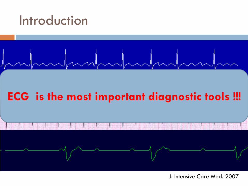
Introduction
Cardiac arrhythmias are a common cause of sudden death.
Symptomatic arrhythmia frequently observed in the ICU hemodynamic compromise
J. Intensive Care Med. 2007
ECG is the most important diagnostic tools !!!
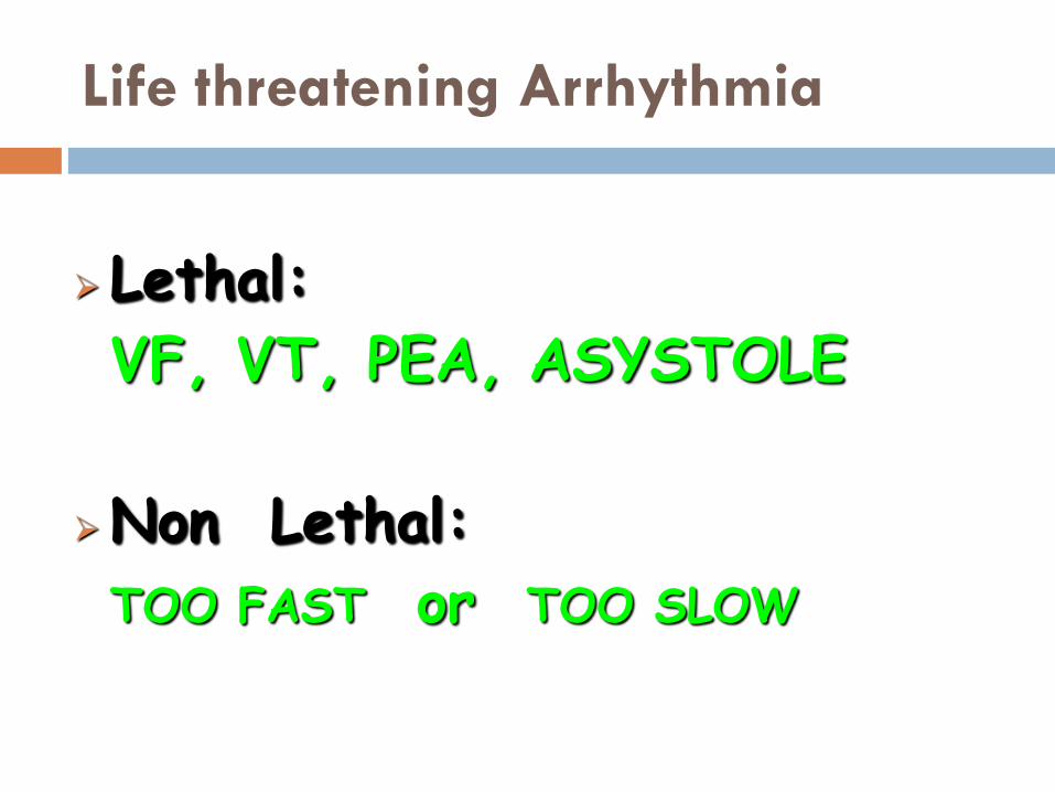
Life threatening Arrhythmia
Lethal:
VF, VT, PEA, ASYSTOLE
Non Lethal:
TOO FAST or TOO SLOW
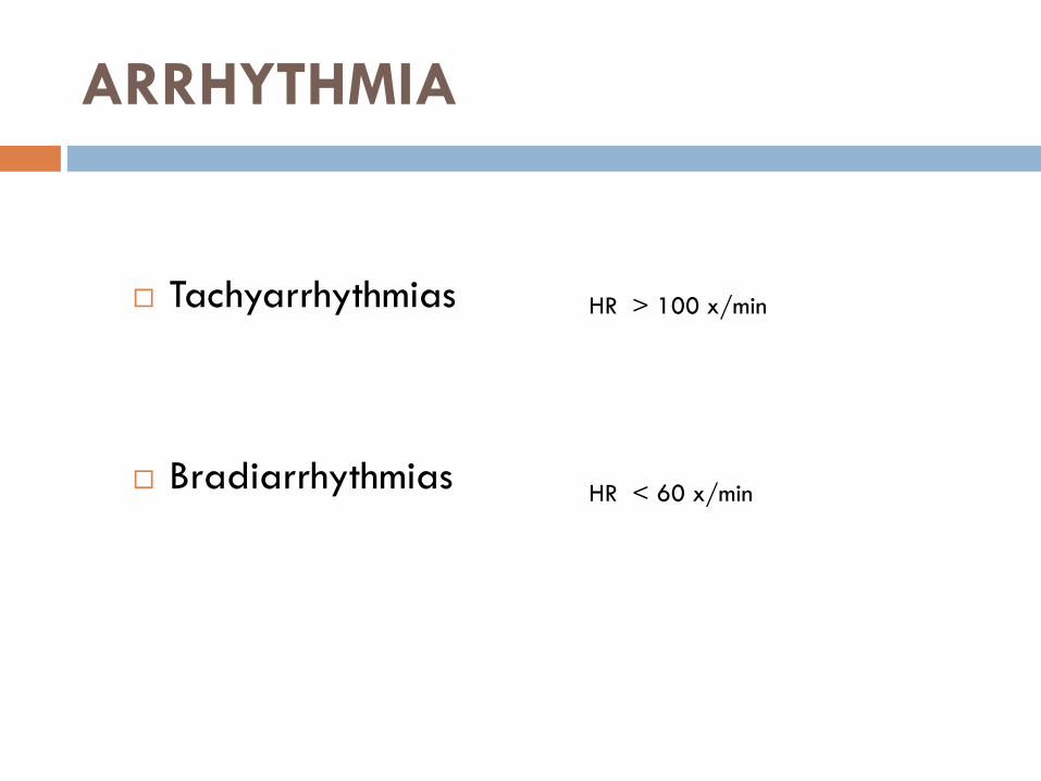
ARRHYTHMIA
Tachyarrhythmias
Bradiarrhythmias
HR > 100 x/min
HR < 60 x/min
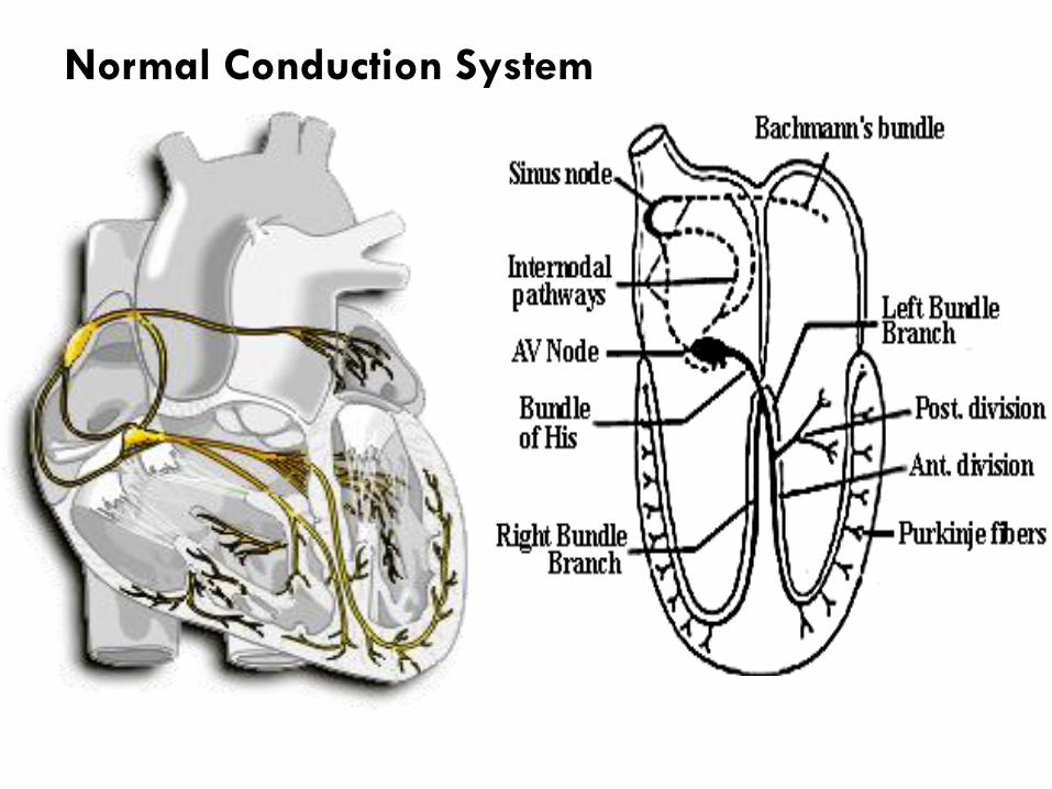
Normal Conduction System
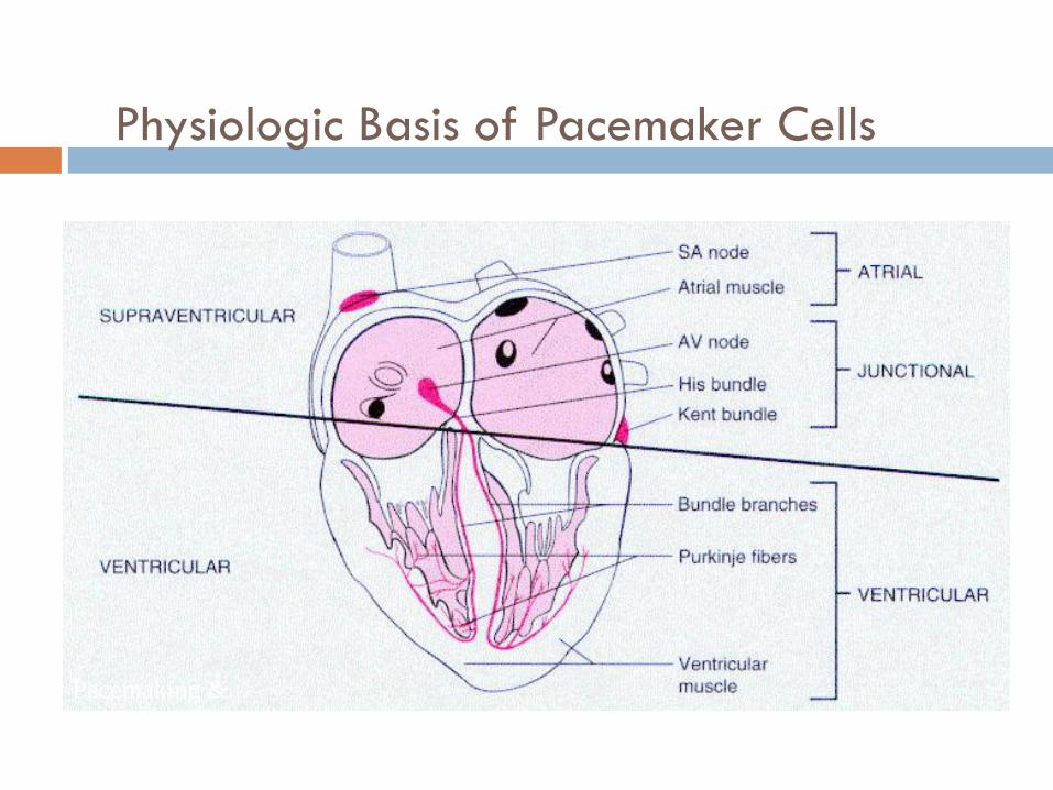
Physiologic Basis of Pacemaker Cells
Pacemaking &
Conduction System
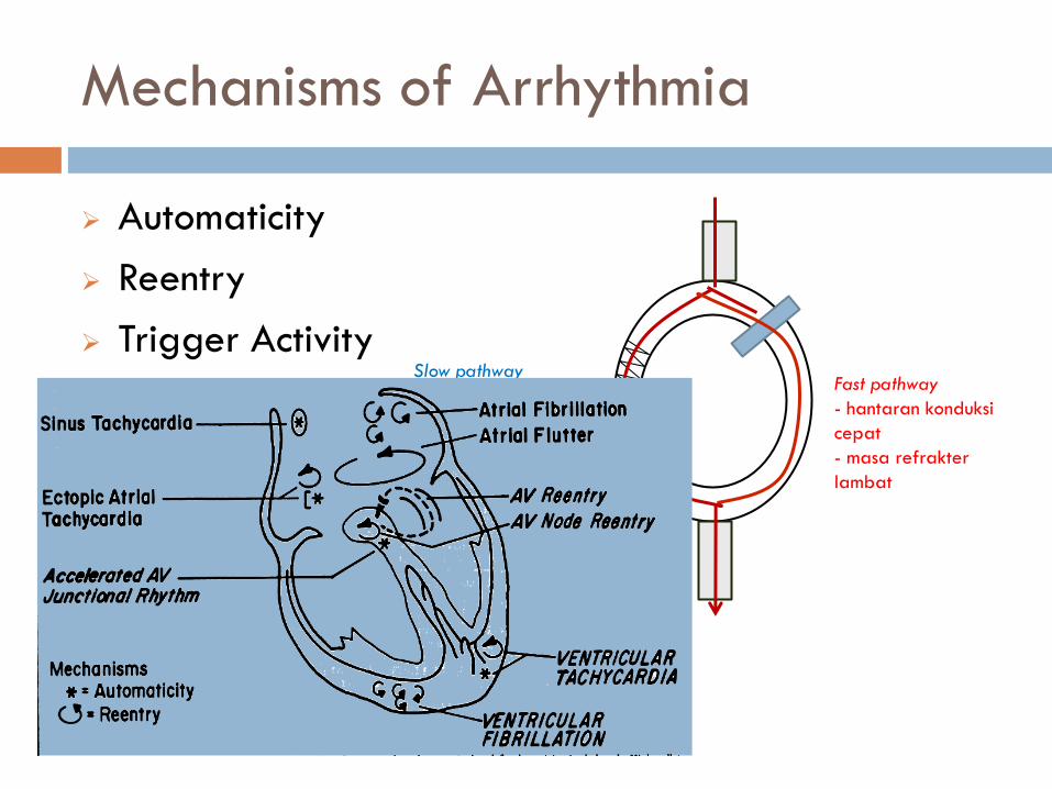
Mechanisms of Arrhythmia
Automaticity
Reentry
Trigger Activity Slow pathway
- hantaran konduksi
lambat
- masa refrakter
cepat
Fast pathway
- hantaran konduksi
cepat
- masa refrakter
lambat
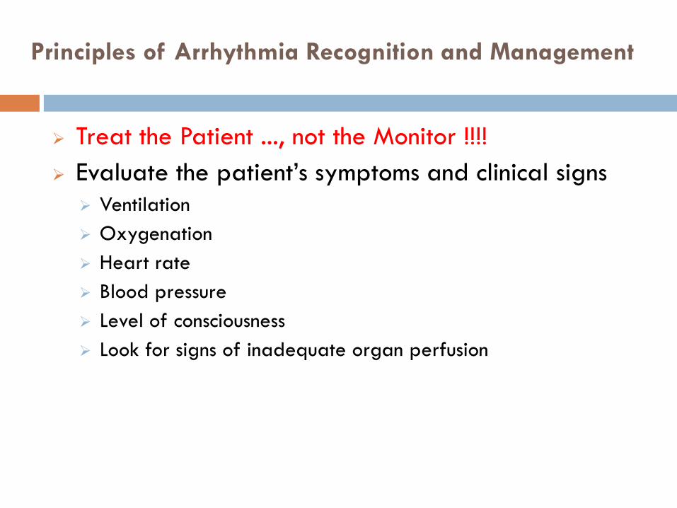
Treat the Patient ..., not the Monitor !!!!
Evaluate the patient’s symptoms and clinical signs
Ventilation
Oxygenation
Heart rate
Blood pressure
Level of consciousness
Look for signs of inadequate organ perfusion
Principles of Arrhythmia Recognition and Management
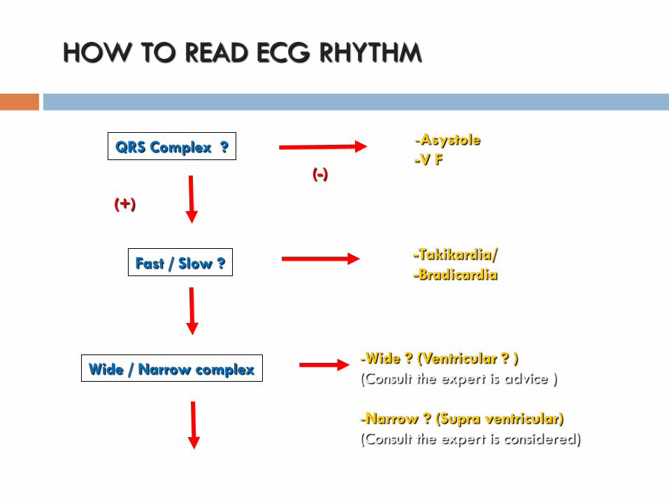
QRS Complex ?
(-)
-Asystole
-V F
(+)
Fast / Slow ? -Takikardia/
-Bradicardia
Wide / Narrow complex -Wide ? (Ventricular ? )
(Consult the expert is advice )
-Narrow ? (Supra ventricular)
(Consult the expert is considered)
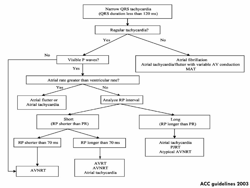
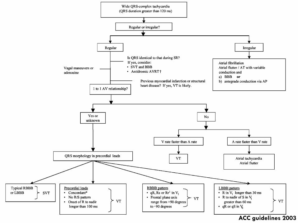
HOW TO READ ECG RHYTHM
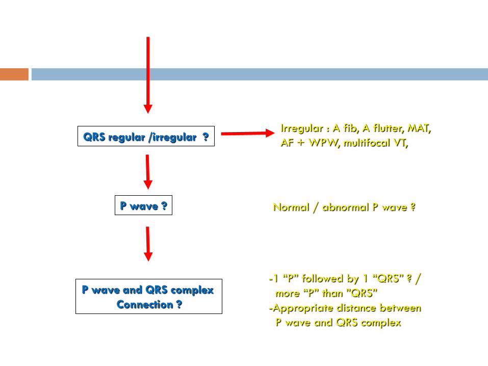
QRS regular /irregular ?
P wave ?
P wave and QRS complex
Connection ?
Normal / abnormal P wave ?
-1 “P” followed by 1 “QRS” ? /
more “P” than ”QRS”
-Appropriate distance between
P wave and QRS complex
Irregular : A fib, A flutter, MAT,
AF + WPW, multifocal VT,
Tachy-Arrhtyhmia
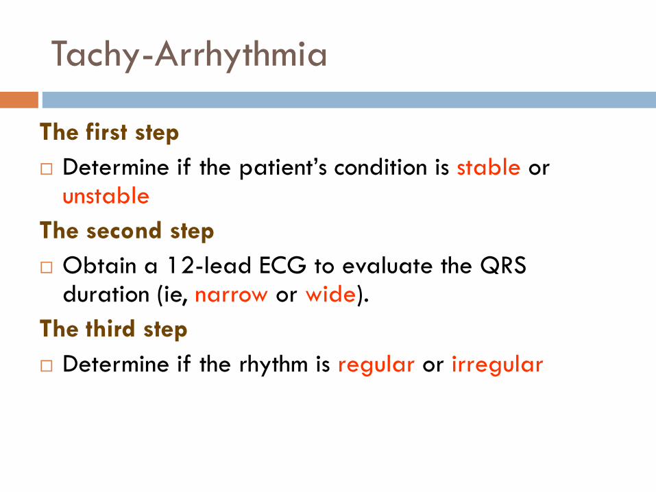
Tachy-Arrhythmia
The first step
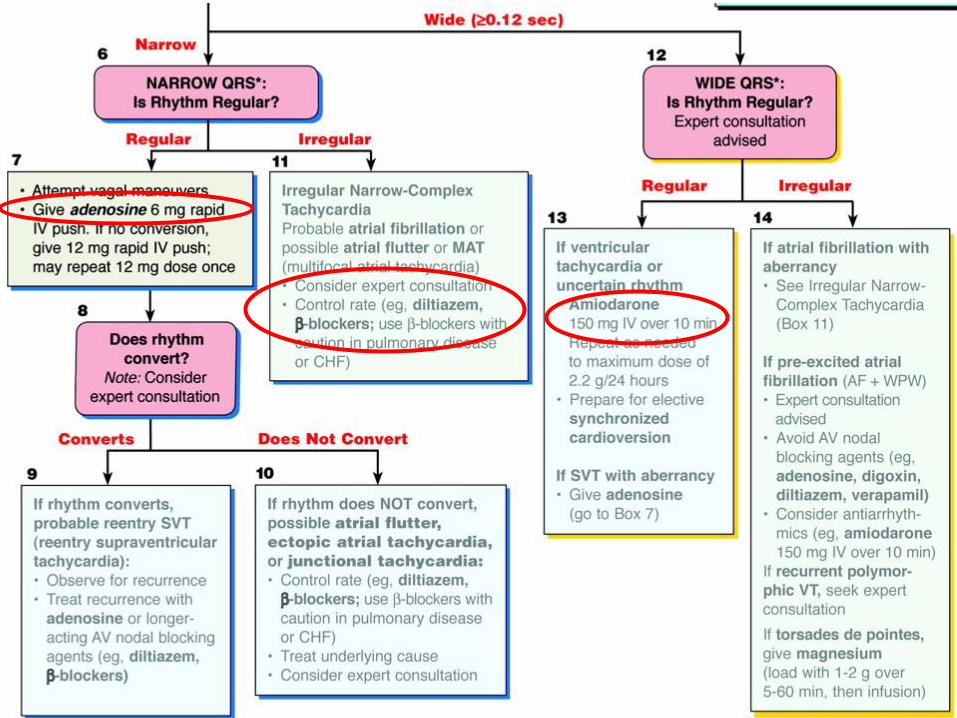
Determine if the patient’s condition is stable or unstable
The second step
Obtain a 12-lead ECG to evaluate the QRS duration (ie, narrow or wide).
The third step
Determine if the rhythm is regular or irregular
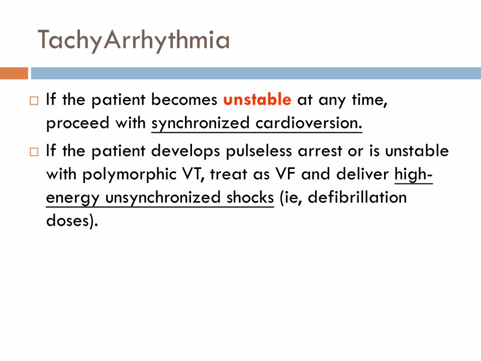
TachyArrhythmia
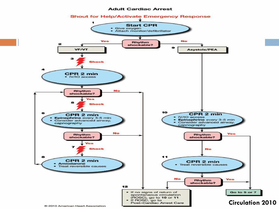
If the patient becomes unstable at any time,
proceed with synchronized cardioversion.
If the patient develops pulseless arrest or is unstable
with polymorphic VT, treat as VF and deliver high-
energy unsynchronized shocks (ie, defibrillation
doses).
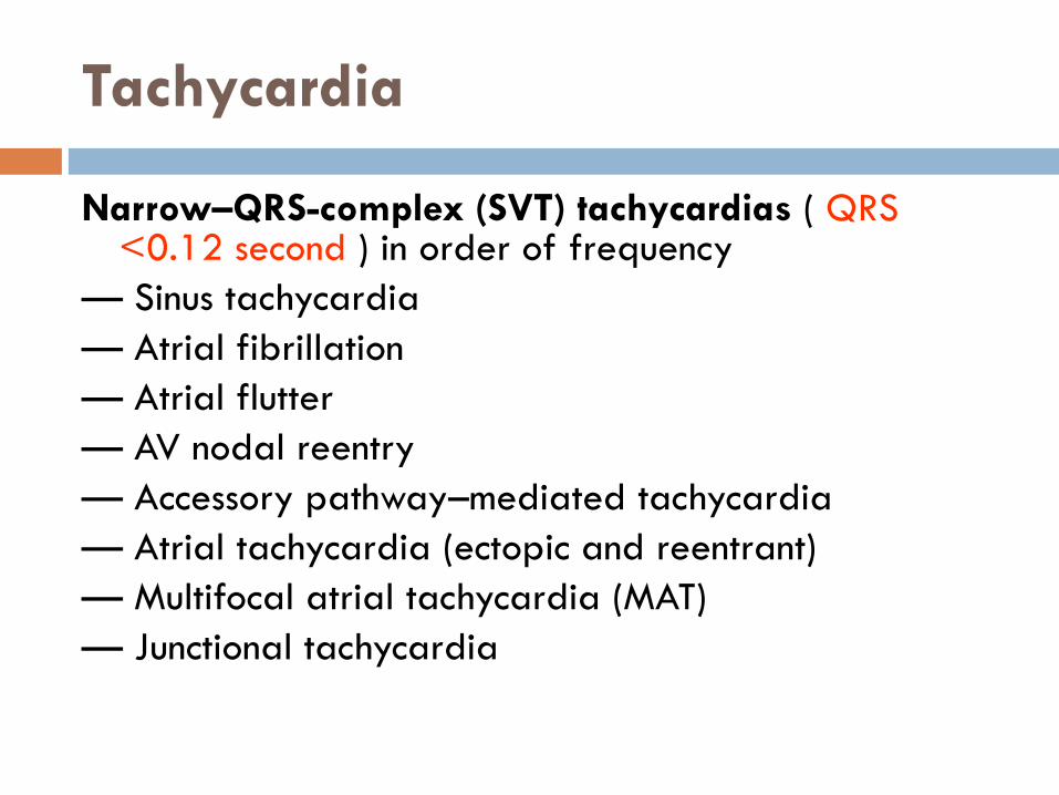
Tachycardia
Narrow–QRS-complex (SVT) tachycardias ( QRS <0.12 second ) in order of frequency
— Sinus tachycardia
— Atrial fibrillation
— Atrial flutter
— AV nodal reentry
— Accessory pathway–mediated tachycardia
— Atrial tachycardia (ectopic and reentrant)
— Multifocal atrial tachycardia (MAT)
— Junctional tachycardia
ACC guidelines 2003
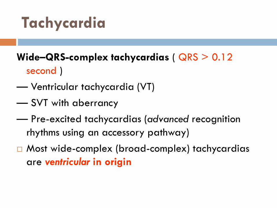
Tachycardia
Wide–QRS-complex tachycardias ( QRS > 0.12
second )
— Ventricular tachycardia (VT)
— SVT with aberrancy
— Pre-excited tachycardias (advanced recognition
rhythms using an accessory pathway)
Most wide-complex (broad-complex) tachycardias
are ventricular in origin
ACC guidelines 2003
Tachycardia
Initial Evaluation and Treatment of Tachyarrhythmias
The evaluation and management of tachyarrhythmias is depicted in the ACLS Tachycardia Algorithm.
Circulation 2010
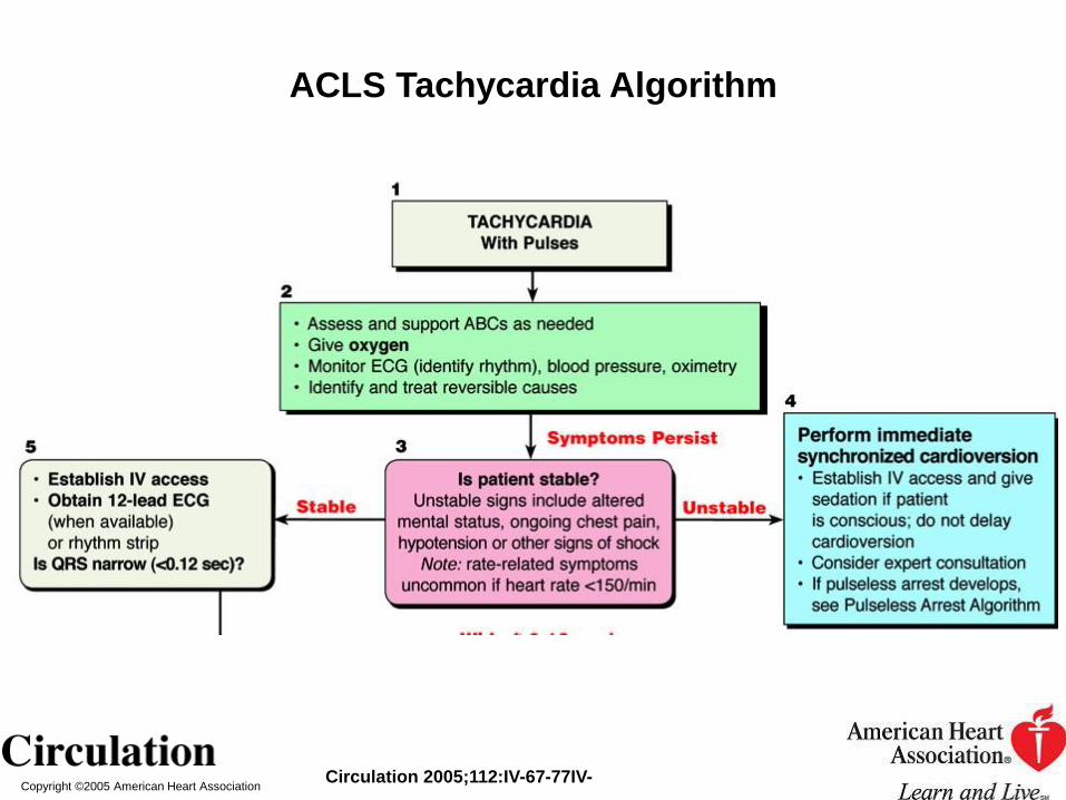
Copyright ©2005 American Heart Association Circulation 2005;112:IV-67-77IV-
ACLS Tachycardia Algorithm
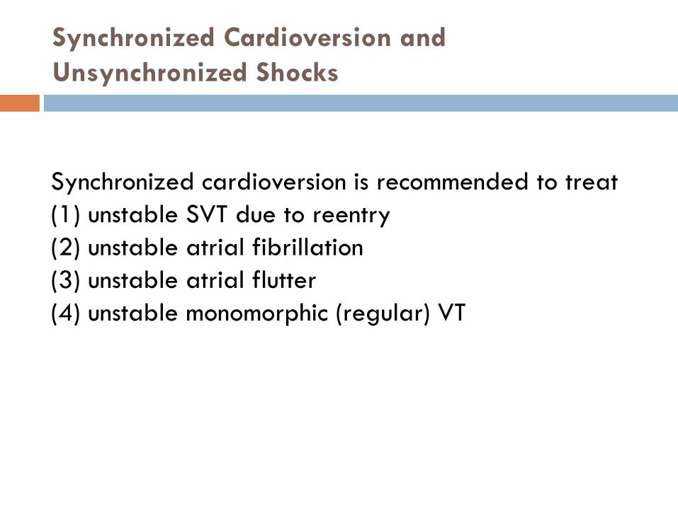
Synchronized Cardioversion and
Unsynchronized Shocks
Synchronized cardioversion is recommended to treat
(1) unstable SVT due to reentry
(2) unstable atrial fibrillation
(3) unstable atrial flutter
(4) unstable monomorphic (regular) VT
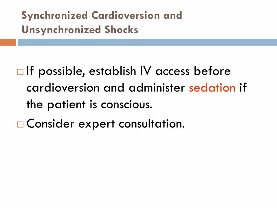
Synchronized Cardioversion and
Unsynchronized Shocks
If possible, establish IV access before
cardioversion and administer sedation if
the patient is conscious.
Consider expert consultation.
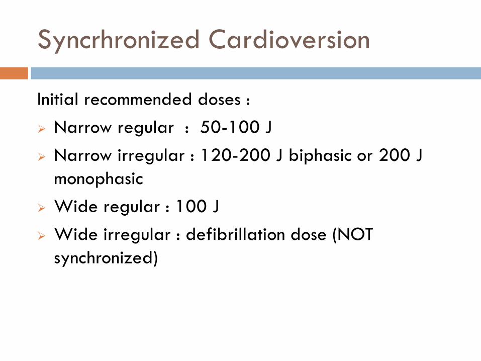
Syncrhronized Cardioversion
Initial recommended doses :
Narrow regular : 50-100 J
Narrow irregular : 120-200 J biphasic or 200 J
monophasic
Wide regular : 100 J
Wide irregular : defibrillation dose (NOT
synchronized)
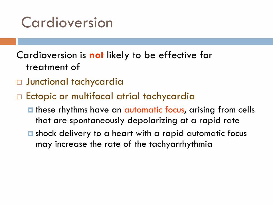
Cardioversion
Cardioversion is not likely to be effective for treatment of
Junctional tachycardia
Ectopic or multifocal atrial tachycardia
these rhythms have an automatic focus, arising from cells that are spontaneously depolarizing at a rapid rate
shock delivery to a heart with a rapid automatic focus may increase the rate of the tachyarrhythmia
Brady-Arrhythmia
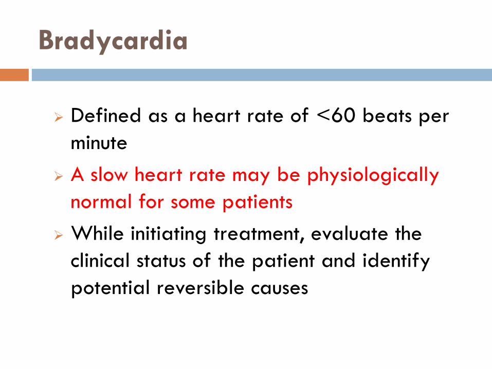
Bradycardia
Defined as a heart rate of <60 beats per
minute
A slow heart rate may be physiologically
normal for some patients
While initiating treatment, evaluate the
clinical status of the patient and identify
potential reversible causes
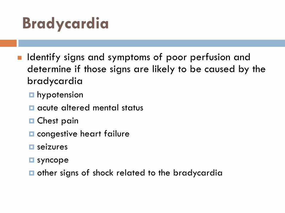
Bradycardia
Identify signs and symptoms of poor perfusion and determine if those signs are likely to be caused by the bradycardia
hypotension
acute altered mental status
Chest pain
congestive heart failure
seizures
syncope
other signs of shock related to the bradycardia
Bradycardia
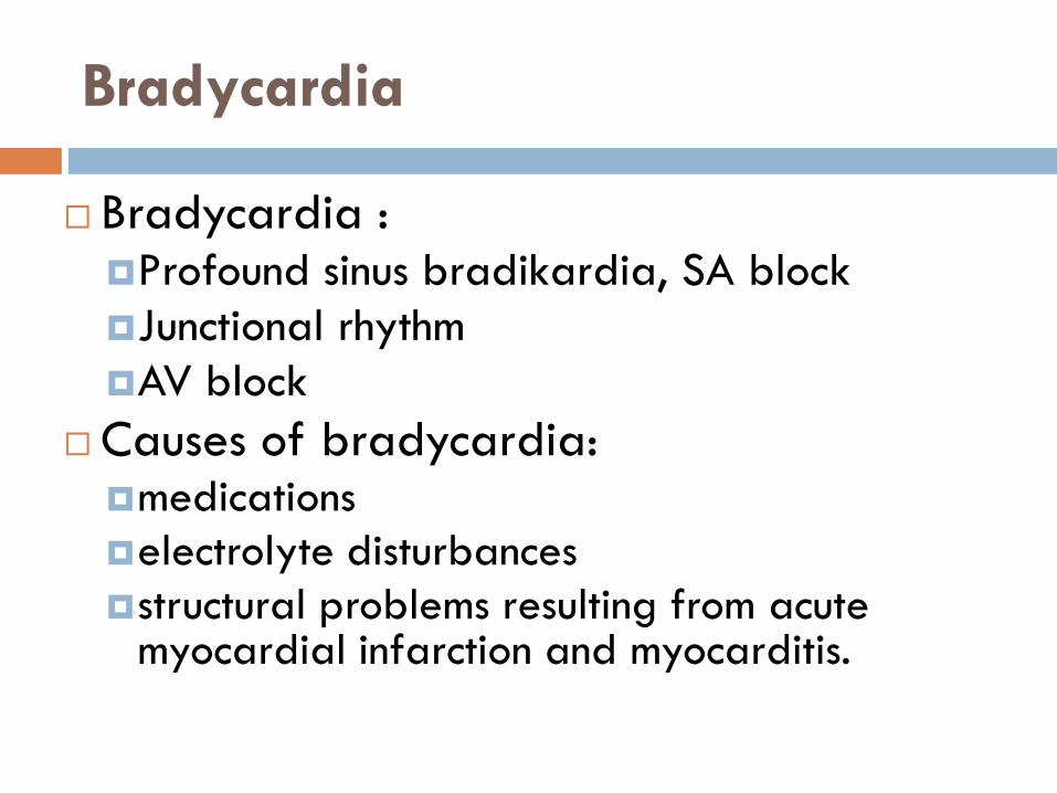
Bradycardia : Profound sinus bradikardia, SA block
Junctional rhythm
AV block
Causes of bradycardia: medications
electrolyte disturbances
structural problems resulting from acute myocardial infarction and myocarditis.
Copyright ©2005 American Heart Association
Circulation 2005;112:IV-67-77IV-
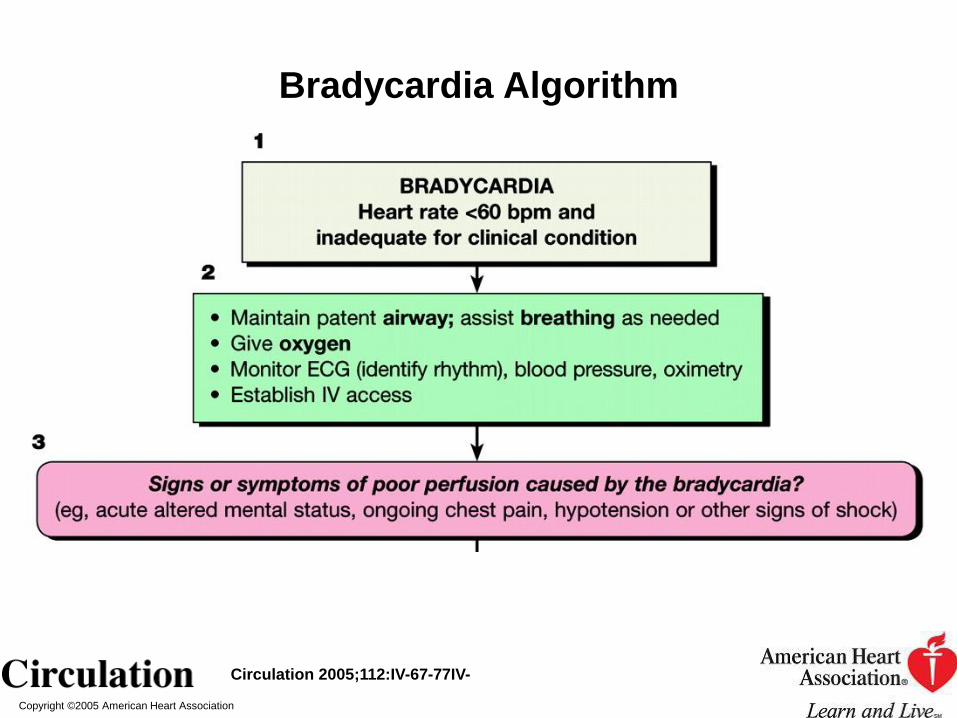
Bradycardia Algorithm
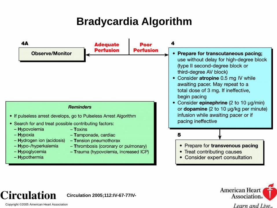
Copyright ©2005 American Heart Association
Circulation 2005;112:IV-67-77IV-
Bradycardia Algorithm
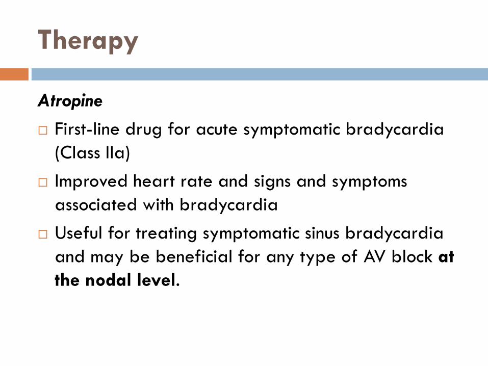
Therapy
Atropine
First-line drug for acute symptomatic bradycardia
(Class IIa)
Improved heart rate and signs and symptoms
associated with bradycardia
Useful for treating symptomatic sinus bradycardia
and may be beneficial for any type of AV block at
the nodal level.
Therapy
Atropine
The recommended dose for bradycardia is 0.5 mg IV every 3 to 5 minutes to a maximum total dose of 3 mg.
Doses <0.5 mg may paradoxically result in further slowing of the heart rate.
Atropine administration should not delay implementation of external pacing for patients with poor perfusion.
Therapy
Atropine
Use cautiously in the presence of acute coronary ischemia or myocardial infarction; increased heart rate may worsen ischemia or increase the zone of infarction.
Atropine may be used with caution and appropriate monitoring following cardiac transplantation. It will likely be ineffective because the transplanted heart lacks vagal innervation.
Therapy
Pacing (Transcutaneous pacing, TCP )
Class I intervention for symptomatic bradycardias
Indication : started immediately for patients Unstable, particularly those with high-degree block
If there is no response to atropine
If atropine is unlikely to be effective
If the patient is severely symptomatic
Therapy
Pacing (Transcutaneous pacing, TCP )
Can be painful and may fail to produce effective
mechanical capture
Use analgesia and sedation for pain control
Verify mechanical capture and re-assess the patient’s
condition
If TCP is ineffective (eg, inconsistent capture)
prepare for transvenous pacing
consider obtaining expert consultation
Therapy
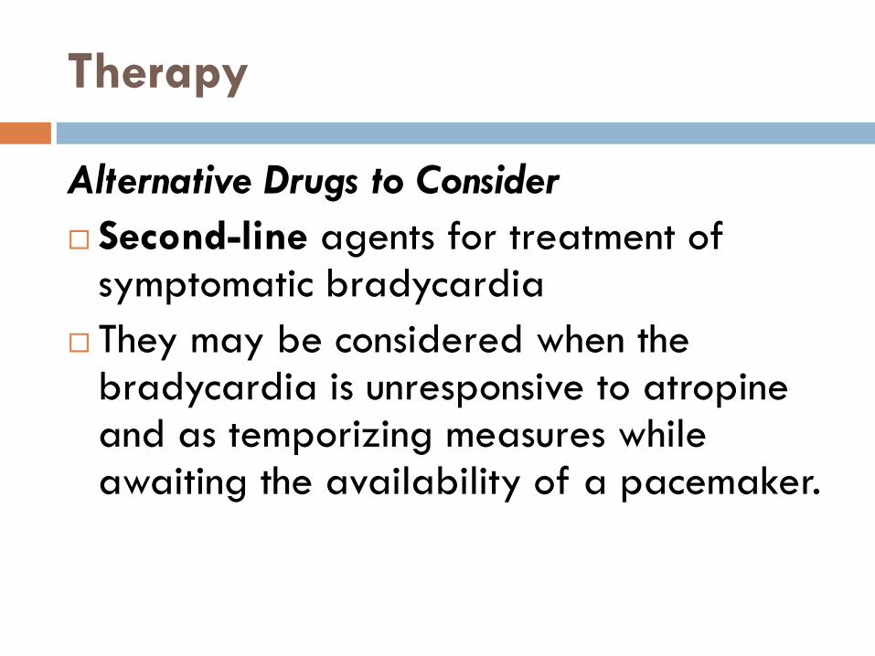
Alternative Drugs to Consider
Second-line agents for treatment of symptomatic bradycardia
They may be considered when the bradycardia is unresponsive to atropine and as temporizing measures while awaiting the availability of a pacemaker.
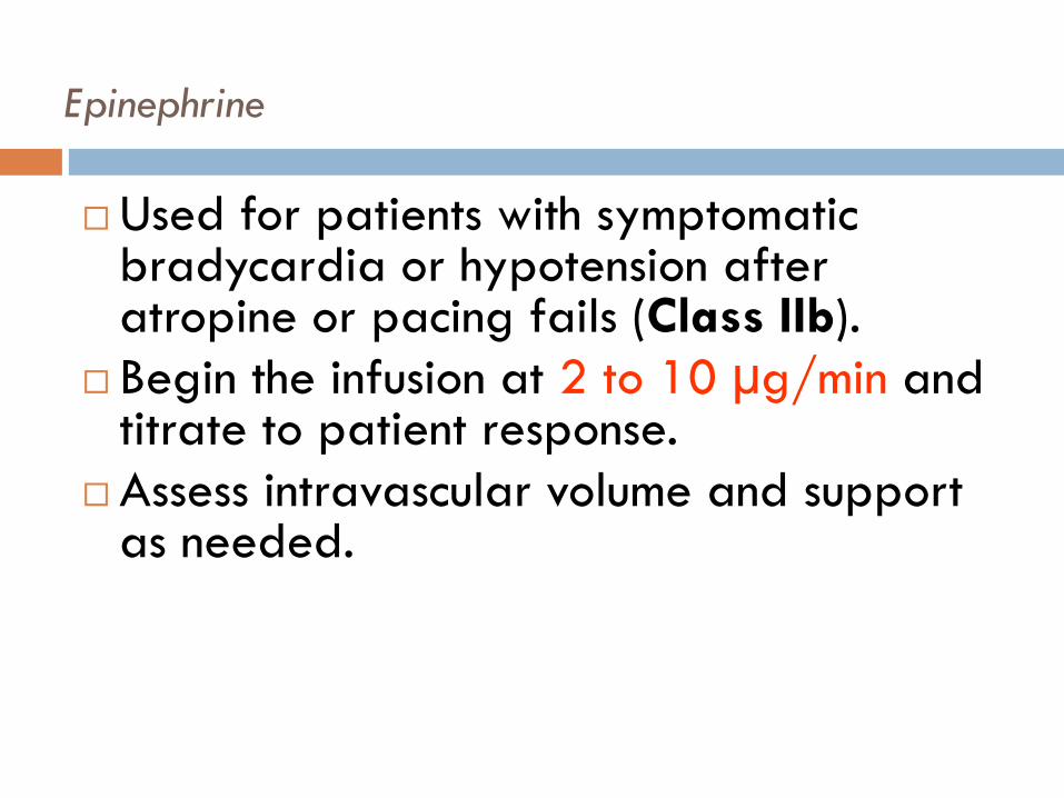
Epinephrine
Used for patients with symptomatic bradycardia or hypotension after atropine or pacing fails (Class IIb).
Begin the infusion at 2 to 10 µg/min and titrate to patient response.
Assess intravascular volume and support as needed.
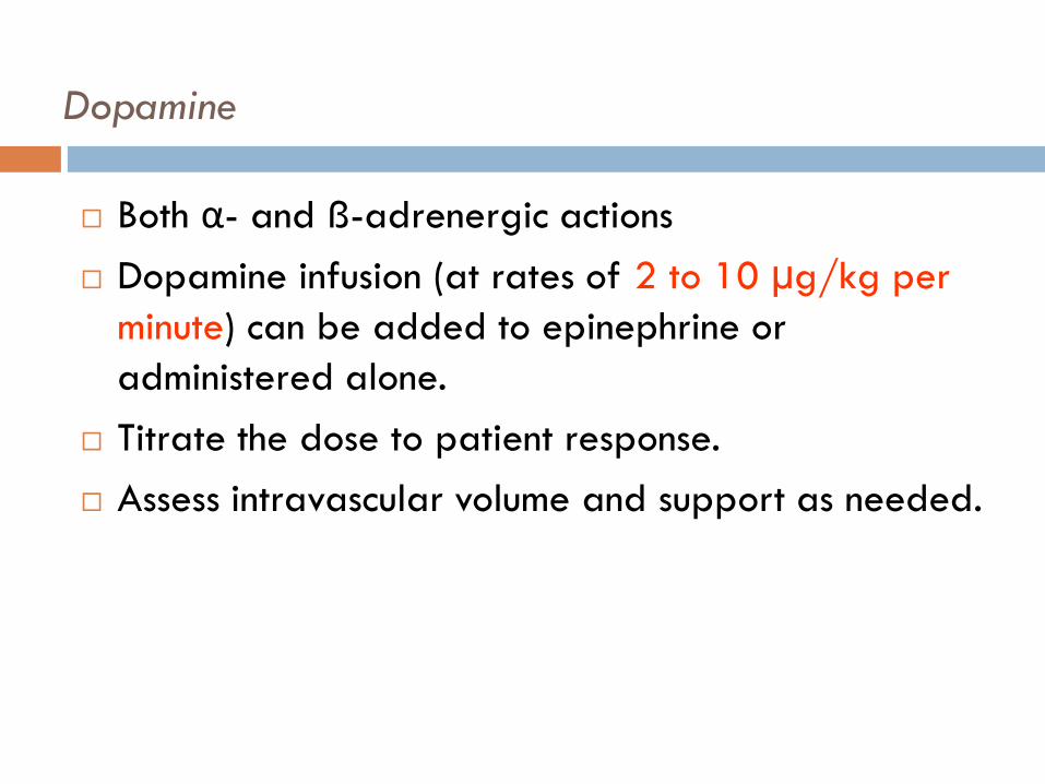
Dopamine
Both α- and ß-adrenergic actions
Dopamine infusion (at rates of 2 to 10 µg/kg per
minute) can be added to epinephrine or
administered alone.
Titrate the dose to patient response.
Assess intravascular volume and support as needed.

Thank you for your attention !!