Embed Size (px)
Citation preview
Ligaments and Tendons Part 3
David Flanigan, MD
Assistant Professor of Clinical Orthopaedics
Team Physician
The OSU Sports Medicine Center

Objectives
Block Objectives Contrast the differences between tendons and ligaments Define the histology and composition of ligaments Identify the function of ligaments Explain the stress/strain curve and how it applies to ligaments Define crimp and anisotrophic and how they apply to ligaments Define the histology and composition of tendons Identify the function of tendons

Difference Ligaments vs Tendons
Resistance to tension Tendon
Uniformly stiff Does not elongate Allows conservation of
energy Ligament
Built-in laxity Allows small forces across
joint Protects joint

What are ligaments?
Fibrous soft tissue Connect bone to bone Allow normal joint motion Prevent abnormal motion,
instability

Role of ligaments
Secondary restraint during normal activity under load Weight-bearing
Guide unloaded motion E.g. knee “screw-home”
Stop abnormal motion to avoid further damage

Function and anatomy
Ropes, not bars Resist tension only
Redundancy across joint Example 1: 4 main
knee ligaments Example 2: ankle
ligaments Block all undesirable
motions loads pass across articulating surfaces

Anisotropic Properties
Strong in tension Weak in compression Anisotropic
Mechanical properties depend on the orientation of the force applied

Muscles are 1st line of defense
Hiking over rocky terrain example
Inversion resisted 1st by peroneus muscles
Ligaments come into play when muscle inactive or too weak

http://www.merck.com/mmhe/print/sec05/ch072/ch072c.html
http://www.eorthopod.com/eorthopodV2/index.php/fuseaction/topics.detail/ID/1e69153b4390c6eff3095daeefe6031a/TopicID/f3734010e47d0fce02d98570d66e2a38/area/19
Ankle Ligaments
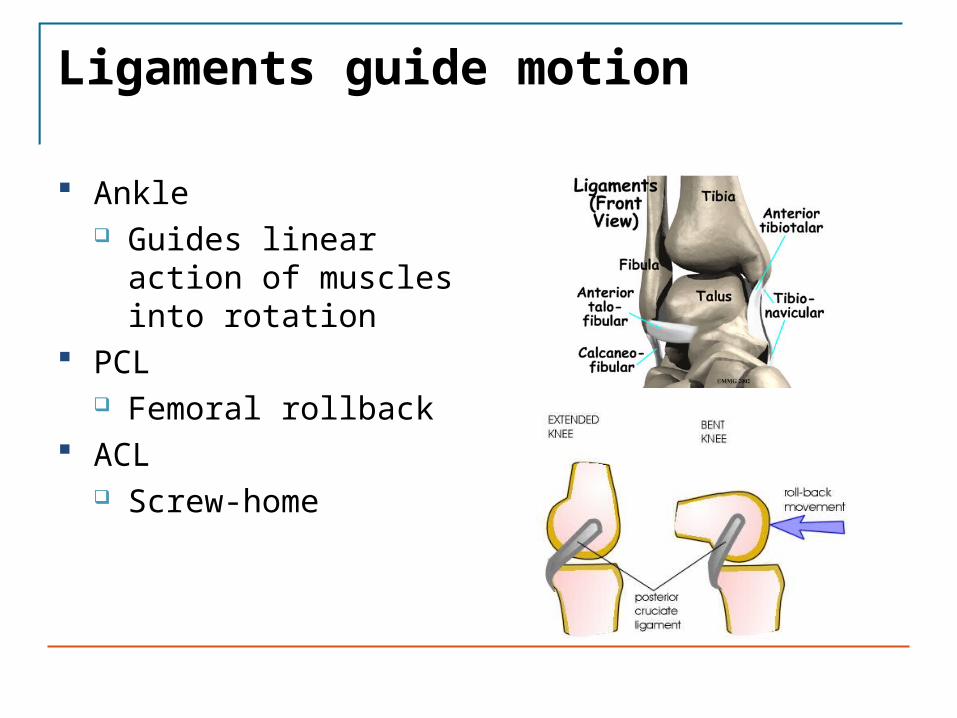
Ligaments guide motion
Ankle Guides linear action of
muscles into rotation PCL
Femoral rollback ACL
Screw-home

Physiologic structure of ligaments
Non-linear stiffness Laxity at low loads High stiffness at high loads
Highly aligned collagen fascicles “Crimp” to provide non-linearity
Straighten out first (low stiffness) Fibrils stretch (high stiffness)
Boorman et al (2006) Journal of Orthopaedic Research 24(4):795.

0
100
200
300
400
0 2 4 6 8 10
Load[N]
Elongation [mm]Reproduced from Woo et al., 2000
Low Stiffness
High Stiffness
Load and Elongation
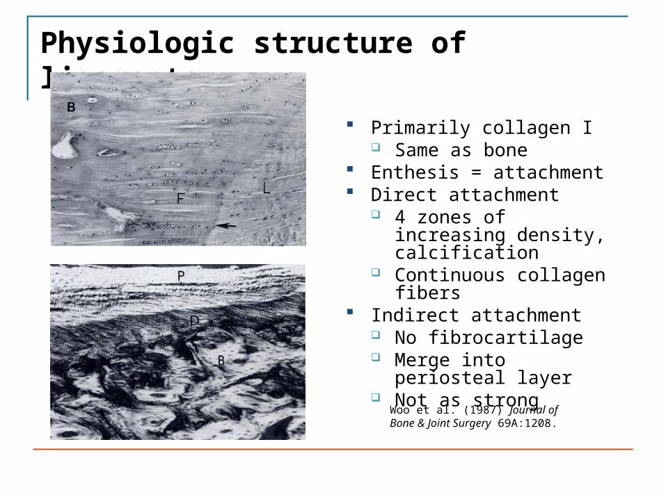
Physiologic structure of ligaments
Primarily collagen I Same as bone
Enthesis = attachment Direct attachment
4 zones of increasing density, calcification
Continuous collagen fibers Indirect attachment
No fibrocartilage Merge into periosteal layer Not as strong
Woo et al. (1987) Journal of Bone & Joint Surgery 69A:1208.

Ligament maintenance
3 principal cell types Fusiform Ovoid, spheroid
Vascular supply Inside—endoligament Outside—epiligament Diffusion of nutrients to
cells
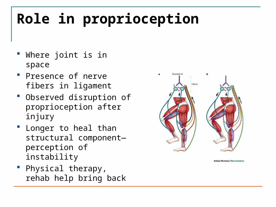
Role in proprioception
Where joint is in space Presence of nerve fibers
in ligament Observed disruption of
proprioception after injury Longer to heal than
structural component—perception of instability
Physical therapy, rehab help bring back

Ligament injury
Grade I—pain, no instability Grade II—some torn fibers, minimal instability Grade III—completely torn Always from excessive tension Dislocations always include ligament injury
I II III
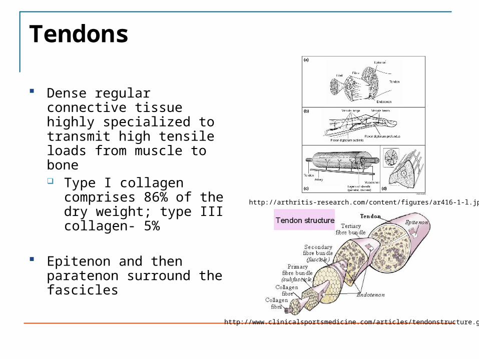
Tendons
Dense regular connective tissue highly specialized to transmit high tensile loads from muscle to bone Type I collagen comprises
86% of the dry weight; type III collagen- 5%
Epitenon and then paratenon surround the fascicles
http://www.clinicalsportsmedicine.com/articles/tendonstructure.gif
http://arthritis-research.com/content/figures/ar416-1-l.jpg

Tendon Insertion
Specialized direct insertion Four zones
Tendon Fibrocartilage Mineralized fibrocartilage Bone
Sharpey’s fibers- Collagen bundles that
extend from the tendon or periosteum into the bone.
http://anatomy.iupui.edu/courses/histo_

Two Types of Tendons
Tendons that pull in a straight line are not enclosed by a sheath but by a paratenon (Achilles tendon), which is loose connective tissue continuous with the tendon
Tendons which are required to bend (flexor tendons of the hand) are enclosed by a tendon sheath which directs the tendon path and acts like a pulley; motion is assisted by synovial fluid produced by epitenon
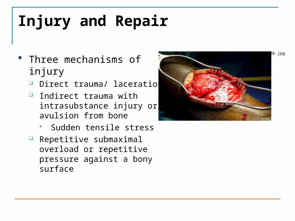
Injury and Repair
Three mechanisms of injury Direct trauma/ laceration Indirect trauma with
intrasubstance injury or avulsion from bone Sudden tensile stress
Repetitive submaximal overload or repetitive pressure against a bony surface
http://www.mccainortho.com/Patella%20Fx%20406b.jpg

Thank You

Survey
We would appreciate your feedback on this module. Click on the button below to complete a brief survey. Your responses and comments will be shared with the module’s author, the LSI EdTech team, and LSI curriculum leaders. We will use your feedback to improve future versions of the module.
The survey is both optional and anonymous and should take less than 5 minutes to complete.
Survey