Embed Size (px)
Citation preview
Proc. Natl. Acad. Sci. USAVol. 92, pp. 5940-5944, June 1995Genetics
Ligand-regulated site-specific recombination(steroid/estrogen/FLP recombinase/inducible gene expression/genetic engineering)
COLIN LOGIE AND A. FRANcIs STEWART*Gene Expression Program, European Molecular Biology Laboratory, Meyerhofstrasse 1, D-69117 Heidelberg, Federal Republic of Germany
Communicated by Lennart Philipson, New York University Medical Center, New York, NY February 21, 1995 (received for review January 9, 1995)
ABSTRACT Site-specific recombination offers a potentialway to alter a living genome by design in a precise and stablemanner. This potential requires strategies which can be usedto regulate the recombination event. We describe a strategy toregulate FLP recombinase activity which relies on expressingFLP as a fusion protein with steroid hormone receptor ligandbinding domains (LBDs). In the absence of a ligand cognateto the LBD, the recombinase activity of the fusion protein isextremely low. Upon ligand administration, recombinase ac-tivity is rapidly induced. These results outline the basis forinducible expression or disruption strategies based on induc-ible recombination. Additionally, we have exploited the con-ditional nature of FLP-LBD fusion proteins to direct inte-gration of a plasmid into a specific genomic site at frequenciesapproaching the frequency of random integration.
Site-specific recombinases of the integrase family catalyzeexcisions, insertions, inversions, or translocations of DNAbetween their recognition target sites. The orientation of thesesites relative to each other and the configuration of thesurrounding DNA determine the product(s) obtained (1, 2).Several integrase family members, including FLP and Crerecombinases, carry all functions required for the recombina-tion reaction in a single polypeptide chain (3-5). Consequently,these recombinases are particularly amenable to applicationsin living systems. They are active when introduced into cellsfrom a broad range of organisms, including Escherichia coli,yeasts, and mammals (2, 3, 6-12). A variety of applicationsbased on the introduction of both the recombinase and ap-propriately disposed recombination target sites have beensuccessful. Full exploitation of site-specific recombination toalter genotype in living systems requires strategies to regulatethe recombination event. In previous studies, regulation hasbeen accomplished by controlled expression of the recombi-nase gene (2) or by introduction of recombinase mRNA orprotein (13, 14). Here we describe a strategy based on regu-lating the activity, rather than the expression, of a site-specificrecombinase.The molecular cloning ofcDNAs for members of the steroid
receptor superfamily revealed that the protein domain respon-sible for binding steroid, the ligand-binding domain (LBD),also inactivates the other functions of the receptor in theabsence of bound steroid (15-17). Inactivation is probablymediated by association of the nonligand-bound LBD with anabundant protein complex containing Hsp9O (18-20). TheLBD has been fused to several transcription factors andoncogenes, imparting the properties of inactivation in theabsence of, and activation by, cognate ligands (21). To testwhether LBD properties could be employed to regulate asite-specific recombinase, we made fusion proteins betweenFLP recombinase and LBDs from the estrogen, glucocorti-coid, or androgen receptor. We show that recombinationmediated by FLP-LBD fusion proteins is strictly regulated in
The publication costs of this article were defrayed in part by page chargepayment. This article must therefore be hereby marked "advertisement" inaccordance with 18 U.S.C. §1734 solely to indicate this fact.
the absence and efficiently induced in the presence of ligandadministration. The strict regulation permits the establishmentof stable cell lines carrying both the expressed fusion proteinand the unrearranged recombination substrate, and recombi-nation is rapidly induced upon ligand administration. Further-more, the presence of expressed FLP-LBD in cells transfectedwith plasmids containing recombinase target sites permitssite-specific integration at efficiencies approaching that ofrandom integration.
MATERIALS AND METHODSCell Culture. All cells were cultured in Dulbecco's modified
Eagle's medium without phenol red containing 10% charcoal-stripped fetal calf serum, 2 mM glutamine, and 100 interna-tional units each of penicillin and streptomycin per ml. Fetalcalf serum was stripped by stirring 500 ml with 25 g of NoritA (Serva) for 30 min, followed by centrifugation at 13,000 xg for 20 min and filtration twice through filters with 0.2-,umdiameter pores.
Transfections. Transfections for transient expression utilizedN-[1-(2,3-dioleoyloxy)propyl]-N,N,N-trimethylammoniummethyl sulfate (DOTAP; Boehringer Mannheim) according tothe manufacturer's instructions with 5 ,ug of plasmids pOG44,pFRT,BGal, p44HER1, p44HGR1, or p44HAR1, as indicated,and 105 E25B2B2 cells grown in 3.5-cm dishes. Stable derivativesof 293 cells (ATCC CRL-1573; American Type Culture Collec-tion) were generated by electroporation. Cells were harvested bytrypsinization and centrifugation, washed one time in phosphate-buffered saline (PBS; 137 mM NaCl/2.7 mM KCI/4.3 mMNa2HPO4/1.4mM KH2PO4), and resuspended in PBS at 107 cellsper ml. Then, 500 ,ul of cell suspension was mixed with 2 ,g oflinearized or 5 ,ug of circular plasmidDNA in 0.4-cm cuvettes andelectroporated at 300 V and 960 ,uF by using a Bio-Rad GenePulser. The cells were then cultured for 24 h before being selected[400 ,ug of Geneticin (G418; GIBCO) and 400 units of hygro-mycin (Calbiochem) per ml].
Cell Lines. E25B2B2 cells were derived by calcium phos-phate cotransfection ofE25B2 cells (ref. 9; Stratagene) with 19,ug of pOG44 and 1 Ag of pOG45, followed by selection forG418 resistance. A site-specific genomic integrant was iden-tified by a lack of ,B-galactosidase expression and by Southernanalysis. Derivatives of 293 cells containing randomly inte-grated pNEO,3GAL were created by transfecting cells withApa I-linearized pNEO3GAL. Four clones that were assessedby Southern analysis to contain independent, single-copyintegrations of the pNEO3GAL transgene were selected fortransfection with pHFE1. Cell lines resistant to hygromycinand G418 were generated by transfection with Xmn I linear-ized pHFE1. Cell lines P1.2B, P1.4B, and Q3.2B were made byplating 100 P1.2, P1.4, or Q3.2 cells onto 10-cm dishes andculturing them in the presence of 100 nM estradiol. Several
Abbreviations: LBD, ligand-binding domain; EBD, estrogen-bindingdomain; FRT, FLP recombinase recognition target.*To whom reprint requests should be addressed.
5940
Proc. Natl. Acad. Sci. USA 92 (1995) 5941
isolated colonies were picked for each and fuconfirmed that all had excised the neomycin-res
Assays. ,B-Galactosidase expression was detecithe method of Sanes et al. (22). f3-Galactosida;levels in cell extracts were determined by theHerbomel et al. (23). Southern analysis was pdescribed (24).
RESULTS
Transient Expression of FLP-LBD Fusion Prous studies with LBD fusion proteins have demcvarious transcription factors and oncoproteinslated by steroid hormones (21). To test whet}fusion-protein strategy could also regulate a sitcombinase, we used a genomic recombination assassay, the open reading frame of the E. coli 1B-(lacZ) gene is interrupted by an expression cassmycin resistance, flanked by directly repeated Fnase recognition targets (FRTs; Fig. 1). FLP-mecbination excises the neomycin resistance gene fmosomal locus, thereby reconstituting the open rof the lacZ gene. In transient-expression experinadded ligand, the FLP-LBD fusions displayed a1% of the activity of unmodified FLP on theexcision target. Addition of the cognate ligandcombinase activity to up to 50% that of unnenzyme (Fig 2).
Stable Expression of FLP-Estrogen-Bind(EBD) Fusion Proteins. Stable cell lines contairunrecombined pNEO,BGAL and FLP-LBD exrmid were established in two steps. First, the rctarget pNEOI3GAL (Fig. 1) was introduced intelectroporation and selected by G418 resistance.
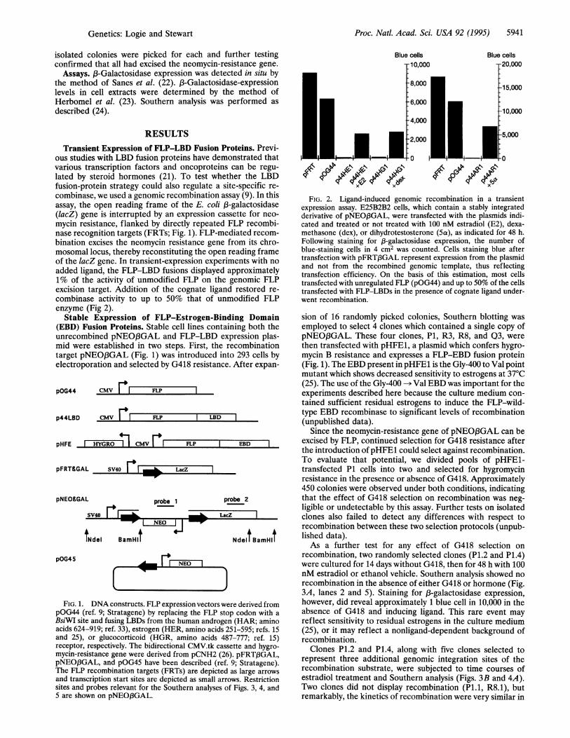
pOG44 CMV FI FLP l
p44LBD CMV I FLP LBD
pHFE HYGRO CMV FLP
pFRT&GAL SV40 | | LacZl
pNEOBGAL
SV40
probe 1
tNdel BamHlI
pOG45 .dM NEO I
prc
MM6 LacZ
FIG. 1. DNA constructs. FLP expression vectors weipOG44 (ref. 9; Stratagene) by replacing the FLP stolBsiWI site and fusing LBDs from the human androgenacids 624-919; ref. 33), estrogen (HER, amino acids 25and 25), or glucocorticoid (HGR, amino acids 487receptor, respectively. The bidirectional CMV.tk cassemycin-resistance gene were derived from pCNH2 (26)pNEO,BGAL, and pOG45 have been described (ref.The FLP recombination targets (FRTs) are depictedand transcription start sites are depicted as small arro
sites and probes relevant for the Southern analyses of5 are shown on pNEO,BGAL.
rther testingistance gene.ted in situ byse-expressione method oferformed as
oteins. Previ-nstrated thatcan be regu-her the LBDte-specific re-ay (9). In thisgalactosidaseiette for neo-'LP recombi-hiated recom-rom its chro-eading framenents with noipproximatelygenomic FLPrestored re-
nodified FLP
Blue cells
T10,000Blue cells
9ip90 4$b 41 s9i p0FIG. 2. Ligand-induced genomic recombination in a transient
expression assay. E25B2B2 cells, which contain a stably integratedderivative of pNEO,3GAL, were transfected with the plasmids indi-cated and treated or not treated with 100 nM estradiol (E2), dexa-methasone (dex), or dihydrotestosterone (5a), as indicated for 48 h.Following staining for 13-galactosidase expression, the number ofblue-staining cells in 4 cm2 was counted. Cells staining blue aftertransfection with pFRT3GAL represent expression from the plasmidand not from the recombined genomic template, thus reflectingtransfection efficiency. On the basis of this estimation, most cellstransfected with unregulated FLP (pOG44) and up to 50% of the cellstransfected with FLP-LBDs in the presence of cognate ligand under-went recombination.
ling Domain sion of 16 randomly picked colonies, Southern blotting wasning both the employed to select 4 clones which contained a single copy ofpression plas- pNEO,BGAL. These four clones, P1, R3, R8, and Q3, wereecombination then transfected with pHFE1, a plasmid which confers hygro-) 293 cells by mycin B resistance and expresses a FLP-EBD fusion proteinAfter expan- (Fig. 1). The EBD present in pHFE1 is the Gly-400 to Val point
mutant which shows decreased sensitivity to estrogens at 37°C(25). The use of the Gly-400 -- Val EBD was important for theexperiments described here because the culture medium con-tained sufficient residual estrogens to induce the FLP-wild-type EBD recombinase to significant levels of recombination(unpublished data).
Since the neomycin-resistance gene of pNEO,BGAL can beEBD excised by FLP, continued selection for G418 resistance after
the introduction ofpHFE1 could select against recombination.To evaluate that potential, we divided pools of pHFE1-transfected P1 cells into two and selected for hygromycinresistance in the presence or absence of G418. Approximately450 colonies were observed under both conditions, indicating
obe 2 that the effect of G418 selection on recombination was neg-ligible or undetectable by this assay. Further tests on isolated
l clones also failed to detect any differences with respect torecombination between these two selection protocols (unpub-
NdeltBamHlt lished data).As a further test for any effect of G418 selection on
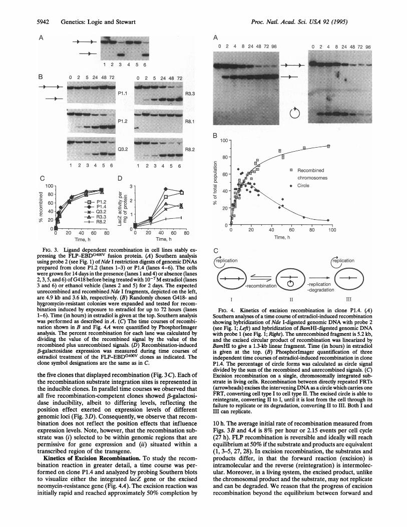
recombination, two randomly selected clones (P1.2 and P1.4)were cultured for 14 days without G418, then for 48 h with 100nM estradiol or ethanol vehicle. Southern analysis showed norecombination in the absence of either G418 or hormone (Fig.3A, lanes 2 and 5). Staining for 03-galactosidase expression,
re derived from however, did reveal approximately 1 blue cell in 10,000 in thep codon with a absence of G418 and inducing ligand. This rare event may(HAR; amino reflect sensitivity to residual estrogens in the culture medium51-595; refs. 15 (25), or it may reflect a nonligand-dependent background of7-777; ref. 15) recombination.wtte and hygro- Clones P1.2 and P1.4, along with five clones selected topFRTanGAL, represent three additional genomic integration sites of the
as large arrows recombination substrate, were subjected to time courses ofws. Restriction estradiol treatment and Southern analysis (Figs. 3B and 4A).Figs. 3, 4, and Two clones did not display recombination (P1.1, R8.1), but
remarkably, the kinetics of recombination were very similar in
Genetics: Logie and Stewart
5942 Genetics: Logie and Stewart
A0 2 4 8 24 48 72 96 0 2 4 8 24 48 72 96
1 2 3 4 5 6
B 0 2 5 24 48 72
P1.1
u- -p0= -aP1.2
_ ~ ._
12 3 4 5 6
C
a)CD
E00a)
Io,
10080
6040
20
0I
0 2 5 24 48 72
00--1 .. . ..._ ..
,- --.
"Wwqkwwp
Q3.2
12 3 4 5 6
D
t-
e)
NoaN m
0 20 40 60 80Time, h Time, h
FIG. 3. Ligand dependent recombination in cell lines stably ex-pressing the FLP-EBDG400V fusion protein. (A) Southern analysisusing probe 2 (see Fig. 1) ofNde I restriction digests of genomic DNAsprepared from clone P1.2 (lanes 1-3) or P1.4 (lanes 4-6). The cellswere grown for 14 days in the presence (lanes 1 and 4) or absence (lanes2, 3, 5, and 6) of G418 before being treated with 10-7M estradiol (lanes3 and 6) or ethanol vehicle (lanes 2 and 5) for 2 days. The expectedunrecombined and recombined Nde I fragments, depicted on the left,are 4.9 kb and 3.6 kb, respectively. (B) Randomly chosen G418- andhygromycin-resistant colonies were expanded and tested for recom-bination induced by exposure to estradiol for up to 72 hours (lanes1-6). Time (in hours) in estradiol is given at the top. Southern analysiswas performed as described in A. (C) The time courses of recombi-nation shown in B and Fig. 4A were quantified by Phosphorlmageranalysis. The percent recombination for each lane was calculated bydividing the value of the recombined signal by the value of therecombined plus unrecombined signals. (D) Recombination-induced,B-galactosidase expression was measured during time courses ofestradiol treatment of the FLP-EBDG400v clones as indicated. Theclone symbol designations are the same as in C.
the five clones that displayed recombination (Fig. 3 C). Each ofthe recombination substrate integration sites is represented inthe inducible clones. In parallel time courses we observed thatall five recombination-competent clones showed f3-galactosi-dase inducibility, albeit to differing levels, reflecting theposition effect exerted on expression levels of differentgenomic loci (Fig. 3D). Consequently, we observe that recom-bination does not reflect the position effects that influenceexpression levels. Note, however, that the recombination sub-strate was (i) selected to be within genomic regions that are
permissive for gene expression and (ii) situated within a
transcribed region of the transgene.Kinetics of Excision Recombination. To study the recom-
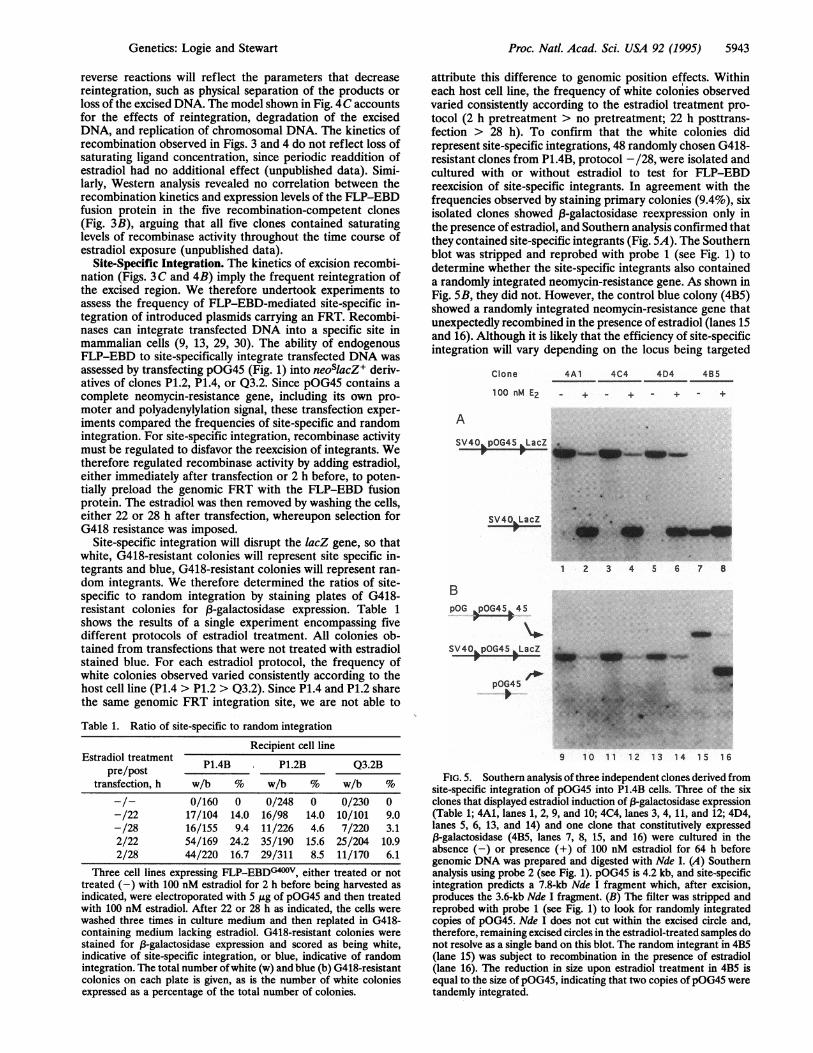
bination reaction in greater detail, a time course was per-formed on clone P1.4 and analyzed by probing Southern blotsto visualize either the integrated lacZ gene or the excisedneomycin-resistance gene (Fig. 4A). The excision reaction wasinitially rapid and reached approximately 50% completion by
C
lication lication
-(b..Ii
crecombinatnn -replication-degradation
11 III
FIG. 4. Kinetics of excision recombination in clone P1.4. (A)Southern analyses of a time course of estradiol-induced recombinationshowing hybridization of Nde I-digested genomic DNA with probe 2(see Fig. 1; Left) and hybridization of BamHI-digested genomic DNAwith probe 1 (see Fig. 1; Right). The unrecombined fragment is 5.2 kb,and the excised circular product of recombination was linearized byBamHI to give a 1.3-kb linear fragment. Time (in hours) in estradiolis given at the top. (B) Phosphorlmager quantification of threeindependent time courses of estradiol-induced recombination in cloneP1.4. The percentage of circle forms was calculated as circle signaldivided by the sum of the recombined and unrecombined signals. (C)Excision recombination on a single, chromosomally integrated sub-strate in living cells. Recombination between directly repeated FRTs(arrowheads) excises the interveningDNA as a circle which carries oneFRT, converting cell type I to cell type II. The excised circle is able toreintegrate, converting II to I, until it is lost from the cell through itsfailure to replicate or its degradation, converting II to III. Both I andIII can replicate.
10 h. The average initial rate of recombination measured fromFigs. 3B and 4A is 8% per hour or 2.15 events per cell cycle(27 h). FLP recombination is reversible and ideally will reachequilibrium at 50% if the substrate and products are equivalent(1, 3-5, 27, 28). In excision recombination, the substrates andproducts differ, in that the forward reaction (excision) isintramolecular and the reverse (reintegration) is intermolec-ular. Moreover, in a living system, the excised product, unlikethe chromosomal product and the substrate, may not replicateand can be degraded. We reason that the progress of excisionrecombination beyond the equilibrium between forward and
A
R3.3
R8.1
R8.2
0B
,o0
0a'a0
0
I'l
ir
Time, h
Proc. Natl. Acad. Sci. USA 92 (1995)
_AOW w _" r.-,-. ..
I
Proc. Natl. Acad. Sci. USA 92 (1995) 5943
reverse reactions will reflect the parameters that decreasereintegration, such as physical separation of the products orloss of the excised DNA. The model shown in Fig. 4C accountsfor the effects of reintegration, degradation of the excisedDNA, and replication of chromosomal DNA. The kinetics ofrecombination observed in Figs. 3 and 4 do not reflect loss ofsaturating ligand concentration, since periodic readdition ofestradiol had no additional effect (unpublished data). Simi-larly, Western analysis revealed no correlation between therecombination kinetics and expression levels of the FLP-EBDfusion protein in the five recombination-competent clones(Fig. 3B), arguing that all five clones contained saturatinglevels of recombinase activity throughout the time course ofestradiol exposure (unpublished data).
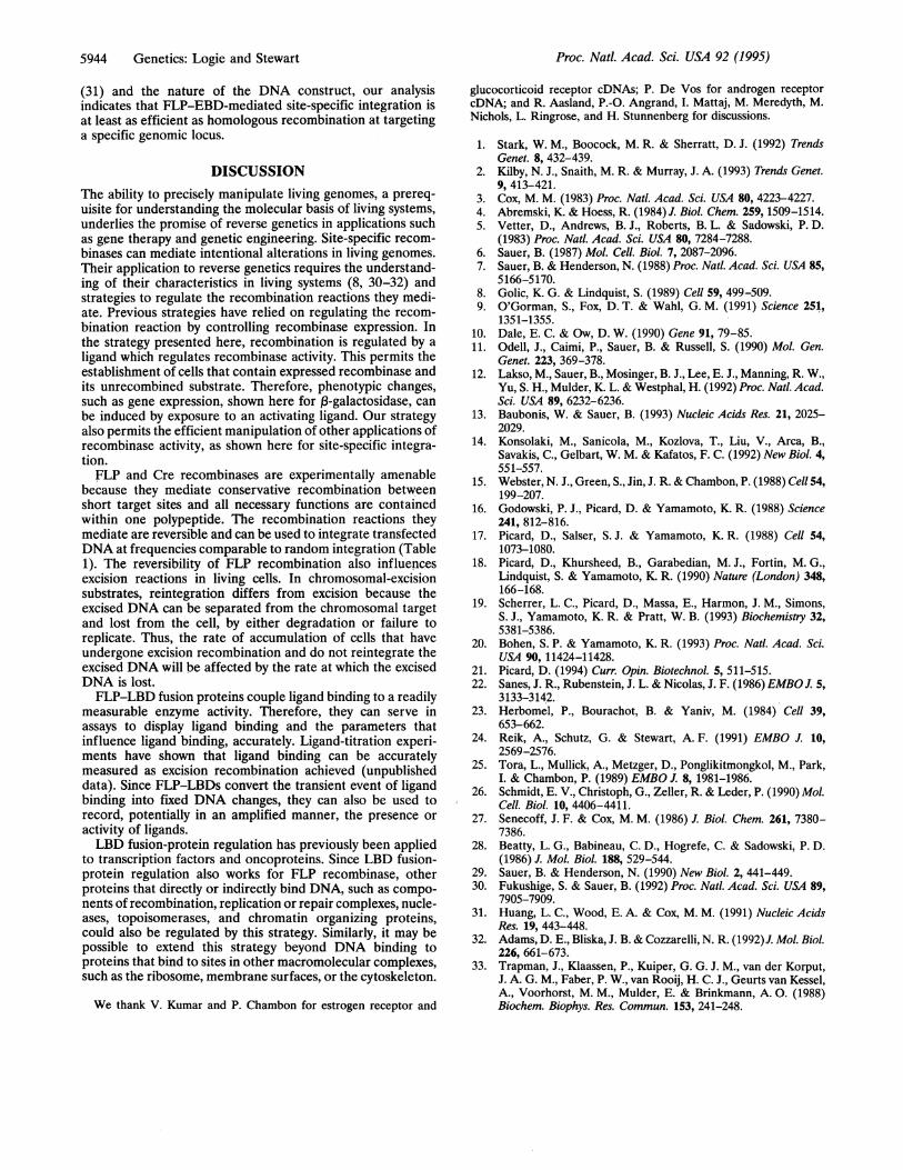
Site-Specific Integration. The kinetics of excision recombi-nation (Figs. 3 C and 4B) imply the frequent reintegration ofthe excised region. We therefore undertook experiments toassess the frequency of FLP-EBD-mediated site-specific in-tegration of introduced plasmids carrying an FRT. Recombi-nases can integrate transfected DNA into a specific site inmammalian cells (9, 13, 29, 30). The ability of endogenousFLP-EBD to site-specifically integrate transfected DNA wasassessed by transfecting pOG45 (Fig. 1) into neoslacZ+ deriv-atives of clones P1.2, P1.4, or Q3.2. Since pOG45 contains acomplete neomycin-resistance gene, including its own pro-moter and polyadenylylation signal, these transfection exper-iments compared the frequencies of site-specific and randomintegration. For site-specific integration, recombinase activitymust be regulated to disfavor the reexcision of integrants. Wetherefore regulated recombinase activity by adding estradiol,either immediately after transfection or 2 h before, to poten-tially preload the genomic FRT with the FLP-EBD fusionprotein. The estradiol was then removed by washing the cells,either 22 or 28 h after transfection, whereupon selection forG418 resistance was imposed.
Site-specific integration will disrupt the lacZ gene, so thatwhite, G418-resistant colonies will represent site specific in-tegrants and blue, G418-resistant colonies will represent ran-dom integrants. We therefore determined the ratios of site-specific to random integration by staining plates of G418-resistant colonies for f3-galactosidase expression. Table 1shows the results of a single experiment encompassing fivedifferent protocols of estradiol treatment. All colonies ob-tained from transfections that were not treated with estradiolstained blue. For each estradiol protocol, the frequency ofwhite colonies observed varied consistently according to thehost cell line (P1.4 > P1.2 > Q3.2). Since P1.4 and P1.2 sharethe same genomic FRT integration site, we are not able to
Table 1. Ratio of site-specific to random integration
Recipient cell lineEstradiol treatment P1.4B P1.2B Q3.2B
pre/posttransfection, h w/b % w/b % w/b %
-/- 0/160 0 0/248 0 0/230 0-/22 17/104 14.0 16/98 14.0 10/101 9.0-/28 16/155 9.4 11/226 4.6 7/220 3.12/22 54/169 24.2 35/190 15.6 25/204 10.92/28 44/220 16.7 29/311 8.5 11/170 6.1
Three cell lines expressing FLP-EBDG40oV, either treated or nottreated (-) with 100 nM estradiol for 2 h before being harvested asindicated, were electroporated with 5 ,gg of pOG45 and then treatedwith 100 nM estradiol. After 22 or 28 h as indicated, the cells werewashed three times in culture medium and then replated in G418-containing medium lacking estradiol. G418-resistant colonies werestained for f3-galactosidase expression and scored as being white,indicative of site-specific integration, or blue, indicative of randomintegration. The total number ofwhite (w) and blue (b) G418-resistantcolonies on each plate is given, as is the number of white coloniesexpressed as a percentage of the total number of colonies.
attribute this difference to genomic position effects. Withineach host cell line, the frequency of white colonies observedvaried consistently according to the estradiol treatment pro-tocol (2 h pretreatment > no pretreatment; 22 h posttrans-fection > 28 h). To confirm that the white colonies didrepresent site-specific integrations, 48 randomly chosen G418-resistant clones from P1.4B, protocol -/28, were isolated andcultured with or without estradiol to test for FLP-EBDreexcision of site-specific integrants. In agreement with thefrequencies observed by staining primary colonies (9.4%), sixisolated clones showed f3-galactosidase reexpression only inthe presence of estradiol, and Southern analysis confirmed thatthey contained site-specific integrants (Fig. 5A). The Southernblot was stripped and reprobed with probe 1 (see Fig. 1) todetermine whether the site-specific integrants also containeda randomly integrated neomycin-resistance gene. As shown inFig. SB, they did not. However, the control blue colony (4B5)showed a randomly integrated neomycin-resistance gene thatunexpectedly recombined in the presence of estradiol (lanes 15and 16). Although it is likely that the efficiency of site-specificintegration will vary depending on the locus being targeted
Clone 4A1 4C4 4D4 485
100 nM E2 + - + - + - +
A
SV40 pOG45 ,LacZ
SV40 LacZ
BpOG pOG45 45
SV40 pOG45 LacZ
pOG45
1 2 3 4 5 6 7 8
9 10 11 12 13 14 15 16
FIG. 5. Southern analysis of three independent clones derived fromsite-specific integration of pOG45 into P1.4B cells. Three of the sixclones that displayed estradiol induction of f-galactosidase expression(Table 1; 4A1, lanes 1, 2, 9, and 10; 4C4, lanes 3, 4, 11, and 12; 4D4,lanes 5, 6, 13, and 14) and one clone that constitutively expressed13-galactosidase (4B5, lanes 7, 8, 15, and 16) were cultured in theabsence (-) or presence (+) of 100 nM estradiol for 64 h beforegenomic DNA was prepared and digested with Nde I. (A) Southernanalysis using probe 2 (see Fig. 1). pOG45 is 4.2 kb, and site-specificintegration predicts a 7.8-kb Nde I fragment which, after excision,produces the 3.6-kb Nde I fragment. (B) The filter was stripped andreprobed with probe 1 (see Fig. 1) to look for randomly integratedcopies of pOG45. Nde I does not cut within the excised circle and,therefore, remaining excised circles in the estradiol-treated samples donot resolve as a single band on this blot. The random integrant in 4B5(lane 15) was subject to recombination in the presence of estradiol(lane 16). The reduction in size upon estradiol treatment in 4B5 isequal to the size of pOG45, indicating that two copies of pOG45 weretandemly integrated.
Genetics: Logie and Stewart
5944 Genetics: Logie and Stewart
(31) and the nature of the DNA construct, our analysisindicates that FLP-EBD-mediated site-specific integration isat least as efficient as homologous recombination at targetinga specific genomic locus.
DISCUSSIONThe ability to precisely manipulate living genomes, a prereq-uisite for understanding the molecular basis of living systems,underlies the promise of reverse genetics in applications suchas gene therapy and genetic engineering. Site-specific recom-binases can mediate intentional alterations in living genomes.Their application to reverse genetics requires the understand-ing of their characteristics in living systems (8, 30-32) andstrategies to regulate the recombination reactions they medi-ate. Previous strategies have relied on regulating the recom-bination reaction by controlling recombinase expression. Inthe strategy presented here, recombination is regulated by a
ligand which regulates recombinase activity. This permits theestablishment of cells that contain expressed recombinase andits unrecombined substrate. Therefore, phenotypic changes,such as gene expression, shown here for ,B-galactosidase, canbe induced by exposure to an activating ligand. Our strategyalso permits the efficient manipulation of other applications ofrecombinase activity, as shown here for site-specific integra-tion.FLP and Cre recombinases are experimentally amenable
because they mediate conservative recombination betweenshort target sites and all necessary functions are containedwithin one polypeptide. The recombination reactions theymediate are reversible and can be used to integrate transfectedDNA at frequencies comparable to random integration (Table1). The reversibility of FLP recombination also influencesexcision reactions in living cells. In chromosomal-excisionsubstrates, reintegration differs from excision because theexcised DNA can be separated from the chromosomal targetand lost from the cell, by either degradation or failure toreplicate. Thus, the rate of accumulation of cells that haveundergone excision recombination and do not reintegrate theexcised DNA will be affected by the rate at which the excisedDNA is lost.FLP-LBD fusion proteins couple ligand binding to a readily
measurable enzyme activity. Therefore, they can serve inassays to display ligand binding and the parameters thatinfluence ligand binding, accurately. Ligand-titration experi-ments have shown that ligand binding can be accuratelymeasured as excision recombination achieved (unpublisheddata). Since FLP-LBDs convert the transient event of ligandbinding into fixed DNA changes, they can also be used torecord, potentially in an amplified manner, the presence or
activity of ligands.LBD fusion-protein regulation has previously been applied
to transcription factors and oncoproteins. Since LBD fusion-protein regulation also works for FLP recombinase, otherproteins that directly or indirectly bind DNA, such as compo-nents of recombination, replication or repair complexes, nucle-ases, topoisomerases, and chromatin organizing proteins,could also be regulated by this strategy. Similarly, it may bepossible to extend this strategy beyond DNA binding toproteins that bind to sites in other macromolecular complexes,such as the ribosome, membrane surfaces, or the cytoskeleton.
We thank V. Kumar and P. Chambon for estrogen receptor and
glucocorticoid receptor cDNAs; P. De Vos for androgen receptorcDNA; and R. Aasland, P.-O. Angrand, I. Mattaj, M. Meredyth, M.Nichols, L. Ringrose, and H. Stunnenberg for discussions.
1. Stark, W. M., Boocock, M. R. & Sherratt, D. J. (1992) TrendsGenet. 8, 432-439.
2. Kilby, N. J., Snaith, M. R. & Murray, J. A. (1993) Trends Genet.9, 413-421.
3. Cox, M. M. (1983) Proc. Natl. Acad. Sci. USA 80, 4223-4227.4. Abremski, K. & Hoess, R. (1984) J. Biol. Chem. 259, 1509-1514.5. Vetter, D., Andrews, B. J., Roberts, B. L. & Sadowski, P. D.
(1983) Proc. Natl. Acad. Sci. USA 80, 7284-7288.6. Sauer, B. (1987) Mo. Cell. Biol. 7, 2087-2096.7. Sauer, B. & Henderson, N. (1988) Proc. Natl. Acad. Sci. USA 85,
5166-5170.8. Golic, K. G. & Lindquist, S. (1989) Cell 59, 499-509.9. O'Gorman, S., Fox, D. T. & Wahl, G. M. (1991) Science 251,
1351-1355.10. Dale, E. C. & Ow, D. W. (1990) Gene 91, 79-85.11. Odell, J., Caimi, P., Sauer, B. & Russell, S. (1990) Mo. Gen.
Genet. 223, 369-378.12. Lakso, M., Sauer, B., Mosinger, B. J., Lee, E. J., Manning, R. W.,
Yu, S. H., Mulder, K. L. & Westphal, H. (1992) Proc. Natl. Acad.Sci. USA 89, 6232-6236.
13. Baubonis, W. & Sauer, B. (1993) Nucleic Acids Res. 21, 2025-2029.
14. Konsolaki, M., Sanicola, M., Kozlova, T., Liu, V., Arca, B.,Savakis, C., Gelbart, W. M. & Kafatos, F. C. (1992) New Bio. 4,551-557.
15. Webster, N. J., Green, S., Jin, J. R. & Chambon, P. (1988) Cell 54,199-207.
16. Godowski, P. J., Picard, D. & Yamamoto, K. R. (1988) Science241, 812-816.
17. Picard, D., Salser, S. J. & Yamamoto, K. R. (1988) Cell 54,1073-1080.
18. Picard, D., Khursheed, B., Garabedian, M. J., Fortin, M. G.,Lindquist, S. & Yamamoto, K. R. (1990) Nature (London) 348,166-168.
19. Scherrer, L. C., Picard, D., Massa, E., Harmon, J. M., Simons,S. J., Yamamoto, K. R. & Pratt, W. B. (1993) Biochemistry 32,5381-5386.
20. Bohen, S. P. & Yamamoto, K. R. (1993) Proc. Natl. Acad. Sci.USA 90, 11424-11428.
21. Picard, D. (1994) Curr. Opin. Biotechnol. 5, 511-515.22. Sanes, J. R., Rubenstein, J. L. & Nicolas, J. F. (1986) EMBO J. 5,
3133-3142.23. Herbomel, P., Bourachot, B. & Yaniv, M. (1984) Cell 39,
653-662.24. Reik, A., Schutz, G. & Stewart, A. F. (1991) EMBO J. 10,
2569-2576.25. Tora, L., Mullick, A., Metzger, D., Ponglikitmongkol, M., Park,
I. & Chambon, P. (1989) EMBO J. 8, 1981-1986.26. Schmidt, E. V., Christoph, G., Zeller, R. & Leder, P. (1990) Mol.
Cell. Biol. 10, 4406-4411.27. Senecoff, J. F. & Cox, M. M. (1986) J. Bio. Chem. 261, 7380-
7386.28. Beatty, L. G., Babineau, C. D., Hogrefe, C. & Sadowski, P. D.
(1986) J. Mo. Biol. 188, 529-544.29. Sauer, B. & Henderson, N. (1990) New Bio. 2, 441-449.30. Fukushige, S. & Sauer, B. (1992) Proc. Natl. Acad. Sci. USA 89,
7905-7909.31. Huang, L. C., Wood, E. A. & Cox, M. M. (1991) Nucleic Acids
Res. 19, 443-448.32. Adams, D. E., Bliska, J. B. & Cozzarelli, N. R. (1992)J. Mol. Biol.
226, 661-673.33. Trapman, J., Klaassen, P., Kuiper, G. G. J. M., van der Korput,
J. A. G. M., Faber, P. W., van Rooij, H. C. J., Geurts van Kessel,A., Voorhorst, M. M., Mulder, E. & Brinkmann, A. 0. (1988)Biochem. Biophys. Res. Commun. 153, 241-248.
Proc. Natl. Acad. Sci. USA 92 (1995)