Embed Size (px)
Citation preview
© 1990 Oxford University Press Nucleic Acids Research, Vol. 18, No. 9 2625
Light affects the structure of Chlamydomonas chloroplastchromosomes
Robert J.Thompson and Gisela MosigDepartment of Molecular Biology, Vanderbilt University, Nashville, TN 37235, USA
Received February 2, 1990, Revised and Accepted March 16, 1990
ABSTRACT
We have analyzed changes in the structure ofchloroplast chromosomes in response to light ingrowing Chlamydomonas cells using a crosslinkingassay based on the intercalation of HMT(4'-hydroxymethyl-4,5',8-trimethylpsoralen) into DNA.Our results show that the structure of chloroplastchromosomes in at least three widely separated regionsis different in light-grown vs. dark-grown cells.Structural changes in chloroplast chromosomes occurwithin 3 hrs after exposure to light or darkness,respectively. The response to light is not inhibited byatrazine and can be elicited by dim blue light incapableof evolving O2, indicating that it does not requirephotosynthesis. Inhibition of cytoplasmic proteinsynthesis with cycloheximide prevents this responseto light, indicating that it depends, at least in part, onproteins imported from the cytoplasm.
INTRODUCTION
Light is a critical factor in development, maintenance and functionof plant cells and their chloroplasts (for reviews see references1-5). Here we have investigated whether the physiologicalresponses of chloroplasts to light or darkness are accompaniedby structural changes in chloroplast chromosomes. We haveprobed for such changes in chloroplast chromosome structurein living cells of the unicellular alga C. reinhardtii, using a DNAcrosslinking assay that combines two methods described byPettijohn and coworkers (6) and by Vos and Hanawalt (7). Thisassay (Fig. 1) is based on the different potential of chromosomesof different structure to intercalate HMT(4'-hydroxymethyl-4,5',8-trimethylpsoralen) (see below).Subsequent brief irradiation with near UV light (UVA) leads tocrosslinking of chloroplast DNA as a function of the extent ofintercalation (8).
Intercalation of psoralens increases with increasing torsionalstress of underwound DNA (6,9). Additionally, DNA bindingproteins can decrease psoralen binding to DNA, either becausethey relieve torsional stress of the DNA (10) or because theyshield the DNA from the intercalator (11-13) or both. Lightmight potentially influence chromosome structure by alteringtorsional stress of the DNA and/or abundance or activity ofcertain DNA binding proteins.
Deproteinized chloroplast DNA is torsionally stressed becauseit is underwound (i.e. it is negatively supercoiled) (14). In vivo,
chloroplast DNA is not bound to histones, but instead appearsto be associated with small, basic proteins, some of which areantigenically related to eubacterial and cyanobacterial proteinsHU (15,16). These and/or other proteins might constrain someof the torsional stress seen in deproteinized chloroplast DNA invitro (10). Chloroplasts also contain DNA tracking proteins aswell as relaxing (17,18) and supercoiling (17,19) topoisomerases.Partial inhibition of the latter enzyme with novobiocin rapidlyreduces torsional stress of the chloroplast DNA (shown here) andalters synthesis of chloroplast transcripts in vivo (20). Theseconsiderations, together, suggest that chloroplast chromosomes,like bacterial chromosomes, maintain a certain homeostasis oftorsional stress.
In contrast to higher plants, C. reinhardtii can develop fullyfunctional chloroplasts in the dark (21). Under these conditionsit can grow when supplied with an appropriate carbon source.Thus, by transferring dark-adapted algal cells into the light andvice versa, direct effects of light on potential HMT-dependentcrosslinking of chloroplast DNA can be studied with fewcomplications resulting from changes in chloroplast pigments,membranes, etc. We show here that exposure to light reducesthe potential for HMT intercalation and DNA crosslinking in atleast three widely separated regions of the chloroplastchromosome. This reduction does not depend on photosynthesis,but it does depend on cytoplasmic protein synthesis. We concludethat light affects the overall structure of the chloroplastchromosomes.
MATERIALS AND METHODSGrowth of the AlgaeC. reinhardtii cw-15 (cell wall-) strain 278gro2 (a derivative ofCC-278 described in reference 22) was used in all experiments.Medium and culture conditions for growing this alga were aspreviously described (22). Since potential HMT crosslinking ofthe chloroplast DNA changes as a function of the growth phaseof the cells (see Fig. 3, below), it is important to compare hght-and dark-adapted cells under otherwise similar physiologicalconditions. To obtain light-adapted cells, starter cultures wereinoculated to 1 x 10⁶ cells/ml and grown overnight in the light(for ca. 2 generations). These cultures were in turn diluted to5X105 cells/ml and incubated an additional 18—24 hrs in thelight. At this time, the light-adapted cells were in the mid-logarithmic growth phase (2.5-3.5X10⁶ cells/ml). To obtaindark-adapted cells, starter cultures were inoculated (with
at Frankfurt Univesity L
ibrary on September 1, 2014
http://nar.oxfordjournals.org/D
ownloaded from

2626 Nucleic Acids Research, Vol. 18, No. 9
stationary phase, light-grown cells) to an initial cell density of4 x 105 cells/ml. They were incubated in the dark for two days(for ca. 3.0 generations). These cultures were in turn diluted to8x 105 cells/ml and incubated an additional 18-24 hrs in thedark. At this time, the dark-adapted cells were also in the mid-logarithmic growth phase (2.5 —3.5X10⁶ cells/ml).
HMT Crosslinking of Chloroplast DNAWe have modified a procedure originally described by Vos andHanawalt (7) to crosslink nuclear DNA in human VA2-6A3cells (Fig. 1A). Approximately 3.5 XlO8 algal cells werewashed in 20 ml of minimal medium (i.e. the medium describedin reference 22, but lacking yeast extract, bactopeptone andsodium acetate) and resuspended in 20 ml of this minimal medium(the complete medium strongly absorbed UVA light). The cellswere chilled, HMT was added to yield a final concentration of0.3-20 /tg/ml (from a 0.25 mg/ml or 2.50 mg/ml stock inethanol), and cells were chilled for 10 more min. The cells werethen poured into 50 ml plastic petri dishes, placed on a sheetof aluminum foil on a rotary shaker and irradiated at roomtemperature for 1.5 min with two fluorescent black-light bulbs(Sylvania, F15T8-BLB, 15 W) while rotating at 90 rpm. Thedose rate, measured with a calibrated silicon photo-diode (PINIOUV) from United Detector Technology, was 3 W/m2 at350 nm. Following irradiation, the cells were pelleted,resuspended in 0.5 ml of Tris buffer, pH 9.5, and lysed byaddition of 60 /tl of 10% SDS. The lysate was extracted twicewith phenol/chloroform/isoamyl alcohol (12:12:1) and then twicewith chloroforrrt/isoamyl alcohol (24:1). One-tenth volume of3 M sodium acetate and three volumes of 95% ethanol wereadded sequentially at room temperature. The precipitated DNAwas collected by spooling onto a glass rod, rinsed twice with70% ethanol and resuspended in 100 /tl of TE buffer. Two /tlof RNAase A (10 mg/ml) were added and the samples wereincubated at 37°C for 30 min. The DNA was reprecipitated,washed as described above and finally resuspended in 50 /tl ofTE (10 mM Tris pH 8.0, 1 mM EDTA).
An aliquot of each DNA sample (1.5 /tg) was restricted withEcoRl, HpaU or Pstl (6 units overnight), 5 fig yeast tRNA wereadded as earner and the nucleic acids were precipitated withethanol at —70°C for 20 min. After centrifugation, the pelletswere washed twice with 70% ethanol and resuspended in 9 /ilof 4% sucrose, 0.25% bromophenol blue. The samples werealkali-denatured by adding 1 /tl of 2 N NaOH at roomtemperature and were then immediately loaded onto 1 % neutralagarose gels in Tris-acetate buffer (40 mM Tris-acetate pH 8.2,2 mM EDTA) at 4°C. Following electrophoresis [for ca. 2 hrsat 100 V in an IBI mini-gel apparatus (Model QSH)], the DNAwas blotted onto nitrocellulose (Schleicher and Schuell). The blotswere probed with chloroplast DNA restnetion fragmentsEcoRl 14, £coRI 19 or HpaU 5, or with a nuclear Pstl fragmentcontaining the calmodulin gene. All probes were ^P-labeled bythe random primer method (23).
The HMT crosslinking assay is illustrated in Figure 1.Autoradiograms of the Southern blots were scanned with a Zeinehsoft laser densitometer and the area under each peak wasdetermined. The number of crosslinks in a restriction fragmentshould follow a Poisson distribution if crosslinking is random(7). Therefore, the average number of crosslinks in the DNArestriction fragment can be estimated using the equation:Po = e~x, where Po is the percentage of the DNA fragment thatwas not crosslinked (i.e. that migrated as single-stranded DNA),
SOtTMFRNBlOT
••
t> =
•
fc-s$—
V TOTAL CTJ.LILAR DNA LIIVT UMVfJ
|E «miti\rrcKossLi\Ks(Xl
DICKST HIIII RUTIIICTMm ENZVMr
iALKAIJ DENATtRF.
\
RAMOtV NEITTRALIZE
I ds
KKS N'EtTRAL AC.AKOSf-QF.LMOT ONTO MTKOCtLLI U » t««>»€ WITH 3*WIFir tTDSA
HHSTRKT1OS FRAIiMKM
12 34 SI
alkali - + - + • -M M T - - - - • •
Figure 1. An assay for measuring the frequency of HMT crosslinking of specificregions of the chloroplast DNA. Panel A Living algal cells were irradiated withUVA light in the presence of HMT to induce interstrand crosslinks in thechloroplast DNA (see Materials and Methods for details) After treatment, totalcellular DNA was isolated and aliquots were restricted with EcoRl or with HpaUThe DNA was then alkali denatured and separated in neutral agarose gels TheDNA was blotted to nitrocellulose and probed with labeled chloroplast restrictionfragment EcoRl 14, £c»RI 19 or HpaU 5 which encompasses promoter PA (42)EcoRl 14 contains the rbcL gene, encoding the large suburut of nbulosebisphosphate carboxylase, the psaB gene, encoding a reaction center protein ofphotosystem I, and the 5' end of the atpA gene, encoding the CF, alpha subunitof the chloroplast ATPase (43) EcoRl 19 encodes tRNA1^ (43) Cp, chloroplastDNA, ds, double-stranded DNA, ss, single-stranded DNA Panel B Map ofChlamydomonas chloroplast DNA showing the location of restriction fragmentsEcofU 14, EcoRl 19 and HpaU 5 used as probes in the crosslinking assay Notethat these fragments are well-separated The nomenclature for the £coRI fragmentsis that of Harris and coworkers (43) Other workers have named these fragmentsdifferently (44) The inverted repeats of the chloroplast DNA are indicated byarrows. Panel C A representative Southern blot probed with chloroplast DNArestriction fragment EcoRl 14 is shown Lane 1, non-denatured EcoRl 14 obtainedfrom plasmid pCP43 (44), lane 2, denatured EcoRl 14, lane 3, non-denatured,6x>RI digested total cellular DNA (1 5 >tg) from light-grown control cells (UVA-lrradjated for 1 5 min in the absence of HMT), lane 4, same as lane 3 exceptthat the DNA was denatured; lane 5, denatured, EcoRl digested DNA from light-grown cells irradiated with UVA light in the presence of HMT (0 5 /jg/ml), lane6, same as lane 5 except that the DNA was not denatured The autoradiogramwas purposely overexposed to show that there was no single-stranded algal DNAunless the DNA had been alkali-denatured, and that there was no double-strandedalgal DNA after denaturation, unless the cells had been treated with HMT andUVA light
and x is the average number of crosslinks in the DNA restrictionfragment. For theoretical and technical reasons this estimate ismost accurate when there are about equal proportions of single-and double-stranded DNA. To compare crosslinking in differentrestriction fragments, these data were expressed as crosslinks per10 kbp.
We expected that crosslinking of chloroplast DNA from a givenalgal culture would increase with increasing HMT concentrations.Within the concentration range used in our experiments, the extentof HMT crosslinking was indeed proportional to the HMTconcentration (Fig. 2). Crosslinking was also dependent on theUVA dose, as expected (data not shown).
at Frankfurt Univesity L
ibrary on September 1, 2014
http://nar.oxfordjournals.org/D
ownloaded from

Nucleic Acids Research, Vol. 18, No. 9 2627
400
0.2 0.4 0.6 0.8
qg/ml HMT1.0 1.2
Figure 2. HMT crosslinking of chloroplast DNA as a function of HMTconcentration The data were obtained by crosslinking the HpaU 5 region of thechloroplast chromosome from dark-adapted cells
Enzymes and ChemicalsRestriction enzymes HpaU, EcoRl and Pstl, and DNApolymerase I Klenow fragment were from Bethesda ResearchLaboratories. RNAase A and random primers were fromBoehringer Mannheim. Cycloheximide, novobiocin and nalidixicacid were from Sigma. Atrazine was from Ciba-Geigy. HMTwas from HRI Associates, Berkeley, CA. [a-32P]dCTP(800 Ci/mol) was from Amersham.
RESULTSHMT Crosslinking of Chloroplast DNA in vivoHMT intercalates into DNA and forms covalent adducts withpynmidines when exposed to UVA light (8). A monoadduct cansubsequently crosslink with an appropriately positionedpyrimidine on the opposite DNA strand. After cutting with arestriction enzyme, alkali denaturation and neutralization,crosslinked restriction fragments will rapidly reanneal and migrateas double-stranded species, whereas the fragments containing nocrosslinks will migrate as single-stranded species (7).
The HMT crosslinking assay is outlined in Figure 1A and arepresentative experiment is shown in Figure 1C. EcoRl-restricted total cellular DNA from HMT-treated cells was alkali-denatured and rapidly renatured. After electrophoretic separation,blotting, and probing of the Southern blot with £coRI 14, onlythe two expected bands corresponding to double- and single-stranded EcoRl 14 hybridized with the labeled EcoRl probe (Fig.1C, lane 5). As expected, non-denatured restriction fragmentsmigrated as double-stranded DNA (Fig. 1C, lanes 1, 3 and 6),and alkali-denatured DNA which was not crosslinked migratedas single-stranded DNA (Fig. 1C, lanes 2 and 4).
For the reasons discussed in Materials and Methods, allsubsequent comparisons were done at optimal HMTconcentrations and UVA doses and we discuss our results in termsof potential HMT-dependent crosslinking of DNA.
Potential HMT Crosslinking of Chloroplast DNA Dependsupon the Growth StateWe suspected from preliminary experiments (not shown) that thepotential for HMT crosslinking of chloroplast DNA is influenced
mIo
100
50
uO
10
1
1•
7>
/J!di
ss
a Ik a
/
1
»• f
li +
2/
//
2 3
-
1 2 3
-
0 20 40 60 80 100HOURS
Figure 3. Potential HMT crosslinking of chloroplast DNA is dependent uponthe growth state Light-adapted cells were diluted to 2 x 10⁵ cells/ml at 0 hrsand growth in the light was continued Cell concentration was measured at theindicated tunes At the indicated points (l e 1, 2 and 3), aliquots of the culturewere used in the HMT crosslinking assay The inset shows a Southern blot,analogous to the one shown in Figure 1C, probed with HpaU 5 The HMTconcentration was 1 0 /ig/ml throughout
by the physiological growth state of the cells. Further studiesshowed that HMT crosslinking of chloroplast DNA increasedwith increasing cell density (Fig. 3). Therefore, all subsequentexperiments were conducted under controlled growth conditions(see Materials and Methods).
White Light Reduces Potential HMT Crosslinking ofChloroplast DNAChloroplast DNA in light-adapted cells experienced less HMTcrosslinking than the DNA in dark-adapted cells (Fig. 4, lanes1 and 2 and Table I, section I). This result was found in threewidely separated regions of the genome, i.e. in the £coRI 19,HpaU 5 and £coRI 14 fragments (Fig. IB and Table I, sectionI). Chloroplast DNA in light-adapted cells which weresubsequently grown in the dark for 3 hrs showed an increase inpotential HMT crosslinking in these same regions (Fig. 4, lanes3 and 4 and Table I, section II). Conversely, chloroplast DNAin dark-adapted cells which were transferred into white light for3 hrs showed a decrease in potential HMT crosslinking (Fig. 4,lanes 5 and 6 and Table I, section HI). The crosslinking ratiosof chloroplast DNA from light- versus dark-grown cells weresimilar (not statistically significantly different) in all three regionsof the genome, regardless of the different conditions of theexperiments shown in Tables 1 and 2 and in many other
at Frankfurt Univesity L
ibrary on September 1, 2014
http://nar.oxfordjournals.org/D
ownloaded from

2628 Nucleic Acids Research, Vol. 18, No. 9
L D
L D LD D L
Table 1. Light reduces potential HMT crosslinking of the chloroplast DNA '
# # #
Figure 4. Light reduces potential HMT crossluiking of the chloroplast DNAA representative Southern blot probed with chloroplast DNA restriction fragmentEcoRl 19 is shown (ds, double-stranded EcoRl 19, ss, single-stranded EcoRl 19)In analogous experiments, HMT crossluiking in the HpaU 5 and EcoRl 14 regionswas analyzed (see Table I for quantitauon) L, light-adapted cells (650 /iW/cm2
white light), D, dark-adapted cells, L—D, light-adapted cells shifted into darknessfor 3 hrs, D—L, dark-adapted cells shifted into white light (650 /iW/cm2) for3 hrs
independent experiments (not shown). These ratios ranged fromabout 0.40 to about 0.75.
An HMT concentration of 1.0 /ig/ml (Fig. 4 and Table I)allowed for the most accurate simultaneous determination of thecrosslinking frequencies in the three regions under the threedifferent light/dark treatments. Increasing or decreasing the HMTconcentration in these and other experiments increased ordecreased, respectively, the absolute frequency of crosslinkingas expected. In spite of this, the effects of light on reducing thepotential crosslinking were similar at different HMTconcentrations (not shown). The much higher HMTconcentrations required to crosslink the nuclear DNA (see below),crosslinked the chloroplast DNA to such an extent thatquantitative comparisons could not be made at the same HMTconcentration.
The light-induced decrease in potential HMT crosslinking wasnot due to increased shielding of chloroplast DNA from UVAlight by pigments since we found no significant change inpigments during the 3 hr light or 3 hr dark treatments (data notshown). There was also no change in thylakoid membranes(electron micrographs not shown) which might sequester thehydrophobic HMT molecules. It should be recalled thatChlamydomonas chloroplasts develop normally and insertfunctional chlorophyll into thylakoid membranes in the dark (21).
EcoRl 19 Hpall 5 EcoRl 14
dark 1 90 1 58 1 04(crosslinks/10 kb)
I light 0 93 0 75 0 79(crosslinks/10 kb)
ratio (light/dark) 0.49' 0.471 0.76'
II
light(crosslinks/10 kb)
3 hrs darkness(crosslinks/10 kb)
ratio (light/dark)
0 88
1 79
0.492
1.11
1 93
0.572
0 74
1 61
0.462
HI
dark(crosslinks/10 kb)
3 hrs light(crosslinks/10 kb)ratio (hght/dark)
2 19
1 07
0.493
1 94
089
0.463
1 70
0 93
0.553
"Autoradiograms like those shown in Figure 4 were scanned with a Zenith softlaser densitometer and the tracings were used to calculate the number ofcrosslinks/10 kb as described in Materials and Methods Superscript numbersindicate ratios obtained from DNA of the same algal culture The HMTconcentration was 1 0 ^g/ml in all cases The cells were exposed to white light(650 /iW/cm2)
Table 2. Photosynthesis is not required for the decrease in potential HMTcrosslinking of chloroplast DNA when cells were grown in the light"
Ratio of Crosslinks'1
EcoRl 14 EcoRl 19 Hpall 5
light, no alrazinedark, no atrazine
light, + atrazinedark, no atrazine
065
051
0 75
0 62
II
III
light,dark,
dark,dark,
bluedark,
+ atrazine+ atrazine
+ atrazineno atrazine
light, no atrazineno atrazine
—
104
0 76
—
102
_
050
_
0 78
'Dark-adapted cells were exposed to white light (650 /iW/cm2) for 3 hrs in theabsence or presence of 5 /iM atrazine (the drug was added 30 mm prior to exposureto light) (I), treated with atrazine for 3.5 hrs in the dark (IF), or exposed to dimblue light (20 /jW/cm2) for 3 hrs (HI) - not determined.'The number of crosslinks/10 kb for each sample was calculated as describedin the legend to Table 1 These numbers were then used to calculate the indicatedratios The HMT concentration was 0 5 jig/ml in all experiments Thisconcentration was used because it allowed for the most accurate quanutation ofthe crosslinking frequencies in DNA from dark-adapted cells.
As a control, we tested the effect of growth in the light onpotential HMT crosslinking of nuclear DNA. Crosslinking wasmeasured in a 3.2 kb Pstl genomic DNA fragment containingthe calmodulin gene together with 5' and 3' flanking sequences(24). In contrast to chloroplast DNA, potential HMT crosslinkingof this nuclear DNA fragment was similar under all growthconditions tested (i.e. continuous dark, dark with subsequent 3 hrslight, or continuous light) (Fig. 5, lanes 3-5). Like in chloroplast
at Frankfurt Univesity L
ibrary on September 1, 2014
http://nar.oxfordjournals.org/D
ownloaded from
Nucleic Acids Research, Vol. 18, No. 9 2629
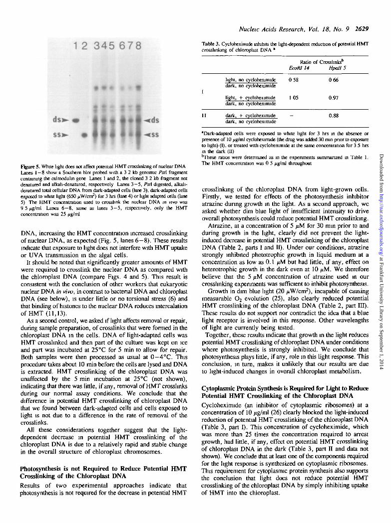
1 2 345 6 7 8
HUi|Figure 5. White light does not affect potential HMT crosslinlang of nuclear DNALanes 1-8 show a Southern blot probed with a 3 2 kb genonuc Pstl fragmentcontaining the calmodulin gene Lanes 1 and 2, the cloned 3 2 kb fragment notdenatured and alkali-denatured, respectively Lanes 3—5, Pstl digested, alkali-denatured total cellular DNA from dark-adapted cells (lane 3), dark-adapted cellsexposed to white light (650 /iW/cm2) for 3 hrs (lane 4) or light-adapted cells (lane5) The HMT concentration used to crosslink the nuclear DNA m vivo was9 5 ng/m\ Lanes 6 - 8 , same as lanes 3 - 5 , respectively, only the HMTconcentration was 25
DNA, increasing the HMT concentration increased crosslinkingof nuclear DNA, as expected (Fig. 5, lanes 6-8) . These resultsindicate that exposure to light does not interfere with HMT uptakeor UVA transmission in the algal cells.
It should be noted that significantly greater amounts of HMTwere required to crosslink the nuclear DNA as compared withthe chloroplast DNA (compare Figs. 4 and 5). This result isconsistent with the conclusion of other workers that eukaryoticnuclear DNA in vivo, in contrast to bacterial DNA and chloroplastDNA (see below), is under little or no torsional stress (6) andthat binding of histones to the nuclear DNA reduces intercalationof HMT (11,13).
As a second control, we asked if light affects removal or repair,during sample preparation, of crosslinks that were formed in thechloroplast DNA in the cells. DNA of light-adapted cells wasHMT crosshnked and then part of the culture was kept on iceand part was incubated at 25°C for 5 min to aJlow for repair.Both samples were then processed as usual at 0—4°C. Thisprocedure takes about 10 min before the cells are lysed and DNAis extracted. HMT crosslinking of the chloroplast DNA wasunaffected by the 5 min incubation at 25°C (not shown),indicating that there was little, if any, removal of HMT crosslinksduring our normal assay conditions. We conclude that thedifference in potential HMT crosslinking of chloroplast DNAthat we found between dark-adapted cells and cells exposed tolight is not due to a difference in the rate of removal of thecrosslinks.
All these considerations together suggest that the light-dependent decrease in potential HMT crosslinking of thechloroplast DNA is due to a relatively rapid and stable changein the overall structure of chloroplast chromosomes.
Photosynthesis is not Required to Reduce Potential HMTCrosslinking of the Chloroplast DNAResults of two experimental approaches indicate thatphotosynthesis is not required for the decrease in potential HMT
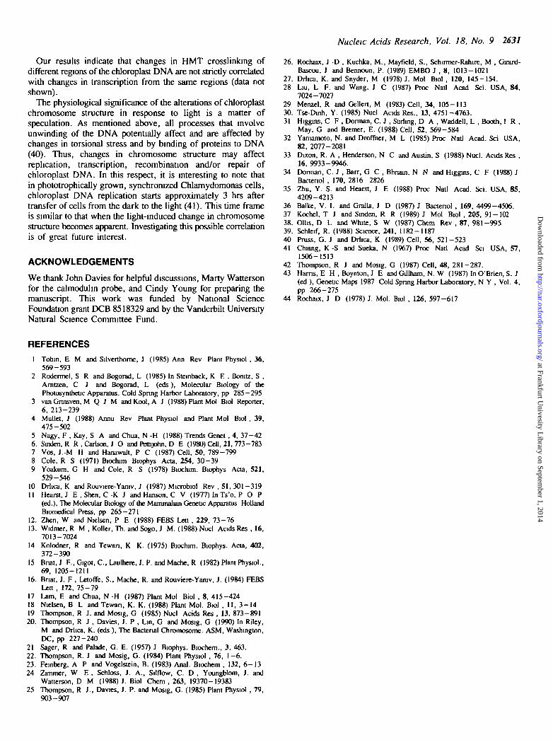
Tabte 3. Cycloheximide inhibits the light-dependent reduction of potential HMTcrosslinking of chloroplast DNA *
Ratio of Crosslinks6
EcoRI 14 Hpall 5
light, no cycloheximidedark, no cycloheximide
0 58 066
II
light, + cycloheximidedark, no cycloheximide
dark, + cycloheximidedark, no cycloheximide
1 05
_
0.97
0.88
'Dark-adapted cells were exposed to white light for 3 hrs in the absence orpresence of 10 /ig/ml cycloheximide (the drug was added 30 min prior to exposureto light) (I), or treated with cycloheximide at the same concentration for 3 5 hrsin the dark (II)'These ratios were determined as in the experiments summarized in Table 1.The HMT concentration was 0 5 jig/ml throughout
crosslinking of the chloroplast DNA from light-grown cells.Firstly, we tested for effects of the photosynthesis inhibitoratrazine during growth in the light. As a second approach, weasked whether dim blue light of insufficient intensity to driveoverall photosynthesis could reduce potential HMT crosslinlang.
Atrazine, at a concentration of 5 /xM for 30 min prior to andduring growth in the light, clearly did not prevent the light-induced decrease in potential HMT crosslinking of the chloroplastDNA (Table 2, parts I and II). Under our conditions, atrazinestrongly inhibited phototrophic growth in liquid medium at aconcentration as low as 0.1 /iM but had little, if any, effect onheterotrophic growth in the dark even at 10 /iM. We thereforebelieve that the 5 /iM concentration of atrazine used in ourcrosslinking experiments was sufficient to inhibit photosynthesis.
Growth in dim blue light (20 /tW/cm2), incapable of causingmeasurable O2 evolution (25), also clearly reduced potentialHMT crosslinking of the chloroplast DNA (Table 2, part m).These results do not support nor contradict the idea that a bluelight receptor is involved in this response. Other wavelengthsof light are currently being tested.
Together, these results indicate that growth in the light reducespotential HMT crosslinking of chloroplast DNA under conditionswhere photosynthesis is strongly inhibited. We conclude thatphotosynthesis plays little, if any, role in this light response. Thisconclusion, in turn, makes it unlikely that our results are dueto light-induced changes in overall chloroplast metabolism.
Cytoplasmic Protein Synthesis is Required for light to ReducePotential HMT Crosslinking of the Chloroplast DNACycloheximide (an inhibitor of cytoplasmic ribosomes) at aconcentration of 10 /tg/ml (26) clearly blocked the light-inducedreduction of potential HMT crosslinking of the chloroplast DNA(Table 3, part I). This concentration of cycloheximide, whichwas more than 25 times the concentration required to arrestgrowth, had little, if any, effect on potential HMT crosslinkingof chloroplast DNA in the dark (Table 3, part II and data notshown). We conclude that at least one of the components requiredfor the light response is synthesized on cytoplasmic ribosomes.This requirement for cytoplasmic protein synthesis also supportsthe conclusion that light does not reduce potential HMTcrosslinking of the chloroplast DNA by simply inhibiting uptakeof HMT into the chloroplast.
at Frankfurt Univesity L
ibrary on September 1, 2014
http://nar.oxfordjournals.org/D
ownloaded from

2630 Nucleic Acids Research, Vol. 18, No. 9
Table 4. HMT crosslinking of chloroplast DNA in novobiocin-treated,nalidixic acid-treated and gamma-ray-irradiated cells.
novobiocinb
nahdixic acid0
gamma-irradiationd
EcoRl
0.32
0 28
0 71
Ratio of Crosslinks*(treated/control)
14 Hpall 5
0 22
-
-
These ratios were determined as in the experiments summarized in Table 1The HMT concentration was 2 0 fig/ml This higher HMT concentrationwas used to insure that measurable proportions of crosslinked chloroplastDNA fragments were present after the novobiocin and nalidixic acidtreatments, both of which greatly reduced potential HMT crosslinking. (Alsonote that in these experiments we used light-adapted cells and not dark-adapted cells as in the experiments summarized in Tables 2 and 3 )bLight-adapted cells were treated with 200 /ig/ml novobiocin for 2 hrscLight-adapted cells were treated with 100 /ig/ml nalidixic acid for 1 hrdLight-adapted cells were irradiated with 12 5 krad of gamma-rays from acobalt source over a 15 min period. The cobalt source available (o us hadconsiderably lower intensity than the source used by Pettijohn andcoworkers (6). The longer exposure time that we had to use also increasedthe chance for repair of the radiation-induced nicks
Novobiocin, Nalidixic Acid and Gamma-Ray Irradiation, Allof Which Reduce Torsional Stress, Reduce Potential HMTCrosslinking of the Chloroplast DNAIn bacteria, the inactivation of DNA gyrase results in an increasein the average linking number (27) and a decrease in the averagetorsional stress of the DNA as measured by psoralen binding (6).As expected, in vivo inhibition of the chloroplast supercoilingtopoisomerase (17,19) with sublethal concentrations of novobiocin(200 /ig/ml) greatly reduced the number of HMT crosslinks bothin the Hpall 5 and in the £coRI 14 region within 2 hrs afteraddition of the drug (Table 4, line 1). Higher novobiocinconcentrations had an even greater effect (data not shown).Similar effects were seen after inhibition of this chloroplasttopoisomerase with nalidixic acid (100 /ig/ml) (17,19) within 1 hrafter addition of the drug (Table 4, line 2).
Gamma-ray irradiation which nicks and thereby partiallyrelaxes the chloroplast DNA also decreased potential HMTcrosslinking in vivo (Table 4, line 3).
These results show directly that the HMT crosslinking assaycan readily detect a reduction of average torsional stress in thechloroplast DNA.
DISCUSSION
We have shown that the potential for HMT crosslinking ofchloroplast DNA in three widely separated regions is higher indark-grown than in light-grown Chlamydomonas cells. Thehigher or lower potential is established within less than three hoursafter growing cells are transferred to the dark or to the light,respectively. Based on these results, we conclude that growthof cells in the light alters the overall structure of the chloroplastchromosome. Trivial explanations for our results can be excludedfrom results of several control experiments.
The different potential for HMT crosslinking of chloroplastDNA cannot be explained by different uptake of HMT into cellsunder these conditions, because there is no difference in HMTcrosslinking of nuclear DNA from light-vs. dark-grown cells eventhough the actual extent of crosslinking depends on the HMT
concentration like that of chloroplast DNA. It is also unlikelythat light decreases the uptake of HMT specifically into thechloroplast because the reduced potential for HMT crosslinkingrequires cytoplasmic protein synthesis. Also, the different HMTcrosslinking potential cannot be explained by significantdifferences in overall chloroplast structure or metabolism. Asmentioned in the Introduction, Chlamydomonas chloroplastsdevelop and green when cells are grown in the dark underheterotrophic conditions. In addition, the potential HMTcrosslinking of chloroplast DNA is reduced when cells aretransferred from the dark to dim blue light or to white light inthe presence of atrazine, i.e. when there is little or nophotosynthesis.
The structural differences between chloroplast chromosomesof light- vs. dark-grown cells, measured by the HMT assay, canbe due to alterations in two major components: (1) unconstrainedtorsional stress of the DNA and (2) DNA binding proteins whichrelieve average torsional stress and/or shield the DNA.
Within the framework of current models on the maintenanceof torsional stress in DNA, especially the twin supercoilingdomain model (28) and the homeostatic balance model (29,30),light might alter torsional stress of the chloroplast DNA in severalways: e.g. by affecting concentrations or activities of one or morechloroplast topoisomerases, by altering concentrations or activitiesof certain DNA binding proteins which constrain torsional stress,and/or by affecting transcription or other DNA-proteininteractions which involve proteins tracking along the DNA.
In bacteria, certain environmental signals activate signal-transduction pathways leading to changes in superhelicity and,by implication, in torsional stress of DNA (31-36).Chlamydomonas cells might contain an analogous transductionpathway that is light-activated, ultimately leading to a partialrelaxation of torsional stress of the chloroplast DNA. Results ofKochel and Sinden (37) show that a two-fold reduction in thesuperhelical density of bacterial plasmid DNA results in anapproximately two-fold reduction in the frequency of HMTcrosslinking of this DNA. Growth in light (vs. dark) results inapproximately two-fold reduction in HMT crosslinking potential.This is less than the reduction induced by chloroplast DNA gyraseinhibitors and indicates that even if light were exclusively toreduce unconstrained torsional stress, it would relax only afraction of it.
The extreme view of a possible alternative explanation is thatlight induces a change in the amount and/or activity of chloroplastchromosomal proteins without causing a significant change inthe torsional stress of the chloroplast DNA. Such proteins couldtheoretically 'coat' a significant fraction of the chloroplast DNAand/or promote a condensation or compaction of the chloroplastchromosomes. In either case, these proteins could reduce thepotential HMT crosslinking of chloroplast DNA by shielding theDNA from HMT or UVA light required for crosslinking, or both.This extreme explanation is probably not a true alternative,because many DNA binding proteins, including histones, alterthe structure of DNA to which they bind (reviewed in references38 and 39).
A light-induced change in binding of DNA sequence-specificrepressor and/or activator proteins or proteins that bind toalternative DNA structures in chloroplast DNA cannot beresponsible for the difference in HMT crosslinking. Such proteinswould not be expected to affect the HMT reactivity of the entirechloroplast chromosome, especially at the low levels of HMTcrosslinking employed in our experiments (i.e. 1—2crosslinks/10 kb).
at Frankfurt Univesity L
ibrary on September 1, 2014
http://nar.oxfordjournals.org/D
ownloaded from
Nucleic Acids Research, Vol. 18, No. 9 2631
Our results indicate that changes in HMT crosslinking ofdifferent regions of the chloroplast DNA are not strictly correlatedwith changes in transcription from the same regions (data notshown).
The physiological significance of the alterations of chloroplastchromosome structure in response to light is a matter ofspeculation. As mentioned above, all processes that involveunwinding of the DNA potentially affect and are affected bychanges in torsional stress and by binding of proteins to DNA(40). Thus, changes in chromosome structure may affectreplication, transcription, recombination and/or repair ofchloroplast DNA. In this respect, it is interesting to note thatin phototrophically grown, synchronized Chlamydomonas cells,chloroplast DNA replication starts approximately 3 hrs aftertransfer of cells from the dark to the light (41). This time frameis similar to that when the light-induced change in chromosomestructure becomes apparent. Investigating this possible correlationis of great future interest.
ACKNOWLEDGEMENTS
We thank John Davies for helpful discussions, Marty Wattersonfor the calmodulin probe, and Cindy Young for preparing themanuscript. This work was funded by National ScienceFoundation grant DCB 8518329 and by the Vanderbilt UniversityNatural Science Committee Fund.
26. Rochaw, J -D , Kuchka, M., Mayfield, S., Schirmer-Rahire, M , Girard-Bascou, J and Bennoun, P. (1989) EMBO J , 8, 1013-1021
27. Mica, K. and Snyder, M (1978) J. Mol Biol , 120, 145-154.28 Liu, L F. and Wang, J C (1987) Proc Natl Acad Sci. USA, 84,
7024-702729 Menzel, R and Gellert, M (1983) Cell, 34, 105-11330. Tse-Dinh, Y. (1985) Nucl Acids Res., 13, 4751-4763.31 Higgins, C F , Dorman, C. J , Stirling, D A , Waddell, L , Booth, I R ,
May, G and Brcmer, E. (1988) Cell, 52, 569-58432 Yamamoto, N. and Droffner, M L (1985) Proc Natl Acad. Sci USA,
82, 2077-208133 Dixon, R. A , Henderson, N C and Austin, S (1988) Nucl. Acids Res ,
16, 9933-9946.34 Dorman, C. J , Barr, G C , Bhnain, N N and Higgins, C F (1988) J
Bactenol , 170, 2816-282635 Zhu, Y. S. and Hearst, J E (1988) Proc Natl Acad. Sci. USA, 85,
4209-421336 Balke, V. L and Gralla, J D (1987) J Bactenol , 169, 4499-4506.37 Kochel, T J and Sinden, R R (1989) J Mol Biol , 205, 91-10238. Ollis, D L and White, S W (1987) Chem Rev , 87, 981-99539. Schleif, R. (1988) Science, 241, 1182-118740 Pruss, G. J and Drlica, K (1989) Cell, 56, 521-52341 Chiang, K -S and Sueka, N (1967) Proc Natl Acad Sci USA, 57,
1506-151342 Thompson, R J and Mosig, G (1987) Cell, 48, 281-287.43 Hams, E H , Boynton, J E and Gillham, N. W (1987) In O'Brien, S. J
(ed ), Genetic Maps 1987 Cold Spring Harbor Laboratory, N Y , Vol. 4,pp 266-275
44 Rochaix, J D (1978) J. Mol. Biol , 126, 597-617
REFERENCES1 Tobin, E M and Silverthome, J (1985) Ann Rev Plant Phystol , 36,
569-5932 Rodermel, S R and Bogorad, L (1985) In Steinback, K E , Bonitz, S ,
Amtzen, C J and Bogorad, L (eds), Molecular Biology of thePhotosynthetic Apparatus. Cold Spring Harbor Laboratory, pp 285-295
3 vanGrmsven, M Q J M and Kool, A J (1988) Plant Mol Biol Reporter,6, 213-239
4 Mullet, J (1988) Annu Rev Plant Physiol and Plant Mol Biol , 39,475-502
5 Nagy, F , Kay, S A and Chua, N -H (1988) Trends Genet , 4, 37-426. Sinden, R R , Carlson, J O and Pettijohn, D E (1980)011,21,773-7837 Vos, J.-M H and Hanawalt, P C (1987) Cell, 50, 789-7998 Cole, R S (1971) Biochim Biophys Ada, 254, 30-399 Yoakum, G H and Cole, R S (1978) Biochim. Biophys Acta, 521,
529-54610 Drlica, K and Rouviere-Yaniv, J (1987) Microbiol Rev , 51, 301-31911 Hearst, J E , Shen, C -K J and Hanson, C V (1977) In Ts'o, P O P
(ed.), The Molecular Biology of the Mammalian Geneuc Apparatus HollandBiomedical Press, pp 265-271
12. Zhen, W and Nielsen, P E (1988) FEBS Lett , 229, 73-7613. Widmer, R M , Koller, Th. and Sogo, J M. (1988) Nucl Acids Res , 16,
7013-702414 Kolodner, R and Tewan, K K. (1975) Biochim. Biophys. Acta, 402,
372-39015 Bnat, J F., Gigot, C , Laulhere, J. P. and Mache, R (1982) Plant Physiol.,
69, 1205-121116. Bnat, J. F , Letoffe, S., Mache, R. and Rouviere-Yaruv, J. (1984) FEBS
Lett , 172, 75-7917 Lam, E and Chua, N -H (1987) Plant Mol Biol , 8, 415-42418 Nielsen, B L and Tewan, K. K. (1988) Plant Mol. Biol , 11, 3-1419 Thompson, R J. and Mosig, G (1985) Nucl Acids Res , 13, 873-89120. Thompson, R J , Davies, J. P , Lin, G and Mosig, G (1990) In Riley,
M and Drlica, K. (eds ), The Bacterial Chromosome. ASM, Washington,DC, pp 227-240
21 Sager, R and Palade, G. E. (1957) J Biophys. Biochem., 3, 463.22. Thompson, R. J and Mosig, G. (1984) Plant Physiol , 76, 1-6.23. Feinberg, A P and Vogelstein, B. (1983) Anal. Biochem , 132, 6 -1324 Zimmer, W E , Schloss, J. A., Silflow, C. D , YoungHom, J. and
Watterson, D M (1988) J. Biol Chem , 263, 19370-1938325 Thompson, R J., Davies, J. P. and Mosig, G. (1985) Plant Physiol , 79,
903-907
at Frankfurt Univesity L
ibrary on September 1, 2014
http://nar.oxfordjournals.org/D
ownloaded from