Embed Size (px)
Citation preview
REVIEW
Light: an indicator of time and placeMichael M. Neff,1,3 Christian Fankhauser,1,4 and Joanne Chory1,2,5
1Plant Biology Laboratory and 2The Howard Hughes Medical Institute, The Salk Institute for Biological Studies,La Jolla, California 92037 USA
To grow and develop optimally, all organisms need toperceive and process information from both their bioticand abiotic surroundings. A particularly important envi-ronmental cue is light, to which organisms respond inmany different ways. The responses can be simple, as inphototactic single-celled organisms, or complex, as inhigher animals, which use visual inputs to modify theirbehavior. Many organisms can also detect cycles of lightand darkness, which are exploited for seasonal and time-of-day responses.
Because they are both photosynthetic and sessile,plants have to be especially plastic in response to theirlight environment. In addition to utilizing light as atime-keeping mechanism, plants are unique in that theyuse light as a source of energy and they analyze light tocontrol such developmental decisions as when to germi-nate and flower. The diverse responses of plants requiresophisticated sensing of intensity, direction, duration,and wavelength of light. The action spectra of light re-sponses have provided assays to identify three photore-ceptors absorbing in the ultraviolet, blue/near ultravio-let, and red/far-red (R/FR)-spectral ranges (Kendrick andKronenberg 1994). The best characterized group of thesephotoreceptors is the red/far-red (R/FR) absorbing phy-tochromes. Phytochromes are photochromic proteinscomposed of a large protein (∼125 kD) covalently at-tached to a linear tetrapyrrole chromophore. Phyto-chromes are synthesized in a red-light-absorbing form, Pr(lmax = 660 nm) which, upon exposure to red light, canbe phototransformed into a far-red-light-absorbing form,Pfr (lmax = 730 nm). Upon exposure to far-red light, Pfris photoconverted to Pr (Kendrick and Kronenberg 1994).Both Pfr and Pr that has been photocycled have beenshown to induce developmental responses (Shinomura etal 2000). Thus, phytochrome acts as a light-controlleddevelopmental switch. Phytochromes have been foundin all taxa of lower and higher plants examined(Mathews and Sharrock 1997; Mathews and Donoghue1999), as well as cyanobacteria (Hughes et al. 1997; Lam-parter et al. 1997; Yeh and Lagarias 1997).
Phytochromes control development throughout the
plant life cycle beginning with seed germination andseedling deetiolation (the transition from growth in thedark to growth in the light). They also control cotyledon/leaf expansion and stem elongation by regulating celldivision and expansion. Phytochromes enable the per-ception of neighboring plants or shade, and influence thetransition to flowering. Because of these profound ef-fects, phytochromes have been studied intensively for>50 years. The impact this family of photoreceptors hashad on the history of Botany is extensively covered in thebook, Pigment of the Imagination (Sage 1992). Many ex-cellent reviews on phytochrome’s role in photomorpho-genesis have been written recently (Mancinelli 1994;Smith 1995; Fankhauser and Chory 1997; Mustilli andBowler 1997; Batschauer 1999; Deng and Quail 1999, andreferences within). In this review we will describe thecomplexities of phytochrome response pathways andhighlight some of the recent accomplishments in eluci-dating the mechanisms by which phytochromes regulateso many downstream responses. Finally, we will exam-ine the interactions between phytochrome and endog-enous developmental programs.
Phytochrome responses
One of the major goals in phytochrome research has beento understand the signal transduction pathways that leadto altered development. Early in the twentieth century itwas shown that a pigment, separate from the activity ofphotosynthesis, was involved in photoperiod detectionand floral induction (Garner and Allard 1920), althoughthe nature of this pigment was not discovered until 30years later. In the 1950’s, phytochrome was character-ized as the pigment that controls lettuce (c.v. GrandRapids) seed germination in red and far-red light (Borth-wick et al. 1952). This developmental response is a clas-sic example of phytochrome’s control of plant develop-ment and is still used today in undergraduate labcourses. Red light stimulates seed germination, but thisinduction can be inhibited by subsequent exposure tofar-red light. The seeds can be cycled through sequentialred or far-red light treatments; however, the final germi-nation response is determined solely by the last lighttreatment. This experiment defined the parameters forpurification of a dichromic photoreceptor that was latertermed phytochrome for “plant color”. Reciprocity, the
Present addresses: 3Department of Biology, Washington University, St.Louis, Missouri 63130 USA; 4Molecular Biology Department, Universitede Geneve, 1211 Geneve 4, Switzerland.5Corresponding author.E-MAIL [email protected]; FAX (858) 558-6379.
GENES & DEVELOPMENT 14:257–271 © 2000 by Cold Spring Harbor Laboratory Press ISSN 0890-9369/00 $5.00; www.genesdev.org 257
Cold Spring Harbor Laboratory Press on June 3, 2018 - Published by genesdev.cshlp.orgDownloaded from
dependence on the total number of photons irrespectiveof the duration of exposure, is exhibited in this response,indicating the involvement of a first-order chemical re-action. Red/far-red reversibility and reciprocity are thehallmarks of the classic phytochrome responses. Thisclass of phytochrome responses is known as the low flu-ence responses (LFRs) and has been described in manydifferent plant systems (Mancinelli 1994).
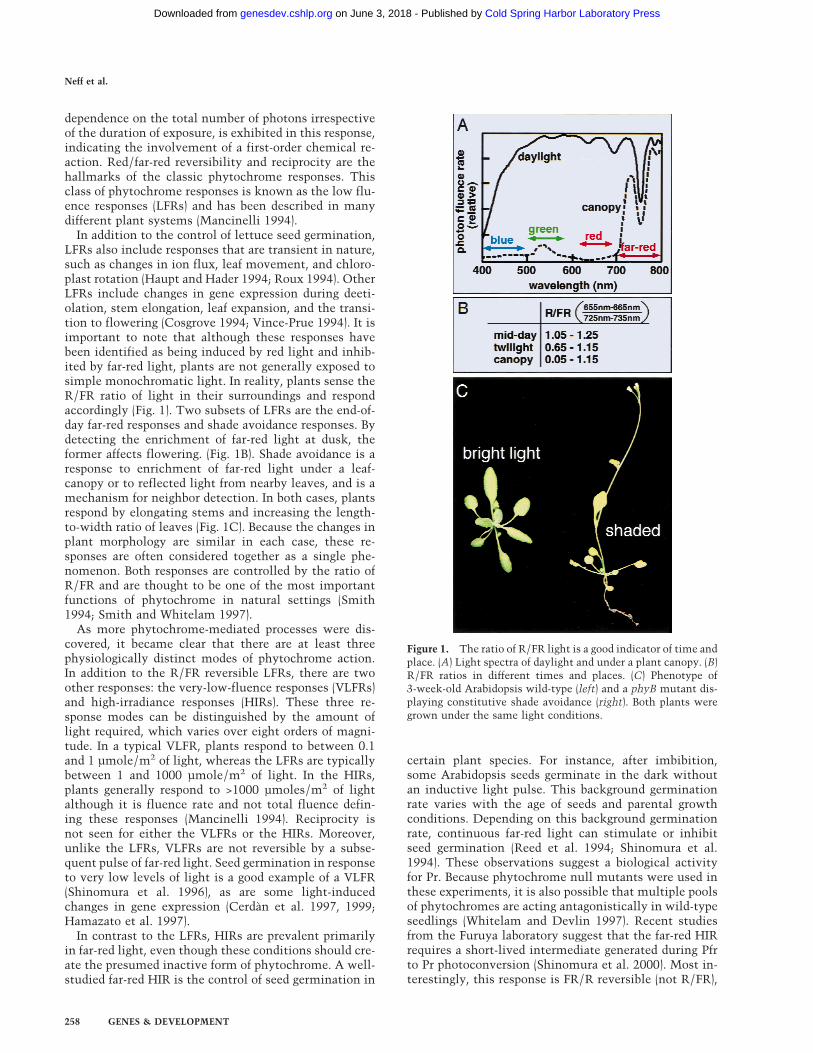
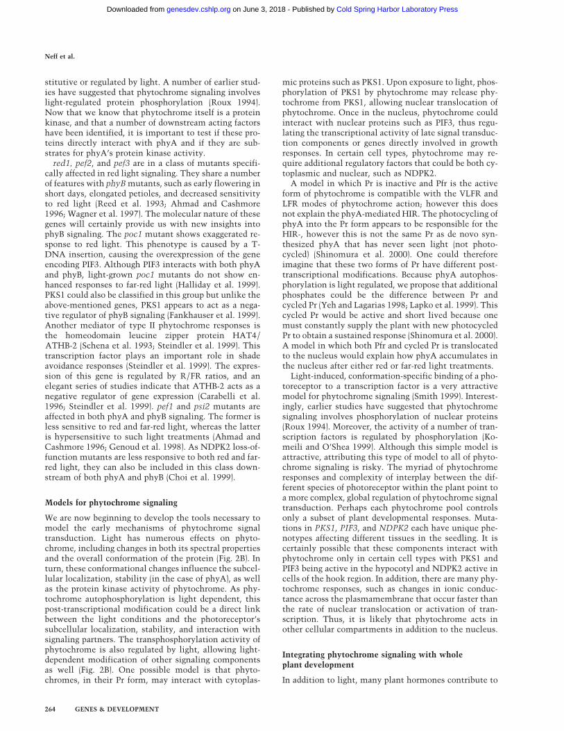
In addition to the control of lettuce seed germination,LFRs also include responses that are transient in nature,such as changes in ion flux, leaf movement, and chloro-plast rotation (Haupt and Hader 1994; Roux 1994). OtherLFRs include changes in gene expression during deeti-olation, stem elongation, leaf expansion, and the transi-tion to flowering (Cosgrove 1994; Vince-Prue 1994). It isimportant to note that although these responses havebeen identified as being induced by red light and inhib-ited by far-red light, plants are not generally exposed tosimple monochromatic light. In reality, plants sense theR/FR ratio of light in their surroundings and respondaccordingly (Fig. 1). Two subsets of LFRs are the end-of-day far-red responses and shade avoidance responses. Bydetecting the enrichment of far-red light at dusk, theformer affects flowering. (Fig. 1B). Shade avoidance is aresponse to enrichment of far-red light under a leaf-canopy or to reflected light from nearby leaves, and is amechanism for neighbor detection. In both cases, plantsrespond by elongating stems and increasing the length-to-width ratio of leaves (Fig. 1C). Because the changes inplant morphology are similar in each case, these re-sponses are often considered together as a single phe-nomenon. Both responses are controlled by the ratio ofR/FR and are thought to be one of the most importantfunctions of phytochrome in natural settings (Smith1994; Smith and Whitelam 1997).
As more phytochrome-mediated processes were dis-covered, it became clear that there are at least threephysiologically distinct modes of phytochrome action.In addition to the R/FR reversible LFRs, there are twoother responses: the very-low-fluence responses (VLFRs)and high-irradiance responses (HIRs). These three re-sponse modes can be distinguished by the amount oflight required, which varies over eight orders of magni-tude. In a typical VLFR, plants respond to between 0.1and 1 µmole/m2 of light, whereas the LFRs are typicallybetween 1 and 1000 µmole/m2 of light. In the HIRs,plants generally respond to >1000 µmoles/m2 of lightalthough it is fluence rate and not total fluence defin-ing these responses (Mancinelli 1994). Reciprocity isnot seen for either the VLFRs or the HIRs. Moreover,unlike the LFRs, VLFRs are not reversible by a subse-quent pulse of far-red light. Seed germination in responseto very low levels of light is a good example of a VLFR(Shinomura et al. 1996), as are some light-inducedchanges in gene expression (Cerdan et al. 1997, 1999;Hamazato et al. 1997).
In contrast to the LFRs, HIRs are prevalent primarilyin far-red light, even though these conditions should cre-ate the presumed inactive form of phytochrome. A well-studied far-red HIR is the control of seed germination in
certain plant species. For instance, after imbibition,some Arabidopsis seeds germinate in the dark withoutan inductive light pulse. This background germinationrate varies with the age of seeds and parental growthconditions. Depending on this background germinationrate, continuous far-red light can stimulate or inhibitseed germination (Reed et al. 1994; Shinomura et al.1994). These observations suggest a biological activityfor Pr. Because phytochrome null mutants were used inthese experiments, it is also possible that multiple poolsof phytochromes are acting antagonistically in wild-typeseedlings (Whitelam and Devlin 1997). Recent studiesfrom the Furuya laboratory suggest that the far-red HIRrequires a short-lived intermediate generated during Pfrto Pr photoconversion (Shinomura et al. 2000). Most in-terestingly, this response is FR/R reversible (not R/FR),
Figure 1. The ratio of R/FR light is a good indicator of time andplace. (A) Light spectra of daylight and under a plant canopy. (B)R/FR ratios in different times and places. (C) Phenotype of3-week-old Arabidopsis wild-type (left) and a phyB mutant dis-playing constitutive shade avoidance (right). Both plants weregrown under the same light conditions.
Neff et al.
258 GENES & DEVELOPMENT
Cold Spring Harbor Laboratory Press on June 3, 2018 - Published by genesdev.cshlp.orgDownloaded from
which is opposite to what is seen in the LFRs such asfar-red inhibition of light-regulated gene expression orlettuce seed germination.
Multiple phytochromes contributeto plant development
The attribution of the variety of light-regulated re-sponses to a single photoreceptor was one of the prob-lems with early phytochrome research. In the 1980’s,spectrophotometric studies indicated that there were atleast two distinct pools of phytochrome, type I (Iightlabile) and type II (light stable). The light-labile pool de-grades fairly rapidly (as fast as a 1-hr half-life, dependingon the plant) upon exposure to red or white light. Al-though there was little spectrophotometrically detect-able phytochrome after prolonged exposure to light,R/FR responses still persisted. That this stable phyto-chrome pool had biological activity was demonstrated inseed germination and end-of-day far-red response experi-ments where the escape times for far-red reversibilitywere three to four times longer than the degradationtimes for light-labile phytochrome (for review, see Man-cinelli 1994).
The cloning of multiple phytochrome apoproteingenes has shed some light on the distinct pools andmodes of action of phytochromes (Abe et al. 1989; Shar-rock and Quail 1989). In Arabidopsis, we now know thatthere are five distinct phytochromes termed phyA–phyE(Sharrock and Quail 1989; Mathews and Sharrock 1997).phyA is a type I phytochrome. phyB–phyE are all type IIphytochromes. In dark-grown tissues, phyA is by far themost abundant phytochrome. After exposure to light,the levels of phyA drop up to 100 fold. Degradation ofphyA is light dependent and requires selective recogni-tion and ubiquitination of Pfr (Clough et al. 1999). PHYAgene expression is also negatively regulated by light(Somers and Quail 1995a). This repression is rapid, oc-curs at the transcriptional level, and requires phyto-chrome (Lissemore and Quail 1988). The regulation ofphyA protein level by light is therefore the result of acoordinated transcriptional and post-translational regu-lation. In light-grown plants, phyB becomes the mostabundant phytochrome; phyC–phyE are less abundanttype II phytochromes (Clack et al. 1994; Hirschfeld et al.1998). All five phytochromes are expressed throughoutthe plant with only minor differences in their expressionpatterns (Somers and Quail 1995b; Goosey et al. 1997).
Although the presence of multiple phytochromes be-gins to address the light labile and light stable pools,assigning precise roles in development for each of thesephotoreceptors required genetic analysis of plants lack-ing one or more of these pigments. Phytochrome apopro-tein mutants have allowed an assessment of the functionof individual photoreceptors. phyA and phyB playunique, redundant, or antagonistic roles in different re-sponses throughout Arabidopsis development (Whitelamand Devlin 1997; Cerdan et al. 1999). phyA is essentialfor de-etiolation in far-red light (light found under acanopy of plants), whereas phyB is the major red light
photoreceptor during seedling development. phyA alsomediates responses to very low fluences of blue, red, andfar-red light. Mutants in phyD and phyE have moresubtle phenotypes, only uncovered in a phyB mutantbackground, demonstrating a degree of redundancy be-tween phyB, phyD, and phyE, with phyB playing themost prominent role of the three (Aukerman et al. 1997;Devlin et al. 1998, 1999). These three phytochromes playa major role in regulating shade avoidance. No muta-tions in phyC have been discovered yet, but overexpres-sion studies suggest a role in primary leaf expansion(Halliday et al. 1997; Qin et al. 1997).
In-depth analysis of plants carrying null mutations fordifferent phytochromes has shown that the major phy-tochrome in Arabidopsis is phyB. Mutations in PHYBhave profound effects on plant development throughoutthe life cycle (Reed et al. 1993). In general, phyB mutantseedlings have long hypocotyls and small cotyledons incontinuous red or white light. They also have less an-thocyanin, chlorophyll, and fewer chloroplasts than thewild type. As adults, these mutants flower early, havelonger petioles and stems, and increased apical domi-nance. These phenotypes are also observed in mutantswith reduced phyB activity from other plant species suchas cucumber, pea, tomato, and rape (Whitelam and Dev-lin 1997). Many of the growth responses regulated byphyB involve cell expansion or elongation. phyB affectsnuclear endoreduplication in hypocotyls of Arabidopsis(Gendreau et al. 1998), a possible mode of control of cellsize (Gendreau et al. 1997). Generally speaking, phyBmutants show constitutive shade avoidance and are al-tered in their end-of-day far-red response (Fig. 1C), indi-cating that it is primarily phyB mediating this process.However, phyB single mutants and phyA phyB doublemutants still show responses to reductions in R/FR ra-tios indicating that other phytochromes play significantroles in plant development (Whitelam and Smith 1991;Robson et al. 1993; Halliday et al. 1994; Devlin et al.1996). This observation was the basis for the geneticscreen that identified phyE mutants (Devlin et al. 1998).
Genetic analysis has shown that there is a complexweb of interactions, not only between the phyto-chromes, but also between phytochromes and the bluelight photoreceptors, cryptochromes. A functional de-pendency of cryptochrome 1 (cry1) on phytochromes hasbeen described based on observations that phyA phyBdouble mutants have a dramatic blue light phenotype(Ahmad and Cashmore 1997). However, phytochromescan absorb blue light (Furuya and Song 1994). Further-more, phytochrome mutants have blue light-mediateddefects in hypocotyl growth inhibition (Whitelam et al.1993), cotyledon expansion (Neff and Van Volkenburgh1994), seed germination (Shinomura et al. 1996), CABgene induction (Hamazato et al. 1997), and light-inducedshrinking of hypocotyl protoplasts (Wang and Iino 1998).Detailed analysis of plants carrying null mutations inmultiple phytochromes and cryptochrome arguesagainst a functional dependency of cryptochromes onphytochromes. Rather, these studies demonstrate a com-plex web of interactions within and between the two
Phytochrome signaling and plant development
GENES & DEVELOPMENT 259
Cold Spring Harbor Laboratory Press on June 3, 2018 - Published by genesdev.cshlp.orgDownloaded from
classes of photoreceptors including redundancy, antago-nism, and effector/modulator relationships (Casal andBoccalandro 1995; Casal and Mazzella 1998; Neff andChory 1998; Wang and Iino 1998; Hennig et al. 1999;Mockler et al. 1999). However, the mechanisms of theseinteractions are not clear and are subject to debate (Ah-mad 1999).
Effector/modulator relationships describe a situationin which a photoreceptor cannot control a growth re-sponse independently yet it can affect that response inthe presence of other, controlling, photoreceptors (Mohr1994). As an example, phototropism is controlled by theblue light-absorbing phototropins, such as NPH1 (Chris-tie et al. 1998), although phytochromes can modulatethis response (Parks et al. 1996; Hangarter 1997; Janoudiet al. 1997). Because pretreatments of omnilateral redlight can enhance the phototropic response to subse-quent exposures of unilateral blue, phytochromes canalso act as preprogrammed amplifiers of this phototro-pin-mediated growth response (Shropshire and Mohr1970; Woitzik and Mohr 1988). A novel photoreceptorwith homology to both phytochrome and NPH1 has re-cently been isolated from the fern Adiantum (Nozue etal. 1998). In this case, the coaction between blue and redlight on phototropism of Adiantum protonema (Hayamiet al. 1986) may be acting through a single photoreceptor.
Molecular properties of phytochrome
One of the major advances made in phytochrome re-search was the ability to partially purify a species of thepigment, allowing the study of its biochemical proper-ties (Butler et al. 1959, 1964). Because of the pioneeringstudies of Butler, full-length phyA holoprotein has beenpurified from multiple plant species. phyA is found as asoluble homodimer with each monomer covalently at-tached to a linear tetrapyrrole chromophore. Each mono-
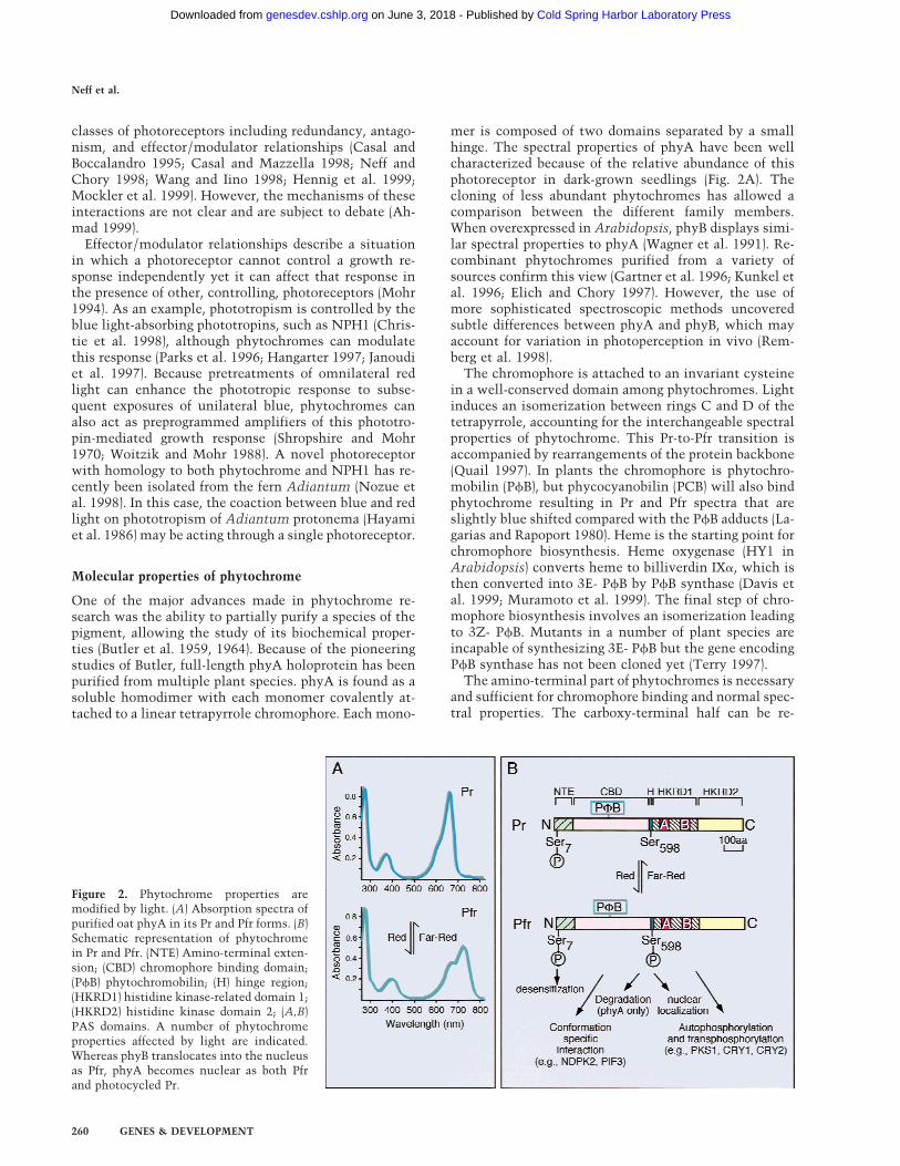
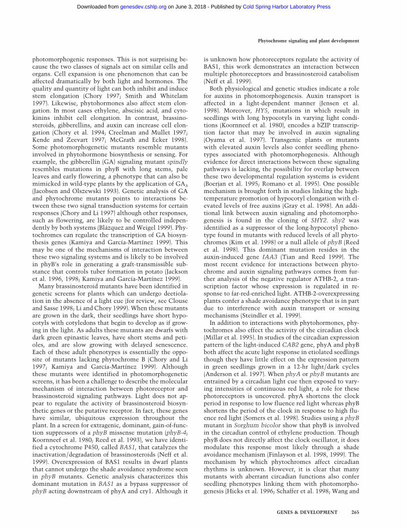
mer is composed of two domains separated by a smallhinge. The spectral properties of phyA have been wellcharacterized because of the relative abundance of thisphotoreceptor in dark-grown seedlings (Fig. 2A). Thecloning of less abundant phytochromes has allowed acomparison between the different family members.When overexpressed in Arabidopsis, phyB displays simi-lar spectral properties to phyA (Wagner et al. 1991). Re-combinant phytochromes purified from a variety ofsources confirm this view (Gartner et al. 1996; Kunkel etal. 1996; Elich and Chory 1997). However, the use ofmore sophisticated spectroscopic methods uncoveredsubtle differences between phyA and phyB, which mayaccount for variation in photoperception in vivo (Rem-berg et al. 1998).
The chromophore is attached to an invariant cysteinein a well-conserved domain among phytochromes. Lightinduces an isomerization between rings C and D of thetetrapyrrole, accounting for the interchangeable spectralproperties of phytochrome. This Pr-to-Pfr transition isaccompanied by rearrangements of the protein backbone(Quail 1997). In plants the chromophore is phytochro-mobilin (PfB), but phycocyanobilin (PCB) will also bindphytochrome resulting in Pr and Pfr spectra that areslightly blue shifted compared with the PfB adducts (La-garias and Rapoport 1980). Heme is the starting point forchromophore biosynthesis. Heme oxygenase (HY1 inArabidopsis) converts heme to billiverdin IXa, which isthen converted into 3E- PfB by PfB synthase (Davis etal. 1999; Muramoto et al. 1999). The final step of chro-mophore biosynthesis involves an isomerization leadingto 3Z- PfB. Mutants in a number of plant species areincapable of synthesizing 3E- PfB but the gene encodingPfB synthase has not been cloned yet (Terry 1997).
The amino-terminal part of phytochromes is necessaryand sufficient for chromophore binding and normal spec-tral properties. The carboxy-terminal half can be re-
Figure 2. Phytochrome properties aremodified by light. (A) Absorption spectra ofpurified oat phyA in its Pr and Pfr forms. (B)Schematic representation of phytochromein Pr and Pfr. (NTE) Amino-terminal exten-sion; (CBD) chromophore binding domain;(PfB) phytochromobilin; (H) hinge region;(HKRD1) histidine kinase-related domain 1;(HKRD2) histidine kinase domain 2; (A,B)PAS domains. A number of phytochromeproperties affected by light are indicated.Whereas phyB translocates into the nucleusas Pfr, phyA becomes nuclear as both Pfrand photocycled Pr.
Neff et al.
260 GENES & DEVELOPMENT
Cold Spring Harbor Laboratory Press on June 3, 2018 - Published by genesdev.cshlp.orgDownloaded from
garded as the output domain. This domain appears to bethe result of the duplication of a bacterial histidine ki-nase-related domain (Schneider-Poetsch 1992; Yeh andLagarias 1998) (Figure 2B). The first of those domainsalso contains two repeats with homology to PER–ARN-T–SIM (PAS) domains (Lagarias et al. 1995; Kay 1997),originally found in basic helix–loop–helix (bHLH) con-taining transcription factors from fly (PER and SIM) andmammals (ARNT and AHR) (Huang et al. 1993). Thesemodules have been found in a wide variety of organismsand play important signaling roles in protein–protein in-teractions, response to small ligands, and changes inlight conditions, oxygen levels, and redox potential (Tay-lor and Zhulin 1999). The majority of missense muta-tions in phyA and phyB cluster in the PAS repeats, dem-onstrating the importance of this domain for phyto-chrome function (Quail et al. 1995). One of the phyBmissense mutations, phyB-101 (Bradley et al. 1996), is inthe second PAS repeat. This mutation affects spectralproperties of the pigment, causing accelerated dark re-version from Pfr to Pr, and alters the end-of-day far-redresponse in seedlings (Elich and Chory 1997). Thus, thePAS repeats may be involved in intramolecular interac-tions within phytochrome itself.
The discovery of phytochromes in cyanobacteriademonstrates that these proteins are not unique toplants (Kehoe and Grossman 1996). Synechocystis Cph1(Cyanobacterial phytochrome 1) has spectral propertiesvery similar to the ones of its higher plant relatives(Hughes et al. 1997). Moreover, Cph1 functions as alight-regulated histidine kinase (Yeh et al. 1997) al-though the biological function of Cph1 in Synechocystisis still unknown. Studies involving prokaryotic phyto-chromes strongly suggest that plant phytochromeshave histidine kinase ancestry. The group of Lagariashas recently shown that two plant phytyochromes arealso light and chromophore-regulated protein kinases,but unlike their cyanobacterial counterparts they auto-phosphorylate on serine/threonine rather than histi-dine/aspartate (Yeh and Lagarias 1998). Phytochromesare not the first eukaryotic serine/threonine kinaseswith histidine kinase ancestry (Harris et al. 1997), andmuch remains to be done to characterize this enzymaticactivity. Some important questions to be answered in-clude: What is the catalytic domain of phytochrome ki-nase? Do different phytochromes, which play distinctroles in vivo, phosphorylate different substrates? And,most importantly, what is the biological relevance ofthis activity?
Interestingly, phyA isolated from plants is a phospho-protein with at least one serine phosphorylated in alight-dependent fashion, suggesting that phosphoryla-tion of this residue results from autophosphorylation orfrom phosphorylation by another phytochrome (Lapko etal. 1999). This serine is also a major phosphoacceptor siteidentified in in vitro phosphorylation studies (Wong etal. 1986). Residues at the very amino-terminus of phyAare also phosphorylated but not in a light-dependentfashion (Lapko et al. 1997, 1999). However, phosphory-lation of this portion of the protein has been implicated
in down-regulation of phyA signaling (Stockhaus et al.1992).
In vitro kinase assays have identified other substratesof phytochrome. Of particular interest are the crypto-chrome blue light receptors cry1 and cry2 (Ahmad et al.1998). Although they are not phosphorylated in a lightdependent fashion in vitro, in vivo analysis shows thatcry1 phosphorylation is stimulated by red light. Theseresults are particularly interesting in view of the largebody of photobiological evidence suggesting an interac-tion between phytochrome and the blue light receptors(Mohr 1994). We have recently identified the first phy-tochrome kinase substrate (PKS1) that is phosphorylatedin a light-dependent manner in vitro (Fankhauser et al.1999). Phosphorylation of PKS1 in vivo also appears to bestimulated by red light, suggesting that phytochrome isthe kinase. PKS1 was identified as a protein that binds tothe carboxyl-terminus of phyA; it also interacts with thecarboxyl-terminus of phyB. The phenotypes of plantsoverexpressing PKS1 are consistent with PKS1 beinga negative regulator of phyB signaling. Fusions withthe jellyfish green fluorescent protein (GFP) (Chiuet al. 1996) show that PKS1 is a cytoplasmic protein(Fankhauser et al. 1999). It will be important to assessthe relevance of cry1, cry2, and PKS1 phosphorylation inphytochrome signaling. Figure 2B presents some of theways this kinase activity may affect phytochrome signaltransduction.
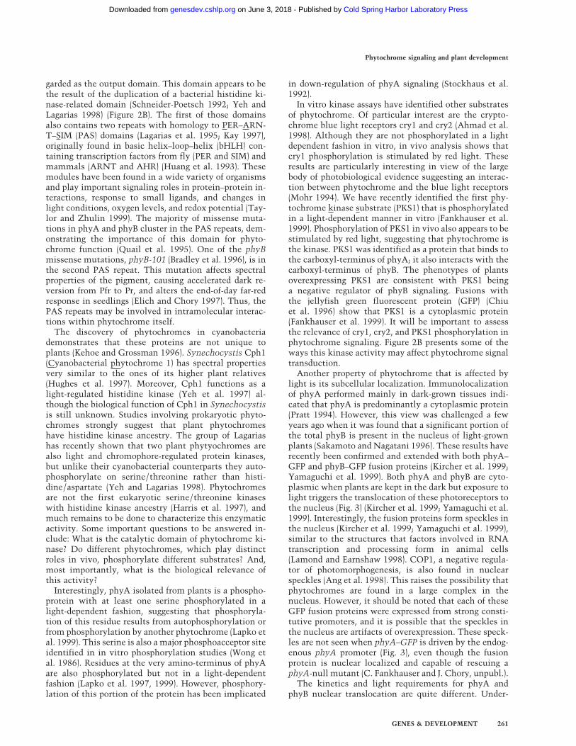
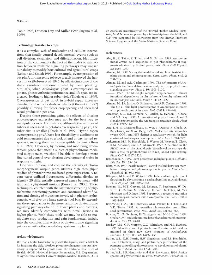
Another property of phytochrome that is affected bylight is its subcellular localization. Immunolocalizationof phyA performed mainly in dark-grown tissues indi-cated that phyA is predominantly a cytoplasmic protein(Pratt 1994). However, this view was challenged a fewyears ago when it was found that a significant portion ofthe total phyB is present in the nucleus of light-grownplants (Sakamoto and Nagatani 1996). These results haverecently been confirmed and extended with both phyA–GFP and phyB–GFP fusion proteins (Kircher et al. 1999;Yamaguchi et al. 1999). Both phyA and phyB are cyto-plasmic when plants are kept in the dark but exposure tolight triggers the translocation of these photoreceptors tothe nucleus (Fig. 3) (Kircher et al. 1999; Yamaguchi et al.1999). Interestingly, the fusion proteins form speckles inthe nucleus (Kircher et al. 1999; Yamaguchi et al. 1999),similar to the structures that factors involved in RNAtranscription and processing form in animal cells(Lamond and Earnshaw 1998). COP1, a negative regula-tor of photomorphogenesis, is also found in nuclearspeckles (Ang et al. 1998). This raises the possibility thatphytochromes are found in a large complex in thenucleus. However, it should be noted that each of theseGFP fusion proteins were expressed from strong consti-tutive promoters, and it is possible that the speckles inthe nucleus are artifacts of overexpression. These speck-les are not seen when phyA–GFP is driven by the endog-enous phyA promoter (Fig. 3), even though the fusionprotein is nuclear localized and capable of rescuing aphyA-null mutant (C. Fankhauser and J. Chory, unpubl.).
The kinetics and light requirements for phyA andphyB nuclear translocation are quite different. Under-
Phytochrome signaling and plant development
GENES & DEVELOPMENT 261
Cold Spring Harbor Laboratory Press on June 3, 2018 - Published by genesdev.cshlp.orgDownloaded from
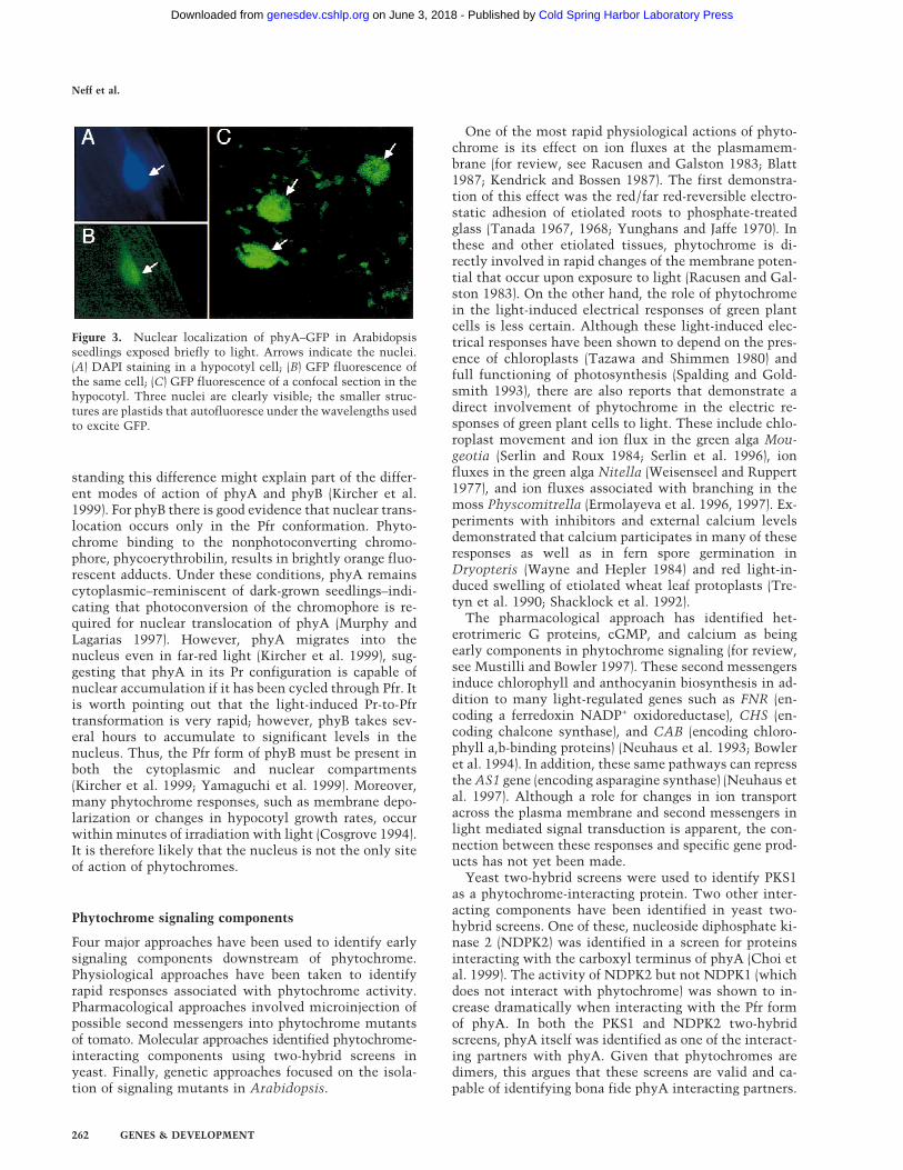
standing this difference might explain part of the differ-ent modes of action of phyA and phyB (Kircher et al.1999). For phyB there is good evidence that nuclear trans-location occurs only in the Pfr conformation. Phyto-chrome binding to the nonphotoconverting chromo-phore, phycoerythrobilin, results in brightly orange fluo-rescent adducts. Under these conditions, phyA remainscytoplasmic–reminiscent of dark-grown seedlings–indi-cating that photoconversion of the chromophore is re-quired for nuclear translocation of phyA (Murphy andLagarias 1997). However, phyA migrates into thenucleus even in far-red light (Kircher et al. 1999), sug-gesting that phyA in its Pr configuration is capable ofnuclear accumulation if it has been cycled through Pfr. Itis worth pointing out that the light-induced Pr-to-Pfrtransformation is very rapid; however, phyB takes sev-eral hours to accumulate to significant levels in thenucleus. Thus, the Pfr form of phyB must be present inboth the cytoplasmic and nuclear compartments(Kircher et al. 1999; Yamaguchi et al. 1999). Moreover,many phytochrome responses, such as membrane depo-larization or changes in hypocotyl growth rates, occurwithin minutes of irradiation with light (Cosgrove 1994).It is therefore likely that the nucleus is not the only siteof action of phytochromes.
Phytochrome signaling components
Four major approaches have been used to identify earlysignaling components downstream of phytochrome.Physiological approaches have been taken to identifyrapid responses associated with phytochrome activity.Pharmacological approaches involved microinjection ofpossible second messengers into phytochrome mutantsof tomato. Molecular approaches identified phytochrome-interacting components using two-hybrid screens inyeast. Finally, genetic approaches focused on the isola-tion of signaling mutants in Arabidopsis.
One of the most rapid physiological actions of phyto-chrome is its effect on ion fluxes at the plasmamem-brane (for review, see Racusen and Galston 1983; Blatt1987; Kendrick and Bossen 1987). The first demonstra-tion of this effect was the red/far red-reversible electro-static adhesion of etiolated roots to phosphate-treatedglass (Tanada 1967, 1968; Yunghans and Jaffe 1970). Inthese and other etiolated tissues, phytochrome is di-rectly involved in rapid changes of the membrane poten-tial that occur upon exposure to light (Racusen and Gal-ston 1983). On the other hand, the role of phytochromein the light-induced electrical responses of green plantcells is less certain. Although these light-induced elec-trical responses have been shown to depend on the pres-ence of chloroplasts (Tazawa and Shimmen 1980) andfull functioning of photosynthesis (Spalding and Gold-smith 1993), there are also reports that demonstrate adirect involvement of phytochrome in the electric re-sponses of green plant cells to light. These include chlo-roplast movement and ion flux in the green alga Mou-geotia (Serlin and Roux 1984; Serlin et al. 1996), ionfluxes in the green alga Nitella (Weisenseel and Ruppert1977), and ion fluxes associated with branching in themoss Physcomitrella (Ermolayeva et al. 1996, 1997). Ex-periments with inhibitors and external calcium levelsdemonstrated that calcium participates in many of theseresponses as well as in fern spore germination inDryopteris (Wayne and Hepler 1984) and red light-in-duced swelling of etiolated wheat leaf protoplasts (Tre-tyn et al. 1990; Shacklock et al. 1992).
The pharmacological approach has identified het-erotrimeric G proteins, cGMP, and calcium as beingearly components in phytochrome signaling (for review,see Mustilli and Bowler 1997). These second messengersinduce chlorophyll and anthocyanin biosynthesis in ad-dition to many light-regulated genes such as FNR (en-coding a ferredoxin NADP+ oxidoreductase), CHS (en-coding chalcone synthase), and CAB (encoding chloro-phyll a,b-binding proteins) (Neuhaus et al. 1993; Bowleret al. 1994). In addition, these same pathways can repressthe AS1 gene (encoding asparagine synthase) (Neuhaus etal. 1997). Although a role for changes in ion transportacross the plasma membrane and second messengers inlight mediated signal transduction is apparent, the con-nection between these responses and specific gene prod-ucts has not yet been made.
Yeast two-hybrid screens were used to identify PKS1as a phytochrome-interacting protein. Two other inter-acting components have been identified in yeast two-hybrid screens. One of these, nucleoside diphosphate ki-nase 2 (NDPK2) was identified in a screen for proteinsinteracting with the carboxyl terminus of phyA (Choi etal. 1999). The activity of NDPK2 but not NDPK1 (whichdoes not interact with phytochrome) was shown to in-crease dramatically when interacting with the Pfr formof phyA. In both the PKS1 and NDPK2 two-hybridscreens, phyA itself was identified as one of the interact-ing partners with phyA. Given that phytochromes aredimers, this argues that these screens are valid and ca-pable of identifying bona fide phyA interacting partners.
Figure 3. Nuclear localization of phyA–GFP in Arabidopsisseedlings exposed briefly to light. Arrows indicate the nuclei.(A) DAPI staining in a hypocotyl cell; (B) GFP fluorescence ofthe same cell; (C) GFP fluorescence of a confocal section in thehypocotyl. Three nuclei are clearly visible; the smaller struc-tures are plastids that autofluoresce under the wavelengths usedto excite GFP.
Neff et al.
262 GENES & DEVELOPMENT
Cold Spring Harbor Laboratory Press on June 3, 2018 - Published by genesdev.cshlp.orgDownloaded from
Plants overexpressing PKS1 have longer hypocotyls inred light, suggesting that PKS1 is a negative regulator ofhypocotyl elongation downstream of phyB. In contrast,NDPK2 appears to be a positive regulator of phyA andphyB signaling. However, hypocotyl elongation is notaffected by this regulatory component. Instead, loss-of-function alleles have a small but significant reduction incotyledon greening and opening of the hypocotyl/coty-ledon hook during deetiolation (Choi et al. 1999).
ndpk2 mutants have altered responses to both red andfar-red light, suggesting that this regulator interacts invivo with both phyA and phyB. NDPK2–GFP fusions ex-pressed in tobacco show both cytoplasmic and nuclearlocalization. Thus, phytochrome may interact withNDPK2 in both the cytoplasm and the nucleus. Red lighthas been shown to stimulate phosphorylation of NDPK2in vivo suggesting that this interacting protein may alsobe a substrate for phytochrome kinase activity (Hamadaet al. 1996; Tanaka et al. 1998; Ogura et al. 1999). ThePAS domains in phytochrome are clearly important asNDPK2 does not interact with a phyA mutated in one ofthe two PAS repeats. The mechanism of action forNDPK2 in plants is not known although studies in othermodel systems implicate this enzyme in many develop-mental processes (Tanaka et al. 1998; Choi et al. 1999).
A third phytochrome-interacting partner is PIF3, anuclear-localized, putative bHLH-containing transcrip-tion factor isolated by virtue of its binding to both phyAand phyB in the yeast two-hybrid system. DecreasingPIF3 levels results in plants that are defective in both redand far-red light sensing, consistent with the view thatthis gene acts downstream of both photoreceptors (Ni etal. 1998). Importantly, PIF3 binds phyB in a light-depen-dent fashion (Ni et al. 1999). As with NDPK2, PIF3 doesnot interact with phytochromes mutated in the PAS do-main. These results again underscore the importance ofthe PAS domain in phytochrome signaling because mu-tations in the PAS domain have major effects on both itsspectral properties (Elich and Chory 1997) and its abilityto interact with downstream signaling partners. It willbe important to test if PIF3 is modified in response tolight, or if binding to phytochrome directly modulatesthe activity of this putative transcription factor. For ex-ample, the activity of CCA1, a DNA-binding protein act-ing downstream of phytochromes, can be modulated byphosphorylation (Sugano et al. 1998). However, caseinkinase 2, but not phytochrome, appears to be the proteinkinase mediating CCA1 phosphorylation.
Genetic and molecular screens have identified a largenumber of genes acting downstream of light receptors.Because different light qualities trigger the same devel-opmental responses using different photoreceptors, it isvery likely that common late-acting signaling interme-diates are used. Mutants in such genes are expected tohave the same phenotypes irrespective of the light qual-ity. Such loci have been identified and they fall into twoclasses: mutants that deetiolate even in the absence oflight and mutants that are defective for their perceptionof light at a variety of different wavelengths. The formerclass is referred to as det/cop/fus mutants based on the
different genetic screens from which they were isolated.These are pleiotropic mutations affecting many aspectsof plant development and these proteins are generallyconsidered to be late signaling components (for review,see Fankhauser and Chory 1997; Deng and Quail 1999;Osterlund et al. 1999; Wei and Deng 1999).
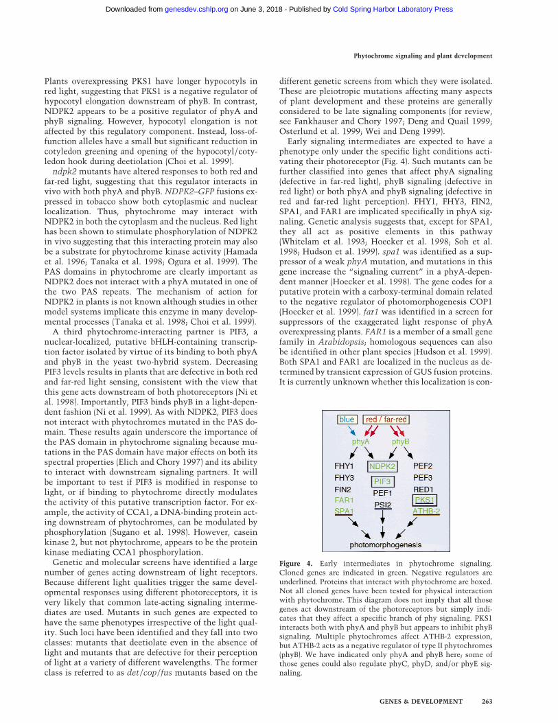
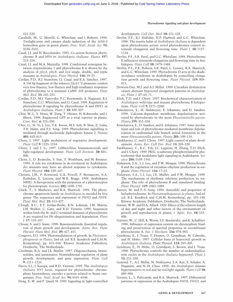
Early signaling intermediates are expected to have aphenotype only under the specific light conditions acti-vating their photoreceptor (Fig. 4). Such mutants can befurther classified into genes that affect phyA signaling(defective in far-red light), phyB signaling (defective inred light) or both phyA and phyB signaling (defective inred and far-red light perception). FHY1, FHY3, FIN2,SPA1, and FAR1 are implicated specifically in phyA sig-naling. Genetic analysis suggests that, except for SPA1,they all act as positive elements in this pathway(Whitelam et al. 1993; Hoecker et al. 1998; Soh et al.1998; Hudson et al. 1999). spa1 was identified as a sup-pressor of a weak phyA mutation, and mutations in thisgene increase the “signaling current” in a phyA-depen-dent manner (Hoecker et al. 1998). The gene codes for aputative protein with a carboxy-terminal domain relatedto the negative regulator of photomorphogenesis COP1(Hoecker et al. 1999). far1 was identified in a screen forsuppressors of the exaggerated light response of phyAoverexpressing plants. FAR1 is a member of a small genefamily in Arabidopsis; homologous sequences can alsobe identified in other plant species (Hudson et al. 1999).Both SPA1 and FAR1 are localized in the nucleus as de-termined by transient expression of GUS fusion proteins.It is currently unknown whether this localization is con-
Figure 4. Early intermediates in phytochrome signaling.Cloned genes are indicated in green. Negative regulators areunderlined. Proteins that interact with phytochrome are boxed.Not all cloned genes have been tested for physical interactionwith phytochrome. This diagram does not imply that all thosegenes act downstream of the photoreceptors but simply indi-cates that they affect a specific branch of phy signaling. PKS1interacts both with phyA and phyB but appears to inhibit phyBsignaling. Multiple phytochromes affect ATHB-2 expression,but ATHB-2 acts as a negative regulator of type II phytochromes(phyB). We have indicated only phyA and phyB here; some ofthose genes could also regulate phyC, phyD, and/or phyE sig-naling.
Phytochrome signaling and plant development
GENES & DEVELOPMENT 263
Cold Spring Harbor Laboratory Press on June 3, 2018 - Published by genesdev.cshlp.orgDownloaded from
stitutive or regulated by light. A number of earlier stud-ies have suggested that phytochrome signaling involveslight-regulated protein phosphorylation (Roux 1994).Now that we know that phytochrome itself is a proteinkinase, and that a number of downstream acting factorshave been identified, it is important to test if these pro-teins directly interact with phyA and if they are sub-strates for phyA’s protein kinase activity.
red1, pef2, and pef3 are in a class of mutants specifi-cally affected in red light signaling. They share a numberof features with phyB mutants, such as early flowering inshort days, elongated petioles, and decreased sensitivityto red light (Reed et al. 1993; Ahmad and Cashmore1996; Wagner et al. 1997). The molecular nature of thesegenes will certainly provide us with new insights intophyB signaling. The poc1 mutant shows exaggerated re-sponse to red light. This phenotype is caused by a T-DNA insertion, causing the overexpression of the geneencoding PIF3. Although PIF3 interacts with both phyAand phyB, light-grown poc1 mutants do not show en-hanced responses to far-red light (Halliday et al. 1999).PKS1 could also be classified in this group but unlike theabove-mentioned genes, PKS1 appears to act as a nega-tive regulator of phyB signaling (Fankhauser et al. 1999).Another mediator of type II phytochrome responses isthe homeodomain leucine zipper protein HAT4/ATHB-2 (Schena et al. 1993; Steindler et al. 1999). Thistranscription factor plays an important role in shadeavoidance responses (Steindler et al. 1999). The expres-sion of this gene is regulated by R/FR ratios, and anelegant series of studies indicate that ATHB-2 acts as anegative regulator of gene expression (Carabelli et al.1996; Steindler et al. 1999). pef1 and psi2 mutants areaffected in both phyA and phyB signaling. The former isless sensitive to red and far-red light, whereas the latteris hypersensitive to such light treatments (Ahmad andCashmore 1996; Genoud et al. 1998). As NDPK2 loss-of-function mutants are less responsive to both red and far-red light, they can also be included in this class down-stream of both phyA and phyB (Choi et al. 1999).
Models for phytochrome signaling
We are now beginning to develop the tools necessary tomodel the early mechanisms of phytochrome signaltransduction. Light has numerous effects on phyto-chrome, including changes in both its spectral propertiesand the overall conformation of the protein (Fig. 2B). Inturn, these conformational changes influence the subcel-lular localization, stability (in the case of phyA), as wellas the protein kinase activity of phytochrome. As phy-tochrome autophosphorylation is light dependent, thispost-transcriptional modification could be a direct linkbetween the light conditions and the photoreceptor’ssubcellular localization, stability, and interaction withsignaling partners. The transphosphorylation activity ofphytochrome is also regulated by light, allowing light-dependent modification of other signaling componentsas well (Fig. 2B). One possible model is that phyto-chromes, in their Pr form, may interact with cytoplas-
mic proteins such as PKS1. Upon exposure to light, phos-phorylation of PKS1 by phytochrome may release phy-tochrome from PKS1, allowing nuclear translocation ofphytochrome. Once in the nucleus, phytochrome couldinteract with nuclear proteins such as PIF3, thus regu-lating the transcriptional activity of late signal transduc-tion components or genes directly involved in growthresponses. In certain cell types, phytochrome may re-quire additional regulatory factors that could be both cy-toplasmic and nuclear, such as NDPK2.
A model in which Pr is inactive and Pfr is the activeform of phytochrome is compatible with the VLFR andLFR modes of phytochrome action; however this doesnot explain the phyA-mediated HIR. The photocycling ofphyA into the Pr form appears to be responsible for theHIR-, however this is not the same Pr as de novo syn-thesized phyA that has never seen light (not photo-cycled) (Shinomura et al. 2000). One could thereforeimagine that these two forms of Pr have different post-transcriptional modifications. Because phyA autophos-phorylation is light regulated, we propose that additionalphosphates could be the difference between Pr andcycled Pr (Yeh and Lagarias 1998; Lapko et al. 1999). Thiscycled Pr would be active and short lived because onemust constantly supply the plant with new photocycledPr to obtain a sustained response (Shinomura et al. 2000).A model in which both Pfr and cycled Pr is translocatedto the nucleus would explain how phyA accumulates inthe nucleus after either red or far-red light treatments.
Light-induced, conformation-specific binding of a pho-toreceptor to a transcription factor is a very attractivemodel for phytochrome signaling (Smith 1999). Interest-ingly, earlier studies have suggested that phytochromesignaling involves phosphorylation of nuclear proteins(Roux 1994). Moreover, the activity of a number of tran-scription factors is regulated by phosphorylation (Ko-meili and O’Shea 1999). Although this simple model isattractive, attributing this type of model to all of phyto-chrome signaling is risky. The myriad of phytochromeresponses and complexity of interplay between the dif-ferent species of photoreceptor within the plant point toa more complex, global regulation of phytochrome signaltransduction. Perhaps each phytochrome pool controlsonly a subset of plant developmental responses. Muta-tions in PKS1, PIF3, and NDPK2 each have unique phe-notypes affecting different tissues in the seedling. It iscertainly possible that these components interact withphytochrome only in certain cell types with PKS1 andPIF3 being active in the hypocotyl and NDPK2 active incells of the hook region. In addition, there are many phy-tochrome responses, such as changes in ionic conduc-tance across the plasmamembrane that occur faster thanthe rate of nuclear translocation or activation of tran-scription. Thus, it is likely that phytochrome acts inother cellular compartments in addition to the nucleus.
Integrating phytochrome signaling with wholeplant development
In addition to light, many plant hormones contribute to
Neff et al.
264 GENES & DEVELOPMENT
Cold Spring Harbor Laboratory Press on June 3, 2018 - Published by genesdev.cshlp.orgDownloaded from
photomorphogenic responses. This is not surprising be-cause the two classes of signals act on similar cells andorgans. Cell expansion is one phenomenon that can beaffected dramatically by both light and hormones. Thequality and quantity of light can both inhibit and inducestem elongation (Chory 1997; Smith and Whitelam1997). Likewise, phytohormones also affect stem elon-gation. In most cases ethylene, abscisic acid, and cyto-kinins inhibit cell elongation. In contrast, brassino-steroids, gibberellins, and auxin can increase cell elon-gation (Chory et al. 1994; Creelman and Mullet 1997;Kende and Zeevart 1997; McGrath and Ecker 1998).Some photomorphogenetic mutants resemble mutantsinvolved in phytohormone biosynthesis or sensing. Forexample, the gibberellin (GA) signaling mutant spindlyresembles mutations in phyB with long stems, paleleaves and early flowering, a phenotype that can also bemimicked in wild-type plants by the application of GA3
(Jacobsen and Olszewski 1993). Genetic analysis of GAand phytochrome mutants points to interactions be-tween these two signal transduction systems for certainresponses (Chory and Li 1997) although other responses,such as flowering, are likely to be controlled indepen-dently by both systems (Blazquez and Weigel 1999). Phy-tochromes can regulate the transcription of GA biosyn-thesis genes (Kamiya and Garcıa-Martınez 1999). Thismay be one of the mechanisms of interaction betweenthese two signaling systems and is likely to be involvedin phyB’s role in generating a graft-transmissible sub-stance that controls tuber formation in potato (Jacksonet al. 1996, 1998; Kamiya and Garcıa-Martınez 1999).
Many brassinosteroid mutants have been identified ingenetic screens for plants which can undergo deetiola-tion in the absence of a light cue (for review, see Clouseand Sasse 1998; Li and Chory 1999). When these mutantsare grown in the dark, their seedlings have short hypo-cotyls with cotyledons that begin to develop as if grow-ing in the light. As adults these mutants are dwarfs withdark green epinastic leaves, have short stems and peti-oles, and are slow growing with delayed senescence.Each of these adult phenotypes is essentially the oppo-site of mutants lacking phytochrome B (Chory and Li1997; Kamiya and Garcıa-Martınez 1999). Althoughthese mutants were identified in photomorphogeneticscreens, it has been a challenge to describe the molecularmechanism of interaction between photoreceptor andbrassinosteroid signaling pathways. Light does not ap-pear to regulate the activity of brassinosteroid biosyn-thetic genes or the putative receptor. In fact, these geneshave similar, ubiquitous expression throughout theplant. In a screen for extragenic, dominant, gain-of-func-tion suppressors of a phyB missense mutation (phyB-4;Koornneef et al. 1980; Reed et al. 1993), we have identi-fied a cytochrome P450, called BAS1, that catalyzes theinactivation/degradation of brassinosteroids (Neff et al.1999). Overexpression of BAS1 results in dwarf plantsthat cannot undergo the shade avoidance syndrome seenin phyB mutants. Genetic analysis characterizes thisdominant mutation in BAS1 as a bypass suppressor ofphyB acting downstream of phyA and cry1. Although it
is unknown how photoreceptors regulate the activity ofBAS1, this work demonstrates an interaction betweenmultiple photoreceptors and brassinosteroid catabolism(Neff et al. 1999).
Both physiological and genetic studies indicate a rolefor auxins in photomorphogenesis. Auxin transport isaffected in a light-dependent manner (Jensen et al.1998). Moreover, HY5, mutations in which result inseedlings with long hypocotyls in varying light condi-tions (Koornneef et al. 1980), encodes a bZIP transcrip-tion factor that may be involved in auxin signaling(Oyama et al. 1997). Transgenic plants or mutantswith elevated auxin levels also confer seedling pheno-types associated with photomorphogenesis. Althoughevidence for direct interactions between these signalingpathways is lacking, the possibility for overlap betweenthese two developmental regulation systems is evident(Boerjan et al. 1995; Romano et al. 1995). One possiblemechanism is brought forth in studies linking the high-temperature promotion of hypocotyl elongation with el-evated levels of free auxins (Gray et al. 1998). An addi-tional link between auxin signaling and photomorpho-genesis is found in the cloning of SHY2. shy2 wasidentified as a suppressor of the long-hypocotyl pheno-type found in mutants with reduced levels of all phyto-chromes (Kim et al. 1998) or a null allele of phyB (Reedet al. 1998). This dominant mutation resides in theauxin-induced gene IAA3 (Tian and Reed 1999). Themost recent evidence for interactions between phyto-chrome and auxin signaling pathways comes from fur-ther analysis of the negative regulator ATHB-2, a tran-scription factor whose expression is regulated in re-sponse to far-red-enriched light. ATHB-2-overexpressingplants confer a shade avoidance phenotype that is in partdue to interference with auxin transport or sensingmechanisms (Steindler et al. 1999).
In addition to interactions with phytohormones, phy-tochromes also effect the activity of the circadian clock(Millar et al. 1995). In studies of the circadian expressionpattern of the light-induced CAB2 gene, phyA and phyBboth affect the acute light response in etiolated seedlingsthough they have little effect on the expression patternin green seedlings grown in a 12-hr light/dark cycles(Anderson et al. 1997). When phyA or phyB mutants areentrained by a circadian light cue then exposed to vary-ing intensities of continuous red light, a role for thesephotoreceptors is uncovered. phyA shortens the clockperiod in response to low fluence red light whereas phyBshortens the period of the clock in response to high flu-ence red light (Somers et al. 1998). Studies using a phyBmutant in Sorghum bicolor show that phyB is involvedin the circadian control of ethylene production. ThoughphyB does not directly affect the clock oscillator, it doesmodulate this response most likely through a shadeavoidance mechanism (Finlayson et al. 1998, 1999). Themechanism by which phytochromes affect circadianrhythms is unknown. However, it is clear that manymutants with aberrant circadian functions also conferseedling phenotypes linking them with photomorpho-genesis (Hicks et al. 1996; Schaffer et al. 1998; Wang and
Phytochrome signaling and plant development
GENES & DEVELOPMENT 265
Cold Spring Harbor Laboratory Press on June 3, 2018 - Published by genesdev.cshlp.orgDownloaded from
Tobin 1998; Dowson-Day and Millar 1999; Sugano et al.1999).
Technology transfer to crops
It is a complex web of molecular and cellular interac-tions that finally control developmental events such ascell division, expansion, and differentiation. Identifica-tion of the components that act at the nodes of interac-tion between multiple signaling pathways may impactour ability to modify horticultural and agricultural crops(Robson and Smith 1997). For example, overexpression ofoat phyA in transgenic tobacco greatly improved the har-vest index (Robson et al. 1996) by alleviating some of theshade avoidance response created by close planting.Similarly, when Arabidopsis phyB is overexpressed inpotato, photosynthetic performance and life span are in-creased, leading to higher tuber yield (Thiele et al. 1999).Overexpression of oat phyA in hybrid aspen increasesdwarfism and reduces shade avoidance (Olsen et al. 1997)possibly allowing for closer crop spacing and increasedwood production per area.
Despite these promising gains, the effects of alteringphotoreceptor expression may not be the best way tomanipulate crops. For instance, although the number oftubers is increased in potatoes overexpressing phyB, thetuber size is smaller (Thiele et al. 1999). Hybrid aspenoverexpressing phyA have lost the ability to acclimate tocold temperatures due to a lack of end-of-day far-red re-sponses, making them more susceptible to frost (Olsenet al. 1997). However, by cloning and modifying down-stream genes that affect a subset of phytochrome-medi-ated output traits, we should be able to develop a morefine tuned control over altering developmental traits inresponse to light.
One way to clone and control the activity of photo-morphogenesis output genes utilizes high throughputstudies of phytochrome-mediated gene expression. A re-cent paper utilized fluorescence differential display toidentify 20 differentially expressed genes between wildtype and a phyA-null mutant (Kuno et al. 2000). Thesetechniques, coupled with the saturated screening of phy-tochrome interacting partners and continued identifica-tion and cloning of mutations involved in photomorpho-genesis, will give us a large genetic tool box. By expand-ing these approaches to the more primitive phytochromesignaling pathways found in lower plants and bacteria,we may identify components with novel actions inhigher plants. With these tools we may be able to ma-nipulate crop production and gain fundamental insightinto the complex interactions of phytochrome signalingpathways with other regulatory systems in plants.
Acknowledgments
We thank Leslie Barden for help with the figures, and Tedd Elichfor inspiring the title. Work on photomorphogenesis in our labo-ratory is supported by grants from the National Institutes ofHealth, (NIH), National Science Foundation, U.S. Departmentof Agriculture, and the Howard Hughes Medical Institute. J.C. is
an Associate Investigator of the Howard Hughes Medical Insti-tute; M.M.N. was supported by a fellowship from the NIH, andC.F. was supported by fellowships from the Human FrontiersScience Program and the Swiss National Science Fund.
References
Abe, H., K. Takio, K. Titani, and M. Furuya. 1989. Amino-ter-minal amino acid sequences of pea phytochrome II frag-ments obtained by limited proteolysis. Plant Cell Physiol.30: 1089–1097.
Ahmad, M. 1999. Seeing the world in red and blue: insight intoplant vision and photoreceptors. Curr. Opin. Plant. Biol. 2:230–235.
Ahmad, M. and A.R. Cashmore. 1996. The pef mutants of Ara-bidopsis thaliana define lesions early in the phytochromesignaling pathway. Plant J. 10: 1103–1110.
———. 1997. The blue-light receptor cryptochrome 1 showsfunctional dependence on phytochrome A or phytochrome Bin Arabidopsis thaliana. Plant J. 11: 421–427.
Ahmad, M., J.A. Jarillo, O. Smirnova, and A.R. Cashmore. 1998.The CRY1 blue light photoreceptor of Arabidopsis interactswith phytochrome A in vitro. Mol. Cell 1: 939–948.
Anderson, S.L., D.E. Somers, A.J. Millar, K. Hanson, J. Chory,and S.A. Kay. 1997. Attenuation of phytochrome A and Bsignaling pathways by the Arabidopsis circadian clock. PlantCell 9: 1727–1743.
Ang, L.H., S. Chattopadhyay, N. Wei, T. Oyama, K. Okada, A.Batschauer, and X.-W. Deng. 1998. Molecular interaction be-tween COP1 and HY5 defines a regulatory switch for lightcontrol of Arabidopsis development. Mol. Cell 1: 213–222.
Aukerman, M.J., M. Hirschfeld, L. Wester, M. Weaver, T. Clack,R.M. Amasino, and R.A. Sharrock. 1997. A deletion in thePHYD gene of the Arabidopsis Wassilewskija ecotype de-fines a role for phytochrome D in red/far-red light sensing.Plant Cell 9: 1317–1326.
Batschauer, A. 1999. Light perception in higher plants. Cell Mol.Life Sci. 55: 153–166.
Blatt, M.R. 1987. Yearly review: Toward the link between mem-brane transport and photoreception in plants. Photochem.Photobiol. 45: 933–938.
Blazquez, M.A. and D. Weigel. 1999. Independent regulation offlowering by phytochrome B and gibberellins in Arabidopsis.Plant Physiol. 120: 1025–1032.
Boerjan, W., M.T. Cervera, M. Delarue, T. Beeckman, W. De-witte, C. Bellini, M. Caboche, H. Van Onckelen, M. VanMontagu, and D. Inze. 1995. Superroot, a recessive mutationin Arabidopsis, confers auxin overproduction. Plant Cell 7:1405–1419.
Borthwick, H.A., S.B. Hendricks, M.W. Parker, E.H. Toole, andV.K. Toole. 1952. A reversible photoreaction controllingseed germination. Proc. Natl. Acad. Sci. 38: 662–666.
Bowler, C., G. Neuhaus, H. Yamagata, and N.-H. Chua. 1994.Cyclic GMP and calcium mediate phytochrome phototrans-duction. Cell 77: 73–81.
Bradley, J.M., G.P. Murphy, G.C. Whitelam, and N.P. Harberd.1996. Identification of phytochrome B amino acid residuesmutated in three new phyB mutants of Arabidopsisthaliana. J. Exp. Bot. 47: 1449–1455.
Butler, W.L., K.H. Norris, H.W. Siegelman, and S.B. Hendricks.1959. Detection, assay, and preliminary purification of thepigment controlling photoresponsive development of plants.Proc. Natl. Acad. Sci. 45: 1703–1708.
Butler, W.L., S.B. Hendricks, and H.W. Siegelman. 1964. Actionspectra of phytochrome in vitro. Photochem. Photobiol. 3:
Neff et al.
266 GENES & DEVELOPMENT
Cold Spring Harbor Laboratory Press on June 3, 2018 - Published by genesdev.cshlp.orgDownloaded from
521–528.Carabelli, M., G. Morelli, G. Whitelam, and I. Ruberti. 1996.
Twilight-zone and canopy shade induction of the Athb-2homeobox gene in green plants. Proc. Natl. Acad. Sci. 93:3530–3535.
Casal, J.J. and H. Boccalandro. 1995. Co-action between phyto-chrome B and HY4 in Arabidopsis thaliana. Planta 197:213–218.
Casal, J.J. and M.A. Mazzella. 1998. Conditional synergism be-tween cryptochrome 1 and phytochrome B is shown by theanalysis of phyA, phyB, and hy4 single, double, and triplemutants in Arabidopsis. Plant Physiol. 118: 19–25.
Cerdan, P.D., R.J. Staneloni, J.J. Casal, and R.A. Sanchez. 1997.A 146 bp fragment of the tobacco Lhcb1*2 promoter confersvery-low-fluence, low-fluence and high-irradiance responsesof phytochrome to a minimal CaMV 35S promoter. PlantMol. Biol. 33: 245–255.
Cerdan, P.D., M.J. Yanovsky, F.C. Reymundo, A. Nagatani, R.J.Staneloni, G.C. Whitelam, and J.J. Casal. 1999. Regulation ofphytochrome B signaling by phytochrome A and FHY1 inArabidopsis thaliana. Plant J. 18: 499–507.
Chiu, W.-I., Y. Niwa, W. Zeng, T. Hirano, H. Kobayashi, and J.Sheen. 1996. Engineered GFP as a vital reporter in plants.Curr. Biol. 6: 325–330.
Choi, G., H. Yi, J. Lee, Y.K. Kwon, M.S. Soh, B. Shin, Z. Luka,T.R. Hahn, and P.S. Song. 1999. Phytochrome signalling ismediated through nucleoside diphosphate kinase 2. Nature401: 610–613.
Chory, J. 1997. Light modulation of vegetative development.Plant Cell 9: 1225–1234.
Chory, J. and J. Li. 1997. Gibberellins, brassinosteroids andlight-regulated development. Plant Cell Environ. 20: 801–806.
Chory, J., D. Reinecke, S. Sim, T. Washburn, and M. Brenner.1994. A role for cytokinins in de-etiolation in Arabidopsis:det mutants may have an altered response to cytokinins.Plant Physiol. 104: 339–347.
Christie, J.M., P. Reymond, G.K. Powell, P. Bernasconi, A.A.Raibekas, E. Liscum, and W.R. Briggs. 1998. ArabidopsisNPH1: A flavoprotein with the properties of a photoreceptorfor phototropism. Science 282: 1698–1701.
Clack, T., S. Mathews, and R.A. Sharrock. 1994. The phyto-chrome apoprotein family in Arabidopsis is encoded by fivegenes: The sequences and expression of PHYD and PHYE.Plant Mol. Biol. 25: 413–427.
Clough, R.C., E.T. Jordan-Beebe, K.N. Lohman, J.M. Marita,J.M. Walker, C. Gatz, and R.D. Vierstra. 1999. Sequenceswithin both the N- and C-terminal domains of phytochromeA are required for Pfr ubiquitination and degradation. PlantJ. 17: 155–167.
Clouse, S. and J. Sasse. 1998. Brassinosteroids: Essential regula-tors of plant growth and development. Annu. Rev. PlantPhysiol. Plant Mol. Biol. 49: 427–451.
Cosgrove, D.J. 1994. Photomodulation of growth. In Photomor-phogenesis in Plants, 2nd ed. (ed. R.E. Kendrick and G.H.M.Kronenberg), pp. 631–658. Kluwer Academic Publishers,Dordrecht, The Netherlands.
Creelman, R.A. and J.E. Mullet. 1997. Oligosaccharins, brassi-nolides, and jasmonates: Nontraditional regulators of plantgrowth, development, and gene expression. Plant Cell9: 1211–1223.
Davis, S.J., J. Kurepa, and R.D. Vierstra. 1999. The Arabidopsisthaliana HY1 locus, required for phytochrome- chromo-phore biosynthesis, encodes a protein related to heme oxy-genases. Proc. Natl. Acad. Sci. 96: 6541–6546.
Deng, X.-W. and P. Quail, H. 1999. Signaling in light-controlled
development. Cell Dev. Biol. 10: 121–129.Devlin, P.F., K.J. Halliday, N.P. Harberd, and G.C. Whitelam.
1996. The rosette habit of Arabidopsis thaliana is dependentupon phytochrome action: novel phytochromes control in-ternode elongation and flowering time. Plant J. 10: 1127–1134.
Devlin, P.F., S.R. Patel, and G.C. Whitelam. 1998. PhytochromeE influences internode elongation and flowering time in Ara-bidopsis. Plant Cell 10: 1479–1488.
Devlin, P.F., P.R. Robson, S.R. Patel, L. Goosey, R.A. Sharrock,and G.C. Whitelam. 1999. Phytochrome D acts in the shade-avoidance syndrome in Arabidopsis by controlling elonga-tion growth and flowering time. Plant Physiol. 119: 909–915.
Dowson-Day, M.J. and A.J. Millar. 1999. Circadian dysfunctioncauses aberrant hypocotyl elongation patterns in Arabidop-sis. Plant J. 17: 63–71.
Elich, T.D. and J. Chory. 1997. Biochemical characterization ofArabidopsis wild-type and mutant phytochrome B holopro-teins. Plant Cell 9: 2271–2280.
Ermolayeva, E., H. Hohmeyer, E. Johannes, and D. Sanders.1996. Calcium-dependent membrane depolarization acti-vated by phytochrome in the moss Physcomitrella patens.Planta 199: 352–358.
Ermolayeva, E., D. Sanders, and E. Johannes. 1997. Ionic mecha-nism and role of phytochrome-mediated membrane depolar-ization in caulonemal side branch initial formation in themoss Physcomitrella patens. Planta 201: 109–118.
Fankhauser, C. and J. Chory. 1997. Light control of plant devel-opment. Annu. Rev. Cell Dev. Biol. 13: 203–229.
Fankhauser, C., K.C. Yeh, J.C. Lagarias, H. Zhang, T.D. Elich,and J. Chory. 1999. PKS1, a substrate phosphorylated by phy-tochrome that modulates light signaling in Arabidopsis. Sci-ence 284: 1539–1541.
Finlayson, S.A., I.-J. Lee, and P.W. Morgan. 1998. PhytochromeB and the regulation of circadian ethylene production in sor-ghum. Plant Physiol. 116: 17–25.
Finlayson, S.A., I.-J. Lee, J.E. Mullet, and P.W. Morgan. 1999.The mechanism of rhythmic ethylene production in sor-ghum. The role of phytochrome B and simulated shading.Plant Physiol. 119: 1083–1089.
Furuya, M. and P.-S. Song. 1994. Assembly and properties ofholophytochrome. In Photomorphogenesis in plants, 2nd ed.(ed. R.E. Kendrick and G.H.M. Kronenberg), pp. 105–140.Kluwer Academic Publishers, Dordrecht, The Netherlands.
Garner, W.W. and H.A. Allard. 1920. Effect of the relative lengthof day and night and other factors of the environment ongrowth and reproduction in plants. J. Agric. Res. 18: 553–606.
Gartner, W., C. Hill, K. Worm, S.E. Braslavsky, and K. Schaffner.1996. Influence of expression system on chromophore bind-ing and preservation of spectral properties in recombinantphytochrome A. Eur. J. Biochem. 236: 978–983.
Gendreau, E., J. Traas, T. Desnos, O. Grandjean, M. Caboche,and H. Hofte. 1997. Cellular basis of hypocotyl growth inArabidopsis thaliana. Plant Physiol. 114: 295–305.
Gendreau, E., H. Hofte, O. Grandjean, S. Brown, and J. Traas.1998. Phytochrome controls the number of endoreduplica-tion cycles in the Arabidopsis thaliana hypocotyl. Plant J.13: 221–230.
Genoud, T., A.J. Millar, N. Nishizawa, S.A. Kay, E. Schafer, A.Nagatani, and N.-H. Chua. 1998. An Arabidopsis mutanthypersensitive to red and far-red light signals. Plant Cell 10:889–904.
Goosey, L., L. Palecanda, and R.A. Sharrock. 1997. Differentialpatterns of expression of the Arabidopsis PHYB, PHYD, and
Phytochrome signaling and plant development
GENES & DEVELOPMENT 267
Cold Spring Harbor Laboratory Press on June 3, 2018 - Published by genesdev.cshlp.orgDownloaded from
PHYE phytochrome genes. Plant Physiol. 115: 959–969.Gray, W.M., A. Ostin, G. Sandberg, C.P. Romano, and M. Es-
telle. 1998. High temperature promotes auxin-mediated hy-pocotyl elongation in Arabidopsis. Proc. Natl. Acad. Sci.95: 7197–7202.
Halliday, K.J., M. Koornneef, and G.C. Whitelam. 1994. Phyto-chrome B, and at least one other phytochrome, mediate theaccelerated flowering response of Arabidopsis thaliana L. tolow red/far-red ratio. Plant Physiol. 104: 1311–1315.
Halliday, K.J., B. Thomas, and G.C. Whitelam. 1997. Expressionof heterologous phytochromes A, B or C in transgenic to-bacco plants alters vegetative development and floweringtime. Plant J. 12: 1079–1090.
Halliday, K.J., M. Hudson, M. Ni, M. Qin, and P.H. Quail. 1999.poc1: An Arabidopsis mutant perturbed in phytochrome sig-naling because of a T-DNA insertion in the promoter ofPIF3, a gene encoding a phytochrome-interacting bHLH pro-tein. Proc. Natl. Acad. Sci. 96: 5832–5837.
Hamada, T., N. Tanaka, T. Noguchi, N. Kimura, and K. Ha-sunuma. 1996. Phytochrome regulates phosphorylation of aprotein with characteristics of a nucleoside diphosphate ki-nase in the crude membrane fraction from stem sections ofetiolated pea seedlings. J. Photochem. Photobiol. B. 33: 143–151.
Hamazato, F., T. Shinomura, H. Hanzawa, J. Chory, and M.Furuya. 1997. Fluence and wavelength requirements for Ara-bidopsis CAB gene induction by different phytochromes.Plant Physiol 115: 1533–1540.
Hangarter, R.P. 1997. Gravity, light and plant form. Plant CellEnviron. 20: 796–800.
Harris, R.A., J.W. Hawes, K.M. Popov, Y. Zhao, Y. Shimomura,J. Sato, J. Jaskiewicz, and T.D. Hurley. 1997. Studies on theregulation of the mitochondrial alpha–ketoacid dehydroge-nase complexes and their kinases. Adv. Enz. Reg. 37: 271–293.
Haupt, W. and D.P. Hader. 1994. Photomovement. In Photo-morphogenesis in plants, 2nd ed. (ed. R.E. Kendrick andG.H.M. Kronenberg), pp. 707–732. Kluwer Academic Pub-lishers, Dordrecht, The Netherlands.
Hayami, J., A. Kadota, and M. Wada. 1986. Blue light-inducedphototropic response and the intracellular photoreceptivesite in Adiantum protonema. Plant Cell Physiol. 27: 1571–1577.
Hennig, L., C. Poppe, S. Unger, and E. Schafer. 1999. Control ofhypocotyl elongation in Arabidopsis thaliana by photore-ceptor interaction. Planta 208: 257–263.
Hicks, K., A. Millar, I. Carr, D. Somers, M. Straume, D.R.Meeks-Wagner, and S. Kay. 1996. Conditional circadian dys-function of the Arabidopsis early-flowering 3 mutant. Sci-ence 274: 790–792.
Hirschfeld, M., J.M. Tepperman, T. Clack, P.H. Quail, and R.A.Sharrock. 1998. Coordination of phytochrome levels in phyBmutants of Arabidopsis as revealed by apoprotein-specificmonoclonal antibodies. Genetics 149: 523–535.
Hoecker, U., Y. Xu, and P.H. Quail. 1998. SPA1: A new geneticlocus involved in phytochrome A-specific signal transduc-tion. Plant Cell 10: 19–33.
Hoecker, U., J.M. Tepperman, and P.H. Quail. 1999. SPA1, aWD-repeat protein specific to phytochrome A signal trans-duction. Science 284: 496–499.
Huang, Z.J., I. Edery, and M. Rosbash. 1993. Pas is a dimeriza-tion domain common to Drosophila period and several tran-scription factors. Nature 364: 259–262.
Hudson, M., C. Ringli, M.T. Boylan, and P.H. Quail. 1999. TheFAR1 locus encodes a novel nuclear protein specific to phy-tochrome A signaling. Genes & Dev. 13: 2017–2027.
Hughes, J., T. Lamparter, F. Mittman, E. Hartmann, W. Gartner,A. Wilde, and T. Borner. 1997. A prokaryotic phytochrome.Nature 386: 663.
Jackson, S.D., A. Heyer, J. Dietze, and S. Prat. 1996. Phyto-chrome B mediates the photoperiodic control of tuber for-mation in potato. Plant J. 9: 159–166.
Jackson, S.D., P. James, S. Prat, and B. Thomas. 1998. Phyto-chrome B affects the levels of a graft-transmissible signalinvolved in tuberization. Plant Physiol. 117: 29–32.
Jacobsen, S.E. and N.E. Olszewski. 1993. Mutations at theSPINDLY locus of Arabidopsis alter gibberellin signal trans-duction. Plant Cell 5: 887–896.
Jaffe, M.J. 1968. Phytochrome-mediated bioelectric potentialsin Mung bean seedlings. Science 162: 1016–1017.
Janoudi, A.K., W.R. Gordon, D. Wagner, P. Quail, and K.L. Poff.1997. Multiple phytochromes are involved in red-light-in-duced enhancement of first-positive phototropism in Arabi-dopsis thaliana. Plant Physiol. 113: 975–979.
Jensen, P.J., R.P. Hangarter, and M. Estelle. 1998. Auxin trans-port is required for hypocotyl elongation in light-grown butnot dark-grown Arabidopsis. Plant Physiol. 116: 455–462.
Kamiya, Y. and J.L. Garcıa-Martınez. 1999. Regulation of gib-berellin biosynthesis by light. Curr. Opin. Plant Biol. 2: 398–403.
Kay, S.A. 1997. PAS, present, and future: clues to the origins ofcircadian clocks. Science 276: 753–754.
Kehoe, D.M. and A.R. Grossman. 1996. Sensor of chromaticadaptation is similar to phytochrome and ethylene recep-tors. Science 273: 1409–1412.
Kende, H. and J.A.D. Zeevart. 1997. The five “classical” planthormones. Plant Cell 9: 1197–1210.
Kendrick, R.E. and M.E. Bossen. 1987. Photocontrol of ionfluxes and membrane properties in plants. In Phytochromeand photoregulation in plants (ed. M. Furuya), pp. 215–224.Academic Press, Tokyo, Japan.
Kendrick, R.E. and G.H.M. Kronenberg. 1994. Photomorpho-genesis in plants 2nd ed. Kluwer Academic Publishers, Dor-drecht, The Netherlands.
Kim, B.C., M.S. Soh, S.H. Hong, M. Furuya, and H.G. Nam.1998. Photomorphogenic development of the Arabidopsisshy2-1D mutation and its interaction with phytochromes indarkness. Plant J. 15: 61–68.
Kircher, S., L. Kozma-Bognar, L. Kim, E. Adam, K. Harter, E.Schafer, and F. Nagy. 1999. Light quality-dependent nuclearimport of the plant photoreceptors phytochrome A and B.Plant Cell 11: 1445–1456.
Komeili, A. and E.K. O’Shea. 1999. Roles of phosphorylationsites in regulating activity of the transcription factor Pho4.Science 284: 977–980.
Koornneef, M., E. Rolff, and C.J.P. Spruit. 1980. Genetic controlof light-inhibited hypocotyl elongation in Arabidopsisthaliana (L.). Heynh. Z. Pflanzenphysiol. 100S: 147–160
Kunkel, T., G. Neuhaus, A. Batschauer, N.-H. Chua, and E.Schafer. 1996. Functional analysis of yeast-derived phyto-chrome A and B phycocyanobilin adducts. Plant J. 10: 625–636.
Kuno, M., T. Muramatsu, F. Hamazato, and M. Furuya. 2000.Identification by large-scale screening of phytochrome regu-lated genes in etiolated seedlings of Arabidopsis thalianausing a fluorescent differential display technique. PlantPhysiol. 122: 15–24.
Lagarias, D.M., S.-H. Wu, and J.C. Lagarias. 1995. Atypical phy-tochrome gene structure in the green alga Mesotaeniumcaldariorum. Plant Mol. Biol. 29: 1127–1142.
Lagarias, J.C. and H. Rapoport. 1980. Chromopeptides from phy-tochrome. The structure and linkage of the Pr form of the
Neff et al.
268 GENES & DEVELOPMENT
Cold Spring Harbor Laboratory Press on June 3, 2018 - Published by genesdev.cshlp.orgDownloaded from
phytochrome chromophore. J. Am. Chem. Soc. 102: 4821–4828.
Lamond, A.I. and W.C. Earnshaw. 1998. Structure and functionin the nucleus. Science 280: 547–553.
Lamparter, T., F. Mittmann, W. Gartner, T. Borner, E. Hart-mann, and J. Hughes. 1997. Characterization of recombinantphytochrome from the cyanobacterium Synechocystis. Proc.Natl. Acad. Sci. 94: 11792–11797.
Lapko, V.N., X.Y. Jiang, D.L. Smith, and P.-S. Song. 1997. Post-translational modification of oat phytochrome A: phos-phorylation of a specific serine in a multiple serine cluster.Biochemistry 36: 10595–10599.
Lapko, V.N., X.-Y. Jiang, D.L. Smith, and P.-S. Song. 1999. Massspectroscopic characterization of oat phytochrome A: iso-forms and post-translational modifications. Protein Sci. 8: 1–11.
Li, J. and J. Chory. 1999. Brassinosteroid actions in plants. J. Exp.Bot. 50: 275–282.
Lissemore, J.L. and P.H. Quail. 1988. Rapid transcriptional regu-lation by phytochrome of the genes for phytochrome andchlorophyll a/b-binding protein in Avena sativa. Mol. CellBiol. 8: 4840–4850.
Mathews, S. and M.J. Donoghue. 1999. The root of angiospermphylogeny inferred from duplicate phytochrome genes. Sci-ence 286: 947–950.
Mathews, S. and R.A. Sharrock. 1997. Phytochrome gene diver-sity. Plant Cell Environ. 20: 666–671.
Mancinelli, A.L. 1994. The physiology of phytochrome actions.In Photomorphogenesis in plants, 2nd ed. (ed. R.E. Kendrickand G.H.M. Kronenberg), pp. 211–270. Kluwer AcademicPublishers, Dordrecht, The Netherlands.
McGrath, R.B. and J.R. Ecker. 1998. Ethylene signaling in Ara-bidopsis: Events from the membrane to the nucleus. PlantPhysiol. Biochem. 36: 103–113.
Millar, A., M. Straume, J. Chory, N.-H. Chua, and S. Kay. 1995.The regulation or circadian period by phototransductionpathways in Arabidopsis. Science 267: 1163–1166.
Mockler, T.C., H. Guo, H. Yang, H. Duong, and C. Lin. 1999.Antagonistic actions of Arabidopsis cryptochromes and phy-tochrome B in the regulation of floral induction. Develop-ment 126: 2073–2082.
Mohr, H. 1994. Coaction between pigment systems. In Photo-morphogenesis in Plants, 2nd ed. (ed. R.E. Kendrick andG.H.M. Kronenberg), pp. 353–376. Kluwer Academic Pub-lishers, Dordrecht, The Netherlands.
Muramoto, T., T. Kohchi, A. Yokota, I. Hwang, and H.M. Good-man. 1999. The Arabidopsis photomorphogenic mutant hy1is deficient in phytochrome chromophore biosynthesis as aresult of a mutation in a plastid heme oxygenase. Plant Cell11: 335–348.
Murphy, J.T. and J.C. Lagarias. 1997. The phytofluors: a newclass of fluorescent protein probes. Curr. Biol. 7: 870–876.
Mustilli, A.C. and C. Bowler. 1997. Tuning in to the signalscontrolling photoregulated gene expression in plants. EMBO. J.16: 5801–5806.
Neff, M.M. and J. Chory. 1998. Genetic interactions betweenphytochrome A, phytochrome B, and cryptochrome 1 duringArabidopsis development. Plant Physiol. 118: 27–35.
Neff, M.M. and E. Van Volkenburgh. 1994. Light-stimulatedcotyledon expansion in Arabidopsis seedlings. Plant Physiol.104: 1027–1032.
Neff, M.M., S.M. Nguyen, E.J. Malancharuvil, S. Fujioka, T.Noguchi, H. Seto, M. Tsubuki, T. Honda, S. Takasuto, S.Yoshida and J. Chory. 1999. BAS1: A gene regulating brassi-nosteroid levels and light responsiveness in Arabidopsis.Proc. Natl. Acad. Sci. 96: 15316–15323.
Neuhaus, G., C. Bowler, R. Kern, and N.-H. Chua. 1993. Cal-cium/calmodulin-dependent and -independent phytochromesignal transduction pathways. Cell 73: 937–952.
Neuhaus, G., C. Bowler, K. Hiratsuka, H. Yamagata, and N.-H.Chua. 1997. Phytochrome-regulated repression of gene ex-pression requires calcium and cGMP. EMBO. J. 16: 2554–2564.
Ni, M., J.M. Tepperman, and P.H. Quail. 1998. PIF3, a phyto-chrome-interacting factor necessary for normal photoin-duced signal transduction, is a novel basic helix-loop-helixprotein. Cell 95: 657–667.
Ni, M., J.M. Tepperman, and P.H. Quail. 1999. Binding of phy-tochrome B to its nuclear signalling partner PIF3 is revers-ibly induced by light. Nature 400: 781–784.
Nozue, K., T. Kanegae, T. Imaizumi, S. Fukuda, H. Okamoto,K.C. Yeh, J.C. Lagarias, and M. Wada. 1998. A phytochromefrom the fern Adiantum with features of the putative pho-toreceptor NPH1. Proc. Natl. Acad. Sci. 95: 15826–15830.
Ogura, T., N. Tanaka, N. Yabe, S. Komatsu, and K. Hasunuma.1999. Characterization of protein complexes containingnucleoside diphosphate kinase with characteristics of lightsignal transduction through phytochrome in etiolated peaseedlings. Photochem. Photobiol. 69: 397–403.
Olsen, J.E., O. Junttila, J. Nielsen, M.E. Eriksson, I. Martinus-sen, O. Olsson, G. Sandberg, and T. Moritz. 1997. Ectopicexpression of oat phytochrome A in hybrid aspen changescritical daylength for growth and prevents cold acclimatiza-tion. Plant J. 12: 1339–1350.
Osterlund, M.T., L.H. Ang, and X.-W. Deng. 1999. The role ofCOP1 in repression of Arabidopsis photomorphogenic devel-opment. Trends Cell Biol. 9: 113–118.
Oyama, T., Y. Shimura, and K. Okada. 1997. The ArabidopsisHY5 gene encodes a bZIP protein that regulates stimulus-induced development of root and hypocotyl. Genes & Dev.11: 2983–2995.
Parks, B.M., P.H. Quail, and R.P. Hangarter. 1996. PhytochromeA regulates red-light induction of phototropic enhancementin Arabidopsis. Plant Physiol. 110: 155–162.
Pratt, L.H. 1994. Distribution and localization of phytochromewithin the plant. In Photomorphogenesis in plants, 2nd ed.(ed. R.E. Kendrick and G.H.M. Kronenberg), pp. 163–186.Kluwer Academic Publishers, Dordrecht, The Netherlands.
Qin, M., R. Kuhn, S. Moran, and P.H. Quail. 1997. Overex-pressed phytochrome C has similar photosensory specificityto phytochrome B but a distinctive capacity to enhance pri-mary leaf expansion. Plant J. 12: 1163–1172.
Quail, P.H. 1997. An emerging molecular map of the phyto-chromes. Plant Cell Environ. 20: 657–665.
Quail, P.H., M.T. Boylan, B.M. Parks, T.W. Short, Y. Xu, and D.Wagner. 1995. Phytochromes: Photosensory perception andsignal transduction. Science 268: 675–680.
Racusen, R.H. and A.W. Galston. 1983. Developmental signifi-cance of light-mediated electrical responses in plant tissue.In Photomorphogenesis (ed. W. Shropshire Jr. and H. Mohr),pp. 687–703. Springer-Verlag, Berlin, Germany.
Reed, J.W., P. Nagpal, D.S. Poole, M. Furuya, and J. Chory. 1993.Mutations in the gene for the red/far red light receptor phy-tochrome B alter cell elongation and physiological responsesthroughout Arabidopsis development. Plant Cell 5: 147–157.
Reed, J.W., A. Nagatani, T.D. Elich, M. Fagan, and J. Chory.1994. Phytochrome A and Phytochrome B have overlappingbut distinct functions in Arabidopsis development. PlantPhysiol. 104: 1039–1049.
Reed, J.W., R.P. Elumalai, and J. Chory. 1998. Suppressors of anArabidopsis thaliana phyB mutation identify genes thatcontrol light signaling and hypocotyl elongation. Genetics
Phytochrome signaling and plant development
GENES & DEVELOPMENT 269
Cold Spring Harbor Laboratory Press on June 3, 2018 - Published by genesdev.cshlp.orgDownloaded from
148: 1295–1310.Remberg, A., A. Ruddat, S.E. Braslavsky, W. Gartner, and K.
Schaffner. 1998. Chromophore incorporation, Pr to Pfr ki-netics, and Pfr thermal reversion of recombinant N-terminalfragments of phytochrome A and B chromoproteins. Bio-chemistry 37: 9983–9990.
Robson, P.R.H. and H. Smith. 1997. Fundamental and biotech-nological applications of phytochrome transgenes. PlantCell Environ. 20: 831–839.
Robson, P.R.H., G.C. Whitelam, and H. Smith. 1993. Selectedcomponents of the shade-avoidance syndrome are displayedin a normal manner in mutants of Arabidopsis thaliana andBrassica rapa deficient in Phytochrome B. Plant Physiol.102: 1179–1184.
Robson, P.R.H., A.C. McCormac, A.S. Irvine, and H. Smith.1996. Genetic engineering of harvest index in tobaccothrough overexpression of a phytochrome gene. Nature Bio-technol. 14: 995–998.
Romano, C.P., P.R.H. Robson, H. Smith, M. Estelle, and H.Klee. 1995. Transgene-mediated auxin overproduction inArabidopsis: hypocotyl elongation phenotype and interac-tions with the hy6-1 hypocotyl elongation and axr1 auxin-resistant mutants. Plant Mol. Biol. 27: 1071–1083.
Roux, S.J. 1994. Signal transduction in phytochrome responses.In Photomorphogenesis in plants, 2nd ed. (ed. R.E. Kendrickand G.H.M. Kronenberg), pp. 187–209. Kluwer AcademicPublishers, Dordrecht, The Netherlands.
Sage, L.C. 1992. Pigment of the imagination: A history of phy-tochrome research. Academic Press, San Diego, CA.
Sakamoto, K. and A. Nagatani. 1996. Nuclear localization ac-tivity of phytochrome B. Plant J. 10: 859–868.
Schaffer, R., N. Ramsay, A. Samach, S. Corden, J. Putterill, I.A.Carre, and G. Coupland. 1998. The late elongated hypocotylmutation of Arabidopsis disrupts circadian rhythms and thephotoperiodic control of flowering. Cell 93: 1219–1229.
Schena, M., A.M. Lloyd, and R.W. Davis. 1993. The HAT4 geneof Arabidopsis encodes a developmental regulator. Genes &Dev. 7: 367–379.
Schneider-Poetsch, H.A.W. 1992. Signal transduction by phyto-chrome: phytochromes have a module related to the trans-mitter modules of bacterial sensor proteins. Photochem.Photobiol. 56: 839–846.
Serlin, B.S. and S.J. Roux. 1984. Modulation of chloroplastmovement in the green alga Mougeotia by the Ca++ iono-phore A23187 and by calmodulin antagonists. Proc. Natl.Acad. Sci. 81: 6368–6372.
Serlin, B.S., R.R. Lew, F. Krasnoshtein, J. Krol, and K.D. Sumida.1996. Phytochrome activation of K+ channels and chloro-plast rotation in Mougeotia: The escape times. Plant CellPhysiol. 37: 175–179.
Shacklock, P.S., N.D. Read, and A.J. Trewavas. 1992. Cytosolicfree calcium mediates red light-induced photomorphogen-esis. Nature 358: 753–755.
Sharrock, R.A. and P.H. Quail. 1989. Novel phytochrome se-quences in Arabidopsis thaliana: Structure, evolution, anddifferential expression of a plant regulatory photoreceptorfamily. Gene & Dev. 3: 1745–1757.
Shinomura, T., A. Nagatani, J. Chory, and M. Furuya. 1994. Theinduction of seed germination in Arabidopsis thaliana isregulated principally by phytochrome B and secondarily byphytochrome A. Plant Physiol. 104: 363–371.
Shinomura, T., A. Nagatani, H. Hanzawa, M. Kubota, M. Wa-tanabe, and M. Furuya. 1996. Action spectra for phyto-chrome A- and B-specific photoinduction of seed germina-tion in Arabidopsis thaliana. Proc. Natl. Acad. Sci. 93:8129–8133.
Shinomura, T., K. Uchida, and M. Furuya. 2000. Elementaryresponses of photoperception by phytochrome A for highirradiance response of hypocotyl elongation in Arabidopsisthaliana. Plant Physiol. 122: 147–156.
Shropshire, W. and H. Mohr. 1970. Gradient formation of an-thocyanin in seedlings of Fagopyrum and Sinapis unilater-ally exposed to red and far-red light. Photochem. Photobiol.12: 145–149.
Smith, H. 1994. Sensing the light environment: the functions ofthe phytochrome family. In Photomorphogenesis in plants,2nd ed. (ed. R.E. Kendrick and G.H.M. Kronenberg), pp. 377–416. Kluwer Academic Publishers, Dordrecht, The Nether-lands.
———. 1995. Physiological and ecological function within thephytochrome family. Annu. Rev. Plant Physiol. Plant Mol.Biol. 46: 289–315.
———. 1999. Phytochromes. Tripping the light fantastic. Na-ture 400: 710–711, 713.
Smith, H. and G.C. Whitelam. 1997. The shade avoidance syn-drome: Multiple responses mediated by multiple phyto-chromes. Plant Cell Environ. 20: 840–844.
Soh, M.S., S.H. Hong, H. Hanzawa, M. Furuya, and H.G. Nam.1998. Genetic identification of FIN2, a far red light-specificsignaling component of Arabidopsis thaliana. Plant J. 16:411–419.
Somers, D.E. and P.H. Quail. 1995a. Phytochrome-mediatedlight regulation of PHYA- and PHYB–GUS transgenes inArabidopsis thaliana seedlings. Plant Physiol. 107: 523–534.
———. 1995b. Temporal and spatial expression patterns ofPHYA and PHYB genes in Arabidopsis. Plant J. 7: 413–427.
Somers, D.E., P.F. Devlin, and S.A. Kay. 1998. Phytochromesand cryptochromes in the entrainment of the Arabidopsiscircadian clock. Science 282: 1488–1490.
Spalding, E.P. and M.H.M. Goldsmith. 1993. Activation of K+
channels in the plasma membrane of Arabidopsis by ATPproduced photosynthetically. Plant Cell 5: 477–484.
Steindler, C., A. Matteucci, G. Sessa, T. Weimar, M. Ohgishi, T.Aoyama, G. Morelli, and I. Ruberti. 1999. Shade avoidanceresponses are mediated by the ATHB-2 HD-Zip protein, anegative regulator of gene expression. Development 126:4235–4245.
Stockhaus, J., A. Nagatani, U. Halfter, S. Kay, M. Furuya, andN.-H. Chua. 1992. Serine-to-alanine substitutions at theamino-terminal region of phytochrome A result in an in-crease in biological activity. Genes & Dev. 6: 2364–2372.
Sugano, S., C. Andronis, R.M. Green, Z.Y. Wang, and E.M. To-bin. 1998. Protein kinase CK2 interacts with and phosphory-lates the Arabidopsis circadian clock-associated 1 protein.Proc. Natl. Acad. Sci. 95: 11020–11025.
Sugano, S., C. Andronis, M.S. Ong, R.M. Green, and E.M. Tobin.1999. The protein kinase CK2 is involved in regulation ofcircadian rhythms in Arabidopsis. Proc. Natl. Acad. Sci. 96:2362–12366.
Tanada, T. 1967. A rapid photoreversible response of barley roottips in the presence of 3-indoleacetic acid. Proc. Natl. Acad.Sci. 59: 376–380.
———. 1968. Short communication: Substances essential for ared, far-red light reversible attachment of mung bean roottips to glass. Plant Physiol. 43: 2070–2071.
Tanaka, N., T. Ogura, T. Noguchi, H. Hirano, N. Yabe, and K.Hasunuma. 1998. Phytochrome-mediated light signals aretransduced to nucleoside diphosphate kinase in Pisum sa-tivum L. cv. Alaska. J. Photochem. Photobiol. B. 45: 113–121.
Taylor, B.L. and I.B. Zhulin. 1999. PAS domains: internal sen-sors of oxygen, redox potential, and light. Microbiol. Mol.
Neff et al.
270 GENES & DEVELOPMENT
Cold Spring Harbor Laboratory Press on June 3, 2018 - Published by genesdev.cshlp.orgDownloaded from
Biol. Rev. 63: 479–506.Tazawa, M. and T. Shimmen. 1980. Direct demonstration of the
involvement of chloroplasts in the rapid light-induced po-tential change in tonoplast-free cells of Chara australis. Re-placement of Chara chloroplasts with spinach chloroplasts.Plant Cell Physiol. 21: 1527–1534.
Terry, M.J. 1997. Phytochrome chromophore-deficient mu-tants. Plant Cell Envirno. 20: 740–745.
Thiele, A., M. Herold, I. Lenk, P.H. Quail, and C. Gatz. 1999.Heterologous expression of Arabidopsis phytochrome B intransgenic potato influences photosynthetic performanceand tuber development. Plant Physiol. 120: 73–82.
Tian, Q. and J.W. Reed. 1999. Control of auxin-regulated rootdevelopment by the Arabidopsis thaliana SHY2/IAA3 gene.Development 126: 711–721.
Tretyn, A., R.E. Kendrick, and M.E. Bossen. 1990. The effect ofa calcium-channel antagonist, nifedipine and agonist BayK-8644, on the phytochrome-controlled swelling of etiolatedwheat protoplasts. Physiol. Plant 78: 230–235.
Vince-Prue, D. 1994. The duration of light and photoperiodicresponses. In Photomorphogenesis in plants, - 2nd edition.(ed. R.E. Kendrick and G.H.M. Kronenberg), pp. 447–490.Kluwer Academic Publishers, Dordrecht, The Netherlands.
Wagner, D., J.M. Tepperman, and P.H. Quail. 1991. Overexpres-sion of phytochrome B induces a short hypocotyl phenotypein transgenic Arabidopsis. Plant Cell 3: 1275–1288.
Wagner, D., U. Hoecker, and P.H. Quail. 1997. RED1 is neces-sary for phytochrome B-mediated red light-specific signaltransduction in Arabidopsis. Plant Cell 9: 731–743.
Wang, X. and M. Iino. 1998. Interaction of cryptochrome 1, phy-tochrome, and ion fluxes in blue- light-induced shrinking ofArabidopsis hypocotyl protoplasts. Plant Physiol. 117: 1265–1279.
Wang, Z.Y. and E.M. Tobin. 1998. Constitutive expression ofthe CIRCADIAN CLOCK ASSOCIATED 1 (CCA1) gene dis-rupts circadian rhythms and suppresses its own expression.Cell 93: 1207–1217.
Wayne, R. and P.K. Hepler. 1984. The role of calcium ions inphytochrome-mediated germination of spores of Onocleasensibilis L. Planta 160: 12–20.
Wei, N. and X.-W. Deng. 1999. Making sense of the COP9 sig-nalosome. A regulatory protein complex conserved fromArabidopsis to human. Trends Genet. 15: 98–103.
Weisenseel, M.H. and H.K. Ruppert. 1977. Phytochrome andcalcium ions are involved in light-induced membrane depo-larization in Nitella. Planta 137: 225–229.
Whitelam, G.C. and P.F. Devlin. 1997. Roles of different phy-tochromes in Arabidopsis photomorphogenesis. Plant CellEnviron. 20: 752–758.
Whitelam, G.C. and H. Smith. 1991. Retention of phytochrome-mediated shade avoidance responses in phytochrome-defi-cient mutants of Arabidopsis, cucumber and tomato. J. PlantPhysiol. 139: 119–125.
Whitelam, G.C., E. Johnson, J. Peng, P. Carol, M.L. Anderson,J.S. Cowl, and N.P. Harberd. 1993. Phytochrome A null mu-tants of Arabidopsis display a wild-type phenotype in whitelight. Plant Cell 5: 757–768.
Woitzik, F. and H. Mohr. 1988. Control of hypocotyl phototro-pism by phytochrome in a dicotyledonous seedling (Sesa-mum indicum L.). Plant Cell Environ. 11: 653–661.
Wong, Y.-S., H.-C. Cheng, D.A. Walsh, and J.C. Lagarias. 1986.Phosphorylation of Avena phytochrome in vitro as a probe oflight-induced conformational changes. J. Biol. Chem. 261:12089–12097.
Yamaguchi, R., M. Nakamura, N. Mochizuki, S.A. Kay, and A.Nagatani. 1999. Light-dependent translocation of a phyto-
chrome B-GFP fusion protein to the nucleus in transgenicArabidopsis. J. Cell Biol. 145: 437–445.
Yeh, K.C. and J.C. Lagarias. 1998. Eukaryotic phytochromes:light-regulated serine/threonine protein kinases with histi-dine kinase ancestry. Proc. Natl. Acad. Sci. 95: 13976–13981.
Yeh, K.C., S.H. Wu, J.T. Murphy, and J.C. Lagarias. 1997. Acyanobacterial phytochrome two-component light sensorysystem. Science 277: 1505–1508.
Yunghans, H. and M.J. Jaffe. 1970. Phytochrome controlled ad-hesion of Mung bean root tips to glass: A detailed character-ization of the phenomenon. Physiol. Plant. 23: 1004–1016.
Phytochrome signaling and plant development
GENES & DEVELOPMENT 271
Cold Spring Harbor Laboratory Press on June 3, 2018 - Published by genesdev.cshlp.orgDownloaded from

10.1101/gad.14.3.257Access the most recent version at doi: 14:2000, Genes Dev.
Michael M. Neff, Christian Fankhauser and Joanne Chory Light: an indicator of time and place
References
http://genesdev.cshlp.org/content/14/3/257.full.html#ref-list-1
This article cites 163 articles, 82 of which can be accessed free at:
License
ServiceEmail Alerting
click here.right corner of the article or
Receive free email alerts when new articles cite this article - sign up in the box at the top
Cold Spring Harbor Laboratory Press
Cold Spring Harbor Laboratory Press on June 3, 2018 - Published by genesdev.cshlp.orgDownloaded from