Embed Size (px)
Citation preview

Light-Dependent Paramagnetic Centers in thePhotosynthesis of Higher Plants
M.G. Goldfeld and L.A. BlumenfeldInstitute of Chemical Physics
Academy of Sciences of the USSRMoscow, USSR
IV
INTRODUCTION 66A. Structure of the Photosynthetic Apparatus of
Green Plants 66B. General Scheme of Electron Transport in the Photo-
synthesis of Higher Plants 67C. The Main Types of ESR Signals Observed in Photo-
synthetic Systems 68D. The Main Problems Solvable by the ESR Technique 70PARMAGNETIC CENTERS AT EARLY STAGES OF LIGHT-INDUCEDCHARGE SEPARATION IN PHOTOSYSTEM I '. 70A. Chlorophyll Functions in Photosynthesis 70B. The Primary Electron Donor in Photosystem I:
Pigment P700 70C. The System of Bound Electron Acceptors of Photo-
system 1 76D. Bound Fe-S Centers on the Reducing Side of Photo-
system 1 83OTHER ELECTRON DONORS AND ACCEPTORS OF PHOTOSYSTEM I 87A. Soluble Ferredoxin 87B. Flavodoxin 88C. Ferredoxin-NADP Reductase 89D. The Reactions on the Oxidizing Side of Photosystem 1 89PRIMARY REACTIONS IN PHOTOSYSTEM II 90A. The Scheme of Electron-Transport Reactions in
Photosystem II 90
B. ESR Signals from Oxidized Chlorophyll in Photo-system II 91
C. Bound Acceptors in Photosystem II 94V. ESR SIGNAL II AND THE REACTIONS ON THE OXIDIZING
SIDE OF PHOTOSYSTEM II 94VI. Mn (II) IONS ANDTHE WATER-SPLITTING SYSTEM 97
A. Charge Accumulation in the Water-Splitting System 97B. ESR Spectra of Hydrated Mn(ll) Ions 98C. Displacement, Binding, and Photooxidation of Mn(ll)
Ions by Chloroplasts 98D. Mn in Chloroplasts as a Relaxant of Water Protons 100E. The Cluster Nature of the Mn Centers in Chloroplasts .... 101F. Alternative Hypotheses on the Structure of the
Water-Splitting Complex 101G. C.I" Ions in the Water-Splitting System and the Mn(ll)
Signal with Superhyperfine Structure 102VII.NONCYCLIC ELECTRON TRANSPORT BETWEEN THE TWO
PHOTOSYSTEMS 103A. Spectral-Kinetic Separation of the Two Photosystems ...103B. The Two-Electron Shutter in the Noncyclic
Electron-Transport Chain 105C. The High-Potential Fe-S Center in a Noncyclic Chain 107
VIII. CONCLUSIONS 108
References 108
I. INTRODUCTION
A. Structure of the PhotosyntheticApparatus of Green Plants
Photosynthetic systems are devices that, with highefficiency, convert the energy of the short-lived excitedstates of chlorophyll and some other pigments into theenergy of stable chemical products. This is the basicprocess of bioenergetics. The highly efficient transfor-mation of light energy over the rather large wavelength
range in which natural photosynthesis takes place canbe used as a model for artificial light-transformationdevices that are now being developed.
The net chemical equation of green-plant photosyn-thesis can be written as follows:
CO, + H2Olight
chlorophyll(CH2O) + O2
This net process is really a long sequence of compart-mented reactions, consisting of light and dark stages. Inthe light stage there is the chlorophyll-sensitized pho-
Duplication of Bulletin of Magnetic Resonance, in whole or in part by any means for any purpose is illegal.
66 Bulletin of Magnetic Resonance

tooxidation of water producing molecular oxygen, O2,the reduction of nicotinamide adenine dinucleotidephosphate (NADP), and the formation of adenosinetriphosphate (ATP) from adenosine diphosphate (ADP)and inorganic phosphate (P.):
Thylakoid
H2O + NADPADP + P.^
lightO2 + NADPHATP + H2O
Then the products of the light stage, NADPH and ATP,are used in a series of dark reactions of CO2 reduction,which are not dependent on chlorophyll and light. Weshall be interested only in the first, light stage, whichoccurs in specialized membrane structures—thylakoidmembranes of chloroplasts and algae.
The minimal functional and structural unit capable ofNADP photoreduction and of ATP synthesis seems to bea thylakoid, which is a more or less osmotically closedsystem, separated by a membrane composed of lipids,proteins, and pigments (Figure 1). The many compo-nents required for the realization of the net processmentioned above are fixed in the thylakoid membrane:pigments (about 5% of the weight of the membrane),proteins containing functional groups capable of redoxtransformations, and plastoquinones. In a mature chlor-oplast from a spinach leaf, one thylakoid disk containsabout 105 molecules of chlorophyll and about 200molecules of plastocyanin. Emerson and Arnold (1)showed that photosynthetic systems can use two con-secutively acting light quanta, if the interval of timebetween them is not less than 20 ms. The O2 yield isabout 1 mole per 2400 moles of chlorophyll per flash. Inbright sunlight every molecule of chlorophyll absorbsone quantum of light per 100 ms, and one quantum per10 s at a moderate but still saturating light intensity.Thus the idea naturally arises that the excitation capturecross section is increased owing to the transfer ofexcitation energy from a large number of pigmentmolecules (not necessarily chlorophyll) to a smallnumber of reaction centers (RC), where the primarychemical transformation takes place. Most of the pig-ment molecules thus form a light-harvesting matrix, andonly a small fraction (less than 1%) undergo a photo-chemical reaction. The light-energy transfer fromhundreds of pigment molecules to a reaction center iscompleted in 100-500 ps (2,3). This means that there isno time for the destructive oxidation processes of thechlorophyll matrix to take place, so the lifetime of achlorophyll molecule in vivo is several days, in whichtime it absorbs 105-106 quanta. Thus, the quantum yieldof the destructive processes is not more than 10"5,whereas the quantum yield of primary photochemicalreactions is almost unity. Not only does the transfer ofenergy to RCs have a high rate, but also the chemical
105 AntennaePigments
200 Electron-transportChains
—NADP*
Inner Space5000 A
Figure 1. Diagram of a thylakoid. The membrane is built up oflipids, proteins, and pigments (chlorophylls and carotenoids).
transformations in the RCs themselves have rather shorthalf-lives; from fluorescence measurements it has beenfound that these times are ca 10'10s.
It has been definitely established that the transfer ofexcitation from antenna chlorophyll to RCs occurs in theform of singlet excitations. The most realistic modelseems to be as follows: the transfer of excitation withinthe limits of small condensed associations of pigmentmolecules takes places through the exciton mechanism,and the transfer between these supramolecular ag-gregates (separated from each other by more than 10 Ais by Forster inductive resonance (4). The physicalheterogeneity of chlorophyll and the chemical hetero-geneity of the pigments results in the absorption of lightby the light-harvesting matrix over a wide spectralrange. The excitation transfer path is shortened owing tothe predominant transfer of energy from short-wavelength to long-wavelength pigment species andfinally to the RCs, which form the longest wavelengthpart of chlorophyll (5).
The high efficiency of the photochemical transforma-tions in the RCs requires that the molecules taking partin this transformation form a tight complex so that thereactions among them are not limited by diffusion. Theonly processes that are truly photochemical are thosein which chlorophyll a molecules take a direct part inthe RCs of both photosystems. The other reactions areordinary dark redox transformations, which take placewithout any light-excited states.
B. General Scheme of Electron Transportin the Photosynthesis of Higher Plants
The most important concept for understanding themechanism of the light stage of photosynthesis is theconcept of electron transport, i.e., the consecutivetransfer of electrons from water, one at a time againstthe gradient of redox potential through a number ofintermediate carriers. Two light-excited chlorophyll
Duplication ofBulletin of Magnetic Resonance, in whole or in part by any means for any purpose is illegal.
Vol. 1,No. 2 67

Bound PS IAcceptors
Ferredoxin-NADP-reductase
Ferredoxin
Ferredoxin -* Cytochrome
Cytochrome f
-»-( Cytochrome
Figure 2. The Z-scheme for electron flow inhigher-plant photosynthesis. The wavy arrowsrepresent the two light reactions:
<710nm<690 nm, PS = photosystem.
O2 + 4H* + 4e"
-0.4 + 0.4
molecules are involved in this electron transport. Chlor-ophyll in its excited state acquires redox properties verydifferent from those exhibited in the ground state andprovides the necessary energy for this unfavorableelectron transfer. Chloroplast membranes can photo-reduce viologen dyes (which replace the natural electronacceptor, NADP) with a standard redox potential of upto -0.6 V and oxidize water with the formation of at least1 mole of ATP. If we remember that in order to oxidizewater the electron acceptor must have a potential atleast 0.1-0.2 V more positive than the potential of theH2O/O2 pair, i.e., about 1.0 V, we can estimate that theexciting light quantum should have an energy of eV. Inview of the inevitable losses needed to stabilize theprimary photochemical products, we must assume thatthis process requires at least two quanta of red light,each with an energy of about 1.8 eV. Independently ofthese energy considerations, many structural results(particularly the study of chloroplast fragments (6)) andthe results of spectral and kinetic measurements (7) atleast confirm the general validity of the concept of theconsecutive transfer of electrons with the participationof the two photochemical systems (8).
Photosystem II includes a chlorophyll-protein com-plex with an action spectrum maximum in the range of680-690 nm. It accepts electrons from water and don-ates them to plastoquinone. Photosystem I also containsa specific arrangement of chlorophyll and protein with
+0.8V
an absorbance maximum at about 700 nm. It oxidizesplastohydroquinone and reduces NADP through anumber of intermediate carriers. Figure 2 shows a widelyaccepted sequence of electron-transfer reactions. Italso shows their redox potentials and the lifetimes (9) ofthe corresponding reactions. Not all the known factssupport this popular concept (for a review see (10)). This"Z-scheme" should be regarded as the kinetically mostaccessible electron path in certain optimal conditions ofthe medium. Electron-path flexibility helps the system toadapt to the environment.
Both photochemical and some dark redox reactionsoccur between closely situated fixed carriers and do notdepend on the diffusion of reagents either within themembrane or in the surrounding solution. The rates ofsome other reactions involving plastoquinone are lim-ited by the mobility of plastoquinone in the membraneand/or by the rate at which ionic equilibria are estab-lished at the membrane interfaces. In the last few years anumber of excellent review articles on the kinetics andmechanisms of photosynthetic electron transport havebeen published (4,9-13).
C. The Main Types of ESR Signals Observedin Photosynthetic Systems
It is convenient to classify the problems relating to themechanisms of photosynthesis into three groups. First,
Duplication of Bulletin of Magnetic Resonance, in whole or in part by any means for any purpose is illegal.
68 Bulletin of Magnetic Resonance

2.0005 flf« 2.0025
Figure 3. The two main sig-nals from preparations con-taining photosystems I and IIrespectively. ESR signals Iand II from mutant Chla-mydomonas reinhardi cellsdeficient in photosystem IIactivity (a) and photosystem Iactivity (b).
20 G
there are the problems pertaining to the energy absorp-tion and transfer of excitation from the matrix pigmentsto the RCs. They require purely physical research tech-niques and are characterized by extremely small timescales (from picoseconds to nanoseconds). Then thereare the problems concerning the mechanisms of theprimary chemical event, which belong to the field ofbiophotochemistry. The last group concerns themechanisms of the dark electron transport among thecarriers along the redox potential gradient, which ischaracterized by times from milliseconds to tens ofmilliseconds. In our review we shall limit ourselves toconsidering problems that can be solved by using theesr technique. It will be seen that these problems arefundamental and sufficiently wide-ranging to justify whatat first sight may seem a purely technique-orientedapproach.
Either all electron carriers in the photosynthetic chainare one-electron redox reagents or else they can takepart in one-electron reactions, and so they can give esrsignals in at least one of their redox states. However,10-15 years ago the use of esr in photosynthetic studieswas mainly limited to observations of two of the esrsignals of the free-radical type (in the system in situ) (14):the signal from oxidized chlorophyll in the RCs of pho-tosystem I (esr signal I) and that from the semiquinonecenter associated with photosystem II (15), which hasnot yet been identified unambiguously (esr signal II) (seeFigure 3). At present it is possible to observe withcertainty the signals of a much larger number ofcomponents. The possibilities of esr spectroscopy in this
field have increased with the introduction of techniquesin the temperature range of liquid helium. The obviousadvantage of esr spectroscopy is its applicability tohighly native preparations of arbitrary or variable opticaldensity. Microwave radiation, unlike visible light inabsorption and fluorescence spectroscopy, does notaffect the state of the objects under study. The sensitivi-ty of the technique is quite comparable to that of differ-ential optical spectroscopy for strongly colored com-pounds (such as chlorophyll), and in many cases it ismuch higher (for compounds of low absorbance, such asiron-sulfur (Fe-S) centers). The study of Fe-S-centers insitu became possible only with the application of esrspectroscopy at liquid-helium temperatures (16).
Apart from the above-mentioned signals of chlor-ophyll radical cations and semiquinone radicals, it is stillpossible at room temperature to observe signals fromMn2+ and in some systems signals from flavosemiquin-one radicals in certain flavoproteins. At temperaturesless then 50 K, esr signals from the oxidized Cu-protein,plastocyanin, can be observed, and at still lower temper-atures signals from reduced Fe-S centers bound inthylakoid membranes and from soluble Fe-S ferrodoxinprotein are observable. In addition, the photosyntheticsystems give a number of unidentified esr signals withboth variable ( -factors and line widths, which presuma-bly indicate the presence of cluster structures formed byferromagnetically coupled particles. Most of the signalsmentioned above depend on light and thus appear to bedirectly connected with the functioning of the photo-synthetic electron-transport chain.
Duplication of Bulletin of Magnetic Resonance, in whole or in part by any means for any purpose is illegal.
Vol. 1,No. 2 69
D. The Main Problems Solvableby the ESR Technique
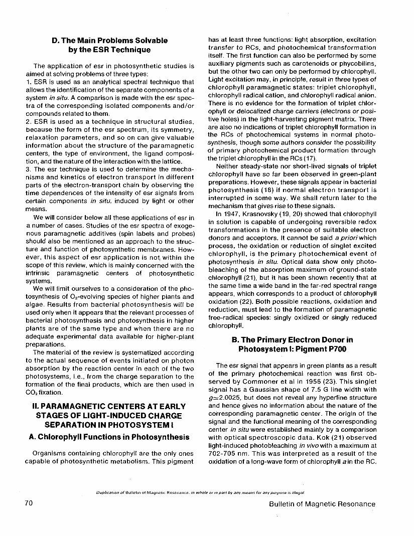
The application of esr in photosynthetic studies isaimed at solving problems of three types:1. ESR is used as an analytical spectral technique thatallows the identification of the separate components of asystem in situ. A comparison is made with the esr spec-tra of the corresponding isolated components and/orcompounds related to them.2. ESR is used as a technique in structural studies,because the form of the esr spectrum, its symmetry,relaxation parameters, and so on can give valuableinformation about the structure of the paramagneticcenters, the type of environment, the ligand composi-tion, and the nature of the interaction with the lattice.3. The esr technique is used to determine the mecha-nisms and kinetics of electron transport in differentparts of the electron-transport chain by observing thetime dependences of the intensity of esr signals fromcertain components in situ, induced by light or othermeans.
We will consider below alJ these applications of esr ina number of cases. Studies of the esr spectra of exoge-nous paramagnetic additives (spin labels and probes)should also be mentioned as an approach to the struc-ture and function of photosynthetic membranes. How-ever, this aspect of esr application is not, within thescope of this review, which is mainly concerned with theintrinsic paramagnetic centers of photosyntheticsystems.
We will limit ourselves to a consideration of the pho-tosynthesis of O2-evolving species of higher plants andalgae. Results from bacterial photosynthesis will beused only when it appears that the relevant processes ofbacterial photosynthesis and photosynthesis in higherplants are of the same type and when there are noadequate experimental data available for higher-plantpreparations.
The material of the review is systematized accordingto the actual sequence of events initiated on photonabsorption by the reaction center in each of the twophotosystems, i.e., from the charge separation to theformation of the final products, which are then used inCO2 fixation.
II. PARAMAGNETIC CENTERS AT EARLYSTAGES OF LIGHT-INDUCED CHARGE
SEPARATION IN PHOTOSYSTEM IA. Chlorophyll Functions in Photosynthesis
Organisms containing chlorophyll are the only onescapable of photosynthetic metabolism. This pigment
has at least three functions: light absorption, excitationtransfer to RCs, and photochemical transformationitself. The first function can also be performed by someauxiliary pigments such as carotenoids or phycobilins,but the other two can only be performed by chlorophyll.Light excitation may, in principle, result in three types ofchlorophyll paramagnetic states: triplet chlorophyll,chlorophyll radical cation, and chlorophyll radical anion.There is no evidence for the formation of triplet chlor-ophyll or delocalized charge carriers (electrons or posi-tive holes) in the light-harvesting pigment matrix. Thereare also no indications of triplet chlorophyll formation inthe RCs of photochemical systems in normal photo-synthesis, though some authors consider the possibilityof primary photochemical product formation throughthe triplet chlorophyll in the RCs (17).
Neither steady-state nor short-lived signals of tripletchlorophyll have so far been observed in green-plantpreparations. However, these signals appear in bacterialphotosynthesis (18) if normal electron transport isinterrupted in some way. We shall return later to themechanism that gives rise to these signals.
In 1947, Krasnovsky (19, 20) showed that chlorophyllin solution is capable of undergoing reversible redoxtransformations in the presence of suitable electrondonors and acceptors. It cannot be said a priori whichprocess, the oxidation or reduction of singlet excitedchlorophyll, is the primary photochemical event ofphotosynthesis in situ. Optical data show only photo-bleaching of the absorption maximum of ground-statechlorophyll (21), but it has been shown recently that atthe same time a wide band in the far-red spectral rangeappears, which corresponds to a product of chlorophylloxidation (22). Both possible reactions, oxidation andreduction, must lead to the formation of paramagneticfree-radical species: singly oxidized or singly reducedchlorophyll.
B. The Primary Electron Donor inPhotosystem I: Pigment P700
The esr signal that appears in green plants as a resultof the primary photochemical reaction was first ob-served by Commoner et al in 1956 (23). This singletsignal has a Gaussian shape of 7.5 G line width withg~2.0025, but does not reveal any hyperfine structureand hence gives no information about the nature of thecorresponding paramagnetic center. The origin of thesignal and the functional meaning of the correspondingcenter in situ were established mainly by a comparisonwith optical spectroscopic data. Kok (21) observedlight-induced photobleaching in wVowith a maximum at702-705 nm. This was interpreted as a result of theoxidation of a long-wave form of chlorophyll a in the RC.
Duplication of Bulletin of Magnetic Resonance, in whole or in part by any means for any purpose is illegal.
70 Bulletin of Magnetic Resonance
This center was called pigment P700. Later it becameclear that this effect was caused by the primary reactionin the centers of photosystem I. That the process is oneof oxidation follows from the similar absorption changesand esr signals, that are obtained by dark oxidationusing different oxidants. Further experiments (24, 25)clearly showed that the esr signal I (P700+) at roomtemperature coincides with optical photobleaching inthe range 702-705 nm in its dependence on the redoxpotential of the medium. The kinetics of the optical effectand the esr signal in dark-light-dark transitions alsocoincide.
1. The Nature of the Light-InducedFree-Radical Center
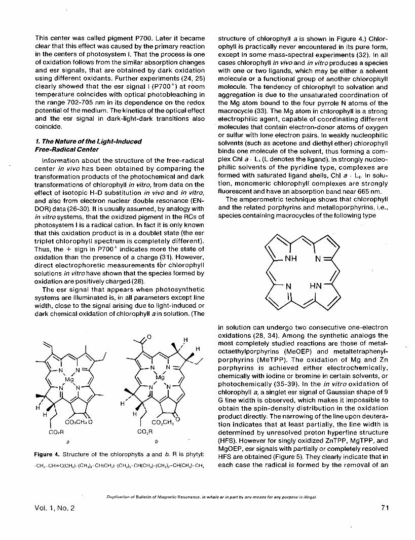
Information about the structure of the free-radicalcenter in vivo has been obtained by comparing thetransformation products of the photochemical and darktransformations of chlorophyll in vitro, from data on theeffect of isotopic H-D substitution in vivo and in vitro,and also from electron nuclear double resonance (EN-DOR) data (26-30). It is usually assumed, by analogy within vitro systems, that the oxidized pigment in the RCs ofphotosystem I is a radical cation. In fact it is only knownthat this oxidation product is in a doublet state (the esrtriplet chlorophyll spectrum is completely different).Thus, the + sign in P700+ indicates more the state ofoxidation than the presence of a charge (31). However,direct electrophoretic measurements fbr chlorophyllsolutions in vitrohave shown that the species formed byoxidation are positively charged (28).
The esr signal that appears when photosyntheticsystems are illuminated is, in all parameters except linewidth, close to the signal arising due to light-induced ordark chemical oxidation of chlorophyll a in solution. (The
H
CO2CH3O COXH O
CO2R CO2R
a b
Figure 4. Structure of the chlorophylls a and b. R is phytyl:
-CHJ-CH=C(CH3)-(CH2)3-CH(CH3HCH2)3-CH(CH3)-(CH !)3-CH(CH3)-CH3
structure of chlorophyll a is shown in Figure 4.) Chlor-ophyll is practically never encountered in its pure form,except in some mass-spectral experiments (32). In allcases chlorophyll in vivo and in vitro produces a specieswith one or two ligands, which may be either a solventmolecule or a functional group of another chlorophyllmolecule. The tendency of chlorophyll to solvation andaggregation is due to the unsaturated coordination ofthe Mg atom bound to the four pyrrole N atoms of themacrocycle (33). The Mg atom in chlorophyll is a strongelectrophilic agent, capable of coordinating differentmolecules that contain electron-donor atoms of oxygenor sulfur with lone electron pairs. In weakly nucleophilicsolvents (such as acetone and diethyl ether) chlorophyllbinds one molecule of the solvent, thus forming a com-plex Chi a • L, (L denotes the ligand). In strongly nucleo-philic solvents of the pyridine type, complexes areformed with saturated ligand shells, Chi a • L2. In solu-tion, monomeric chlorophyll complexes are stronglyfluorescent and have an absorption band near 665 nm.
The amperometric technique shows that chlorophylland the related porphyrins and metalloporphyrins, i.e.,species containing macrocycles of the following type
in solution can undergo two consecutive one-electronoxidations (28, 34). Among the synthetic analogs themost completely studied reactions are those of metal-octaethylporphyrins (MeOEP) and metaltetraphenyl-porphyrins (MeTPP). The oxidation of Mg and Znporphyrins is achieved either electrochemically,chemically with iodine or bromine in certain solvents, orphotochemically (35-39). In the in vitro oxidation ofchlorophyll a, a singlet esr signal of Gaussian shape of 9G line width is observed, which makes it impossible toobtain the spin-density distribution in the oxidationproduct directly. The narrowing of the line upon deutera-tion indicates that at least partially, the line width isdetermined by unresolved proton hyperfine structure(HFS). However for singly oxidized ZnTPP, MgTPP, andMgOEP, esr signals with partially or completely resolvedHFS are obtained (Figure 5). They clearly indicate that ineach case the radical is formed by the removal of an
Duplication of Bulletin of Magnetic Resonance, in whole or in part by any means for any purpose is illegal.
Vol. 1,No. 2 71

10G
Figure 5. First derivative esr spectra of (a) MgOEP+' ^and (b) (MgOEP-meso-dA)+' ClO^in methanol at - 5 0 c C .Second derivative esr spectra of (c) MgTPP + ' CIO^ and (d)(MgTPP-d20)
+' CIO4 (perdeuterated phenyl groups) in chloro-form. From ref. (28) with permission.
electron from the porphyrin molecular orbital. However,esr spectra of the cation radical of MgOEP and MgTPPdiffer greatly in their HFS patterns. For MgTPP, the HFSfrom the four equivalent N atoms and from phenylprotons was observed, whereas for MgOEP a signalconsisting of five hyperfine lines from four equivalentmeso protons is observed, while the splitting on thenitrogen nuclei is absent. This assignment agrees withthe data obtained from H-D substitution. From theseresults it follows that the radicals have x-electron struc-ture and that two types of distribution of spin density inradical cations of metalloporphyrins are possible.
The optical spectra of these two oxidized porphyrinsare quite different (28). The molecular orbital (MO)calculations show that for oxidized chlorins two almostdegenerate states separated by an energy gap corre-sponding to 2000 cm"1 are possible (40). The two cal-culated spin densities on structurally distinct atoms inmetalloporphyrin radical cations agree with the exper-imentally obtained hyperfine constants from esr spectraof MgOEP+ and MgTPP+. A comparison of the data formetalloporphyrins producing esr signals with resolvedHFS with the data for oxidized chlorophyll in vitroproducing an unsplit singlet signal, has shown that theelectronic structures of all these oxidized forms are verysimilar. All these compounds behave in a similar way
under electrochemical oxidation and have similar redoxpotentials. Metal coordination has a rather weak effecton the chemical, electrochemical, and spectral proper-ties of the compounds.
The esr spectra obtained for synthetic metalloporphy-rins made it possible to conclude that one-electronoxidation leads to the formation of ir-radical cations. Itfollows that in the case of chlorophylls a and b ir-radicalcations are also formed. This conclusion is confirmed byENDOR data (30, 40, 41). Figure 6 shows the ENDORspectrum of radical cations of chlorophyll a at 108 K.Hyperfine interaction constants determined by thistechnique are in agreement with the prediction that theradical cation of chlorophyll a is in the state M 2(symmetry group C2v), as are ZnTPP+ and MgTPP+. Thehyperfine splitting on /3-protons in radical cations ofchlorophyll a decreases as the temperature increases.This also happens in the case of ZnTPC+ (40). Thus, thedistribution of spin density depends on the externalconditions (temperature and possibly solvent). It istherefore possible that a radical cation can exist as amixture of electron tautomers whose equilibrium isdetermined by the parameters of the medium.
2. Metalloporphyrin Radical Anions
As will be seen below, apart from radical cations ofoxidized chlorophyll, radical anions of reduced chlor-
Figure 6. The ENDOR spectra of the blue-green algaSynechococcus lividus oxidized by ferricyanide at 108 K (A) iscompared with that of in vitro chlorophyll a+ in CDCI3-CD3OD(4:1) oxidized by l2 (B). Numerical values of the peaks are inmegahertz (2.8 MHz = 1 G). Only the high-frequency half ofeach spectrum is shown. The hyperfine coupling constant istwice the difference between the frequencies of the peak andthe center of the spectrum. From ref. (30) with permission.
Duplication of Bulletin of Magnetic Resonance, in whole or in part by any means for any purpose is illegal.
72 Bulletin of Magnetic Resonance

ophyll a and pheophytin a may be formed in photosyn-thetic systems. They also have been obtained electro-chemically as well as photolytically and radiolytically invitro (42-44). The properties of these reduced formsindicate that these particles are also ir-radicals. The esrspectrum of Chi a radical anions at low temperatures is asinglet with a p-factor that is practically the same as theg-factor of radical cations. Its line width is about 13 G,i.e., much greater than the line width of a singlet linefrom radical cations (9 G). The unpaired electron densitydistribution in the porphyrin ring is confirmed by abroadening of the signal when the isotopic substitution12Q _» 13Q j S m a c |e pujita et al (44) have recently shownthat radical anions of pheophytin a and of Chi a can beobtained by electrolysis in highly purified anhydroussolvents under strictly anaerobic conditions and arestable for several months. The reaction is reversible. Acomparison of esr and ENDOR spectra of these speciesin dimethylformamide and dimethylformamide-c/? hasshown that there is no proton exchange between thesolvent and radical cations even on prolonged standing.Structureless esr signals of Chi a 'and Pheoa'becomehighly saturated as the microwave power increases andthe saturation effect increases with decreasing tempera-ture, which makes observation of these signals in situmore difficult.
MO calculations (44-47) for metalloporphyrin andmetalloporphyrin radical anions have led to predictionson the distribution of spin density, in accordance withENDOR spectra of radical anions (40, 44) of naturalisotopic composition as well as of deuterated deriva-tives. The calculations predict a considerable splitting ofthe signal on N nuclei of pyrrole rings II and IV and aremarkable spin density on the Mg atom. Hyperfineinteraction with Mg can in principle be demonstratedexperimentally by observing the esr or ENDOR spectraof oxidized pigments containing 25Mg (/ = 5/2). Thiswould make it possible to differentiate the signals ofChi a" and Pheo a" in vivo and in vitro. This approachhas already been used for radical cations of bacterialchlorophyll and for some synthetic metalloporphyrinradical cations for which the spin density on the centralmetal atom was much smaller than that assumed forradical anions (48).
3. Chlorophyll Dimers in Reaction Centers
The esr signal I of oxidized P700+ centers in manyrespects coincides with the esr signal of radical cationsof chlorophyll a in vitro, but differs from it in one sig-nificant parameter, the line width, which is 9-10 G formonomeric chlorophyll in vitroand 7.5 G in vivo(23, 49).H-D substitution narrows the lines (50), but the ratio ofthe line widths in v/Voand in vitro remains the same. The
difference in esr line widths corresponds to the shift ofthe absorption band from 665 nm for monomeric chlor-ophyll to 704 nm for chlorophyll in the RCs of photo-system I (21). These differences have proved to be quiteuseful for determining the nature of the photoreactioncenters of photosystem I. Fifty years ago the long-waveshift in the absorption spectra was thought to be due tothe influence of the environment of the chlorophyll inv/Vo(51). Later it was regarded as a confirmation of thechlorophyll-aggregated state in the photosyntheticmechanism (5, 52). The narrowing of the esr line ofoxidized chlorophyll in situ as compared to monomericchlorophyll in solution was explained by the unpairedelectron density, which is uniformly distributed in twochlorophyll molecules forming a dimeric structure. Muchresearch has been devoted to the theoretical analysis ofpossible dimeric and oligomeric structures and to ob-taining and investigating the properties of model as-sociates that simulate the properties of the pigment inthe reaction center of photosystem I (30). These modelsare constructed on the basis of the above-mentionedunsaturated coordination of the Mg atom in the chlor-ophyll molecule and the presence of electron-donorester and keto groups in chlorophyll.
Chlorophyll oligomers can be divided into two groups.In oligomers of (Chi a) composition (n = 2-20), the sidegroups of other chlorophyll molecules are directly boundto the Mg atom. The other group of model aggregates ismade up of oligomers in which the bond between anytwo chlorophyll molecules is formed by a bifunctionalligand molecule such as water. In the presence ofequimolar quantities of Chi a and pyrazine in CCI4, forexample, these associations can reach colloidal dimen-sions (30, 33). The addition of water to solutions ofchlorophyll a in aliphatic hydrocarbons leads to achange of color (the absorption maximum shifts from670-680 nm to 740 nm) and also to some changes in theinfrared spectrum, which indicates C=O- • • Mg bondfission. If Chi a is replaced by ethylchlorophyllide, inwhich the phytyl chain is substituted for ethyl, the con-densed hydrate can be obtained in a crystal form. Itsstructure is determined by X-ray diffraction (53, 54). Thisstructure is formed by parallel layers of the samegeometry as the monolayer chlorophyll at the solution-air interface. These chlorophyll films also absorb at 735nm (55). Under photochemical oxidation of any of thesesystems, a product with singlet esr signals of Gaussianform of different line widths is obtained (56). Light-induced signals seemed to be observed only forchlorophyll-water adducts and were unstable in thedark. The 740 nm adduct esr signal is unusually narrow(of about 1 G line width). This is very unusual for such alarge molecule as chlorophyll, with many structurallydifferent H and N atoms. The narrowing of the signal is
Duplication of Bulletin of Magnetic Resonance, in whole or in part by any means for any purpose is illegal.
Vol. 1, No. 2 73
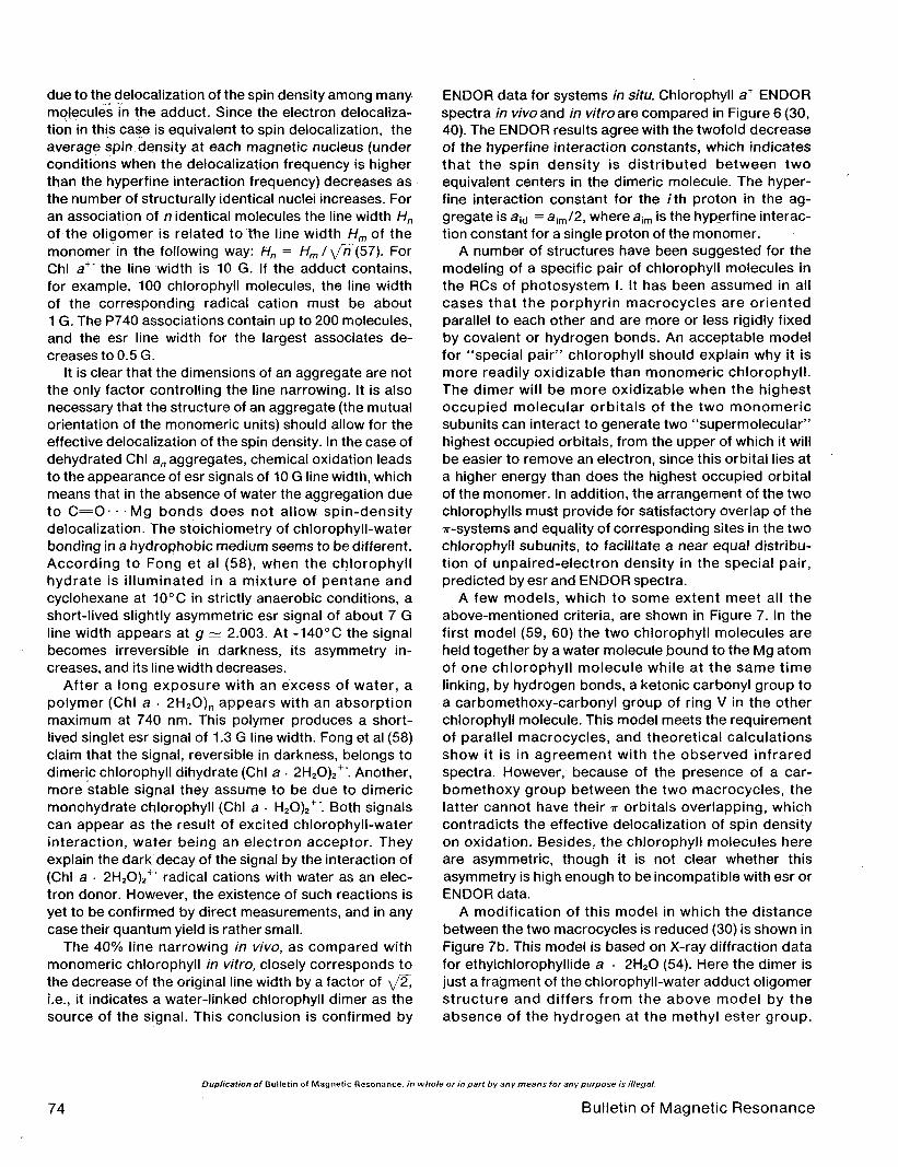
due to the delocalization of the spin density among manymolecules in the adduct. Since the electron delocaliza-tion in this case is equivalent to spin delocalization, theaverage spin density at each magnetic nucleus (underconditions when the delocalization frequency is higherthan the hyperfine interaction frequency) decreases asthe number of structurally identical nuclei increases. Foran association of n identical molecules the line width Hnof the oligomer is related to the line width Hm of themonomer in the following way: Hn = Hml \fn(b7). ForChi a+' the line width is 10 G. If the adduct contains,for example, 100 chlorophyll molecules, the line widthof the corresponding radical cation must be about1 G. The P740 associations contain up to 200 molecules,and the esr line width for the largest associates de-creases to 0.5 G.
It is clear that the dimensions of an aggregate are notthe only factor controlling the line narrowing. It is alsonecessary that the structure of an aggregate (the mutualorientation of the monomeric units) should allow for theeffective delocalization of the spin density. In the case ofdehydrated Chi an aggregates, chemical oxidation leadsto the appearance of esr signals of 10 G line width, whichmeans that in the absence of water the aggregation dueto C = O - M g bonds does not allow spin-densitydelocalization. The stoichiometry of chlorophyll-waterbonding in a hydrophobic medium seems to be different.According to Fong et al (58), when the chlorophyllhydrate is illuminated in a mixture of pentane andcyclohexane at 10°C in strictly anaerobic conditions, ashort-lived slightly asymmetric esr signal of about 7 Gline width appears at g ~ 2.003. At -140°C the signalbecomes irreversible in darkness, its asymmetry in-creases, and its line width decreases.
After a long exposure with an excess of water, apolymer (Chi a • 2H2O)n appears with an absorptionmaximum at 740 nm. This polymer produces a short-lived singlet esr signal of 1.3 G line width. Fong et al (58)claim that the signal, reversible in darkness, belongs todimeric chlorophyll dihydrate (Chi a • 2H2O)2
+\ Another,more stable signal they assume to be due to dimericmonohydrate chlorophyll (Chi a • H2O)2
+\ Both signalscan appear as the result of excited chlorophyll-waterinteraction, water being an electron acceptor. Theyexplain the dark decay of the signal by the interaction of(Chi a • 2H2O)2
+' radical cations with water as an elec-tron donor. However, the existence of such reactions isyet to be confirmed by direct measurements, and in anycase their quantum yield is rather small.
The 40% line narrowing in vivo, as compared withmonomeric chlorophyll in vitro, closely corresponds tothe decrease of the original line width by a factor of \/2,i.e., it indicates a water-linked chlorophyll dimer as thesource of the signal. This conclusion is confirmed by
ENDOR data for systems in situ. Chlorophyll a+ ENDORspectra in vivo and in vitro are compared in Figure 6 (30,40). The ENDOR results agree with the twofold decreaseof the hyperfine interaction constants, which indicatesthat the spin density is distributed between twoequivalent centers in the dimeric molecule. The hyper-fine interaction constant for the /th proton in the ag-gregate is aid = aim/2, where aim is the hyperfine interac-tion constant for a single proton of the monomer.
A number of structures have been suggested for themodeling of a specific pair of chlorophyll molecules inthe RCs of photosystem I. It has been assumed in allcases that the porphyrin macrocycles are orientedparallel to each other and are more or less rigidly fixedby covalent or hydrogen bonds. An acceptable modelfor "special pair" chlorophyll should explain why it ismore readily oxidizable than monomeric chlorophyll.The dimer will be more oxidizable when the highestoccupied molecular orbitals of the two monomericsubunits can interact to generate two "supermolecular"highest occupied orbitals, from the upper of which it willbe easier to remove an electron, since this orbital lies ata higher energy than does the highest occupied orbitalof the monomer. In addition, the arrangement of the twochlorophylls must provide for satisfactory overlap of the7r-systems and equality of corresponding sites in the twochlorophyll subunits, to facilitate a near equal distribu-tion of unpaired-electron density in the special pair,predicted by esr and ENDOR spectra.
A few models, which to some extent meet all theabove-mentioned criteria, are shown in Figure 7. In thefirst model (59, 60) the two chlorophyll molecules areheld together by a water molecule bound to the Mg atomof one chlorophyll molecule while at the same timelinking, by hydrogen bonds, a ketonic carbonyl group toa carbomethoxy-carbonyl group of ring V in the otherchlorophyll molecule. This model meets the requirementof parallel macrocycles, and theoretical calculationsshow it is in agreement with the observed infraredspectra. However, because of the presence of a car-bomethoxy group between the two macrocycles, thelatter cannot have their T orbitals overlapping, whichcontradicts the effective delocalization of spin densityon oxidation. Besides, the chlorophyll molecules hereare asymmetric, though it is not clear whether thisasymmetry is high enough to be incompatible with esr orENDOR data.
A modification of this model in which the distancebetween the two macrocycles is reduced (30) is shown inFigure 7b. This model is based on X-ray diffraction datafor ethylchlorophyllide a • 2H2O (54). Here the dimer isjust a fragment of the chlorophyll-water adduct oligomerstructure and differs from the above model by theabsence of the hydrogen at the methyl ester group.
Duplication of Bulletin of Magnetic Resonance, in whole or in part by any means for any purpose is illegal.
74 Bulletin of Magnetic Resonance

Figure 7. Several models for "chlorophyll special pair": a) a molecule of water is coordinated to the Mg atom of one chlorophylland simultaneously hydrogen-bonded to the ketonic C=O and carbomethoxy C=O functions of the second chlorophyll (59). Themodel is asymmetric. The two chlorophylls are not equivalent, as the lower Chi a is acting only as acceptor, b) An asymmetricmodel based on the structure of crystalline ethylchlorophyllide a-2H2O (30, 54). c) Another version of the same model (61); the twoChi a molecules in the special pair are equivalent and are related by translational symmetry, d) Model of Fong (62). In this modelthe two Chi a molecules are crosslinked by interaction with the carbomethoxy C=O functions. The arrangement has C symmetry,but the Chi a macrocycles are rotated at a 60° angle and are 6 A apart, e) Special pair model with C^ symmetry as proposed byShipman et al (63). In this version the ketonic C=O functions are hydrogen-bonded. Various nucleophiles R'XH, where X = O, N, orS and R' = H or alkyl can act as crosslinking agents.
Exciton calculations (61) have shown that for this kind ofdimer there must be a shift of the red absorption max-imum up to 693 nm, which agrees with the data for P700in vivo. The chlorophyll molecules are not quite the samehere either. The macrocycles are separated by a dis-tance of 3.6 A, which is smaller than the sum of their vander Waals radii, i.e., the required overlap of the %systems is ensured. Another pair of molecules may bedistinguished in the same structure, as is shown inFigure 7c. They form a dimer consisting of completely
identical, but not quite symmetrical subunits. In bothcases the third molecule disturbs the structure of theacceptor chlorophyll molecule, so that the other twomolecules, regarded as a dimer, become more similar.Fong (62) suggested a completely symmetrical C2 struc-ture, shown in Figure 7d. The two chlorophyll moleculesare held together by two water molecules. Each watermolecule is at the same time bound to the Mg atom anda ring V methyl ester group of the other chlorophyll.Ketonic groups do not take part in the dimer formation.
Duplication of Bulletin of Magnetic Resonance, in whole or in part by any means for any purpose is illegal.
Vol. 1, No. 2 75

However, the construction of molecular models hasshown that the distance between the macrocycles in thiscase is 5.7 A, i.e., greater than the sum of the van derWaals radii. Calculations of the optical properties of thisstructure do not give the experimentally observed long-wave shift (30, 63). The ketonic groups, which havestronger electron-donor properties than does themethyl ester group, are not used at all. Experimentallyobtained dimers (64), which it was suggested corre-spond to this model, were constructed in such a way thatthe carbomethoxy functions (according to infraredspectra) did not in fact take part in the dimerization (65).
Shipman et al (66) and Boxer and Closs (67) havesuggested a model which seems to overcome the dif-ficulties of Fong's model, but which meets the require-ment of maximal symmetry of the monomers (Figure7e). This last model is attractive also because it allowscoordination not only with water, but with other nucleo-philes. In situ these could be nucleophilic protein sidegroups. It opens up the possibility of binding RC dimericchlorophyll to the proteins that are isolated with dimericchlorophyll from plant material during fragmentationand purification of reaction center preparations. Thedimer structure is maintained by hydrogen bondsbetween keto functions and water (or some other nu-cleophilic group -SH,-NH2,-OH); 7r-system overlap isalso achieved. An experimental realization of the model(66) seems to be a system containing 0.1 chlorophyll ain toluene in the presence of 1.5 M excess ethanol. Asthe solution cools, the initially observed absorptionmaximum at 668 nm disappears and is replaced by amaximum at 702 nm. According to spectral data, neither
Figure 8. Covalently linked chlorophyll molecules in the foldedconfiguration. Dotted lines indicate H bonds.
the keto nor ester carbonyls take part in coordination atroom temperature. However, in the cooling process aketo group becomes bound by a hydrogen bond, andthe ester group remains free.
A comparatively new direction in the modeling of thein vivo chlorophyll dimer is the synthesis of compoundsin which two porphyrin rings are linked by one or twocovalent bridge structures (67-71). Between the opticalproperties of Zn-porphyrin dimers with peptide bridgesand corresponding monomers, there are no differencesthat are comparable to the differences between themonomeric chlorophyll and dimeric chlorophyll in pho-tosystem I. The problem of photosynthetic dimer model-ing has stimulated much research into synthesis, whichhas been highly successful. For example, bis(pyrochlor-ophyllide a) ethylene glycol diester was obtained (67). Itsabsorption maximum in benzene or carbon tetrachlor-ide in the presence of excess water or primary alcoholswas observed at 694 nm. Wasielewski et al (72) havedeveloped synthetic methods that have resulted in thepreparation of covalently bound dimeric species ofchlorophyll a (Figure 8). The ethylene glycol bridges areflexible enough not to interfere with the formation ofhydrated symmetrical structures containing their mac-rocycles at the closest possible position. At the sametime they assist in stacking by constantly keeping thetwo monomeric subunits close together. In benzene inthe presence of water, this compound had a spectrumwith an absorption band at 694 nm and a minor peak at677 nm. This spectrum is close to the chlorophyll spec-trum in vivo. The esr spectrum of the correspondingoxidation product does not appear to have been inves-tigated in detail, but there is little doubt that the spec-trum would be similar to that of radical cations of P700+
dimeric chlorophyll.
C. The System of Bound ElectronAcceptors of Photosystem I
It has been firmly established that chlorophyll a is aprimary photoexcited electron donor in photosystem Iand forms a dimer of a specific structure. Thus, theproblem of determining the primary charge-separationmechanism is essentially reduced to that of establishingthe nature of the earliest electron acceptor(s). Afterexcitation by a laser pulse, chlorophyll a, (P700) isoxidized in less than 20 ns (41). However, it is known thatquantum stabilization occurs much more quickly, i.e., inless than 30 ps (2, 3). Thus, the early electron transferoccurs at a very high rate, incompatible with anyprocesses limited by diffusion. Evidently, the par-ticipants in this reaction are constantly forming a com-plex, and indeed the electron transfer in this complex
Duplication of Bulletin of Magnetic Resonance, in whole or in part by any means for any purpose is illegal.
76 Bulletin of Magnetic Resonance

occurs independently of the temperature, even at thetemperature of liquid helium.
In some early publications it was suggested that achlorophyll molecule is an early electron acceptor (73).For a long time there was no experimental support forthis hypothesis. ESR data showed that the early accep-tor is an Fe-S center with a more negative potential thanthat of free ferredoxin. This problem is still being inten-sively investigated; however, the latest experimentsagain lead to the conclusion, though on quite differentexperimental grounds, that the primary acceptor mustbe a pigment molecule. This conclusion is equally validfor all types of reaction centers, for the RCs of photo-systems I and II and those of bacterial photosynthesis aswell.
For bacterial photosynthesis it was established fromoptical and esr data that the earliest acceptor is notubiquinone, as was thought earlier, but bacterio-pheophytin, a Mg-free analogue of bacteriochlorophyll.When reduced, this acceptor forms a radical anion,which is identified by its optical and esr spectra (74).Later Klevanik et al (75, 76) established that in photo-system II pheophytin takes part as an intermediatecarrier in the electron transfer between the pigmentP680 in the reaction center and plastoquinone, which fora long time was regarded as the primary electron accep-tor of photosystem II. Finally it was also shown forphotosystem I by optical spectroscopy and esr thatthere exists at least one more carrier between the Fe-Scenter and pigment P700. Some indirect data indicatethat chlorophyll a molecules might be this carrier.Energy considerations make this assumption quiteprobable.
Redox titration data of the P700+esr signal and thesignals of the bound Fe-S centers and the photosystemII early acceptor have shown that the P700 redox poten-tial is about + 0.4 V (22, 77), whereas the potential of thepigment in the photosystem II reaction center, P680,cannot be less than +0.8 V (since photosystem IIbrings about the decomposition of water into O2, then£H2O/O2
= 0-8 V). The half-wave potentials in electro-chemical oxidation of chlorophyll a and pheophytin a (indimethylformamide) are -0.88 and -0.64 V (44). Inphotosystem I the reaction
P700 + Chi a light P700+'+Chla~
must be accompanied by a free-energy change AE =1.3 eV (-0.9 to +0.4V), whereas for the hypotheticalreaction
P700 + Pheo alight
P700+#+ Pheo a"
In photosystem II the change in free energy for thehypothetical reaction
P680 + Chi alight
+~P680+' + Chi a '
would be AE = 1.7 eV, while for the reaction
P680 + Pheo a light •*-P680+'+ Pheo a '
the change is AE = 1 eV( - 0.6 to + 0.4 V).
the change is AE = 1.4 eV. Since 700 nm and 680 nmphotons absorbed by photosystems I and II have ener-gies of 1.77 and 1.82 eV, respectively, then taking intoaccount the losses necessary for the stabilization of theseparated charges, it follows that the energy of a quan-tum is more completely used in the transfer of an elec-tron to pheophytin in photosystem II or to chlorophyll inphotosystem I. The pheophytin redox potential (-0.64V) and the chorophyll redox potential ( - 0.88 V) are quitesufficient for reduction of the plastoquinone (E = - 0.2V) and the bound Fe-S chloroplast center (E = - 0.6 V),respectively. Independent of these energy considera-tions, experiments have shown that in the reactioncenter of photosystem I there are several componentsamong which, at any temperature, electron transfer ispossible in the forward direction (i.e., from the light-excited chlorophyll dimer) and among some of whichback transfer is possible (charge recombination). Ininvestigations of early acceptor properties, one trend isclearly seen. As techniques of measuring short-lived andweak changes in the optical absorption and esr spectrabecome better, the measured redox potentials of theearliest acceptors become greater, thus approachingthe calculated redox potential of excited chlorophyll. Atthe same time the rate of charge recombination of thedimeric chlorophyll radical cation (the oxidized primarydonor) with the component considered to be a primaryacceptor also increases over a wide temperature range.The main results are obtained by studying chloropiastsand their fragments by the application of esr and opticalspectroscopy at liquid-nitrogen and liquid-heliumtemperatures (4-77 K) (77-91).
1. The Composition of the Acceptor Complexof Photosystem I
Malkin and Bearden (78) observed esr signals of Fe-Scenters (Figure 9) from illuminated chloropiasts at 10 K.These centers produced signals at ^-values of 2.05,1.94, and 1.86—close to those of reduced soluble fer-redoxin. Illumination at 715 nm was as effective as at615 nm. From this it was concluded (and confirmed bylater experiments) that these centers are related tophotosystem I. A quantitative comparison of the P700+
content and the Fe-S centers, as well as a comparison of
Duplication of Bulletin of Magnetic Resonance, in whole or in part by any means for any purpose is illegal.
Vol. 1, No. 2 77

1
0«2.O5
-AJJ
\ i \
fif«1.94 p«1.85
J I
f . DARK
7 1 LIGHT
f 1 715/7^at10oK JsAJ*r*<
i l I
Figure 9. Photoreduction of abound Fe-S center in intact spinachchloroplasts after illumination at10 K. From ref. (16), with per-mission.
3200 3300 3400 3500Magnetic Field (gauss)
their kinetic characteristics in the interval of 10-100 K,shows that there is a good correlation between P700oxidation and Fe-S center reduction in chloroplasts andin the particles enriched in photosystem I (79-81).
In addition, in frozen chloroplasts exposed to light asignal with p-factors of 2.05,1.93, and 1.89 appears; thissignal does not change in the dark at low temperatures.This signal can also be obtained by dark reduction bydithionite. Estimates of the redox potential of this centervary between - 530 and - 580 mV (81-83). Independentoptical data on the bound acceptor were obtained by Keet al (82), who found short-lived photobleaching at 430nrn (P430) and established a correlation between P430and low-temperature signals with g -factors of 2.05,1.95, and 1.87. This component was regarded as possi-bly being the primary acceptor in photosystem I. How-ever, data were subsequently obtained that indicated anintermediate electron carrier (or carriers) between P700and the Fe-S centers with these g-iactors.
Mclntoshi and Bolton (84) and Evans et al (89) ob-served at about 10 K a new esr signal with g values of2.07, 1.86, and 1.78 (Figure 10); this signal appearsreversibly either under an intense low-temperatureillumination of photosystem I fragments in the reducingmedium (simultaneously with the esr I signal) or understrong dark reduction in the presence of certain redoxmediators (the corresponding component was called the"X center"). The redox potential of this center is about
3600
- 730 mV. Later, Demeter and Ke (86), studying reversi-ble and irreversible absorption changes induced by lightin a strongly reducing medium, also concluded thatthere was an intermediate carrier between P700 andP430. The charge separation between these compo-nents is reversible even at 20 K, and the degree ofreversibility is determined by the fraction of reducedP430 present and reaches 100% when the potential ofthe medium is about —670 mV. This potential exceedsthe potentials of viologen dyes, which still can be photo-reduced by chloroplasts. It seems that an electron istransferred onto the intermediate acceptor and thenonto P430 if the latter has not been previously reduced.The charges of both P700+ and this intermediate accep-tor also recombine quickly even at liquid-helium temper-ature, if further transfer of electrons onto more distantacceptors is impossible.
However, an esr signal from the reduced X center (theA2 center (87)) can also be observed, with g«1.78,1.88,and 2.08, without pretreatment by strong reducingagents. In this case a stable steady-state signal of thesame type is observed when the sample is graduallyfrozen in strong light. The signal remains stable indarkness and is not accompanied by an esr signal I fromthe P700+ centers. Evidently, this result can be ex-plained by the fact that in the process of cooling, theelectron transfers on the oxidizing side of photosystem Iare stopped at a higher temperature than that of X
Duplication of Bulletin of Magnetic Resonance, in whole or in part by any means for any purpose is illegal.
78 Bulletin of Magnetic Resonance

reduction. Thus, the states with reduced P700, oxidizedsecondary donor (plastocyanin), and reduced acceptorX" become fixed. The combination of charges [Pc+
P700 X~] is stable at cryogenic temperatures.Recently Mathis et al (92) studied photosystem I
chlorophyll-protein complexes obtained by dodecylsul-fate treatment, in which no photochemical activity hadbeen observed before (93) or in which charge separationwas very ineffective (94). In these preparations theydetected P700 oxidation induced by a short light pulsse;after this, P700+ reduction by charge recombinationwith an electron acceptor occurred, in 0.5 ms at 5 K andin 10 ms at 294 K. Later a similar result was obtained forchloroplasts incubated in the presence of a reducingagent. Thus, the reversibility of low-temperature P700oxidation (and acceptor reduction) can be obtained bothby the reduction of the majority of the bound acceptorsor by their removal or denaturation.
Using very sensitive, high-speed equipment, Sauer etal (95) studied laser-induced reactions of photosystem Iin subchloroplast fragments at room temperature andhave shown that the electron capacity of the acceptorpool is four electrons for one P700 center:
P700 •*• P430 [AB]
If all the components of this pool are oxidized, twoflashes are required for complete P430 reduction. Thiscenter is identified with the bound Fe-S center foundearlier, whose redox potential is about - 500 mV (P430).If all the P430 are initially reduced, the flash will reducethe A2 component, which corresponds to the center witha more negative redox potential (about - 700 mV). Theesr signal from this center was determined by Mclntoshand Bolton (84) and by Evans et al (84, 89) at a tempera-ture close to that of liquid helium. If this acceptor is alsopreviously reduced, the charge separation is limited bytwo components, P700 and A,. The times for chargerecombination between P700 and each of the boundacceptors at room temperature are: 30 ms for P430~,250 fis forAa and 3/us for A," These components are allcapable of taking part in photochemical charge separa-tion at liquid-nitrogen and liquid-helium temperatures,and the charges recombine between P700+ and A,"or A2~at a very high rate. At 5 K the reoxidation of A,"due to itsreaction with P700+ takes about 3 ms, though the reac-tion of P700+ with P430" is practically inhibited. Ac-cording to data that Ke presented in a report deliveredat an international seminar in Moscow in September1978, the absorption spectra of the earliest intermediateproducts of the photosystem I reaction contain featurescharacteristic of both P700+ and the chlorophyll aradical anion. It is suggested that the esr signal ob-served when chloroplasts in a strongly reducing medium
2.2
1
2. 1 2.0
i
g1.9
Valuei
1.8
i
1.7
i
300 320 340 360 380
Magnetic Field (mT)
Figure 10. ESR spectrum of component X (A2). Photosystem Iparticles frozen with Fe-S centers A and B reduced (a), orcenters A and B and component X reduced (b), and theirdifference corresponding to the spectrum of X (c). From ref.(91), with permission.
are illuminated in the temperature range of 4-77 K,contains a contribution from radical anions of the chlor-ophyll acceptor. This assumption is confirmed by theasymmetry of the esr signal that has been observed atthese reducing potentials. The signal is distorted by thesuperposition of the signal from the radical anionprimary acceptor. Thus, the A, component appears tobe chlorophyll a. *
2. Radical Pairs as an Early Productof the Photochemical Reaction
Experiments using the chemically induced dynamicelectron spin polarization (CIDEP) technique provideanother source of information about the early reactions
'Recently the observation of an esr signal of 14gline width hasbeen reported in photosystem I particles illuminated at roomtemperature and frozen in the light in the presence of a strongreducing agent. This may correspond to the electron acceptorA2. [P. Heathcote, K.N. Timofeev, and M. C. W. Evans, FEBSLett. 101, 105(1979)].
Duplication of Bulletin of Magnetic Resonance, in whole or in part by any means for any purpose is illegal.
Vol. 1, No. 2 79

Figure 11. Calculated (solid lines) and experimental esrspectra for the oriented and unoriented polarized signal fromspinach chloroplasts. Solid triangles are experimental intensi-ties for flow-oriented chloroplasts. Open circles are exper-imental intensities for unoriented chloroplasts. From ref. (99),with permission. >
in photosystem I centers (96-99). The study of signalsfrom triplet chlorophyll in situ provides a third source.The CIDEP in photosynthetic systems was discovered byBlankenship et al (100) and by Mclntosh and Bolton(101) and was investigated in detail in Sauer's lab-oratory (98,99).
ESR signals observed from various photosystem Ipreparations in the first few microseconds of pulsedexcitation display spin polarization. The pulse inducesan esr signal with a p-value of 2.0025, which coincideswith the p-value of the esr signal I from P700+-centers,but with an anomalous intensity distribution along themagnetic field (Figure 11). Microwave radiation emis-sion prevails in the low-field region, whereas in thehigh-field region enhanced absorption occurs. Thismeans that the free-radical center with this esr signalhas a nonequilibrium spin distribution. The shape of thissignal changes with the orientation of chioroplasts in theflow. These effects were analyzed (99) according to thespin polarization theory developed earlier by Adrian(102) for radical pairs in solution. The spin-state popula-tion changes because of coherent mixing of the tripletand the singlet states of the weakly coupled partners in aradical pair. This mixing, caused by the presence of local
magnetic fields and by spin exchange, takes place morequickly than the incoherent spin-lattice relaxation. Thesign of the effect (emission or enhanced absorption ofmicrowave radiation) can give information about thestate preceding the radical pair. CIDEP data give infor-mation about both partners of a radical pair, thoughsometimes only the esr signal from one of them can beobserved directly.
In addition, the dependence of the spectrum with spinpolarization on the orientation of the membrane in themagnetic field is useful in studying the anisotropy of themagnetic interactions within the radical pair and theorientation of the free-radical species relative to oneanother and to the membrane. Since the spin polariza-tion occurs before the establishment of equilibrium withthe lattice, fast kinetic techniques must be used inexperiments. In practice a photosynthetic system, suchas a chloroplast suspension, is illuminated by short lightpulses in the esr cavity, and then the kinetics of thechanges in the amplitude of the first derivative of the esrabsorption are observed at various fixed field intensitiesnear the absorption maximum. Then the esr spectra,corresponding to various times after the pulse, areconstructed on the basis of the kinetic data. It is possiblein practice to reduce the esr spectrometer dead time to2 us. Hence, of course, the earliest processes are lostsince the primary photochemical reaction has a muchshorter lifetime. Nevertheless, two different esr signalcomponents are observable, one of which has acomparatively long lifetime of 30 ms and the usual shapeof an esr I signal, while its amplitude corresponds to thecontents of P700 in the specimen. The other component,with a significantly larger amplitude and shorter lifetime,has an anomalous shape, which indicates the occur-rence of CIDEP. The shapes of the long-lived componentand of the steady-state esr I signal do not depend onchloroplast orientation in the magnetic field. However,the shape of the anomalous signal does depend to agreat extent on the chloroplast orientation in the mag-netic field (Figure 11). Also the shape of the anomalouslypolarized signal depends on the resolution time of thespectrometer. Thus, it was concluded that the relaxationrate is different for different hyperfine lines of the free-radical center.
Mclntosh and Bolton (96, 97) noticed the possibility ofesr spectra distortions in CIDEP experiments because athigh modulation frequencies (100-1000 kHz) conditionsfor slow passage through the resonance are not fulfilled:even at room temperatures the spin-lattice relaxationtime of P700+ centers may be of the order of 1 /xs. Thesedistortions may be similar to the anomalous polarizationof the esr signal. Because of this they observed only thedirect esr absorption signal, without high-frequencymodulation. This made it possible to reduce the dead
Duplication of Bulletin of Magnetic Resonance, in whole or in part by any means for any purpose is illegal.
80 Bulletin of Magnetic Resonance

time of the instrument to 0.4 us. The results showed thatthe anomalous intensity distribution in the esr spectrumthat appears immediately after excitation of the systemby a short pulse is really caused by the CIDEP. Thespectrum at 100 K contained both absorption and emis-sion components, situated asymmetrically relative to thecenter of the esr signal I.
One of the main peculiarities of the signal was a shiftin the direction of the weak field, as compared to signalsobserved at room temperature. Mclntosh et al (97)suggest that the existence of short-lived signals with arange of p-values of 2.045-2.065 indicates the participa-tion of an alternative electron acceptor in charge separ-ation, apart from the chlorophyll a suggested by Dis-muckes et al (98), the lifetime of which in a reduced stateincreases as the temperature decreases. It is possiblethat these two intermediate acceptors, functioningbetween P700 and A2 (component X in the terminologyof Mclntosh and Bolton (84)), are situated in differentphotosystems I, i.e., the reaction centers of photosys-tems I are in fact heterogeneous. However, this problemrequires further research.
According to redox titrations the amplitudes of theshort-lived emission and enhanced absorption signalsdecrease with an increase in the redox potential of themedium in the interval 400-600 mV. The amplitude of thesteady-state esr signal I simultaneously increases. Thespin polarization was also observed ip subchloroplastfragments of photosystem I, though the orientationeffects did not occur in these preparations (the particlesare approximately spherical and so do not orient in theflow). The steady-state esr signal I and the polarizedsignal have the same p-value. There is no doubt now thatboth signals belong to oxidized chlorophyll a, theprimary electron donor of photosystem I. The observedpolarization and orientation effects can be explained bysupposing that electron transfer from dimeric chlor-
ophyll P700 to the primary acceptor causes the forma-tion of radical pairs in which both radicals are closeenough to make possible a partial overlapping of theirorbitals and an effective spin exchange. The initial spinconfiguration is the same as that of the initial excitedP700 from which electron transfer occurs. This meansthat the configuration is a singlet and that no polariza-tion can take place in the initial state. However, in aweakly coupled radical pair there is a coherent mixing ofthe singlet and the triplet states due to local magneticfields (hyperfine and spin orbital), different for eachradical. In solutions and, it seems, in membrane struc-tures, this mixing leads to spin polarization. Since theP700+ has an isotropic g-tensor (the correspondingsteady-state signal is isotropic and does not depend onthe orientation) while the anomalously polarized signaldepends appreciably on the orientation, it must beassumed that this anisotropy is determined by theacceptor component of the radical pair. The anisotropyof the signal disappears since the electron transportseparates the oxidized primary donor and the sequen-tially reduced acceptors more rapidly than the spinrelaxation takes place (98,99).
The previously suggested triplet mechanism for theformation of the anomalous polarized signal (100,101)does not explain the signs of the polarization for differ-ent hyperfine lines of the P700+ esr signal (i.e., thedependence of the amplitude on the field). A detailedtheory of the effect has been developed in the work ofFriesner et al (99), who examined a model of the radicalpairs that considers the influence of the anisotropy ofthe g-tensor of the primary and secondary electronacceptors, as well as the fixed orientation of all threeparticipants in this reaction in the membrane. Theresults obtained can be described, both qualitativelyand quantitatively, on the basis of the electron-transferscheme:
P700 —*- A, (Chlorophyll a) —*- A2 (Fe-S center X) —*~ P430 (binuclear Fe-S center [g] )
The first of these acceptors is, apparently, an organicmolecule with a practically isotropic g-tensor very closeto those of P700+. This acceptor is most probably thechlorophyll a molecule. The secondary acceptor A2 musthave a highly anisotropic p-tensor and be rather rigidlyoriented relative to the plane of the membrane (which isconfirmed by direct measurements of esr signals fromthis center in oriented chloroplasts; see below). Neitherthe model that takes into account only the spin ex-change between P700+ and acceptor A (a small organicmolecule with an isotropic g tensor) nor the model that
considers only the interaction between P700+ andacceptor A2 (which has an anisotropic p-tensor) providesan explanation of the experimental results. Only a two-site model is adequate for a quantitative interpretationof both the intensity of the polarized signals and theirorientational dependence. Such a model accounts forthe consecutive electron migrations from P700 to A, andfurther to A2, and both acceptors must be in a state ofspin exchange with P700+ (with appreciably differentvalues for the exchange integral, the exchange beingmore effective for A,~ than for A2~). Some details are still
Duplication of Bulletin of Magnetic Resonance, in whole or in part by any means for any purpose is illegal.
Vol. 1 , N o . 2 81

p-Value
22 21 20 19
Dark
Light-Dark
29 30 32 34 36
H{kG)Figure 12. ESR spectra at liquid-helium temperature ofchromatophores from chromatium D. The redox potential was- 260 mV. The lower trace is the difference between light anddark signals. Peaks A and E result from microwave absorptionand emission respectively. This spectrum is similar to thetriplet bacteriochlorophyll in solution. From ref. (103), withpermission.
unclear. For example, the polarized signal in non-oriented samples is narrower than the signal from P700+
(5.6 G instead of 7.5 G), but the mechanism is still to beexplained. New experiments and theoretical studies willbe necessary for the lifetimes of the reduced inter-mediate acceptors to be estimated by the CIDEP ap-proach and for the nature of A, and A2 to be finallyestablished.
The theory also has given a satisfactory explanationof the esr triplet signals, which are observed in certainphotosynthetic systems when electron transport isblocked in the vicinity of the reaction centers (Figure 12).So far no one has been able to observe triplet chlor-ophyll esr signals in preparations from higher plants oralgae. These signals were observed only in preparationsof reaction centers from photosynthesizing bacteria inconditions of vigorous reduction. They displayedproperties rather unusual for triplet states (103,104).
As is well known, the energy levels of a tripletmolecule are determined by dipole interaction betweentwo unpaired electrons and in esr experiments by theinteraction with the external magnetic field. In this casethe triplet substates are denoted by | 7+,), | 7"0>, and| T-i), and for each orientation of the triplet moleculethere is a system consisting of three levels betweenwhich two transitions are possible Ams = ± 1. Evalua-tions of the zero-field-splitting parameters, Dand E,which characterize the coupling between two unpairedelectrons as well as the symmetry of the system, havebeen obtained from esr spectra of triplet chlorophyllboth in wVoand in vitro (Table I) (103-111). The tripletsignal of bacteriochlorophyll in situ differs from thesignals of various chlorophylls in vitro in two respects.First, the Dand Eparameters are smaller in situ than invitro. This corresponds to a spin-density distributionoutside a single molecule different from that for chlor-ophyll triplets in dilute solution.
The other important characteristic of esr triplet spec-tra is the kinetics of the population and depopulation oftriplet sublevels. In this respect, chlorophylls in situ andin vitro also differ. Generally speaking, all the spectra oftriplet chlorophylls display spin polarization due tonon-Boltzmann populations of the levels. This featurecan be explained by the polarization of singlet-triplettransitions (109,110) and is related to the fact that tripletchlorophyll appears as a transformation product of anexcited singlet molecule. However, in situ the centralsublevel To is more populated for all molecular orienta-tions than the other two sublevels. Katz et al (30,111)have suggested an interpretation 6f the spectra of tripletchlorophyll in situ. This interpretation is also based onradical pairs that are considered as precursors of tripletchlorophyll in the reaction center. As in CIDEP theory, itis suggested that during light-induced charge separa-
Duplication of Bulletin of Magnetic Resonance, in whole or in part by any means for any purpose is illegal.
82 Bulletin of Magnetic Resonance

tion an ion-radical pair is initially formed in the singletstate, similar to the initial singlet state of excited chlor-ophyll. This pair then turns into normal products if other,more distant electron acceptors are present. If these areinitially reduced, the charges recombine within theradical pair.
In principle, the radical pair that is initially formed inthe singlet state also has a triplet state with three spinsublevels, 7+ (RP), 7"0 (RP), and T. (RP). As the distancebetween two unpaired electrons in the radical pair isquite large, the states TQ (RP) and S, (RP) are quite closeto each other in energy, whereas the levels T. (RP) and7+ (RP) lie respectively higher and lower than To (RP),and the degree of mixing due to local field inhomogene-ity is higher for the states S, (RP) with To (RP) than for thesame singlet S, (RP) with the two other triplet substates.The T+ (RP) and T_ (RP) levels can be populated bynonresonance mixing with other levels. However,reverse electron transport, i.e., transition from the stateTo (RP) to the state To (D) (triplet of the donor molecule)proceeds faster than does nonresonance relaxation.This leads to a more preferable population of the 7"0 (D)state, whereas the T+ (D) and T (D) levels remainunpopulated.
The nature of this triplet is not clear at the presentstage of research. It may be a complex with the changetransfer between two pigment molecules in the dimerP+-P ~\ i.e., essentially a biradical ion pair, or else it is acomplex with supermolecular orbitals with two parallelspins, as in the case of triplet monomeric chlorophyll(30). It is also not clear what kinetic peculiarities of thechemical transformations (i.e., electron transfer) and/orspin relaxation are preventing observation of tripletstates in the photochemical reaction centers of higherplants, unlike the case for bacterial photosynthesis.
D. Bound Fe-S Centers on the Reducing Sideof Photosystem I
The first Fe-S protein, ferredoxin, was isolated fromspinach leaves by Tagawa and Arnon in 1962 (112). Atfirst it was supposed that it was the only protein of thistype that took part in photosynthetic electron transport.Later, however, it became clear that ferredoxin is easilywashed out from photosynthetic membranes and thatafter its removal the chloroplasts still contain about 30nmole of acid-labile sulfur and nonheme iron per 1 mg ofchlorophyll, or 15 moles of Fe-S centers per 1 mole ofphotochemical reaction centers, i.e., more than theheme iron (5-10 nmole per 1 mg of chlorophyll). Tech-niques exist for a direct chemical determination ofnonheme iron and acid-labile sulfur, but they do notallow the determination of the composition of the corre-
Table 1. Zero-Field-Splitting Parameters for in vitro andin vivo Triplet States.
Species D(104cm E(104cm
Chi a 270-320*B Chi a 224Rhodospirillum rubrum
cells 185Rhodopseudomonas sphaeroides
cells 182
4053
33
35
* Dependent on solvent and concentration
sponding centers. Optical methods are not sensitiveenough for experiments in situ since proteins of theferrodoxin type have weak absorption bands in the350-450 nm region (« « 10 mM 1cm~1)that are maskedby strong absorption of chlorophyll (100 mM"'cm"1),which is present in great excess. The investigation of thebound Fe-S centers (i.e., centers not removable bywashing) in photosynthetic membranes became possi-ble only due to the application of esr. The reduced Fe-Scenters at a temperature lower than 80 K producecharacteristic esr signals sensitive to light and providingdefinite information about the structure of paramagneticcomplexes. These signals are caused by the antifer-romagnetic interaction between high-spin Fe (III) andhigh-spin Fe (II) (with electron spins 5/2 and 2 respec-tively), which results in the formation of a paramagneticcenter with a total effective spin of 1/2 and a p-value ofabout 2.0. The g -values of the strongest correspondingesr lines are usually within the range 1.89-1.96, thecommonest being 1.94, hence this signal is usuallyreferred to as "the signal 1.94." Reduced Fe-S centersgive spectra of rhombic (gx ^ gy =t gz) or axialsymmetry (g\\ ¥= g ±). Signals from different Fe-Scenters often overlap. They can be separated by redoxtitration, i.e., by recording signals from frozen suspen-sions of photosynthetic membranes in light or in dark, inthe presence of oxidants or reductants that determinethe redox potential of the medium and mediators thatequilibrate the Fe-S centers with the medium.
The reduction of the membrane-bound Fe-S center inlight gives rise to an esr signal (Figure 9) close in fiF-valueto the signal from the reduced soluble ferredoxin. How-ever, the signals from bound Fe-S centers differedgreatly from the esr signal of soluble ferredoxin in theirline widths (15 G and 50 G, respectively). A similar signalwas observed later from illuminated photosystem Isubchloroplast particles (77, 83, 92,94, 113,114) as wellas from many other photosynthetic systems: greenalgae (115), blue-green algae (115,116), etc. In subchlor-oplast preparations enriched in photosystem II, thesesignals have not been observed (117).
Duplication of Bulletin of Magnetic Resonance, in whole or in part by any means for any purpose is illegal.
Vol. 1,No. 2 83

A stoichiometric ratio of 1 : 1 (118, 119) was estab-lished between the number of P700+ paramagneticcenters and the number of reduced Fe-S centers ap-pearing under the illumination of photosynthetic prepar-ations at low temperatures. The reaction induced bylight at 77 K is irreversible. At temperatures above 80 Kit becomes more or less reversible (120,121). The 40%decrease in the P700+ esr signal at 120 K is followed bya corresponding decrease in the Fe-S-center signal. Inphotosynthetic preparations obtained by treatment withsodium dodecyl sulfate, no stable charge separation atlow temperature was observed, though at 300 K in thesepreparations the photooxidation of P700 took place,producing a corresponding esr signal (94,122). Thephotooxidation of P700 was inhibited by the treatmentof chloroplasts with concentrated urea solutions in thepresence of ferricyanide (123). Here labile sulfur isoxidized to free sulfur. The P700 pigment itself is appar-ently not changed by this treatment, since its chemicaloxidation can still be observed in darkness and is ac-companied by the same esr signal as is the photooxida-tion of untreated preparations. Dithiothreitol, whichrestores the structure of the active Fe-S center, alsorestores the P700 photooxidation.
After dark reduction of the Fe-S centers at redoxpotentials of the medium below - 540 mV, stable light-induced charge separation at cryogenic temperaturesbecomes impossible. At more negative potentials of themedium (below -590 mV), the observed spectrumchanges: its ^-values become 1.89, 1.92, 1.94, and 2.05.These four signals cannot belong to a single Fe-S centerwith a total spin of 1/2, because one center can produceno more than three signals. Therefore it may be conjec-tured that in this case there is a superposition of at leasttwo partially overlapping signals. The comparison ofvarious data leads to the conclusion that on the acceptorside of photosystem I there are two types of Fe-Scenters: A centers, with g values of 1.86. 1.94, and 2.05and a redox potential of - 540 mV, that are capable ofaccepting electrons from P700 at cryogenic tempera-tures, and B centers with g -values of 1.89, 1.92, and2.05 and a redox potential of -590 mV. At cryogenictemperatures B centers are less able to accept electronsfrom P700 than A centers. According to Malkin andBearden (16), in photosystem I particles the number of Bcenters photoreduced at 10 K is 10-25% of the numberof photoreduced A centers. In Dunaliella parva theseauthors observed about equal photoreduction of A andB centers. Both centers can be photoreduced at 300 K inthe presence of electron donors to photosystem I in themedium (e.g., reduced dichlorophenol indophenol). Inthe dark these donors do not reduce A and B.
Many aspects concerning the composition and func-tional role of these two Fe-S centers are not yet clear. Ke
et al (82) and Evans et al (83) obtained contradictorydata on the number of electrons accepted during thereduction of A and B. The data of the latter, according towhich this reduction is one-electron, seem more plausi-ble on the basis of the known properties of isolated Fe-Sproteins. Further, it is not clear why B-center reductionis accompanied by the inhibition of the signal with thep-value of 1.86. A comparison of the intensities of all theesr signals of the reduced A centers with the totalintensities of reduced A and B signals shows that thedisappearance of the line at g *=» 1.86 may be related tothe strengthening of the line g «s 1.89, to which it isshifted. Evans et al (81) suggested that the disappear-ance of the 1.86 signal is caused by the total change ofshape of the signal of the reduced A center, due to spininteractions with the paramagnetic B center. For thiseffect to be possible, these centers must be situatedquite close to each other, presumably in the sameprotein. In fact, nobody has succeeded in obtainingphotochemically active preparations that contain eitherA centers only or B centers only. Attempts to isolatesuch a protein have failed, too. It is possible that thesetwo Fe-S clusters, initially identical and bound with thesame protein, are quite similar in their properties, butthe reduction of one of the centers slightly changes theproperties (the redox potential, kinetic availability,electron structure) of the other center. There is noinformation about the possibility of the consecutiveelectron transfer from A to B or vice versa.
A number of speculations have been made about thefunctions of A and B centers. Bolton (124) suggestedthat the B center takes part in the cyclic electron flowand that the A center takes part in the reduction ofNADP. This would mean that the A and B centers arecontained in different proteins and, probably, even ondifferent sides of the membrane. Arnon et al (116), onthe basis of esr data on blue-green algae fragments,suggested that only B centers take part in the electrontransfer to NADP. This conclusion was based on thechange in the steady-state intensity of the line with g «1.89. However, they did not take into account the con-tribution of the A center through the shift of the signalwith g~ A .86. Finally, according to the data of Dismukesand Sauer (87), the peak with g « 1.86 belongs only tothe A center, and its increase upon reduction of the Bcenter is caused by the interaction between thesecenters. The signal with g «* 1.96 is here considered asone of the components of the esr signal from the Bcenters.
The chemical nature of membrane Fe-S centers canbe determined by various techniques, mainly bycomparison of the esr spectra of photosynthetic prepar-ations with the esr spectra of synthetic cluster structurescontaining Fe and S and soluble Fe-S proteins of known
Duplication of Bulletin of Magnetic Resonance, in whole or in part by any means for any purpose is H/egaf,
84 Bulletin of Magnetic Resonance

structure (125,126). In isolated proteins the activecenters can contain either two or four Fe atoms. Cam-mack and Evans (127) compared the esr spectra ofphotosystem I fragments reduced after 80% dimethylsulfoxide treatment and similarly treated soluble Fe-Sproteins in which the structure of the active center wasknown, namely chloroplast ferredoxin containing two Featoms per center, and soluble bacterial ferredoxins withtwo centers, each containing four atoms of Fe. The dataobtained lead to the conclusion that the properties ofthe bound Fe-S centers in the chloroplast are close tothose of bacterial ferredoxins and that one center alsocontains four Fe atoms. This interpretation is compli-cated by the fact that dimethyl sulfoxide facilitatesdimer-tetramer transitions in some synthetic Fe-S clus-ters, modeling the active center of ferredoxin (128). Thisanalogy to the structure of the Fe-S centers of bacterialferredoxins was taken as the basis for the presentconcept of the structure of the bound Fe-S centers inphotosystem I. The spatial structures of the 4Fe-4Scenters in Chromatiumierredoxin (four Fe atoms) and inPeptococcus aerogenes and Micrococcus aerogenesferredoxins were established by X-ray diffraction (129,130). In all cases the structures shown in Figure 13proved to be the same. Each Fe atom is situated in thetetrahedral environment of sulfur ligands. Fe is coor-dinated with three bridge atoms of inorganic sulfur andone S atom of a cysteine residue. ,ln P. aerogenesprotein there are two clusters that are situated at adistance of 11.5 A from each other.
The compounds (Fe4S4(SR)4)4~, where R = CH2C6H5 orC6H5 (131,132), have been synthesized as models of4Fe-4S active centers. X-ray diffraction has confirmedthe similarity between their structures and the structuresof the active centers, soluble Fe-S proteins with four Featoms. Thus it is quite probable that membrane-boundFe-S centers have the same structure. This is importantas these centers cannot be investigated by directstructural methods. The esr and Mossbauer spectra ofsynthetic analogues and soluble Fe-S proteins are alsoquite similar. Low-molecular-weight analogues werealso obtained directly from proteins by substitution ofcysteine residues for benzyl thiol ligands (128).
The other intermediate electron carrier, with esr linesat gx = 1.78, gy = 1.88, and gz = 2.08 (the X center, A2),could not be identified with the same certainty. Itscontent in chloroplasts is equal to that of P700 (91). Theredox potential of this center, which is about - 700 mV,is appreciably more negative than the potentials of the Aand B centers. Such a high negative potential is nottypical for Fe-S proteins. Usually the redox potentials of2Fe-2S-, 4Fe-4S-, and 8Fe-8S- proteins are from- 400 to - 550 mV. However, there are some exceptionsto this rule. The "superreduced" form 4Fe-4S-
Figure 13. The three-dimensional structures of soluble2Fe-2S plant ferredoxin (a) and of the 4Fe-4S center (b). Theblack cirlces are Fe atoms, the open circles are inorganic Satoms.
ferredoxin from the photosynthesizing bacteria Chro-matium has Em = -640 mV in the presence of 70%dimethyl sulfoxide (133). This center is paramagnetic inthe oxidized state and diamagnetic in the reduced state.Superoxidized ferredoxins are also known (134). Forexample, the 4Fe-4S protein from Chromatium has aredox potential of +350 mV.
Carter et al (129) have suggested that 4Fe-4S proteinscan be found in three redox states. The first is anoxidized paramagnetic state C+ with total spin S = 1/2,which is observed for the high-potential Chromatiumprotein. The second is a diamagnetic state C (S = O)which is equivalent to the oxidized state of all the otherFe-S proteins. The third and most highly reduced para-magnetic state is typical for the majority of the reducedFe-S proteins containing 4Fe-4S centers. These pro-teins have an esr signal close to g « 1.94. Thesereduced states have been observed for the Fe-S centersof various C/osfr/c//a( 134), for hydrogenases and solubleferredoxins of various bacteria (135), and for model
Duplication of Bulletin of Magnetic Resonance, in whole or in part by any means for any purpose is illegal.
Vol. 1,No.2 85

low-molecular-weight complexes of similar structure(136). However, the redox potentials of model com-plexes are much lower than those of proteins. Thisshows the significant role of the protein environment inthe redox properties of Fe-S centers. In particular theeffect of the hydrogen bonds between cysteine sulfurand the proton-donor protein groups, and also the effectof a hydrophobic environment on the redox propertiesof Fe-S centers have been considered (130,133).
The states of the Fe atoms in four- and eight-nuclearFe-S centers have not been established. In theMossbauer spectra of bacterial Fe-S proteins with fouror eight Fe atoms, signals characteristic of Fe(ll) andFe(lll) have not been separated (137). Only an increase inchemical shift upon reduction has been found. The for-mal valence state of the three forms of Fe-S proteins canbe written as follows: C+ (3 Fe(lll) + 1 Fe(ll)); C (2 Fe(lll)+ 2 Fe(ll)); C" (1 Fe(lll) + 3 Fe(ll)). However, the effectiveantiferromagnetic coupling among the Fe atoms to agreat extent averages the states of Fe. It has been sug-gested that the center with g «s 1.78, 1.83, and 2.08 inhigher-plant photosynthesis belongs to the "super-reduced" Fe-S clusters mentioned above. The absenceof a signal from its initially oxidized form is probablycaused by relaxation broadening. The alternative sug-gestion has been made by Bolton (124) who assumedthat this center is a nonheme-iron complex with thequinone of the same type as the Fe-ubiquinone complexin the reaction centers of photosynthesizing bacteria.
AOutside
50-100 A
Thylakoid Membrane'X—y
P700
Intrathylakoid PCYSpace
Figure 14. The orientation of principal p-axes of component Xand of the bound Fe-S center B, Fd(B), relative to the thylakoidmembrane and a possible arrangement of these and otherphotosystem I components. PCy is plastocyanin. From ref.(87), with permission.
This suggestion agrees with the fact that photosystem Ipreparations always contain a certain amount of plas-toquinone, but there are no data indicating the par-ticipation of this plastoquinone in the early reactions ofphotosystem I.
Additional information on the consumption and rela-tive orientation of photosystem I acceptor centers hasbeen recently obtained by Dismukes and Sauer (87)from the angular variation effects of the correspondingesr signals in chloroplasts. This work opens a new areaof research that seems to be very promising. The exper-iments are based on the orienting effect of a strongmagnetic field (10-20 kG) on chloroplasts, discovered byCeacintov et al (138). An analysis of optical absorptionchanges and chlorophyll fluorescence polarization inoriented chloroplasts leads to the conclusion that theplanes of the thylakoid membranes of the chloroplasts(which have the shape of prolate ellipsoids) are mostlyoriented normal to the magnetic field vector. If thesuspension of chloroplasts oriented by the magneticfield is frozen, then the orientation induced by themagnetic field remains even after the field is removed.For the Fe-S center A, orientation dependence has notbeen found, but the lines corresponding to center Bwere significantly changed when the sample, frozen in amagnetic field of 9 to 20 kG, was rotated by 90° relativeto the initial orientation. In this case the line at gx = 1.89reaches its maximum when the membrane is oriented atright angles to the magnetic field. From this it is con-cluded that the x-axis of the g -tensor lies in the mem-brane plane, and the y-axis is oriented normal to themembrane plane.
The questions arise: How can the highly symmetriccubic structure of the 4Fe-4S cluster form a paramag-netic center with the esr spectrum, which depends onthe orientation in the magnetic field? And, why do thetwo apparently structurally similar centers A and Bbehave differently? If we assume that the electron in thereduced center A can reside on any two Fe atoms form-ing the diagonal of the cube, there can be no angulardependence of the esr signal because on the average inhalf of the centers A, the ^-tensor will be oriented withrespect to the membrane (and the field) in one way, andin the other half, in the other way. However, the center Bcan be reduced only after reduction of the center A, andthis possibly creates the nonequivaience of the two pairsof Fe atoms, which causes the anisotropy of the orienta-tion relative to the membrane.
The intensities of all three components of the esrsignal A2 (or X) of the carrier were found to be dependenton the orientation, no matter how the signal is obtained.It was shown that for this center the minor axis of theg -tensor {gx = 1.78) is oriented normal to the mem-brane. Upon rotation of the sample practically no line
Duplication of Bulletin of Magnetic Resonance, in whole or in part by any means for any purpose is illegal.
86 Bulletin of Magnetic Resonance

shifts were observed, only some changes in the relativeintensity of the lines. The strong orientational depen-dence of esr signals from center X is in agreement withthe fact that this component is closely associated withthe donor P700 center and that it determines the orien-tational dependence of CIDEP signals from radical pairs.
Since the ordering of the thylakoid membranes them-selves is far from ideal (the ordering cannot be perfect inclosed membrane vesicles because of edge effects) thedegree of ordering of the Fe-S centers relative to themembrane must be very high. The orientation of thep-tensors of some photosystem I components, obtainedfrom angular dependence data of esr signals is shown inFigure 14.
III. OTHER ELECTRON DONORS ANDACCEPTORS OF PHOTOSYSTEM I
In contrast to the very complicated system of boundelectron acceptors of photosystem I, further electrontransfer takes place with the participation of only twosoluble and distinct components, ferredoxin and theflavoprotein enzyme ferredoxin-NADP reductase.
A. Soluble Ferredoxin
The physiological properties of soluble ferredoxinswere studied by Arnon and his colleagues (reviewed in(10)), who note its many functions as an electron donoracting in various metabolic pathways (in the reduction ofNADP, nitrite, and sulfite, as a cyclic electron transportmediator, and as a participant in other reactions). Thisprotein, with a molecular weight of 12,000 and a redoxpotential of -420mV, has been isolated in crystallineform (139). The structure of its active center has beenexamined in detail. It has been shown that plant fer-redoxin molecules contain two Fe atoms and two Satoms (140). A model of the spatial structure of thiscenter is shown in Figure 13. The binuclear centerof plant ferredoxin contains two atoms of labile sulfurand four cysteine residues in the ligand environment ofeach iron atom. Synthetic low-molecular-weight com-pounds of (FeS(SR)2)22~ composition are suggested asmodels. The structure of one of these compounds,(FeS(SCH2)2C6H4)2
2~ was established by X-ray diffraction(141). The optical, nmr, and Mossbauer spectra of thiscompound are similar to the corresponding ferredoxinspectra. At low temperatures oxidized ferredoxins withtwo Fe atoms are diamagnetic, due to the zero totalelectron spin caused by the antiferromagnetic interac-tion between the two Fe(lll) atoms with spin S = 5/2.When the temperature increases, they exhibit paramag-netism. This is caused by admixing excited paramagne-
tic states with total spins of 1, 2 , . . . , to the initial stateof the Fe-S complex having zero total spin. The reducedferredoxin contains one atom of Fe(lll) (S = 5/2) and oneatom of Fe(ll) (S = 2), so the total spin is 1/2. The esrspectrum observed at temperatures below 50 K has arhombic symmetry with gx = 1.89, gy = 1.95, and g^ =2.05. A comparison of the esr spectra of reduced solubleferredoxins Of natural isotopic composition with thespectra of ferredoxins labeled with 57Fe and 33S hasshown that the unpaired electron interacts by the hyper-fine interaction mechanism with iron, with acid-labilesulfur, and with one or more S atoms in cysteine (142,143). ;
The ENDOft technique has given important informa-1
tion about the structure of the active center (142). It hasrevealed that the two Fe atoms of reduced ferredoxin, inspite of the effective antiferromagnetic interactionbetween them, are not equivalent. ENDOR measure-ments of spinach ferredoxin containing 57Fe have giventhe following values of the hyperfine interaction con-stants with iron nuclei:
Fe(lll): Ax = 51 G, Ay = 50 G, Az= 42 GFe(ll):>4z=35.5G.
Additional information on the electron structure of theiron environment has been obtained from the esr andENDOR spectra of Synechococcus lividus cultivatedwith D2O (144). It has been shown that the protein con-tains several classes of protons strongly interacting withthe active center and nonexchangeable with themedium. There are also four types of protons that areexchangeable with the medium. These results correlatewith 1H nmr data, according to which there also existeight types of protons, nonexchangeable with themedium, whose magnetic relaxation is significantlyaccelerated upon ferredoxin reduction due to the ap-pearance of a paramagnetic center near these protons.The corresponding 1H nmr signals are identified asarising from four methylene groups of cysteine residuesin the ligand environment of the iron. Their chemicalshifts do not completely coincide with one another anddepend on the temperature in different ways. From thisit also follows that an unpaired electron is unequallydistributed between two iron atoms (145). Electronexchange between Fe atoms, according to these data,takes place at a low rate, which does not cause either abroadening or a coalescence of nmr signals.
The Mossbauer spectrum of oxidized plant ferredoxincontaining "Fe consists of two overlapping doublets,determined by quadrupole splitting. The splitting con-stants and chemical shifts show that both atoms ofhigh-spin iron in oxidized ferredoxin are situated insimilar environments. Upon reduction the Fe-S signalremains practically unchanged; however, the entireMossbauer spectrum becomes more complicated
Duplication of Bulletin of Magnetic Resonance, in whole or in part by any means for any purpose is illegal.
Vol. 1, No. 2 87

l/h
1.8
1.6
1.4
1.2
1.0
0.8
0.6
m
; / *
• i i i i i i i i i
12 14 16 18 20Temperature (K)
a
22
1.6
1.4
1.2
1.0
0.8
10 12 14 16 18 20 22Temperature (K)
Figure 15. The intensity of the line at grx«1.89 in esr spectra ofsoluble ferredoxin (a) of bean chloroplasts (b) as a function ofthe temperature: 1) nonequilibrium states; 2) after relaxationto equilibrium. From ref. (150), with permission.
because of the appearance of a new doublet whichbelongs to an Fe(ll) atom, also in a high-spin state.
The values of the exchange integrals, /, characterizingthe interaction between the iron atoms in spinach fer-redoxin are -183 cm"1 for oxidized protein and-100 cm"1 for reduced protein (146). The Fe-S fer-redoxin centers are situated inside the protein globuleand are inaccessible to solvent molecules. This conclu-sion is based on the fact that ferredoxin does not ac-celerate the magnetic relaxation of water protons asmeasured by 1H nmr, either in the oxidized or in thereduced state (147). Ferredoxin in vivo acts as a one-
electron carrier, but in vitro a different, two-electronreduction is possible (148,149).
For soluble ferredoxin and bound Fe-S centers inVicia faba bean chloroplasts, some differences in thestate of the active center and its close environment havebeen found that are caused by the structural nonequilib-rium of the system (150). The esr spectra of ferredoxinin solution and Fe-S centers in chloroplasts were ob-served in the temperature range 10-30 K. The reductionof Fe-S centers was achieved by two methods: 1) bydithionite treatment in the presence of methylviologen;2) by solvated electrons generated by radiolysis at 77 K.The observed esr spectra of Fe-S proteins reduced bythese two methods did not differ from one another inshape. However, the temperature dependence of theintensity of the resonance signal with a p-value of 1.89 isquite different (Figure 15). The exchange integral is verysensitive to small distortions of the geometry of theparamagnetic center and its close environment. Thedecrease of the exchange integral when the structure ofthe center departs from equilibrium may appreciablyenhance the spin-lattice interaction. At fixed tempera-ture the esr signal will be saturated at higher microwavepower, and at fixed power the saturation will beachieved at a lower temperature.
It has been suggested that during intense electronflow in vivoXhe rate at which electrons leave the excitedP700 centers will be so great that the environment of theFe-S center will not be able to relax after each reductionand reoxidation, and that in the steady state the proteinconformation near the Fe-S center is nonequilibrium.The situation is somewhat similar for Fe-S center reduc-tion in low-temperature radiolysis. The nonequilibriumchemical properties of proteins may be quite differentfrom their equilibrium properties. This has been shownfor many proteins in v/Yro(151) and is of importance forbiological applications, particularly those involving thebiochemistry of electron transfer.
B. Flavodoxin
The functions of soluble ferredoxin, at least in somephotophysiological reactions, can be performed byflavodoxin, a protein that contains flavin mononucleo-tide (FMN) as a cofactor. Some algae use this protein asa photosystem I electron acceptor when there is an Fedeficiency in the medium (152). The reduction of thisprotein is accompanied by the appearance of an esrsinglet signal with a p-value of 2.002 from flavosemiquin-one radicals. The esr signal from ferredoxin is observedonly at quite low temperatures, but that from flavodoxinsemiquinone radicals is observed at room temperature.This opens new possibilities, not yet fully exploited, ofusing the esr technique to study photoinduced
Duplication of Bulletin of Magnetic Resonance, in whole or in part by any means for any purpose is illegal.
88 Bulletin of Magnetic Resonance

processes in vivo without fixing the redox state byfreezing.
C. Ferredoxin-NADP Reductase
The next component of the chain accepting electronsfrom ferredoxin is ferredoxin-NADP reductase, whichcontains flavin adenine nucleotide (FAD). This proteinhas been isolated in crystalline form and studied in detailby spectral and biochemical techniques (153, 154).Although flavosemiquinone radicals are one of theclassical objects of esr study, radiospectroscopy has notyet made any noticeable contribution to the study of itsfunctions in situ. This can be partially explained by thehigh rate of dismutation of ferredoxin-NADP-reductaseflavosemiquinone radicals and by the impossibility ofgenerating these radicals at low temperatures, as in thiscase electron transfer on flavin does not occur.Ferredoxin-NADP-reductase flavosemiquinone radicalsare formed (at room temperature) in photosystem I by apulse lasting less than 1 us. The subsequent dismutationof semiquinone radicals with the formation of diamagne-tic particles occurs with a half-life of about 300 uswhereas the reoxidation of flavin by NADP requiresabout 500 MS (155). Evidently at the level of this compon-ent various electron-transfer chains effectively ex-
change electrons, though the mechanism of such anexchange is still unknown.
With that, we complete our survey of reactions occur-ring in the light stage of photosynthesis on the reducingside of photosystem I. Further processes occur amongthe soluble components in chloroplast stroma. Thoughthere is little doubt that at least in some of these reac-tions paramagnetic states appear as intermediateproducts, ttiese states have not been found by esr insitu, and their study is beyond the scope of this review.
D. The Reactions on the OxidizingSide of Photosystem I
The reactions on the oxidizing side of photosystem Iconsist of the reduction of P700+ centers due to theinteraction with secondary electron donors, also fixed inthe thylakoid membranes of the chloroplast. Twomembrane-bound proteins, cytochrome f and plas-tocyanin, were considered to be possible immediateelectron donors to P700+. The problem of the relativelocalization of these carriers in the chain was the subjectof some controversy, which was reflected in the litera-ture (156). The sequence of electron transfer now ap-pears to be as follows (157, 158):
20 200plastohydroquinone- ->- cytochrome f plastocyanin
20 MS
-»-P700
though direct electron transfer from plastohydroquin-one to plastocyanin, is also possible (155,156). Severalmilliseconds after short-pulse illumination of Chlorellacells, the contact between cytochrome f and plastocy-anin breaks and is reestablished only after 0.5 s (159).Electron pathway variability at the cytochrome f level isconsidered in connection with the differences in therelative contents of these proteins in the thylakoidmembranes, which form grana, and in the agranalregions (159). This is consistent with the assumption thatcytochrome f participates in cyclic electron flow as theelectron donor for plastocyanin and in noncyclic elec-tron transport, plastohydroquinone acts as a directelectron donor for plastocyanin. For some lower plantsvariability in donor composition (plastocyanin andcytochromes) for P700 was found, dependent on Fe andCu content of the medium (160).
All the cytochromes in chloroplasts (cytochromes f,b 559, b s) are low-spin complexes and so do not give esrsignals. The recent results of Beinert et al (161, 162) formitochondrial cytochromes show, however, that duringelectron transfer, high-spin complexes can also beformed. The corresponding esr signals at 10 K have veryhigh g -values.
Plastocyanin, a copper protein discovered by Katoh(163,164), is easily separated from the thylakoid mem-branes by sonication or detergent treatment, but unlikesoluble ferredoxin it cannot be washed out from chloro-plasts by isotonic solutions. This is explained by the factthat plastocyanin is located mainly near the inner sur-face of the thylakoid membranes, while ferredoxin is lo-cated on the outer surface. X-ray diffraction shows thatthe copper complex in plastocyanin forms a distortedtetrahedral structure in which the cysteine SH group,the methionine thioester group, and the two imidazole Natoms of histidine are copper ligands (165). It is difficultto follow the redox reactions of plastocyanin in situ by itsabsorption spectra (a wide band centered at 597 nm)because of its low extinction coefficient and overlappingwith some other more intense bands of other compon-ents of the system. Hence the esr technique is valuablefor studying plastocyanin function in photosynthesis.
The esr spectrum of oxidized plastocyanin is char-acterized by axial symmetry with g±= 2.05, g\\ = 2.2, anda small hyperfine splitting, which is unusual for coppercomplexes. Only the signal corresponding to g\\ is dis-tinctly split into four lines. This is due to hyperfineinteraction with the copper nucleus (/= 3/2) (Figure 16).
Duplication of Bulletin of Magnetic Resonance, in whole or in part by any means for any purpose is illegal.
Vol. 1, No. 2 89

3200 3400 3600
2800 3000 3200
Magnetic Field (gauss)
Figure 16. ESR spectrum of oxidized isolated plastocyanin (a)and of membrane-bound plastocyanin in spinach chloroplasts(b) at 25 K. Bottom: the low-field portion of the spectra of thesesamples. From ref. (166) with permission.
The location of plastocyanin between the two pho-tosystems agrees with the redox potential of this protein(340 mV) and the dependence of its redox state in situ onthe wavelength of the light. The oxidized plastocyaninesr signal appears on illumination of chloroplasts byfar-red light, which mainly excites photosystem I(166,167), and disappears with weak red light, whichexcites both photosystems (168). For the esr signal ofplastocyanin in chloroplasts, no orientation dependencehas been observed (87).
Plastocyanin oxidation in chloroplasts in light isobserved only at room temperature, but not at 77 K,
which shows that plastocyanin reactions with photo-chemical centers are not primary, but secondary events.
Malkin and Bearden, who first observed the esr signalfrom oxidized plastocyanin in chloroplasts in s/fi/(168),showed that the phosphorylation uncoupler NH4CI,which accelerates the electron flow, also stimulatesplastocyanin reduction, as do ADP and Pj. Thus, it wasshown that the coupling site of electron transport andphosphorylation is situated at a point of the chainpreceding plastocyanin. Diuron, which blocks electrontransport at the level of the bound acceptor of photo-system II, promotes plastocyanin oxidation.
At present it is not clear whether plastocyanin is theonly copper protein in chloroplasts. According to Katohet al (169), plastocyanin contains only about half of theCu in chloroplasts. Malkin and Bearden did not find anyother esr signals from Cu proteins in chloroplasts.However, this does not exclude the possibility that otherCu complexes are present but are not revealed in the esrspectra. In spite of the ease of the quantitative removalof plastocyanin from chloroplasts, subchloroplast frag-ments containing photosystem I and photosystem IIalways contain a certain amount of Cu. This can beobserved in the characteristic esr spectra of the Cucomplexes, which appear when plant material is treatedwith xanthate(ROCS2 )or fructose after acid degradation(170). In heavy subchloroplast particles isolated bytreatment with digitonin, triton X-100, and NaCI solu-tions and thoroughly purified of P700 contamination, asignal from a Cu complex was observed. The tempera-ture dependence of the esr signal from oxidized Cuprotein in chloroplasts indicates the heterogeneity ofthis complex: a part of its esr signal is inhibited as thetemperature increases from 13 to 50 K, and another partwas observed at an even higher temperature, up to 77 K(171).
IV. PRIMARY REACTIONS INPHOTOSYSTEM II
A. The Scheme of Electron-TransportReactions in Photosystem II
In photosystem II electron transfer takes place fromwater to plastoquinone. Many experiments done on thisfragment of the electron-transport chain have made itpossible to describe in detail some phenomenologicalproperties of reactions both on the oxidizing and on thereducing sides of photosystem II. However, the im-mediate participants in the primary photochemical eventare far from being as completely characterized as theircounterparts in photosystem I. It is now firmly estab-lished (mainly by application of high-speed differencespectroscopy and fluorescence techniques) that the
Duplication of Bulletin of Magnetic Resonance, in whole or in part by any means for any purpose is illegal
90 Bulletin of Magnetic Resonance
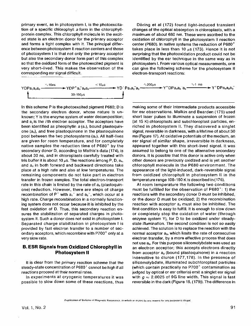
primary event, as in photosystem I, is the photoexcita-tion of a specific chlorophyll a form in the chlorophyll-protein complex. This chlorophyll molecule in the excit-ed state is an electron donor for the primary acceptorand forms a tight complex with it. The principal differ-ence between photosystem II reaction centers and thoseof photosystem I is that not only the primary acceptorbut also the secondary donor form part of this complexso that the oxidized form of the photoexcited pigment isvery short-lived. This makes the observation of thecorresponding esr signal difficult.
Doring et al (172) found light-induced transientchanges of the optical absorption in chloroplasts, with amaximum of about 680 nm. These were ascribed to theoxidation of chlorophyll in the photosystem II reactioncenter (P680). In native systems the reduction of P680+
takes place in less than 10 us (173). Hence it is notsurprising that the photooxidation product could not beidentified by the esr technique in the same way as inphotosystem I. From various optical measurements, onecan give the following scheme for the photosystem IIelectron-transport reactions:
YDPa,a2a3-'VIOns <10Ms
i a2a3-
30-150MS
In this scheme P is the photoexcited pigment P680; D isthe secondary electron donor, whose nature is un-known; Y is the enzyme system of water decomposition;and a, is the /th electron acceptor. The acceptors havebeen identified as pheophytin a (aO, bound plastoquin-one (a2), and free plastoquinone in the plastoquinonepool between the two photosystems (a3). All half-livesare given for room temperature, and for completelynative samples the reduction time of P680+ by thesecondary donor D, according to Mathis's data (174), isabout 30 ns, and in chloroplasts carefully treated withtris buffer it is about 10 us. The reactions among P, D, a1;
and a2 in both forward and backward directions takeplace at a high rate and also at low temperatures. Theremaining components do not take part in electrontransfer in frozen samples. The total electron-transportrate in this chain is limited by the rate of a3 (plastoquin-one) reduction. However, there are steps of chargerecombination of P+ with a,~ and a2", which occur at ahigh rate. Charge recombination in a normally function-ing system does not occur because it is inhibited by thefast oxidation of D. Thus, this secondary reaction en-sures the stabilization of separated charges in photo-system II. Such a donor does not exist in photosystem I.Separated charge stabilization in photosystem I isprovided by fast electron transfer to a number of sec-ondary acceptors, which recombine with P700+ only at avery slow rate.
B. ESR Signals from Oxidized Chlorophyll inPhotosystem II
It is clear from the primary reaction scheme that thesteady-state concentration of P680+ cannot be high if allreactions proceed at their normal rates.
In experiments at cryogenic temperatures it waspossible to slow down some of these reactions, thus
-*-YD+Pa1a2 a3- •*-Y+DPa1a2 a3- -•Y+DPa1a2a3
making some of their intermediate products accessiblefor esr observations. Malkin and Bearden (175) usedshort laser pulses to illuminate a suspension of frozen(at 15 K) chloroplasts and subchloroplast particles, en-riched in photosystem II. They discovered a singletsignal, reversible in darkness, with a lifetime of about 30ms (Figure 17). At oxidative potentials of the medium, anesr signal of similar shape, irreversible in darkness,appeared together with this short-lived signal. It wasassumed to belong to one of the alternative secondarydonors. It is possible that this donor is active only whenother donors are previously oxidized and is yet anotherchlorophyll molecule in the P680 environment. Theappearance of the light-induced, dark-reversible signalfrom oxidized chlorophyll in photosystem II in thetemperature range 108-180 K is described in (176).
At room temperature the following two conditionsmust be fulfilled for the observation of P680 + : 1) thereactions with the secondary donor D must be inhibited,or the donor D must be oxidized; 2) the recombinationreaction with acceptor a2 must also be inhibited. Thefirst condition is easy to fulfill. It is enough to slow downor completely stop the oxidation of water (throughenzyme system Y), for D to be oxidized under steady-state illumination. The second condition is not so easilyachieved. The solution is to replace the reaction with thenormal acceptor a3, which limits the rate of consecutiveelectron transfer, by a more effective process that doesnot use a3. For this purpose silicomolybdate was used asan electron acceptor; this accepts electrons directlyfrom acceptor a2 (bound plastoquinone) in a reactioninsensitive to diuron (177,178). In the presence ofsilicomolybdate, illuminated subchloroplast particles(which contain practically no P700+ contamination asjudged by optical or esr criteria) emit a singlet esr signalwith g as 2.0025 of 9G line width. This signal is fastreversible in the dark (Figure 18, (179)). The difference in
Duplication of Bulletin of Magnetic Resonance, in whole or in part by any means for any purpose is illegal.
Vol. 1,No. 2 91

5m M Ascorbate
10 ms
Figure 17. The kinetics of flash-induced change of the esr signal atg«2.0025 at 35 K. Chloroplastfragments were incubated with fer-ricyanide or ascorbate prior to flash ,activation at 660 nm. The reversiblepart of the signal has been attribut-ed to P680+. The dark decay corre-sponds to the back reactionbetween P680+ and the reducedprimary acceptor of photosystem II.From ref. (175), with permission.
660 nm Flash
line widths between the light-induced signal of photo-system II (9G) and the esr signal I (P700+, 7.5 G) was notrelated to the electron acceptor's nature: photosystem Iparticles in light with silicomolybdate gave a normal esr Isignal. With other acceptors, such as ferricyanide,photosystem II particles did not give rise to a signal inlight that was reversible in darkness. The esr signal ofphotosystem II was appreciably different from the esr Isignal from chloroplasts in its dependence on the lightintensity (Figure 19). Its dark decay at 0° C can beapproximated by a bi-exponential function with char-acteristic times 13.5 s and 2.1 s. Initially the preparationcontained approximately equal amounts of these two
kinetically distinct centers. The steady-state intensity ofthe light-induced signal from photosystem II was deter-mined by the rate of its dark decay both when thetemperature varied and when electron donors of pho-tosystem II (hydroxylamine, Mn2+ ions) were introduced.This is consistent with the assumption that the signalappears as a result of photooxidation, and not as theresult of secondary dark oxidation of any componentacting as an electron donor for P680+ centers. Even if asecondary electron donor is the source of the signal, itmust be very close, in both its structure and in its func-tional properties, to the primary photooxidizable elec-tron donor of photosystem II.
Figure 18. ESR signal at 0«s2.OO25in photosystem II fragments in thelight and the kinetics of its riseunder saturating light (12 W/m2)and dark decay at 0°C in the pres-ence of 1m/W silicomolybdate.[From ref. (182)]
Duplication of Bulletin of Magnetic Resonance, in whole or in part by any means for any purpose is illegal.
92 Bulletin of Magnetic Resonance

20 40 60
Intensity (%)80 100
Figure 19. The light saturation curves of esr signals: 1) signal I from diuron-treated chloroplasts; 2,3) silicomolybdate-dependentsignal from photosystem II particles; 1,3) at 22°C; 2) at 2°C; 100% of light corresponds to 12 W/m2. Intensity is in percent, and(d%"/d H )max is the amplitude of the first derivative of esr absorption.
The accessibility of the bound electron acceptor Q (orA2) for external electron donors and acceptors increasesas the p H of the medium decreases (180). At p H 4 thisled to the appearance in light of a singlet signal fromoxidized chlorophyll, which quickly decays in the dark(181). This chlorophyll may either be the photooxidizedprimary donor or one of the alternative secondary do-nors of photosystem II.
Silicomolybdate does not lead to the appearance ofan esr singlet signal under illumination of photosystem IIpreparations at 77 K. Evidently, this is related to the factthat at 77 K silicomolybdate does not oxidize the donorwhich is accessible for the P680+ centers. The proper-ties of the silicomolybdate-dependent esr signal can bestudied at low temperatures, if this signal is "frozen in"by cooling the samples in light. At room temperature thephotosystem II signal does not differ from the P700+
signal in its relaxation properties. However, at 77 K themicrowave-saturation curves of these two signals arequite different (182). The esr signals were compared withthe optical effects induced by light in photosystem IIpreparations in the presence of silicomolybdate, and itwas shown (183) that the appearance of the signalcorrelates with photobleaching, with maxima at 685 nmand 435 nm. This spectrum coincides with that obtained
by Doring et al (172), who attributed it to the photoox-idized pigment in the photosystem II reaction center.Optical changes were compared with the intensity of thephotoinduced singlet signal calibrated by the esr sig-nal of nitroxyl radical, and the molar extinction of thecorresponding center was evaluated (0.66 x 10⁵
W ' c m " 1 (184), coinciding with the molar extinction ofP700 pigment, from the data of Hijama and Ke (185)).
When studying reactions induced by light or oxidants,it is always necessary to consider the possibility ofpigment oxidation in the light-harvesting matrix. Thisprocess does in fact occur at room temperature in thepresence of certain combinations of oxidants. Forexample, if a mixture of silicomolybdate and potassiumchloroiridate is present in photosynthetic preparations,including those that contain no P700, an esr signalappears in light and is stable for tens of minutes indarkness (182). On the background of this dark signal, adark-reversible signal appears from photosystem IIfragments in light; the latter is identical with the onedescribed above. A mixture of chlorophylls a and b\n anaqueous-alcohol medium in the presence of thiscombination of oxidants gives the same dark-stablesignal. A similar signal was obtained from a suspensionof photosystem II particles that were pretreated at 80°C
Duplication of Bulletin of Magnetic Resonance, in whole or in part by any means for any purpose is illegal.
Vol. 1,No. 2 93

for 10 min. The esr signal reversible in darkness was notobserved in this case.
Pigment oxidation in the light-harvesting matrix wasalso observed in photosystem I preparations at low-temperature illumination, in the presence of a largeexcess of oxidants (77). The esr signal differs in itsrelaxation parameters from the esr signal I from theP700 centers. Its appearance in light at 15 K is notaccompanied by the reduction of acceptor Fe-S centers.This fact can be used to differentiate the P700+ signaland the signal from oxidized antenna chlorophyll (85). Inphotosystem II preparations at cryogenic temperaturesin the presence of excess ferricyanide, an esr signal alsoappears in light (186), which must come from oxidizedmatrix chlorophyll or from a secondary donor of chlor-ophyll nature. All these signals are stable in darkness.
C. Bound Acceptors in Photosystem II
The role of plastoquinone as one of the participants inthe earliest photosystem II reactions induced by lightabsorption at very low temperatures was shown usingesr techniques by Knaff et al (187). After vigorous ex-traction of plastoquinone from photosystem II subchlor-oplast particles, the reversible component of the pho-toinduced esr signal from oxidized chlorophyll (whichhad previously been observed for oxidizing potentials ofthe medium at 15 K) was lost. This component reap-pears upon reconstruction with plastoquinone. Thecharge recombination time of P680+ and the boundplastosemiquinone radical indicated by these data isabout 3-5 ms at 15 K. The formation of plastosemiquin-one radicals by photosystem II photoexcitation wasshown earlier by differential absorption spectroscopy(188). These results, however, do not exclude the exis-tence of other carriers between the P680 center and thebound plastoquinone, which recombine with P680 atrates too high for esr observations.
According to Klevanik et al (75, 76), the photosystemII primary acceptor is pheophytin a, which forms a tightcomplex with P680. Its reduction product, the pheophy-tin radical anion, is identified by its differential opticalspectrum. The accumulation of pheophytin radicalanions upon steady-state illumination of photosystem IIsubchloroplast fragments in the presence of diuron,which blocks the oxidation of bound plastosemiquinoneat negative redox potentials of the medium ( - 200 mV),occurs through the formation of a complex in the state[P680 pheo a~'Pq~"]. The recombination of P680+ andpheo a is inhibited by a not very effective but everoperative process, the reduction of P680+ by anexogenous electron donor present in excess. However,attempts to observe the esr signal from the pheophytinradical anion in these conditions have so far been unsuc-
cessful as have attempts to observe the esr signal fromthe plastosemiquinone radical anion of the reducedacceptor Q~. The reason for this is probably the cou-pling among the various paramagnetic centers (forexample, between the pheophytin radical anion and theplastosemiquinone radical anion) or the formation of acomplex with a transition metal ion, such as iron. Asimilar problem was solved earlier in connection with thecharge-separation mechanism in bacterial photosynthe-sis (74). A signal from the ubisemiquinone radical anionin bacteria grown on a medium deficient in Fe in thepresence of oxidants was observed at room tempera-ture (24). The esr signal from the bacteriopheophytinradical anion was observed at temperatures below 17 K(189). It had a fir-factor of 2.003 and was split intocomponents separated by intervals of 63 G.
It now seems probable that iron components notidentical with the components related to photosystem Iare present near the reaction centers of photosystem I.As was mentioned above, no esr signals typical for Fe-Scenters on the acceptor side of photosystem I werefound in preparations of subchloroplast particles ofphotosystem II. Nevertheless, these preparations con-tain iron in significant amounts (190). This is not surpris-ing in itself, since heme iron is contained in photosystemII as cytochrome thsg. However, a part of the iron in thesepreparations is clearly not contained in a heme complex.This is shown by the formation of the nitrosyl-Fe com-plex in the presence of nitrite and cysteine with char-acteristic esr signals at g =» 2.03 at 77 K(191).
V. ESR SIGNAL II AND THE REACTIONS ONTHE OXIDIZING SIDE OF PHOTOSYSTEM II
The esr signal II at g ~ 2.0046 with partially resolvedhyperfine structure (Figure 3) is observed in all pho-tosynthesizing organisms capable of oxygen evolution.The most convenient particles for studying this signalare subchloroplast fragments enriched in photosystem IIand purified from P700, algae mutants inactive in pho-tosystem I reaction, and chloroplasts adapted to dark-ness, especially in anaerobic conditions. The esr signal IIis not observed in photosynthesizing bacteria and algaemutants incapable of oxygen evolution. These are thefacts that support the assertion that the esr signal IIcomes from photosystem II.
A whole range of modern methods has been used inthe study of the paramagnetic centers responsible forthe esr signal II. Methods have involved comparison ofvarious mutants and fractions of photosynthetic organ-isms, variations in the illumination and composition ofthe medium, the use of isotopically pure media forgrowing algae, the application of inhibitors, electron
Duplication of Bulletin of Magnetic Resonance, in whole or in part by any means for any purpose is illegal.
94 Bulletin of Magnetic Resonance

donors and acceptors, the use of kinetic measurementsand models in vitro. Earlier experiments have beenreviewed by Kohl (192). It has been shown that thepartially resolved hyperfine structure of the esr signal IIis caused by protons, since it disappears for algae grownon D2O. It now seems sure that the sources of the esrsignal II are plastosemiquinone radicals that arise from acertain fraction of the plastoquinones present in chloro-plasts and algae in great excess compared with theother electron carriers in thylakoid membranes. Themost convincing argument in favor of this identificationis the fact that the esr signal II from chloroplasts, whichdisappears when plastoquinone is extracted, is restoredto its original shape when exogenous plastoquinone isintroduced. If fully deuterated exogenous plastoquinoneis introduced, the signal becomes narrower andstructureless (193). Nevertheless, it was found impossi-ble to reproduce completely the asymmetric shape ofthe esr signal observed in vivo by using isolated plasto-semiquinone in vitro.
The esr signal II is not related to noncyclic electrontransport between the two photosystems, as it is stablein darkness for many hours, nor is it related to the earlyreactions in the photochemical centers. This is becausewhen preparations adapted to darkness are excited atroom temperature by short light pulses, the esr signal IIincreases within seconds. Recently, however, the ap-pearance of a light-induced esr signal with the char-acteristics of signal II has been reported from subchloro-plast particles enriched in photosystem II at cryogenictemperatures.*
The plastoquinones, the source of this signal, are agroup of structurally related compounds (Figure 20). Inchloroplasts, plastoquinone A is the main component ofthis group, about 50 moles per mole of reaction center(194). About 6-10 plastoquinone molecules take part inthe electron transfer between the two photosystems.(This process will be considered in the next section.) Thefunctions of the remaining part of the plastoquinonepool are not yet clear. Plastoquinone is easily extractedfrom chloroplasts. This destroys some obviously secon-dary electron-transport reactions. Plastoquinone, whichseems to be the only low-molecular-weight carrier inthylakoid membranes, is thought to be capable of trans-ferring electrons between donors and acceptors fixed inthe membrane and separated by a hydrophobic barrier.This property of plastoquinone has been recently illus-trated in model membranes separating donor and ac-ceptor solutions (195).
Semiquinones are a traditional object of esr research.Since Michaelis (196), it has been accepted that the
Plastoquinone A
H,C
Plastoquinone B
Vitamin
Plastochromanol
*J. H. A. Nugent and M. C. W. Evans, FEBS Lett. 101, 101(1979).
Figure 20. Structures of some prenylquinones.
redox reactions involving quinones proceed by a one-electron mechanism, i.e., semiquinone radicals orradical anions are formed as intermediates. The obser-vation of the redox transformation in situ is made dif-ficult, however, because of the absence of intenseabsorption bands in both the oxidized and the reducedform. The esr signals from plastosemiquinone radicals insitu overlap with the strong esr signals from oxidizedchlorophyll. The lifetime and the steady-state concen-tration of plastosemiquinone radicals are rather smallsince equilibrium in their dismutation reaction is shiftedfar towards the diamagnetic products, unless there arespecial reasons, such as limited mobility in membranes,that interfere with the establishment of this equilibrium.
Due to the recent work of Sauer and co-workers(197-204), significant progress has been made in the
Duplication of Bulletin of Magnetic Resonance, in whole or in part by any means for any purpose is illegal.
Vol. 1,No. 2 95

Figure 21. The esr signal II from bean chloroplasts cooled to— 120°C in a magnetic field of ca 10 kG in the initial orienta-tion, and after rotation of the sample by 90°.
understanding of plastoquinone functions and the na-ture of the esr signal II. Three kinetic components of thesignal II were found: Sconst, SS|0W, and $as t. The esr signal" Sconst is observed in darkness and does not decay evenafter leaves are aged for several days in darkness.Chloroplast aging, heating with CI-CCP [carbonyl cya-nide m-chlorophenyl hydrazone], incubation with 0.8 Mtris-HCI and divalent cations, higher alcohols (205-208),other treatments causing the inactivation of the systemof water decomposition, and other reactions on theoxidizing side of photosystem II, induce the decay ofsignal II Sconst.
After long adaptation to darkness the signal SS|OW
appears from chloroplasts in light. When illumination isby a series of flashes there is a periodic component inthe increase of the esr signal II, with maxima at thesecond and sixth flashes. It follows that the free radicalarises from the interaction of the diamagnetic precursorwith certain of the intermediate states of the waterdecomposition system (states S2, S3 in Kok's termin-ology) (210). At the same time a monotonic increase ofthe signal in response to the flashes shows that it cannotbe identified with any particular S, state, since theconcentration of each of them must oscillate with aperiod of four flashes.
The anisotropy of the esr signal II (Sconst and SS|OW) is initself an indication of the fact that the corresponding freeradical centers are not in a state of rapid rotation, i.e.,
the anisotropic hyperfine structure is not averaged.Experiments with magnetically oriented chloroplastsshow that free radical centers are not only immobilizedbut also rigidly oriented relative to the thylakoid mem-brane (98,211). Figure 21 shows the esr signals from peachloroplasts in suspensions cooled to - 120°C in amagnetic field of about 10 kG both at the initial orienta-tion and after a 90° rotation. The disappearance of thelow-field shoulder when the sample is rotated seems tomean that the A-tensor component corresponding tomaximal splitting is oriented normal to the plane of thethylakoid membrane. Similar results were obtained withwhole cells of the alga Chlamydomonas reinhardi (mu-tant deficient in photosystem II (212)).
There are no orientation effects in photosystem IIsubchloroplast particles because shape asymmetry islost upon fragmentation. The dependence of the shapeof the esr signal II on chloroplast orientation in themagnetic field was also observed at room temperaturefor the chloroplasts oriented by hydrodynamic flow (98).
In chloroplasts with water splitting inactivated byremoval of Mn2+, an esr signal was observed whose pa-rameters were close to those of the steady-state esrsignal H. The signal appears in light in less than 100 MSand decays in darkness after about 2 s (II Sfast (201)).This signal is inhibited by diuron, which is an inhibitor ofnoncyclic electron transport. In its intensity this fastsignal II approximately corresponds to the content of theP700 centers. In fully active chloroplasts a much moreshort-lived signal of roughly similar shape (T1/2 ** 100 MSfor decay) was observed instead of II S(ast (203-204). Thissignal is saturated by low microwave power. At such apower no saturation of other components of the esrsignal II has been observed. The changes in relaxationand kinetic characteristics seem to be caused by theremoval of paramagnetic Mn2+ ions. The substitution ofNi2+ for Mn2+ partially restored the signal's initial depen-dence on microwave power without any change in itslifetime. The data obtained make it possible to attributethe function of the physiological electron donor ofphotosystem II to the diamagnetic precursor of thecenter responsible for the fast or very fast esr signal II. Itdoes not seem to be the direct electron donor for P680+
pigment, but it acts on the later stages of electrontransport on the oxidation side of photosystem II.
The problem of the nonhomogeneity of the paramag-netic centers responsible for the esr signal II has alsobeen tackled by means of radiospectroscopy. In highlynative samples (whole leaves of higher plants (213)) thesignal is saturated at room temperature at a microwavepower of about 25 mW, and the saturation is accompa-nied by a change in the signal shape. The shape changeunder saturation was also observed for chloroplastsfrom various sources (214).
The electron spin-echo resonance technique has
Duplication of Bulletin of Magnetic Resonance, in whole or in part by any means for any purpose is illegal.
96 Bulletin of Magnetic Resonance

been recently used by Nishimura et al (209) for theinvestigation of esr signal II. They also concluded thatsignal II consists of at least three "dark" componentsthat differ in their relaxation times.
The centers responsible for the esr signal II do notseem to be readily accessible for exogenous electrondonors and acceptors. Nevertheless, in the presence ofcertain electron-transport mediators and in combina-tion with other treatments destabilizing the intermediate
dx"/d//
40 60 80 100 160s
Figure 22. Some typical recordings of the increase of the esrsignal II from photosystem II particles under illumination andits dark decay: 1) without additives; 2) in the presence of1 m/W ferricyanlde, 3) in the presence of 1 m/W 2,6-dichloro-phenolindophenol; 4) with both oxidants in the medium.
states of the water-splitting system, electron donorstend to inhibit the esr signal II. In this connection it isnatural to suppose that it is formed by plastohydroquin-one oxidation and not by plastoquinone reduction. Inphotosystem II fragments dichlorophenolindophenol, aneffective electron-transport mediator, inhibits the esrsignal II in darkness, but does not interfere with itsincrease in light. A characteristic feature of the esr signalII is its inhibition in light and in darkness in variousphotosynthetic preparations (chloroplasts, photosystemII fragments, algae) in the presence of silicomolybdate(179,182). Evidently, redox agents can inhibit the esrsignal II both by the oxidation of plastosemiquinoneradicals (silicomolybdate) and by their reduction(reduced dichlorophenolindophenol). The high reactivityof silicomolybdate towards plastosemiquinone radicalsis in agreement with its ability to accept electrons fromthe reduced bound acceptor Q of photosystem II, whichis also a plastosemiquinone radical. Hydrophilic redoxagents and redox agents not permeating the membrane(ferricyanide, ascorbate) do not affect the intensity of theesr signal II in darkness. In light, ferricyanide induces anincrease of the esr signal II. This light-induced signalquickly decays in darkness. When the kinetics of thelight-dark transitions of the esr signal II in the presenceof dichlorophenolindophenol and ferricyanide, are com-pared, it becomes evident that the reactions in thepresence of both these acceptors are quite independentfrom each other and involve different centers contribut-ing to the total esr signal II (Figure 22). It is interestingthat the relaxation characteristics of these two types ofparamagnetic centers are also different, as follows froma comparison of the shape and microwave saturation ofcorresponding signals at 77 K.
VI. Mn(ll) IONS AND THE WATER-SPLITTINGSYSTEM
The distribution of metals in photosynthetic mem-brane fractions (168, 170, 190, 215), the effect of chelat-ing agents on O2 evolution by chloroplasts in the pres-ence of silicomolybdate (216, 217), and some otherfacts indicate that transition ions participate in reactionson the oxidizing side of photosystem II. Most attentionhas been given to the study of the function of Mn in thewater-splitting system.
A. Charge Accumulation in theWater-Splitting System
Water oxidation with O2 formation requires the ac-cumulation of four oxidizing equivalents. Joliot's exper-iments (218) showed that when chloroplasts and algaeare illuminated by short light flashes, O2 release is aperiodic function of the number of the flashes (with a
Duplication of Bulletin of Magnetic Resonance, in whole or in part by any means for any purpose is illegal
Vol. 1, No. 2 97

period of four flashes). Hence it follows unambiguouslythat oxidizing equivalents are accumulated one by one inevery electron-transport chain and that there is nocooperation among different photosystem II centers inthe process of water splitting. These postulates form thebasis of the phenomenological scheme due to Kok et al(210), according to which water splitting goes through asequence of five states with the absorption of four lightquanta:
+ 4H++4e"
Many experimental results have given more informa-tion about the details of this scheme, such as the in-dividual properties of the S, states, their lifetimes, theirdependence on the redox potentials of the medium, andso on. However, the nature of these states is still notclear, the chemical composition of the complexes inwhich the positive charges accumulate is not known,and attempts to isolate the water-splitting complex fromthe photosynthetic membranes have failed. Mn par-ticipation in photosynthetic O2 production and theindependence of other electron-transport steps on thepresence of Mn is reviewed in ref. (219). Mn redoxreactions are thought to play a central role in the sug-gested models of water splitting (220).
B. ESR Spectra of Hydrated Mn(ll) Ions
ESR signals from hydrated Mn(ll) ions in photosyn-thetic systems are observable at room temperature inhighly native specimens (leaves, algae, chloroplasts).Almost all the experimental data obtained show that thissignal does not come from functionally active Mn in thewater-splitting system. Thus, functionally active Mndoes not give any esr signal in normal observationconditions, and its presence can be proved only aftercell (chloroplast) destruction and Mn extraction bytreatments that destroy the water-splitting system.Among these are treatments with acids (down to pH 4),cyanide, 0.8 /Wtris-HCI, and high concentrations ofdivalent cations (221-223).
The esr spectrum of hydrated Mn(ll) ions in solution isa sextet of lines of approximately equal intensity, causedby hyperfine splitting on the 55Mn nucleus with nuclearspin / = 5/2. The Mn2+ esr spectrum is described by aspin-Hamiltonian
added to this spin-Hamiltonian, corresponding to zerofield splitting
3C = + AIS
3C z F S = S(S + 1)] S 2\y I
where g = 2.003, A = 94.5 G. If the symmetry of thesystem is lower than octahedral, a quadratic term is
where Dand Eare the zero-field-splitting parameters. Itleads to the appearance of five groups of lines due to thefine structure, corresponding to transitions A/Ws = ± 1between the levels Ms = ±5/2, ±3/2, ± 1/2. However,these transitions, except between Ms = ± 1/2, arehighly anisotropic and the corresponding lines arebroadened in powder samples so that they becomeunobservable. In this case, the spectrum looks like thefamiliar sextet, caused by the transitions between thelevels Ms= ± 1/2.
In biological systems, as well as in solutions of Mn(ll)complexes, molecular rotation can average the anisot-ropy of the zero field splitting, if TC~1>3CZFS (where TC isthe rotational correlation time and 3CzFS is in frequencyunits). In this case the whole spectrum collapses to thecenter, and its observed intensity is increased due to thecontribution of the transitions between other spin levels.This situation occurs also with hydrated Mn(ll) ions. Forthe complexes with macromolecules rc~' is often lessthan 3CzFS. In this case there is no averaging of theanisotropy of the zero field splitting, and only the linescorresponding to the transitions between the levelsMs= ±1/2 are observed, i.e., the spectrum decreasesto about one fifth of its total intensity. Siderer et al (224)have obtained data that indicate that at least a part ofthe Mn(ll) bound in chloroplasts is in the form ofmolecular complexes with TC < 3CzFS. The intensity of theMn(ll) sextet in lettuce chloroplasts was four to five timesless than the signal from the same quantity of Mn(ll) inwater solution. In our laboratory, measurements havebeen made of the Mn(ll) esr signal from chloroplastsuspensions, from supernatants of centrifuged chloro-plasts, and from bound Mn in chloroplasts (in the lastcase measurements were made after chloroplasts weretreated with acid). These measurements did not revealsignificant contribution of bound Mn to the total signalfrom the suspension (225).
The above-mentioned approach is not applicable inthe case of highly effective spin-orbital coupling. For thiscase intense groups of lines occur typically in the lowfield region (226, 227). This type of signal has beenobserved at room temperature in concentrated chloro-plast preparations from bean leaves (225).
C. Displacement, Binding, and Photo-oxidation of Mn(ll) Ions by Chloroplasts
The study of the state and the functions of Mn(ll) inphotosynthetic systems follows different lines, viz., 1)
Duplication of Bulletin of Magnetic Resonance, in whole or in part by any means for any purpose is illegal.
98 Bulletin of Magnetic Resonance

Mnb (mole/mole Chi)1CT1 -
5.0
4.0
3.0
2.0
1.0
Mnb (mole/mole Chi x102)
10' 10" 10" 5.0 10.0
Figure 23. Binding curves of exogeneous Mn(ll) by chloropiasts from bean leaves; 1) chloropiasts treated with 0.8 M tris-HCI, pH7.8; 2)intact chloropiasts; 3) tris-pretreated chloropiasts with a constant total cation concentration, [Mn2+] + [Mg2+] = 0.8 m/W.
the binding of exogenous Mn(ll) by chloropiasts, eitherintact or after displacement of bound Mn by mild treat-ments, which allow many types of photosynthetic activityto be preserved, 2) the electron-donor function of Mn inchloropiasts, 3) the search for conditions that enableobservation of intrinsic Mn esr signals in photosyntheticsystems. In addition, the technique of nmr relaxation ofwater protons has recently been suggested for the studyof the Mn state in chloropiasts (228).
Spinach and bean chloropiasts, washed out by theusual incubation solution and actively evolving O2 inlight, contain six to ten Mn atoms per electron-transportchain. Further extraction of Mn leads to inactivation ofO2 release, though the ability to oxidize various artificialelectren donors to photosystem II is still preserved.Treatments mild enough to allow reactivation by theintroduction of exogenous Mn(ll) do not lead to thedisplacement of all endogenous Mn. However, pho-tosynthetic membranes can bind much greater amountsof Mn(ll) (225). Figure 23 shows the binding of exoge-nous Mn(ll) by intact chloropiasts and by chloropiastsinactivated by treatment with tris. The maximum amountof bound Mn is about 80 atoms per electron-transportchain, i.e., it is much higher than the amount of Mn(ll)necessary for O2 evolution. The characteristic feature of
the binding curve is its S shape, which indicates thecooperativity of the binding. For chloropiasts treatedwith 0.8 M tris-HCI, the point of inflection shifts in thedirection of smaller free Mn(ll) concentrations, but it isstill distinctly seen. The S shape is not a nonspecificcation effect; in fact, the Mn(ll) sorption curve in thepresence of Mg2+, providing constant total cation con-centration in the solution, has the same S shape as forthe binding of Mn(ll) in the absence of Mg. The nature ofthe cooperativity is not yet clear. At the present stage ofresearch two hypotheses can be suggested: 1) Thebinding of the first Mn(ll) ions causes a change in themembrane structure that is favorable for further bind-ing. 2) Binding is accompanied by a change in the oxida-tion state of Mn, and stable oxidation and binding takeplace only when there are no less than two Mn ions at thegiven membrane locus. For example, Mn(ll) is oxidizedto Mn(lll), but the latter is strongly held by membranesonly after dismutation into Mn(ll) and Mn(IV). Thisprocess is probable because in neutral solutions theMn(IV) state is more stable than the Mn(lll) state. For thedismutation reaction to be possible at a given mem-brane locus there must be at least two Mn(ll) ions pres-ent. This explains the inflection of the binding curve.The oxidation of Mn during its binding under conditions
Duplication of Bulletin of Magnetic Resonance, in whole or in part by any means for any purpose is illegal.
Vol. 1,No. 2 99

of chloroplast reactivation seems to take place. Itrequires illumination of short duration at least (229).
Conclusions about the electron-donor properties ofMn(ll) in chloroplasts were first made from observationsof the activation of electron transfer onto an exogenousacceptor after addition of Mn(ll) salts (230, 231). The esrtechnique has made it possible to observe directly Mn(ll)disappearance in illuminated chloroplasts or chloroplastparticles of photosystem II both under steady-rate andpulsed illumination (182, 232). The quantitative corre-spondence between the Mn consumed and the reducedelectron acceptor in steady-state experiments orbetween the Mn signal and esr signal I decay in pulseexperiments, and also the competitive inhibition ofMn(ll) oxidation by other electron donors of photosys-tem II, show that in these experiments actual Mn(ll)oxidation takes place rather than nonspecific cationbinding. The free Mn(ll), but not the bound Mn, isoxidized upon illumination, and the decrease in theMn(ll) esr signal is partially reversible in darkness after aflash. This effect is caused by the removal of a part of thebound and esr-inactive Mn from chloroplasts. The donorfunction of exogenous Mn(ll) manifests itself only afterthe degradation of the activity of the water-splittingsystem (by aging or tris-HCI treatment of chloroplasts orin photosystem II particles). This agrees with the sugges-tion that the binding of exogenous Mn(ll) during thereactivation of the O2-releasing system of chloroplasts isaccompanied by its oxidation and with the observationsthat for the tight binding of Mn in the O2-releasingsystem it is necessary that at least some of the atomsforming the catalytic complex should be oxidized. How-ever, these arguments are all indirect ones, and there isno unambiguous information about the valence state ofMn in chloroplasts in the rest state.
D. Mn in Chloroplasts as a Relaxantof Water Protons
Mn (II), as a paramagnetic ion, enhances the magneticrelaxation of water protons. This phenomenon is used insome recent experiments (228, 233-235). The dataobtained can be summarized as follows. Chloroplastsincrease the rate of relaxation of water protons as shownby increase in 7T1 and T2~\ where 7", and T2 are thespin-lattice and spin-spin relaxation times respectively.This effect is caused by endogenous Mn in the water-splitting system. There is a correlation between thedegree of displacement of Mn by Mg, the decrease inO2-releasing activity, and the decrease in the en-hancement effect of proton relaxation. Other chloroplastparamagnetic centers do not give any significant con-tribution to this effect because they are well shielded
from water. The dependence of 7", 1 on the resonancefrequency (233) shows that the relaxation rate is deter-mined by the electron spin-relaxation time (TS about 1ns). This agrees with the suggestion that the protonrelaxant is Mn(ll), since Mn(lll), Fe(ll), and Fe(lll) (forwhich Ts^iO"10 to 10~11 s (236)) cannot effectivelyenhance proton relaxation. When chloroplasts wereexcited by light pulses, changes in T2~* have beenobserved with a period of four flashes (228), whichindicates that the state of the Mn in the system alsochanges with the same period. The reason for thischange is not known: it may be caused by a Mn valencychange and the change in its accessibility for waterprotons. Thus, these measurements have also made itpossible to establish correlation between the states ofthe water-splitting system (S0-S4) and the state of Mn inchloroplasts. It should be noted that the proton-exchange rate between bulk water and water in thecoordination sphere of endogenous Mn is quite high (thefree induction decay is simply described by an exponen-tial function). Thus 7"2~
1 changes cannot be explained byvariations in the exchange rate.
To summarize the information available for Mn inchloroplasts, four pools of Mn in chloroplasts can bedistinguished. The "zero" pool, which cannot be iso-lated by nondestructive treatments, consists of two orthree Mn atoms per electron-transport chain. It is notclear whether this tightly bound Mn has anything to dowith the water splitting.* There is also a Mn pool innormal chloroplasts, which can be isolated by compara-tively mild treatments that provide reversible inactiva-tion of the O2-releasing system. Two more pools arefilled only in the presence of Mn(ll) ions in the medium.The filling of one of them is specific for Mn and is char-acterized by the cooperativity mentioned above, whilethe filling of the other one is a nonspecific effect ofdivalent cations. There exists a slow Mn(ll)-ion exchangeamong the last three pools. At high concentration ofdivalent cations other than Mn(ll) in the medium, thisexchange causes the loss of Mn(ll) from the poolspecifically related to O2 evolution. In complete absenceof Mn2+ in the medium, this pool loses Mn, which alsoleads to the inactivation of O2 release upon chloroplastaging. This inactivation occurs more quickly than withother types of electron-transport activity. Evidently, thewater-splitting system can function for a long time onlywhen a certain background concentration of Mn2+ ionsis present in water, since there exists a dynamic equilib-rium between the Mn in the catalytically active complexand Mn in the solution or in other membrane loci (207).
*lt may be related to the superoxide dismutase activity of thechloroplast membrane [C. H. Foyer and D. O. Hall, FEBS Lett.101,324(1979)].
Duplication of Bulletin of Magnetic Resonance, in whole or in part by any means for any purpose is illegal.
100 Bulletin of Magnetic Resonance

E. The Cluster Nature of the Mn Centersin Chloroplasts
Water oxidation, as was mentioned above, requiresthe accumulation of four oxidative equivalents in the onelocus. That is why in different models of this process it isassumed that several Mn ions form a complex, and it isnatural to suppose that a magnetic coupling can occuramong these ions. The properties of Mn as a paramag-netic proton relaxant must change if the Mn ions form acluster with correlated spins. ESR signals of quite a newtype were observed in bean chloroplasts at liquid-heliumtemperature with g ca 3, which depends on the room-temperature preillumination (171). A similar signal wasfound independently in spinach chloroplasts by Slabasand Evans (237). The signal with g ca 3 appears inchloroplasts adapted to darkness at observationtemperatures below 40 K. The signal characteristics(g-factor, line width, intensity) depended on the numberof pulses at preillumination of the sample and, what isparticularly interesting, on the orientation of the samplerelative to the magnetic field in the esr spectrometer.The temperature dependence of the p-factor is consis-tent with the conclusion that the signal is caused by acertain complex, evidently containing Mn, in whichferromagnetic coupling among the metal ions takesplace (237). Further experiments (238) show that thesignal with these properties appears onty when thesample is gradually cooled to a temperature of about23 K in a magnetic field of H = 2300 G. The dependenceof the signal on temperature and magnetic field duringfreezing indicates that this process is of the phase-transition type. The system examined belongs to theclass of spin glasses, i.e., magnetically diluted structureswith cooperative magnetic coupling, which occur withina certain interval of temperatures and external magneticfields (239). This effect should give research workers anew technique for studying cluster structures formed byparamagnetic ions, which play an important role in manybiocatalytic processes, including photosynthesis.
F. Alternative Hypotheses on the Structure ofthe Water-Splitting Complex
Some data indicate that Mn is not the only, andprobably not the indispensable, component of thewater-splitting system. There is evidence that Cu ionsparticipate in the reactions of photosystem II, includingwater splitting. This possibility is especially interesting ifO2 release is regarded as the reverse of terminal oxida-tion in respiration. It is well known that the enzyme thatcatalyzes O2 binding contains Cu in its active center.
Holdsworth and Arshad (240) isolated a pigment-proteincomplex from the diatomic alga Phaeodactylum tri-cornutum, which contained 8 moles of Cu and 2 molesof Mn per 40 moles of chlorophyll. This photochemicallyactive complex displayed a number of reactions typicalof photosystem II, and its esr spectrum containedcomponents that were identified as signals of Mn2+ andCu2+. There is other information about the isolation ofMn-containing complexes (241, 242), but none of theexperiments proved that the formation of these com-plexes is not an artifact caused by the treatment of theinitial material, and in any case these complexes did notshow catalytic activity in the water-splitting reaction.Finally, recent experiments have shown that the in-troduction of Cu ions (in the form of an albumin complex)into potato chloroplasts, pretreated with 0.8 /Wtris-HCI,restores the O2-evolving activity (243).
What is the nature of the other Mn ligands, apart fromwater, in the water-splitting complex? This question isclosely connected with the existing models of the cata-lytic complex (220). It is usually assumed that a catalyticcomplex contains two Mn(ll) ions oxidized to Mn(IV), orfour Mn(ll) ions oxidized to Mn(lll). In this case it is alsoassumed that Mn(lll) or Mn(IV) oxidizes the ligand watermolecules into H2O
+ or OH'. As the formation of reactiveparticles like OH radicals in the organic medium wouldbe quite toxic (any organic molecules would have tohydroxylate in the presence of OH radicals), it is as-sumed that these particles are stabilized in the coordin-ation shell of Mn until their number becomes largeenough for O2 release to be possible. In some variants ofthe model it is supposed that H2O2 is the intermediateform (this suggestion is indirectly supported by theability of chloroplasts to use H2O2 as an electron donor inphotosystem II (244)).
It should be noted that the redox potential of theMn(ll)/Mn(lll) pair is 1.5 V, and although a quantum ofred light has sufficient energy to transport an electronagainst this potential, the formation of such a strongoxidant in a biological system is highly improbable, andthe potential is still not enough for a reaction such asH2O -»• OH (A Em ca 2V). Evidently the ligand shell con-tains components that lower the potential of this pair.Data indicate that electron-transport reactions inhigher-plant chloroplasts on the oxidizing side of pho-tosystem II are inhibited by the removal of plastoquin-ones. Along with the properties of the transient esr sig-nal II, this fact leads to the hypothesis that the plasto-quinone in photosystem II, as in the noncyclic chain, isan electron carrier across the hydrophobic barrier sep-arating Mn complex from the reaction center. Onecan suppose (245) that plastoquinone, which acts aselectron-transfer mediator between Mn2+ and P680+,forms a charge-transfer complex (an ion pair)
Duplication of Bulletin of Magnetic Resonance, in whole or in part by any means for any purpose is illegal
Vol. 1, No. 2 101

Mn3+ — Pq ' whose potential (only slightly more than 0.8V) is not sufficient to oxidize water into OH radicals.Hence these ion pairs can exist long enough for all fourMn ions to form similar ion pairs during consecutiveabsorption of four quanta by photosystem II. Only at thelast step, when an energy of more than 3.2 eV (sufficientfor the transformation 2H2O ->• O2 + 4H+ + 4e~) hasaccumulated in the center, does water oxidation takeplace. In this case no superreactive particles are formed;hence some of the above problems disappear.
The tight interaction of semiquinone radicals withparamagnetic Mn3+ ions explains the absence of an esrsignal corresponding to this paramagnetic product. Thespin-lattice relaxation time decreases so much that theline width of the semiquinone radical signal, accordingto the Heisenberg principle, increases to values thatmake resonant absorption impossible to observe. Thismechanism keeps the reaction center of photosystem IIin a state that is practically independent of the state ofthe water-splitting complex. Thus, the principle of spa-tial separation is realized here. It is generally quitecharacteristic of membrane biochemical systems andprovides the stabilization of the reaction products afterthe reaction event occurs.
G. Cl Ions in the Water-SplittingSystem and the Mn(ll) Signal
with Superhyperfine Structure
Physiological experiments show that for O2 evolutionin chloroplasts chloride ions must be present, as must
9±: ;2.02
[Mn]-CI
! I I I[Mn]-CI
Mn2+ ions (246, 247). It is not known whether theserequirements are related and whether any direct inter-action between Mn2+ and Cl" ions takes place in thecatalytic complex, although recent results make thisinteraction probable.
Figure 24 shows the esr spectrum of Mn in cypressneedles (248). The main feature of this spectrum is thequartet super-splitting presumably caused by inter-action with a nucleus with / = 3/2. The usual six-component esr signal from Mn2+ ions is also observed.The quartet superhyperfine structure is connected withthe appearance of weak lines in the low field, which areapparently caused by gn. Evidently, the quartet split Mnsignal, unlike the signal of free Mn2+, is characterized bya certain anisotropy of its p-tensor. Similar signals havebeen found in "iany other photosynthesizing species, inthe leaves of some citrus plants, black currant, thuja,and pine. In at least two cases (thuja and pine) thepresence of these paramagnetic centers in chloroplastshas been proved. Superhyperfine structure of this typehas been observed in the signal from photosyntheticallyactive lettuce chloroplasts (224). This signal is notobserved in ordinary preparations from standard pho-tosynthetic species, such as bean or spinach chloro-plasts and leaves. However, if the chloroplasts are
[Mn]-F
Figure 24. The esr signal from cypress needles (Cupressussempervirens, var. pyramidalis) at 77 K.
Mn(ll)aq
Figure 25. The esr signal from cypress needles pretreated tosubstitute F ligand for Cl ligand.
Duplication of Bulletin of Magnetic Resonance, in whole or in part by any means for any purpose is illegal.
102 Bulletin of Magnetic Resonance
isolated from aging bean plants, quartet superhyperfinestructure is also observed. It should be mentioned thatfor Mn super-splitting of the hyperfine structure lines onother nuclei is generally not at all typical, as it is very rarethat Mn forms strong high-spin complexes with ligandsother than water. Quartet superhyperfine structure canindeed be caused only by a Cl ligand, because only Cl ofthe common elements has an isotope with a nuclearspin of 3/2. The nuclear spin of Cu is also 3/2, butbinuclear complexes containing magnetically interact-ing Mn and Cu produce signals of quite a different type(as does, for example, the binuclear complex of Mn andCu with pyridine oxide, due to the antiferromagneticinteraction between Mn2+ and Cu2+ ions) (249).
The assignment of the superhyperfine structure asthat caused by splitting on Cl is confirmed by the pos-sibility of substitution of F~ for Cl" leading to the doubletsplitting on the 19F nucleus (Figure 25) (250). It is reason-able to assume that the Mn-CI complex in photosyn-thetic membranes is a product of the partial catabolismof the water-splitting active structure, which can ac-cumulate in sufficient quantity only in certain species.Certainly, this product is not identical with the water-splitting complex itself. However, this end product ofdegradation preserves some features of the initial struc-ture and indicates the presence of the Mn-CI link.
VII. NONCYCLIC ELECTRON TRANSPORTBETWEEN THE TWO PHOTOSYSTEMS
A. Spectral-Kinetic Separation of the TwoPhotosystems
Electron transfer between the reaction centers of thetwo photosystems is the slowest process of the entirelight stage of photosynthesis and so limits its rate. Thisprocess, in which P680 in photosystem II centers is anelectron donor and P700+ in the photosystem I center isan acceptor, proceeds with the participation of plasto-quinone, cytochrome f, plastocyanin, and probably oneother component, a high-potential Fe-S center (251,252).
The reaction of plastocyanin with P700+ takes placeat high rate (T1/2 <=« 20 /xs at room temperature), whereasthe reduction of plastocyanin and cytochrome f uponpulse excitation of photosystem II is characterized byT1/2 = 6-150 ms (253). In the study of the kinetics andmechanism of the slowest steps of noncyclic transport,P700+ can be considered a direct electron acceptorfrom plastohydroquinone because of the significantdifference between these times. This is the basis for theapplication of the esr signal I from P700+ to study thekinetics of noncyclic electron transport. The otherparticipant in this process, plastoquinone, as was men-
tioned above, does not reveal itself in the esr spectra,though there is no doubt that during its one-electronreduction and reoxidation, semiquinone radicals orradical anions are formed.
The oxidation of P700+, which can be observed by theesr technique in any photosynthetic system of higher-plant type exposed to far-red light, and its reduction innear-red light, which mainly excites photosystem II,provide one of the most simple, direct, and easily ob-servable confirmations of spectral separation of the twophotosystems and of sequential electron transfer(Figure 26) (254-257). The dependence of the esr signalon the intensity of near-red or of white light (Figure 27)indicates that the limiting stage of the whole electron-transport system is that between the two photosystems.In weak light the electrons from photosystem II com-pletely cancel the positive charge on the P700 thatappears upon photosystem I excitation. In bright light,when the interval between two consecutive excitationsof photosystem I is smaller than the time required forelectron transfer in the limiting step, P700 centers are inthe oxidized state most of the time. This causes theappearance of the steady-state esr signal I.
The kinetics of the increase of the esr signal in far-redlight depend mainly on the preillumination of the pho-tosynthetic system. In bright light, which excites bothphotosystems, the plastoquinone pool between them isreduced because the limiting step of the electron trans-port is between plastohydroquinone and P700 + . As aresult, with subsequent far-red illumination the increasein the esr signal I is retarded. Diuron, an inhibitor ofnoncyclic electron transport between the two photo-systems, removes the qualitative differences in theeffects of red and far-red light on the esr signal I, as wellas the lag phase. The "memory" of the preceding il-lumination in intact systems such as higher-plant leavesis preserved in darkness for tens of seconds. Hence thesteady state of leaves and chloroplasts is usually farfrom equilibrium under the medium's conditions (255).
Noncyclic electron transport is not the only factor thatdetermines the steady-state intensity of esr signals.Cyclic electron flow in photosystem I and reactions ofthese centers with endogenous reductants (which forman inner redox buffer and react with P700+ via plas-toquinone, plastocyanin and cytochrome f) also lead tothe reduction of P700+ (258-262). They slow down theincrease of the esr signal I in far-red light and accelerateits dark decay. Both these processes of P700 reductionare inactivated when chloroplasts are exposed for a longtime in aerobic conditions, which result from the exhaus-tion of the reductants and the transfer of all the compo-nents of the cyclic chain to the oxidized state. With longdark incubation the lag phase of the signal rises becauseof the reduction of the plastoquinone pool by near-red
Duplication of Bulletin of Magnetic Resonance, in whole or in part by any means for any purpose is illegal.
Vol. 1,No. 2 103

Figure 26. Chromatic transi-tions in the esr signal I inten-sities from the leaf ofTrachycarpus fortunei sp.Open triangles: 710 nm lightswitching on and off. Solidtriangles: the same for 634nm light. The light intensitiesare given as percentages ofthe maximum intensity (2.0W/m2 for 710 nm, 40 mW/m2
for 634 nm).
light and decreases due to plastohydroquinone interac-tion with atmospheric oxygen. As a result, the excitationof chloroplasts by a single pulse of less than 1 us dura-tion is sufficient for P700 oxidation. This proves theone-quantum mechanism of charge separation in pho-tosystem I (262).
There are some typical transitory effects that accom-pany changes in the spectral composition of light actingon the leaves of higher plants (255, 256). Switching on634 nm light (which excites both photosystem I andphotosystem II) over a constant 710 nm background(which excites mostly photosystem I) induces a rapiddecrease in the esr signal, with a subsequent transitionto a plateau (Figure 26). The times of nonmonotonictransition effects are of the order of seconds, which ismuch more than the characteristic times of any elemen-tary steps of electron transfer. Analysis of these effectsand comparison with other data such as inductionphenomena in chlorophyll a fluorescence, changes of
accessibility of the donor centers of photosystem II forsilicomolybdate (178), and the inhibitors of electron-transport reactions and membrane modifiers in pho-tosystem II (263, 264), lead to the conclusion that theselong-duration transitory processes are at least partiallycaused by structural changes in the photosyntheticmembranes. These influence the efficiency of the energytransfer between the two photosystems and/or the rateconstants of the slowest elementary steps of electrontransfer, which depend on the spectral composition ofthe light and the degree of reduction of the componentsof the electron-transport chain (255). Detailed computercalculations that assume all the kinetic constants to beinvariable do not give a quantitative description of thetransitory effects observed under changed illumination(265, 266). The structural-kinetic lability of the electron-transport chain increases the adaptation and regulatoryabilities of photosynthetic membranes to changingexternal conditions.
710nm
J i i i • i
7-mnrr. ^ i i g T1
f / 634nm
VV i i i i i i0 1 3 0 20 40 0 0.4 0.8 1.2
Figure 27. The esr signal I intensity (arbitrary units) from Hibiscus sp. leaf as function of light intensity (W/m2). Right curves: theleaf pretreated with diuron.
Duplication of Bulletin of Magnetic Resonance, in whole or in part by any means for any purpose is illegal.
104 Bulletin of Magnetic Resonance

B. The Two-Electron Shutter in the NoncyclicElectron-Transport Chain
Under steady-state illumination the pigment P700 isan intermediate electron carrier, and the fraction ofoxidized P700 centers is determined by the ratio of theoxidation and reduction rates. Thus, the esr signal Icannot be used to obtain a direct estimate of theelectron-transport rate. However, short light pulsesprovide a single excitation of the reaction centers ofeach of the two photosystems, and the kinetics of thechanges in the esr signal I induced by a flash allow theevaluation of the rate of P700+ reduction due to non-cyclic electron transport and other reductive processes,and also the determination of the quantity of reducingequivalents reaching the P700+ centers at a singleexcitation of photosystem II. This approach, calledesr-flash photolysis (258), was used for studying thevarious reactions of P700+ reduction. In this section weshall consider one aspect of noncyclic electron trans-port, which concerns the coupling of one- and two-electron reactions in a noncyclic chain.
All the carriers acting in a noncyclic chain can bedivided into one-electron carriers (reaction centers,cytochromes, plastocyanin) and two-electron carriers(plastoquinones). Plastoquinone forms the pool be-tween the two photosystems (6-10 moles of plasto-quinone per electron transport chain). Optical absorp-tion data indicate that in the process of electron trans-port plastoquinone forms a product of twofold reduc-tion, plastohydroquinone. Under steady-state illumina-tion semiquinone radicals are not accumulated in anysignificant amounts. The problem is: How does twofoldreduction of plastoquinone occur by its interaction witha one-electron donor, the photosystem II reactioncenter? In principle, two ways of coupling are possible:1) Electron-transport chains are joined together in pairsat the level of the photosystem II secondary acceptor, sothat a flash of saturating intensity (i.e., one that excitesall the photosystems II simultaneously) leads tosynchronized two-electron reduction of a plastoquinonemolecule, which forms a complex with two reactioncenters of photosystem II. A variant of such cooperationbetween the chains is a simultaneous reduction of twoplastoquinone molecules to plastosemiquinone radicalsin the neighboring chains and subsequent dismutationof these plastosemiquinone radicals into a pair ofdiamagnetic particles, plastohydroquinone and plasto-quinone:
2Pq"+ 2H+ = Pq + PqH2
If the system is excited by a sequence of short flashes, asimilar quantity of reducing equivalents in the form of
Figure 28. The reduction of P700 centers measured by esrspectroscopy. 710 nm background is 2.0 W/m2. Zigzag arrowsindicate flashes of 1 us duration. Curve a shows the expectedbehavior of the signal (not to scale) and curve to the experimen-tal result.
plastohydroquinone molecules appears in response toevery flash.2) A one-electron donor and a two-electron acceptor arecoupled in a different way if the chains do not cooperateat the acceptor level. In this case, the result of excitationby a single short flash depends on the environment ofthe acceptor at the moment before the flash. If at thatmoment there is a plastosemiquinone radical close tothe acceptor (plastoquinone), the flash will cause theformation of a second semiquinone, which dismutateswith the first one into a quinone-hydroquinone pair. Ifinitially there are no semiquinone radicals close to theacceptor, the flash will cause the formation of a singleplastosemiquinone.
The results of the flash are quite different for the twocases because the reactivity, mobility, charge, and otherproperties of plastohydroquinone and plastosemiquin-
Duplication of Bulletin of Magnetic Resonance, in whole or in part by any means for any purpose is illegal.
Vol. 1, No. 2 105

one radicals (or radical anions) are very different. Thesituation is, to some degree, similar to that of the water-splitting system; the periodicity in O2 evolution indicatesconsecutive charge accumulation on the oxidizing sideof photosystem II, while a lack of periodicity wouldcorrespond to the cooperation of several photosystem IIreaction centers in the synchronized oxidation of twowater molecules. The observation of changes in the esrsignal I from P700+ centers exposed to short light pulsesenabled the correct alternative to be selected (267-269).Experiments of two types were performed, one with aweak red background constantly supporting P700 in itsoxidized state and the other in the absence of the far-redbackground. In the first case, at the instant of the flashthe P700 centers are initially oxidized, and the onlyresult of the flash can be P700+ reduction by the elec-trons injected into the chain by photosystem II. This isshown in Figure 28. The second flash, which occursabout 1 s after the first one, oxidizes all the reducedP700 centers and excites photosystem II. Thus, the netresult of the two flashes would not be different from thatof one flash if the amounts of reducing equivalentscoming to the P700 center in response to each flashwere the same. However, the experiment showed thatthe second flash is always more effective than the firstone. The criterion of the reducing efficiency of the flashis the difference between the level of the esr signal I insaturating far-red light (with complete oxidation of P700)and its level immediately after the flash. This result doesnot depend on the water-splitting system (water as adonor can be replaced by diphenylcarbazide or Mn(ll)ions). Oxidation of the exogenous donor (Mn2+), alsoobservable by the esr technique, occurs uniformly inresponse to each light flash (232). The reactions on the
36 s nm
20 si 1
Figure 29. Oscillations of the esr signal I from bean chloro-plasts under pulsed illumination. Right trace is the signalvariation for flash fired with constant 710 nm background (2.0W/m2).
oxidizing side of photosystem I have nothing to do withthe effect, since all the experiments were conducted insaturating concentration of the photosystem I electronacceptor (methylviologene).
The result is not related to interchain interaction sincethe inhibition of noncyclic transport in some of thechains by varying the concentration of the photosystemII inhibitor, diuron, had no effect on the ratio of thereducing efficiencies of the first and second flashes.Typical recordings that correspond to the second typeof experiment without the far-red background areshown in Figure 29. With chloroplast excitation byseveral light flashes, the esr signal I oscillates with aperiod of 2 flashes. Since in this case also the differencebetween the levels of the esr signal I in saturating far-redlight and the level after the flash corresponds to thequantity of reducing equivalents that reach the P700+
centers, this result shows that the conductivity of theelectron-transport chain oscillates. The first flash, whichacts after chloroplast adaptation to far-red light (forcomplete oxidation of the plastoquinone pool) and ashort adaptation to darkness (for lowering the esr signalI), does not induce the reduction of P700+ centers. Thusthe reducing equivalents generated by photosystem IIdo not reach the P700+ centers. The second flashcauses the complete reduction of the P700+, the thirdcauses its partial oxidation, etc. The oscillations aredamped quickly. These observations indicate the exis-tence of a two-electron shutter in the electron-transportchain, i.e., a component that accepts electrons one byone and that is able to transfer them further to the chainonly after twofold reduction. Independent data confirm-ing the existence of the two-electron shutter were ob-tained by the coulometric technique (270) and by chlor-ophyll a fluorescence measurements (271).
What is this component? Some authors have sug-gested the existence of a special unidentified carrierwith unusual electron-transport properties (271). In themodel proposed (267-269) the differences in the proper-ties of plastoquinone in its three possible redox statesare used. The one-electron reduction of plastoquinoneinduced by a flash causes the formation of a plasto-semiquinone radical bound with the reaction center ofphotosystem II. This radical with p K ca 5-6.5 (the usualp K interval of benzosemiquinone radicals, slightlydependent on the substituents) is not protonated at theexpected values of p H, i.e., it is a radical anion. Theplastosemiquinone radical anion, because of its charge,remains bound at the site where it is formed. Its mobilityin the hydrophobic region of the membrane is apprecia-bly lower than that of the unchanged plastoquinonespecies, and it is unable to transfer a reducingequivalent to the photosystem I reaction center. As aresult, the first flash causes P700+ oxidation, which is
Duplication of Bulletin of Magnetic Resonance, in whole or in part by any means for any purpose is illegal.
106 Bulletin of Magnetic Resonance

not compensated by its reduction by electron transferfrom photosystem II. The second flash generates asecond radical anion that by dismutation with the firstone, produces a quinone-hydroquinone pair. Non-charged plastohydroquinone (the p K of substitutedhydroquinones is always higher than 10) freely passesthrough a hydrophobic barrier and reduces the P700 +
center. The process then repeats itself, but the ac-cumulation of unused reducing equivalents on the innerside of a membrane (in the form of plastosemiquinoneradical anions) leads to rapid damping of the oscillationsof esr signal I.
This model is somewhat oversimplified. The interac-tion of plastohydroquinone and plastosemiquinone withatmospheric oxygen and with the redox buffer of thestroma together with the nonzero rate of recombinationof semiquinone radicals simultaneously formed in neigh-boring electron-transport chains, makes the amplitudeof oscillations rather variable. The amplitude valuedepends on the redox potential of the medium, thechloroplast state, and the pH value (272, 273). Theidentification of oscillations as the stepwise reduction ofpiastoquinone is in accord with the oscillations of theoptical absorption at the maximum of the piastoquinoneabsorption band (274). This model also enables periodiceffects of the proton uptake under pulse illumination ofchloroplasts to be explained (275) because the dismuta-tion reaction is accompanied by the uptake of twoprotons from the external medium, and the reoxidationof plastohydroquinone on the inner side of the mem-brane must be accompanied by the injection of twoprotons into the inner thylakoid space.
The two-electron shutter based on the properties ofthe prenylquinones is a quite common phenomenon.Some indications of two-electron oscillations ofelectron-transport-chain conductivity have also beenobserved in photosynthetic bacteria (276, 277). Thenonobservability of the esr signal from plastosemiquin-one radicals at the sites of photoreduction on the exter-nal side of the membrane and of reoxidation on theinternal side of the membrane probably occurs in bothcases because these species are coupled to other para-magnetic centers, most probably containing iron. Thepresence of iron in the photosystem II reaction centers,where piastoquinone photoreduction takes place, hasalready been discussed. Now we shall consider somedata that indicate that plastohydroquinone is reoxidizedalso at the site containing iron.
C. The High-Potential Fe-S Center in aNoncyclic Chain
Malkin and Aparicio (251) found in spinach chloro-plasts an esr signal with gz «» 2.02, gy =» 1.89, and gx ~
1.78, observable at liquid-helium temperature. As two ofthese lines overlap with the signals from other paramag-netic centers, this center can be most conveniently iden-tified by the g « 1.89 line. The esr properties and redoxcharacteristics of the 1.89 center are similar to those ofthe paramagnetic center discovered earlier by Rieske etal (278) in mitochondria and submitochondrial particles.As with other Fe-S centers, the reduced form of the 1.89center in chloroplasts is paramagnetic. Reduction isachieved by using mild reductants such as hydroquin-one. The center's redox potential is +290 mV (252) anddoes not depend on the p H in the p H interval 6.0-8.0.
The exact location of this Rieske-type center in chlo-roplasts has not yet been determined. By analogy withthe Rieske center in mitochondria, which interacts withcytochrome c, it is suggested that the 1.89 center inchloroplasts also takes part in electron transport in theregion of cytochrome f.
Durohydroquinone effectively reduces the 1.89 cen-ter, and this reaction is inhibited by dibromothymo-quinone, a piastoquinone antagonist, but it is not sensi-tive to diuron (279). Cytochrome f is reduced simulta-neously. As the cytochrome f redox potential is muchhigher than that of the 1.89 center (350-380 mV), itsposition in the electron-transpprt chain can be expectedto be as follows:
plastohydroquinone -* 1.89 center -»• cytochrome f
This localization of the 1.89 center between plastohy-droquinone and cytochrome f is also supported by thefact that in mutants of duckweed (genus Lemna) in whichelectron transport between plastohydroquinone andcytochrome f\s blocked (252), the chloroplasts arecapable of photooxidation, but not of photoreduction ofcytochrome f, and no Rieske-type-center reduction isobserved, whereas in chloroplasts from the wild strainthese reactions occur in the same way as in spinachchloroplasts. A Rieske-type center has been discoveredalso in etioplasts before greening (280), as have cyto-chrome f, plastocyanin, soluble ferredoxin, andferredoxin-NADP reductase, but low-potential Fe-Scenters A and B have not been reduced in etioplasts.Their photoreduction is observed only after severalhours of greening in light, and simultaneously etioplastsacquire the ability to oxidize P700 at 15 K. It should bementioned that the site between plastohydroquinoneand cytochrome f is one of the coupling sites betweenchloroplast electron transport and photophoshorylation.Hence it is quite probable that the process of biologicalenergy transformation takes place in chloroplasts withthe direct involvement of the 1.89 center. This center isrigidly fixed in the thylakoid membrane, and the corre-sponding esr signal depends on the chloroplast orienta-tion in the magnetic field (87).
Duplication of Bulletin of Magnetic Resonance, in whole or in part by any means for any purpose is illegal.
Vol. 1, No. 2 107

The Fe-S centers of the Green N2 complex of mito-chondria reduced by excess substrate in anaerobicconditions differ in their conformation states in coupledand uncoupled organelles. This difference, which cannotbe detected by comparing the shapes of the esr spectrafrom the reduced centers at any fixed temperature, canbe detected by comparing the temperature depen-dences of the esr spectra from the Fe-S centers in theinterval 10-40 K. This effect probably reflects the par-ticipation of conformationally constrained protein statesin the coupling site during the biological energy transfor-mation (150), as considered in Section III A. It may beexpected that the same differences will be observed forthe 1.89 center in coupled and uncoupled chloroplasts, ifthis center is really situated at the coupling site betweenthe electron transport and the phosphorylation.
VIII. CONCLUSIONS
The information contained in this review clearly showsthat the esr technique has played a most important roleat all stages of investigation of the light processes inphotosynthesis. With its help the fundamental problemof establishing the nature of the primary electron donorsand acceptors in photochemical reaction centers hasbeen solved. The discovery and investigation of Fe-Scenters, a most important class of electron carriers,have become possible only due to the application of theesr technique. Many features of the secondary electron-transport processes, including the mechanisms of theirindividual steps and the structural-kinetic lability of theprocess as a whole can also be studied by esr spectros-copy. The investigation of photosynthesis by the esrtechnique is at present going through a period of intensedevelopment. The sensitivity of esr spectral parametersto subtle variations of the local environment of para-magnetic centers opens up the possibility of studyingthe nonequilibrium states of individual electron carriersin situ. This approach, which has already given initialresults in the study of respiratory metabolism, can alsobe expected to be successful in photosynthetic inves-tigations. The properties of some recently discoveredparamagnetic centers, the composition and physio-logical functions of which are still unknown, also requirefurther consideration.
A retrospective glance at the development of esrresearch into photosynthesis shows that not only has theesr technique made it possible to achieve valuableresults in this area, but the problems themselves stimu-lated the development of esr spectroscopy. This hap-pened both in the experimental field (the decrease of thedead time of the equipment, the increase of sensitivity,the widening of the temperature interval of measur-
ements, etc.) and in the theoretical interpretation ofspectral data, relaxation effects, and kinetic results. Thismakes the study of photosynthesis an extremely satisfy-ing field of application of esr spectroscopy.
ACKNOWLEDGEMENTS
We are grateful to Drs. Helmut Beinert, Robert Blan-kenship, James Bolton, Donald Borg, R. Cammack,Michael Evans, Joseph Katz, Richard Malkin, KennethSauer, and Joseph Warden for providing us with reprintsor preprints of their papers on the subject. Thanks aredue to Mr. Geoffry Gordon for his help in the prepara-tion of the English version of the manuscript.
References
'R. Emerson and N.J. Arnold, J. Gen. Physiol. 15,391 (1932).2A. Yu. Borisov and M.D.II'ina, Biochim. Biophys. Ada 305, 364
(1973).3A.B. Rubin, Photochem. Photobiol.28,1021 (1978).4K. Sauer, Ace. Chem. Res. 11, 257(1978).5F.F. Litvin and V.A. Syneschov, in Bioenergetics of Photo-
synthesis, Govindjee, Ed., Academic Press, New York, 1975, p. 619.6N.K. Boardman, Ann. Rev. Plant. Physiol.2t, 115(1970).7 R. Emerson and C M . Lewis, Am. J. Bot. 30, 165 (1943).SR. Hill and F. Bendall, A/a/ore186,136(1960).9 H.T. Witt, Quart. Rev. Biophys. 4,365 (1971).
10D.I. Arnon, in Photosynthesis-1, M. Avron and A. Trebst, Eds.,Springer, Berlin, 1977, p. 7.
"W. Junge, in Photosynthesis-1, M. Avron and A. Trebst, Eds.,Springer, Berlin, 1977, p. 59.
12M. Avron, in Bioenergetics of Photosynthesis, Govindjee, Ed.,Academic Press, New York, 1975, p. 374.
"Govindjee and R. Govindjee, in Bioenergetics of Photosynthesis,Govindjee, Ed., Academic Press, New York, 1975, p. 2.
14 E.C. Weaver, Ann. Rev. Plant. Physiol. 19,283 (1968).15 E.C. Weaver and N.I. Bishop, Science 140, 1095 (1963).16 R. Malkin and A.J. Bearden, Biochim. Biophys. Ada 505, 147
(1978).17D.L. Vander Meulen and Govindjee, J. Sci. Indus. Res. 32, 62
(1973).18R.L. Dutton, J.S. Leigh, and M. Seibert, Biochem. Biophys. Res.
Commun. 46, 406 (1972).19A.A. Krasnovsky, Dokl. Akad. NaukSSSR 58,855(1947).20A.A. Krasnovsky and G.P. Brin, Dokl. Akad. Nauk SSSR63, 163
(1948).21B. Kok, Biochim. Biophys. Ada 48, 527 (1961).22 P. Mathis and A. Vermeglio, Biochim. Biophys. Ada 369, 371
(1975).23B. Commoner, J.J. Heise, and J. Townsend, Proc. Natl. Acad. Sci.
t/.S.A42,710(1956).24J.T. Warden and J.R. Bolton, Ace. Chem. Res.7, 189(1974).25 R.A. Baker and E.C. Weaver, Photochem. Photobiol. 18, 237
(1973).2eD.C. Borg, J. Fajer, R.H. Felton, and D. Dolphin, Proc. Natl. Acad.
Sci. U.S.A. 67, 813(1970).27D.C. Borg, J. Fajer, A. Forman, R.H. Felton, and D. Dolphin, in
Proc. 4th Intern. Biophys. Congress, Moscow, 1973, p. 528.
Duplication of Bulletin of Magnetic Resonance, in whole or in part by any means for any purpose is illegal.
108 Bulletin of Magnetic Resonance

M D. Dolphin and R.H. Felton, Ace. Chem. Res.7, 26(1974).29 D.C. Borg, A. Forman, and J. Fajer, J. Am. Chem. Soc. 98, 6889
(1976).30J.J. Katz, J.R. Norris, L.L. Shipman, M.C. Thurnauer, and M.R.
Wasielewski, Ann. Rev. Biophys. Bioenerget. 7,393 (1978).31 J.R. Norris, H. Sheer, and J.J. Katz, Ann. N. Y. Acad. Sci. 244, 261
(1975).32R.C. Dougherty, J. Saphon, P. Dreyfus, J.J. Katz., cited in ref.(30).33 J. Katz, in Inorganic Biochemistry, G.L. Eichhorn, Ed., Elsevier,
Amsterdam, 1973, v. 2, p. 1022.34A. Stanienda and G. Biebl, Z. Phys. Chem. 52,254(1967).35 J.H. Fuhrop and D. Mauzerall, J. Am. Chem. Soc. 90, 3875 (1968);
91,4174(1969).36 R.H. Felton, D. Dolphin, D.C. Borg, and J. Fajer, J. Am. Chem. Soc.
91, 196(1969).37 J. Fajer, D.C. Borg, A. Forman, D. Dolphin, and R.H. Felton, J. Am.
Chem. Soc.92,3451 (1970).38A.N. Sidorov, V. Ye. Kholmogorov, R.P. Yevstigneyeva, and G.N.
Koltzova, Biofizika12, 143(1968).M J . Fajer, D.C. Borg, A. Forman, R.H. Felton, L. Veigh, and D.
Dolphin, Ann. N. Y. Acad. Sci. 206,349 (1973).40 J. Fajer, M.S. Davis, D.C. Brune, L.D. Spaulding, D.C. Borg, and A.
Forman, Brookhaven Symp. Biol. 28, 74 (1976).41 J.R. Norris, H. Scheer, M. Druyan, and J.J. Katz, Proc. Natl. Acad.
Sci. U.S.A. 71, 4897 (1974).42 A.I. Bobrovsky and V. Ye. Kholmogorov, Opt. Spectrosc. 30, 32
(1971).43T. Saji and A.J. Bard, J. Am. Chem. Soc.99,2235 (1977).4 41. Fujita, M.S. Davis, and J. Fajer, J. Am. Chem. Soc. 100, 6280
(1978).45J. Fajer, A. Forman, M.S. Davis, L.D. Spaulding, D.C. Brune, and
R.M. Felton, J. Am. Chem. Soc.99,4134(1977).""L.K. Hanson, M.S. Davis, J. Fujita, and J. Fajer, Biophys. J. 21,
I95a(1978).47L.K. Hanson and J. Fajer, unpublished, cited in rfef. (44).48M.C. Thurnauer, M.K. Bowman, B. Cope, and J.R. Norris, J. Am.
Chem. Soc. 100, 1965(1978).49J.R. Norris, R.A. Uphaus, H.L. Crespi, and J.J. Katz, Proc. Natl.
Acad. Sci. U.S.A.6S, 625(1971).50D.H. Kohl, J. Townsend, B. Commoner, H.L. Crespi, R.C. Dough-
erty, and J.J. Katz, Nature2Q6,405(1965).5 'V.N. Lubimenko, Rev. Gen. Bot.39,347(1927).52A.A. Krasnovsky, Ann. Rev. Plant. Physiol.n, 363(1960).53H.C. Chow, R. Serlin, and C.F. Strouse, J. Am. Chem. Soc. 97,
7230,7237(1975).54C.E. Strouse, Prog. Inorg. Chem.22, 159(1976).55G.L. Gaines, W.D. Bellamy, and A.G. Tweet, J. Chem Phys. 41,
2572(1964).56M. Garcia-Morin, R.A. Uphaus, J.R. Norris, and J.J. Katz, J. Phys.
Chem.73, 1066(1969).57 L.A. Blumenfeld, V.V. Voevodsky, and A.G. Semenov, Chemical
Applications of ESR, Novosibirsk, 1962, p. 132.seF.K. Fong, A.J. Hoff, and F.A. Brinkman, J. Am. Chem. Soc. 100,
619(1978).53 J.J. Katz and J.R. Norris, Curr. Topics Bioenerget. 5,41 (1973).60 K. Ballschmitter, and J.J. Katz, Nature 220,1231 (1968).61 L.L. Shipman and J.J. Katz, J. Phys. Chem. 81, 577(1977).62 F.K. Fong, Proc. Natl. Acad. Sci. U.S.A. 71,3692 (1974).63 L.L. Shipman, J.C. Dmicki, and H.A. Sheraga, J. Phys. Chem. 78,
2055(1974)."F.K. Fong and V.J. Koester, Biochim. Biophys. Acta423, 52(1976).65T.M. Cotton, P.A. Loach, J.J. Katz, and K. Ballschmitter,
Photochem. Photobiol. 27, 735 (1978).66 L.L. Shipman, T.M. Cotton, J.K. Norris, and J.J. Katz, Proc. Natl.
Acad. Sci. U.S.A.73, 1791 (1976).
67S.G. Boxer and G.L. Closs, J. Am. Chem. Soc. 98, 5406 (1976).68F.P. Schwarz, M. Gouterman, Z. Muljiani, and D.H. Dolphin,
Bioinorg. Chem.2, 1 (1972).69 J.A. Anton, J. Kwong, and P.A. Loach, J. Heterocycl. Chem. 13,
717(1976).70J.P. Collman, C M . Elliott, T.R. Halbert, and B.S. Tovrog, Proc.
Natl. Acad. Sci. U.S.A.74, 18(1977).71H. Ogoshi, H. Sugimoto, and Z.Y. Yoshida, Tetrahedron Lett. 2,
169(1977)."M.R. Wasielewski, M.H. Studies, and J.J. Katz, Proc. Natl. Acad.
Sci. U.S.A.73,4282(1976).73M.D. Kamen, in Light and Life, W.D. McElrog and B.D. Glass, Eds.,
Johns Hopkins, Baltimore, 1961, p. 483.74P.L. Dutton, Photochem. Photobiol. 24,655(1976).75A.V. Klevanik, V.V. Klimov, V.A. Shuvalov, and A.A. Krasnovsky,
Dokl. Akad. NaukSSSR 236,241 (1977).76V.V. Klimov, A.V. Klevanik, V.A. Shuvalov, and A.A. Krasnovskii,
FEBSLett.82, 183(1977)."M.C.W. Evans, C.K. Sihra, and A.R. Slabas, Biochem. J. 162, 75
(1977).78 R. Malkin and A.J. Bearden, Proc. Natl. Acad. Sci. U.S.A. 68, 16
(1971).79 A.J. Bearden and R. Malkin, Biochem. Biophys. Res. Commun. 46,
1299(1972).80M.C.W. Evans, A. Telfer, and A.V. Lord, Biochim. Biophys. Acta
267,530(1972).81B. Ke, K. Sugahara, E.R. Shaw, R.E. Hansen, W.D. Hamilton, and
H. Beinert, Biochim. Biophys. Acta 368,401 (1974).82B. Ke, R.E. Hansen, and H. Beinert, Proc. Natl. Acad. Sci. U.S.A.
70, 2941 (1973).83M.C.W. Evans, S.G. Reeves, and R. Cammack, FEBSLett.49, 111
(1974).84A.R. Mclntosh and R. Bolton, Biochim. Biophys. Acta 430, 555
(1976).85D.L. Williams-Smith, R. Heathcote, C. K. Sihra, and M.C.W. Evans,
Biochem. J. 170,365 (1978).86 S. Demeter and B. Ke, Biochim. Biophys. Acta 462,770 (1977).87G.C. Dismukes and K. Sauer, Biochim. Biophys. Acta 504, 413
(1978).88 R. Malkin and A. Bearden, in Coordination Chemistry Reviews, in
press.89M.C.W. Evans, C.K. Sihra, and R. Cammack, Biochem. J. 158, 71
(1976).90P. Heathcote, D.L. Williams-Smith, C. K. Sihra, and M.C.W. Evans,
Biochim. Biophys. Acta 503,333 (1978).91 P. Heathcote, D.L. Williams-Smith, and M.C.W. Evans, Biochem.
J. 170, 373 (1978).92 P. Mathis, K. Sauer, and R. Remy, FEBS Lett. 88, 275 (1978).93 R. Remy, J. Hoatan, and J.C. Lecterc, Photochem. Photobiol. 26,
151(1977).MR. Malkin, A.J. Bearden, F.A. Funter, R. Alberte, and J.P.
Thornber, Biochim. Biophys. Acta 389,394 (1976).95K. Sauer, P. Mathis, S. Acker, and J.A. Van Best, Biochim.
Biophys. Acta 503,120(1978).96 A.R. Mclntosh and J.R. Bolton, submitted to Reviews of Chemical
Intermediates.97A.R. Mclntosh, M. Manikovski, S.K. Wong, C.P.S. Taylor, and J.R.
Bolton, Biochem. Biophys. Res. Commun., in press.98G. Ch. Dismuckes, A. McGuire, R. Blankenship, and K. Sauer,
Biophys. J. 21, 239 (1978).99 R. Friesner, G. C. Dismukes, and K. Sauer, Biophys. J., in press.
100 R.A. Blankenship, A. McGuire, and K. Sauer, Proc. Natl. Acad. Sci.U.S.A. 72,4943 (1975).
""A.R. Mclntosh and J.R. Bolton, Nature 263,443(1976).102 F. J. Adrian, J. Chem. Phys. 54,3918 (1971).
Duplication of Bulletin of Magnetic Resonance, in whole or in part by any means for any purpose is illegal.
Vol. 1,No. 2 109
103P.L. Dutton and J.S. Leigh, Biochem. Biophys. Res. Commun. 46,414(1972).
104C.A. Wraight, J.S. Leigh, P.L. Dutton, and R.K. Clayton, Biochim.Biophys. Acta333, 401 (1974).
tosw Hagele, D. Schmid, and H.C. Wolff, Z Naturforsch. 33A, 83, 94(1978).
106R.H. Clarke, R.E. Connors, and H.A. Frank, Biochem. Biophys.Res. Commun. 71, 671 (1976).
107M.C. Thurnauer and J.R. Norris, Biochem. Biophys. Res.Commun. 73, 501(1976).
108 R.H. Clarke, R.E. Connors, and J. Keegan, J. Chem. Phys. 66, 358(1977).
109M. Schwoer and H. Sixl, Z. Naturforsch.24A, 952(1969).l10H.Sixland M. Schwoer, Z. Naturforsch.25k, 1383(1970)." 'M.C. Thurnauer, J.J. Katz, and J.R. Norris, Proc. Natl. Acad. Sci.
U.S.A. 72, 3270 (1975).112 K. Tagawa and D.Y. Arnon, A/atore195, 537 (1962).113 R. Malkin, Arch. Biochem. Biophys. 169, 77(1975).114 B. Ke, K. Sugahara, and E.R. Shaw, Biochim. Biophys. Ada 408,
12(1975)." s M.C.W. Evans, S.G. Reeves, and A. Telfer, Biochem. Biophys.
Res. Commun. 51, 593 (1973).116D.Y. Arnon, H.Y. Tsujimoto, and T. Hiyama, Proc. Natl. Acad. Sci.
U.S.A. 74, 3826(1977).117B. Ke, S. Sahu, E.R. Shaw, and H. Beinert, Biochim. Biophys. Acta
347,36(1974).118A.J. Bearden and R. Malkin, Biochim. Biophys. Acta 283, 456
(1972)."9M.C.W. Evans, P. Heathcote, and D.L. Williams-Smith, Biochem.
J. 170, 365 (1978).120 R. Malkin and A.J. Bearden, Fed. Proc. 33,378 (1974).121 J.W.M. Visser, K.P. Rijgersberg, and J. Amesz, Biochim. Biophys.
Acta368, 235(1974).122 N. Nelson, C. Bergis, B.L. Silver, D. Getz, and M.C.W. Evans, FEBS
Lett. 58, 363(1975). '123 J.H. Golbeck, S. Lien, and A. San Pietro, Biochem. Biophys. Res.
Commun. 71,452 (1976).124 J.R. Bolton, in Primary Processes in Photosynthesis, J. Barber,
Ed., Elsevier, Amsterdam, 1977, p. 433.125 R.M. Sands and W.R. Dunham, Quart. Rev. Biophys. 7,443 (1974).t26A.F. Vanin and D. Sh. Burbaev, Journal Vses. Chimitch.
obschestva2-\, 672(1976).127R. Cammack and M.C.W. Evans, Biochem. Biophys. Res.
Commun. 67,463 (1975).128L. Que, R.H. Holm, and L.E. Mortenson, J. Am. Chem. Soc.97,463
(1975).129C.W. Carter, J. Kraut, S.T. Freer, R.A. Alden, L.C. Sicker, E.
Adman, and L.M. Jensen, Proc. Natl. Acad. Sci. U.S.A.69,3526(1972).t30C.W. Carter, J. Kraut, S.T. Freer, and R.A. Alden, J. Biol. Chem.
249,6339(1974).131L. Que, J.R. Anglin, M.A. Bobrik, A. Davison, and R.H. Holm, J.
Am. Chem. Soc. 96, 6042 (1974).132T. Heskovitz, B.A. Averill, R.M. Holm, W.D. Phillips, and J.F. Weier,
Proc. Natl. Acad. Sci. U.S.A. 69, 2437 (1972).133 R. Cammack, Biochem. Biophys. Res. Commun. 54, 548(1973).134W.V. Sweeney, A.J. Bearden, and J.C. Rabinovitz, Biochem.
Biophys. Res. Commun.59, 188(1974).135D.L. Erbes, R.H. Burris, and W.H. Orme-Johnson, Proc. Natl.
Acad. Sci. U.S.A. 72,4795 (1975).136 B.V. De Pamphilis, B.A. Averill, T. Herskovitz, and R.H. Holm, J.
Am. Chem. Soc. 96,4159 (1974).137C.V. Thompson, C.E. Johnson, D.P.E. Dickson, R. Cammack, D.
Hall, U. Weser, and K.K. Rao, Biochem. J. 139, 97 (1974).138N.E. Ceacintov, F. Van Nostrand, J.F. Becker, and J.B. Tinkel,
Biochim. Biophys. Acta267, 65(1972).
139A. Mitsui and D.I. Arnon, Physiol. Plant.25,135(1971).140 K.T. Fry and A. San Pietro, Biochem. Biophys. Res. Commun. 9,
218(1967).141 J.J. Myaerle, R.B. Frankel, R.M. Holm, J.A. Ibers, W.D. Phillips,
and J.F. Weiers, Proc. Natl. Acad. Sci. U.S.A. 70, 2429 (1973).142J. Fritz, R. Anderson, J. Fee, G. Palmer, R.H. Sands, J.C.M.
Tsibris, I.C. Gunsalus, W.H. Orme-Johnson, and H. Beinert, Biochim.Biophys. Acta 253,110 (1971).
I43W.H. Orme-Johnson, R.E. Hansen, and H. Beinert, Fed. Proc. 158,336(1969).
144R.E. Anderson, W.K. Dunham. R.H. Sands, A.J. Bearden, and H.L.Crespi, Biochim. Biophys. ActatoB, 306(1975).
145M. Pol, W.D. Phillips, J.D. Glickson, C.C. McDonald, and A. SanPietro, Proc. Natl. Acad. Sci. U.S.A.68, 68(1971).
146G. Palmer, W.K. Dunham, J.A. Feo, R.H. Sands, T. Jizuka, and T.Yonetani, Biochim. Biophys. Acta245,201 (1971).
147A.S. Mildvan, W. Estabrook, and G. Palmer, in Magnetic Reson-ance in Biological Systems, London, Pergamon, 1969, p. 175.
148F.R. Whatley, K. Tagawa, and D. Arnon, Proc. Natl. Acad. Sci.U.S.A. 49, 266(1963).
149 T. Horio and A. San Rietro, Proc. Natl. Acad. Sci. U.S.A. 51, 1226(1964).
150 L.A. Blumenfeld, D. Sh. Burbaev, A.V. Lebanidze, and A.F. Vanin,Stud. Biophys.63, 143(1977).
151 L.A. Blumenfeld, in Molecular Interactions and Activity inProteins, Ciba Foundation Symp., 60 (new series), 1978, p. 47.
152J. Norris, H. Crespi, and J.J. Katz, Biochem. Biophys. Res.Commun.49, 139(1972).
153H. Bothe, in Photosynthesis-1, M. Avron and A. Trebst, Eds.,Springer, Berlin, 1977, p. 217.
154G. Forti, in Photosynthesis-1, M. Avron and A. Trebst, Eds.,Springer, Berlin, 1977, p. 222.
155 B. Bouges-Boquet, FEBS Lett. 94,95 (1978).156 E.N. Muzafarov and E.A. Akalova, Izv. Akad. Nauk SSSR, Ser.
Biol., 1975,253.157W. Haehnel, Biochim. Biophys. Acta 423, 199 (1976); 459, 418
(1977).158W. Haehnel, Proceedings, 3rd Intl. Congress on Photosynthesis,
Rehovot, 1974, Vol. 1, M. Avron, Ed., Elsevier, New York, 1975, p. 557.159B. Bouges-Boquet, Biochim. Biophys. Acta 462,371 (1977).160 P. Wood, Eur. J. Biochem. 87,219 (1978).161 R.W. Shaw, R.E. Hansen, and H. Beinert, Biochim. Biophys. Acta
504,187(1978).t62H. Beinert and R.W. Shaw, Biochim. Biophys. Acta 462, 121
(1977).163S. Katoh, Nature 186,533 (1960).164S. Katoh, in Photosynthesis-1, M. Avron and A. Trebst, Eds.,
Springer, Berlin, 1977, p. 247.165P.M. Colman, H.C. Freeman, J.M. Guss, M. Murata, V.A. Norris,
J.A. Ramshaw, and M.P. Venkatappa, Nature 272,319(1978).166 R. Malkin and A.J. Bearden, Ann. N. Y. Acad. Sci. 222, 846 (1973).187J.W.M. Visser, J. Amesz, and B.F. van Gelder, Biochim. Biophys.
Acta333, 279(1974).168 R. Malkin and A.J. Bearden, Biochim. Biophys. Acta 292, 169
(1977).169S. Katoh, I. Suga, I. Shiratori, and A. Takamija, Arch. Biochem.
Biophys. 94, 136 (1961).170M.G. Goldfeld, R.I. Halilov, and A.F. Vanin, Biofizika, 24, 542
(1979).171 L.A. Blumenfeld, M.G. Goldfeld, and A.I. Tzapin, Paramagnetic
Centers and Charges in Bacterial Photosynthesis and in Photosystem Iof Higher Plants, Chernogolovka, 1975.
172G. Doring, H.H. Stiehl, and H.T. Witt, Z Naturforsch. 22B, 639(1967).
Duplication of Bulletin of Magnetic Resonance, in whole or in part by any means for any purpose is illegal.
110 Bulletin of Magnetic Resonance
•A

" 3 M . Glaser, C. Wolff, and G. Renger, Z. Naturforsch. 31C, 712(1976).
174 P. Mathis, Report to the Symposium on the Primary Mechanismsin Photosynthesis, Moscow, November, 1978.
175 R. Malkin and A.J. Bearden, Biochim. Biophys. Ada 396, 250(1975).
176 J.W. Visser, C.P. Rijgersberberg, and P. Gast, Biochim. Biophys.Acta 460,36(1977).
177 R.T. Giaquinta and R.A. Dilley, Biochim. Biophys. Acta 387, 288(1975).
178M.G. Goldfeld, V.D. Mikoyan, and A.I. Tzapin, Dokl. Akad. NaukSSSR 229, 1251(1976).
179 M.G. Goldfeld, R.I. Halilov, Dokl. Akad. Nauk SSSR 237, 1494(1977).
180S. Itoh and M. Nishimura, Biochim. Biophys. Acta 460, 381(1977).
181M. Pulles, H. van Gorkom, and G. Vershoor, Biochim. Biophys.Acta 440,98(1976).
182M.G. Goldfeld, R.I. Halilov, and S.V. Hangulov, Mol. Biol. (Mos-cow), 13, 324(1979).
183 M.G. Goldfeld, R.I. Halilov, S.V. Hangulov, A.A. Kononenko, andP.P. Knox, Biochem. Biophys. Res. Commun.SS, 1199(1978).
184M.G. Goldfeld, A.A. Kononenko, P.P. Knox, R.I. Halilov, and S.V.Hangulov, Biofizika 24, 162(1979).
185T. Hijama and B. Ke, Biochim. Biophys. Acta 267, 160 (1972).186A.J. Bearden and R. Malkin, Biochim. Biophys. Acta 266, 325
(1973).187D. Knaff, R. Malkin, J.C. Myron, and M. Stoller, Biochim. Biophys.
Acta 459,402(1977).188H.H. Stiehl and H.T. Witt, Z Naturforsch. 23B, 220(1968).189 R.C. Prince and J.P. Thornber, FEBS Lett. 81,233, (1977).190M.G. Goldfeld and R.I. Halilov, Biofizika 24, 542 (1979).191 A.F. Vanin, Biochimia 32,277(1967).192 D. Kohl, in Biological Applications of Electron Spin Resonance,
H.M. Swartz, J.R. Bolton, and D.C. Borg, Eds., Wiley, New York, 1972,p. 213.
193D. Kohl and P. Wood, Plant Physiol.44, 1439(1969).194 J. Amesz, Biochim. Biophys. Acta 301, 35 (1973).195 G. Hauska, FEBS Lett. 79,345 (1977).196L. Michaelis, J. Biol. Chem.96, 703(1932).197G.T. Babcock and K. Sauer, Biochim. Biophys. Acta 325, 504
(1973).198 G.T. Babcock and K. Sauer, Biochim. Biophys. Acta 325, 483
(1973).199 R.E. Blankenship, G.T. Babcock, and K. Sauer, FEBS Lett. 51, 287
(1975).200G.T. Babcock and K. Sauer, Biochim. Biophys. Acta 376, 329
(1975).201 G.T. Babcock, R.E. Blankenship, and K. Sauer, FEBS Lett. 61, 286
(1976).202G.T. Babcock and K. Sauer, Biochim. Biophys. Acta 376, 315
(1976).203 J.T. Warden, R.E. Blankenship, and K. Sauer, Biochim. Biophys.
Acta 423,462(1976).204 R.E. Blankenship, A. McGuire, and K. Sauer, Biochim. Biophys.
Acta 459,617(1977).M5B.R. Velthuys and J.N.M. Visser, FEBSLett.SS, 109(1975).206R.H. Lozier and W. Butler, Photochem. Photobiol. 17, 133 (1973).207M.G. Goldfeld, A.I. Tzapin, L.V. Vozvyshaeva, and R.I. Halilov,
Biofizika23,266(1978).208A. Esser, Photochem. Photobiol.20, 167, 173(1974).2C9N.N. Nishimura, A.J. Hoff, J. Schmidt, and J.H. Van der Waals,
Chem. Phys. Lett. 58, 164(1978).210B. Kok, B. Forbush, and M. McGloin, Photochem. Photobiol. 11,
457(1970).
211 M.G. Goldfeld, V.P. Timofeev, and R.I. Halilov, Dokl. Akad. NaukSSSR 247,235(1979).
212M.G. Goldfeld, V.I. Lodygin, and R.I. Halilov, Physiol. Rastenii, inpress.
213L.A. Blumenfeld, M.G. Goldfeld, and A.I. Tzapin, ParamagneticCenters and Charges in Photosystem II and in the Electron TransportChain of Higher Plant Photosynthesis, Chernogolovka, 1975.
214E.K. Ruuge, A.N. Tichonov, and L.A. Blumenfeld, Biofizika 19,1034(1974).
215J.M. Anderson, N.K. Boardman, and D.I. David, Biochem.Biophys. Res. Commun. 17,685 (1964).
216L.V. Vosvyshaeva, M.G. Goldfeld, and A.I. Tzapin, Biofizika23,918(1978).
217R. Barr and F.L. Crane, Plant. Physiol. 57,450 (1976).218 P. Joliot and A. Joliot, Biochim. Biophys. Acta 153,625 (1968).219B.A. Diner and P. Joliot, in Photosynthesis-I, M. Avron and A.
Trebst, Eds., Springer, Berlin, 1977, p. 187.220G. Renger, FEBSLett.81, 223(1977).221T. Yamashitaand W. Butler, Plant Physiol. A3, 1978(1968).222M. Takahashi and K. Asada, Eur. J. Biochem. 64,445 (1976).223 K.Y. Chen and J. Wang, Bioinorg. Chem. 3, 339 (1974).224Y. Siderer, S. Malkin, R. Poupko, and Z. Luz, Arch. Biochem.
Biophys. 179,174(1977).225V.E. Yushmanov and M.G. Goldfeld, Molec. Biol. (Moscow), in
press.226T. Castner, G.S. Newell, W.C. Holton, and C.P. Slichter, J. Chem.
Phys. 32,668(1960).227R.B. Birdy, G. Brun, D.M.L. Goodgame, and M. Goodgame, J.
Chem. Soc. Dalton Trans. 1979, 149.228T.J. Wydrzynski, N. Zumbulyadis, P.G. Schmidt, H.S. Gutowsky,
and Govindjee, Proc. Natl. Acad. Sci. U.S.A. 73,96 (1976).229G.M. Cheniae and I.F. Martin, Biochim. Biophys. Acta 502, 321
(1978).230R.H. Kenten and P.I. Mann, Biochem. J.61, 279 (1955).231G. Ben Hayim and M. Avron, Biochim. Biophys. Acta 205, 86
(1970).232S.V. Hangulov and M.G. Goldfeld, Biofizika 23, 272 (1978).233T.J. Wydrzynski, S.B. Marks, P.G. Schmidt, Govindjee, and H.S.
Gutovsky, Biochemistry 17,2155(1978).234M.G. Goldfeld, L.V. Vosvyshaeva, and V.E. Yushmanov, Biofizika
24,264(1978).235-p Wydrzynski, N. Zumbulyadis, P.G. Schmidt, and Govindjee,
Biochim. Biophys. Acta 408, 349(1975).236 R.A. Dwek, Nuclear Magnetic Resonance in Biochemistry. Oxford,
Clarendon Press, 1973, Chaps. 9 and 10.237A.R. Slabas and M.C.W. Evans, Nature 270, 169 (1977).238 L.A. Blumenfeld, D. Sh. Burbaev, S.V. Hangulov, and A.I. Tzapin,
BiofizikalZ, 614(1978).239 A.P. Murani, J. Magnetism and Magnetic Materials 5, 95(1977).240E.S. Holdsworth and J.H. Arshad, Arch. Biochem. Biophys. 183,
361(1977).241 J.P. Thornber, R.P.F. Gregory, C.A. Smith, and J. Leggett-Bailey,
Biochemistry 6,391 (1967).242B. LagoutteandJ. Duranton, FEBS Lett. 51, 21 (1975).243 N.K. Ramaswany and P.M. Nair, Plant Sci. Lett. 13, 383(1978).244B. Velthuys and B. Kok, Biochim. Biophys. Acta 502, 211 (1978).245M.G. Goldfeld, L.A. Blumenfeld, L.G. Dmitrovsky, and V.D. Mi-
koyan, Molec. Biol. (Moscow), in press.246 P.M. Kelley and S. Izawa, Biochim. Biophys. Acta 502, 198 (1978).247S. Izawa, R.L. Heath, and G. Hind, Biochim. Biophys. Acta 180,
388(1969).248M.G. Goldfeld, A.F. Vanin, I.I. Hailova, and A.G. Chetverikov,
Dokl. Akad. Nauk SSSR 236, 210 (1977).249 D.A. Krost and G.L. McPherson, J. Am. Chem. Soc. 100, 987
(1978).
Duplication of Bulletin of Magnetic Resonance, in whole or in part by any means for any purpose is illegal.
Vol. 1, No. 2 111
250M.G. Goldfeld, I.I. Hailova, and A.F. Vanin, Biofizikai\, 561 (1979).251R. Malkin and P.J. Aparicio, Biochem. Biophys. Res. Commun. 63,
1157(1975).252 R. Malkin and H.B. Posner, Biochim. Biophys. Acta 501, 552
(1978).253V. Siggel, Bioelectrochem. Bioenergetics 3,302(1976).254E.C. Weaver, Photochem. Photobiol.7,93(1968).255LA. Blumenfeld, M.G. Goldfeld, A.Y. Tzapin, and S.V. Hangulov,
Photosynthetica 8, 168(1974).. 256R. Bochmann, L.A. Blumenfeld, A.K. Kukushkin, and E.C. Ruuge,StudiaBiophys.28, 165(1971).
257 A.N. Tichonov, E.K. Ruuge, V.K. Subchinsky, and L.A. Blumenfeld,Physiol. Rastenii 22, 5 (1975).
258J.T. Warden and J.R. Bolton, Photochem. Photobiol. 20, 263(1974).
259 J.T. Warden, Biochim. Biophys. Acta 440, 89 (1976).26°M.G. Goldfeld, S.V. Hangulov, M.V. Gussev, and K.A. Nikitina,
Biofizika 19,697(1974).261 M.G. Goldfeld, S.V. Hangulov, R.I. Halilov, and A.I. Tzapin,
Biofizika 20,254(1975).262 J.T. Warden, Proc. Natl. Acad. Sci. U.S.A.73, 2773(1976).263 L.J. Prochaska and R.A. Dilley, Arch. Biochem. Biophys. 187, 61
(1978).264 Y. Kobayashi, Y. Inoue, and K. Shibata, Plant Cell Physiol. 18, 453
(1977).265A.K. Kukushkin, A.N. Tichonov, L.A. Blumenfeld, and E.K, Ruuge,
Physiol. Rastenii 22, 241 (1975).
266S.V. Kuprin, A.N. Tichonov, and A.K. Kukushkin, Biofizika 22,161,353,(1977).
267S.V. Hangulov, M.G. Goldfeld, and L.A. Blumenfeld, Stud.Biophys. 48, 85(1975).
266S.V. Hangulov, M.G. Goldfeld, and L.A. Blumenfeld, Dokl. Akad.NaukSSSR 218,726(1974).
269 M.G. Goldfeld, S.V. Hangulov, and L.A. Blumenfeld, Photo,synthetical, 21 (1978).
270 B. Bouges-Boquet, Biochim. Biophys. Acta 314, 250 (1973).271 B.R. Velthuys and J. Amesz, Biochim. Biophys. Acta 333, 85
(1974).272M.G. Goldfeld and S.V. Hangulov, Biofizika 23,467(1978).273M.G. Goldfeld, V.D. Mikoyan, M.V. Gussev, and K.A. Nikitina,
Dokl. Akad. NaukSSSR 239, 121 (1978).274M.P.J. Pulles, H.J. van Gorkom, and J.G. Willemsen, Biochim.
Biophys. Acta 449,536(1976).275C.F. Fowler, Biochim. Biophys. Acta 459,351 (1977).276 C.A. Wraight, Biochim. Biophys. Acta 459, 525(1977).277 B.G. de Grooth, R. van Grondelle, J.C. Romijn, and M.P.J. Pulles,
Biochim. Biophys. Acta 503,480 (1978).278J.S. Rieske, R.E. Hansen, and W.S. Zaugg, J. Biol. Chem. 289,
3017(1964).279C.C. White, R.K. Chain, and R. Malkin, Biochim. Biophys. Acta
502, 127(1978).280B.G. Baltimore and R. Malkin, Plant. Physiol.60, 76(1977).
Duplication of Bulletin of Magnetic Resonance, in whole or in part by any means for any purpose is illegal.
112 Bulletin of Magnetic Resonance


![Chapter 10 Physiology: Photosynthesis · 7 Light-Dependent Reactions ... • Critical to food production, ... Slide set 7 - CH 10 -Physiology - Photosynthesis [Compatibility Mode]](https://img.pdfslide.net/doc/110x75/5afb63e67f8b9a2d5d8f8925/chapter-10-physiology-photosynthesis-light-dependent-reactions-critical.jpg)