Embed Size (px)
Citation preview
Light sources for phototherapy
Light sources for phototherapy2
Contents
3 Preface
4 The human being and sunlight in history
6 Light in prevention, therapy and rehabilitation
12 Side effects
14 Characteristics of optical radiation
16 Optical properties of the skin
18 Artificial light sources
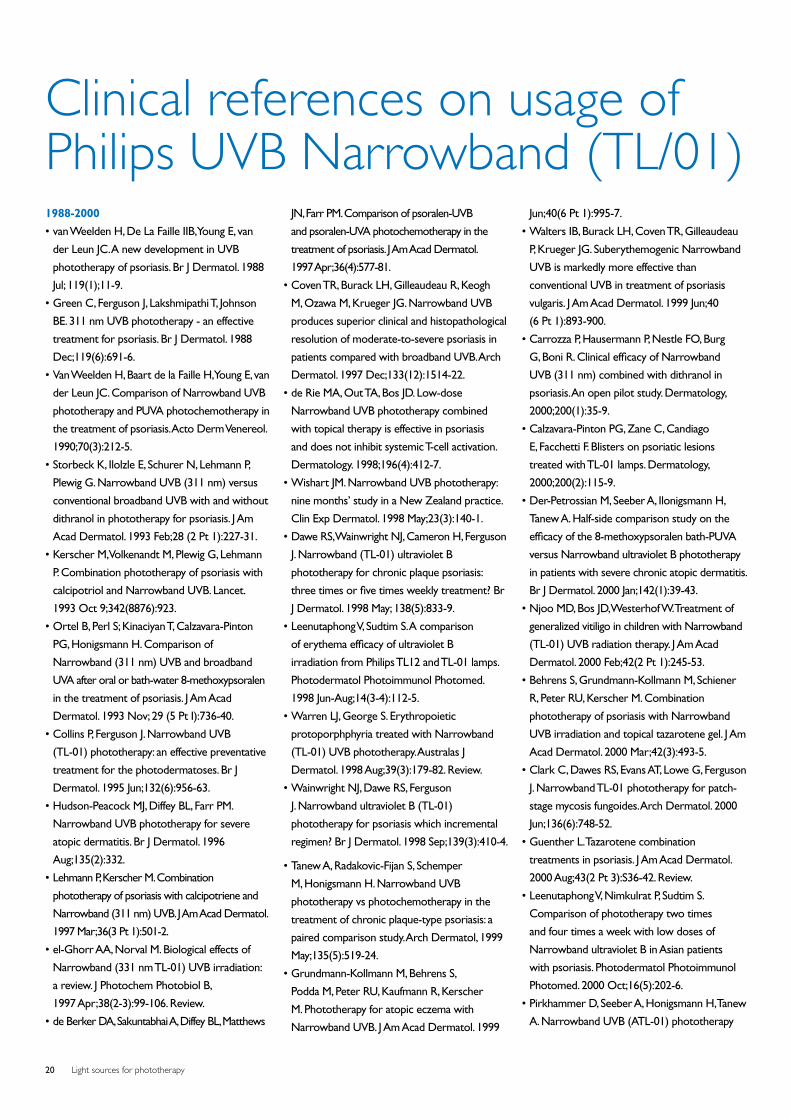
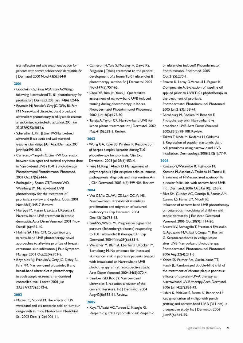
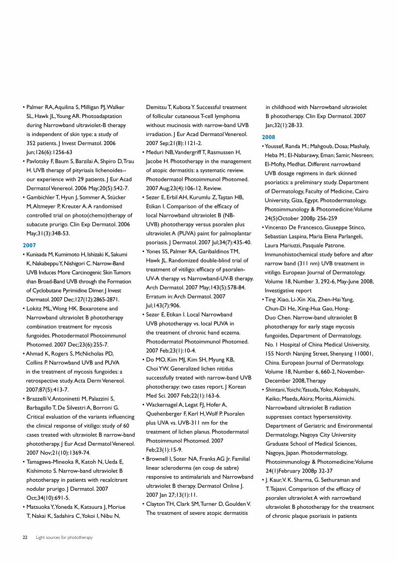
20 Clinical references on usage of Philips UVB Narrowband (TL/01)
24 Pertinent references
26 Lamps and their applications
4th. revised edition 2009
Light sources for phototherapy 3
Philips Lighting is the ideal partner
This publication provides a review of the
history and the current state of affairs
regarding photomedical applications and a
summary of the products now available.
There is always the possibility that new
products emerge from the close cooperation
between scientists and lamp manufacturers.
The more accurately the application, its action
spectrum or the dosage is defined, the better
the light source can be optimized as to its
efficacy and economy.
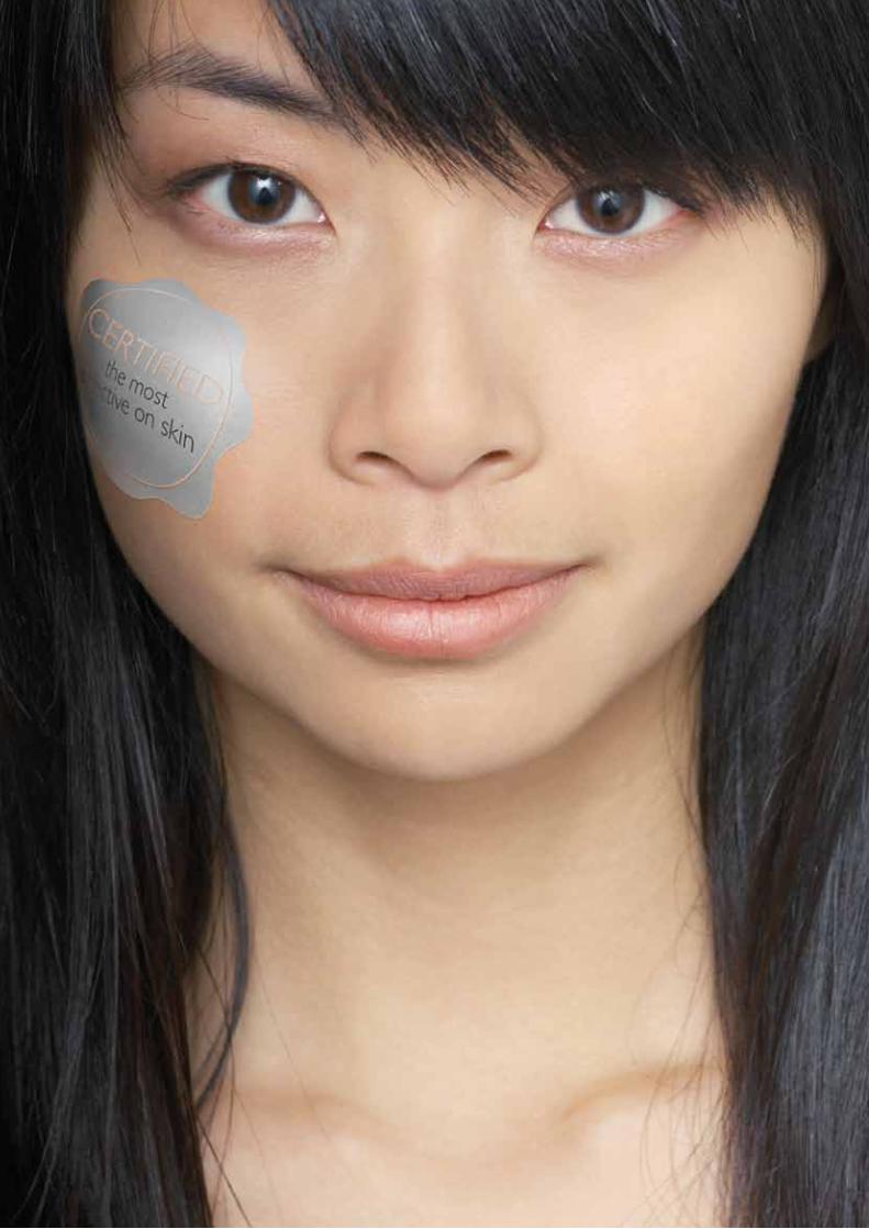
Proven to be the most effective on skinFor more then 100 years, Philips has been one of the pioneers in lighting. Today, as the world’s largest
manufacturer of lighting products, we employ a wealth of experience to satisfy lighting requirements
for a multitude of applications. For photobiological and therapeutical purposes, Philips Lighting
can provide a broad variety of lamps. Not only did we introduce the very first UVB lamps we’ve
continued to develop and improve them for the last 20 years. Our application laboratory carries
out research in close cooperation with universities and clinics throughout the world. Independent
clinical studies have proven that Philips UVB narrowband phototherapy lamps are the most effective
and safest for clinical use in the treatment for psoriasis, Vitiligo and other skin diseases*. Based on this
unmatched combination of experience and knowledge, Philips Lighting can offer the best advice to
equipment manufacturers on any possible application.
Don’t settle for poor imitations that could be harmfull to patients. Insist on Philips. The only UVB
narrowband phototherapy lamps that are certified to be the safest on skin.
*Reference available at www.philips.com/phototherapy
Light sources for phototherapy4
Far back in history, sun was considered
a source of life. Indeed, it was often
elevated to the status of a god and
men believed in the healing powers
of its rays. All over the world evidence
has been found of cults worshipping
sun-gods.
In ancient history the sun worship of the
Pharaoh Akhenaton (1350 B.C.) was very
important. He built temples dedicated to
the light god, Aton. These temples were very
unusual for the time as they had no roof,
so the sunlight could freely fill the space
inside. As an example to their co-religionists,
Akhenaton and his family took off their
clothes to benefit from the healing effects
of the rays of the sun. The priests remained
rather skeptical about this “enlightened”
religion of Akhenaton. It flourished at the
expense of their mystical and darker cults.
After the death of Akhenaton, the sun
temples were soon pulled down. However,
“sunbathing” continued to exist through the
centuries in Egypt. The historian Herodotus
(5th century B.C.) found this so remarkable
that he described it in his chronicles: “The
health-promoting properties of sunlight
have been recognized from the beginning
of civilization as a natural intuitive desire
which causes humans, when in poor health,
to be attracted by our largest optical
radiation source: the sun.” In these early
times, phototherapy (heliotherapy) was born
and guided by experience rather than any
scientific basis for the treatment of certain
ailments. The Greek doctor and “father” of
medical science Hippocrates (born in 460 B.C.
on the island of Cos) had, on his many travels
in Egypt, studied the sunlight treatment which
was practiced there. On his return to Greece,
he set up a clinic and medical school on the
island of his birth thus breaking away from
the medicine as practiced at the time by the
priests. He was practicing medicine for the
first time as a real empirical science. He wrote
books on the surgery of fractures, hygiene and
diets. In his sanatorium with its open gallery
facing south he treated patients on a scientific
basis. He is, with good reason, considered to
be the father of light therapy.
Later the Greeks and Romans continued
this light therapy, otherwise known as
heliotherapy. In the Roman baths (therms),
famous throughout history, it was also
possible to sunbathe in a solarium. The
concept of the solarium dates from this
time, indicating that use was being made of
natural sunlight. Nowadays the word solarium
refers to the use of an artificial sun, i.e.
equipment containing special lamps. With the
decline of the Roman Empire, heliotherapy
disappeared. In the Dark Ages and with
the spread of Christianity, medicine and
hygiene declined, creating a situation where
epidemics of cholera, plague and smallpox
could easily break out. Also with the rise of
Christianity, attention to the body and display
of nakedness was considered sinful. All baths
disappeared from houses and public bath
houses were closed. The Swiss Arnold Rikli
(1823-1906) reintroduced the positive effects
of sunlight forgotten for many centuries, and
used this effects as the basis of successful
natural healing methods. He practiced for
more than 50 years. He was responsible for
developing therapeutic guidelines and ideas
which are still valid today. His motto “Water
is good, air is better and light is best of all”
is at the core of heliotherapy. The Danish
doctor Niels Ryberg Finsen (1860-1904)
initiated an emphatic rebirth of light therapy
in 1898. In that year he established a sun
garden in Copenhagen (attached to the Finsen
Institute) for his patients, where they could
sunbathe completely naked. At the start, only
natural sunlight was used, but because sunlight
at this latitude (55°N) is not so plentiful, he soon
The human being and sunlight in history
Light sources for phototherapy 5
changed over to the use of artificial light sources.
Consequently he discovered that the ultraviolet
part of the sunlight spectrum had a beneficial
influence on the human body. In 1893 he
demonstrated that red light was beneficial
for healing the skin of smallpox patients. With
artificially generated ultraviolet rays he could
cure patients suffering from skin tuberculosis.
In 1903, one year before his death, he
received the Nobel Prize for Medicine.
It is clear that in the millions of years of
evolution our bodies have become adapted
and make use of the complete solar spectrum
to regulate various body functions. The
beneficial effects of ultraviolet rays were
researched and valued much more in East
European countries, such as Russia, than
in the Western medical world. It is a great
pity that in our Western society’s attention
is given almost exclusively to the negative
effects of solar radiation. These negative
acute and chronic effects only occur when
the body is excessively exposed to this
radiation. In general, no mention is made of
the great benefits of UV radiation which can
be received in full measure when it is used
in moderation. An important reason for this
is that illnesses which were previously cured
with the help of UV radiation are now treated
with drugs including antibiotics. An example of
this is skin tuberculosis (lupus vulgaris) which
was formerly treated (discovered by Finsen)
with UV radiation. This was later replaced
by drugs and so treatment with ultraviolet
radiation was quickly forgotten. With the
present rapid increase in the use of medicines
and the many objections which this has given
rise to, the prophylactic and therapeutic
effects of optical radiation deserve to receive
much more attention.
In view of the long history of the relationship
between man and the sun’s rays, in this
brochure written in a non-technical style,
we are trying to create more interest in the
positive effects of non-visual optical radiation.
Since prehistoric times the evolution of all life
on earth has taken place under the radiation
of the sun.
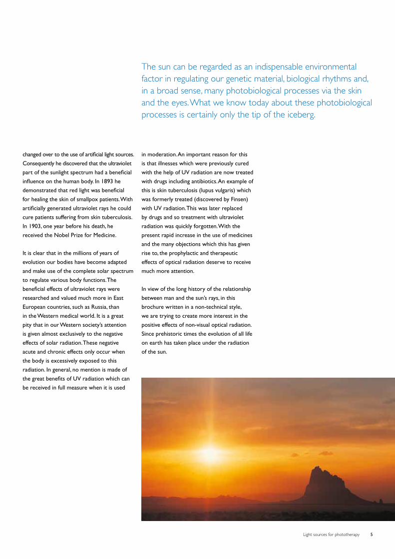
The sun can be regarded as an indispensable environmental factor in regulating our genetic material, biological rhythms and, in a broad sense, many photobiological processes via the skin and the eyes. What we know today about these photobiological processes is certainly only the tip of the iceberg.
Light sources for phototherapy6
Light in prevention, therapy and rehabilitationNowadays, prevention, therapy and rehabilitation of certain diseases and conditions with optical
radiation are the result of clinical studies guided by the progress in physics, chemistry and molecular
biology. We have a greater understanding of the science behind what our ancestors did intuitively in
previous centuries.
research and photomedicine; this reflects the
expanding potential of optical radiation for
prevention and therapy. Of course, adequate
knowledge and experience with handling
optical radiation (ultraviolet, visible light and
infrared) are essential if full advantage is to be
taken of all potential uses. The aim is always
to maximize the benefit whilst minimizing the
level of risk. The correct dosage is the most
important step. This means that phototherapy
must always be carried out under the
supervision of a doctor.
Modern photomedicine started about
100 years ago with the already mentioned
publication (1899) of Niels Finsen(1) in
which he described the treatment of lupus
vulgaris by ultraviolet radiation. At that time
Finsen’s results were attained by the rays of
a strong carbon arc. Now, after 100 years of
development, more sophisticated lamps are
available for prophylactic and therapeutic
purposes(2).
Knowledge as the fundamental basis
The past 30 years has seen an increase of
publications concerning photobiological
Light sources for phototherapy 7
Light protection
It is one of the most important functions of
the skin to build up its own light protection(3).
Melanogenesis and skin thickening can be
activated by sunlight. However there is a
possibility of sunburn, especially if there is
an individual (genetic) disposition, such as in
people with skin-types 1 to 4, (e.g. Caucasians).
In this group the time to build up photo-
protection needs to be extended with lower
UV dosages. Thus the UV dosage must be in
line with individual response and sensitivity
according to skin-type. Sunlight has the
highest intensities in the visible and near
infrared region. The reflection in these regions
is about 50% for all skintypes. This is also a
natural protection mechanism. In calculating
dosages one has to take this into account!
Psoriasis
Psoriasis is a (primarily) genetically
determined, multi-causal and chronic skin
disease with wide spread red, raised skin
lesions covered with silvery, white scales,
which occurs worldwide, but is more
prevalent in the temperate climatic zone. It
affects 2% of all light-skinned people. It is a
disease which is still incurable; moreover, no
effective chemotherapy without side effects
has yet been developed for treating such
diseases. Photochemotherapy is one way of
treating the disorder with a high success rate.
PUVA photochemotherapy
A new era in therapeutic photomedicine
was initiated at the start of the 1970’s
when, on the basis of research work
carried out in USA and Austria, Parrish et
al.(4) described the systemic treatment of
psoriasis by psoralens and irradiation with
UVA, so-called PUVA therapy. At this time
the concept of photochemotherapy was
introduced. In photochemotherapy, the
combination of a photosensitizing chemical
compound and optical radiation is used
to bring about a therapeutically beneficial
result not produced by either the radiation
or a drug alone. The drug may be applied
topically or orally to reach the skin by blood
circulation and is subsequently activated
by irradiation with UVA. In practice, PUVA
photochemotherapy is not only used for the
treatment of psoriasis but for many other
skin diseases as well (being in common use
for more than 20 indications at present). It
is applied by using a UV-sensitizing medicine
and combining it with UVA lamps (‘TL’/09
and sometimes filtered HPA). The natural,
UV-sensitizing psoralens (8 MOP, 5 MOP
or others) are available in the market
under various brand names. Recently it has
been found that chronic and high dosage
use of PUVA chemophototherapy in the
treatment of psoriasis has serious negative
side effects. Under the conditions indicated,
the application of PUVA increases the risk
of obtaining skin cancers including malignant
melanoma. As a consequence there is a shift
in preferred treatment protocols in favor of
TL/01 phototherapy(5).
UltravioletHere are the up to date aspects of
UV “light” regarding prevention and
therapy of various skin diseases.
Light sources for phototherapy8
Broadband UVB/Narrowband
UVB phototherapy
Phototherapy of psoriasis or other diseases
of the skin is a type of therapy without
any photo-sensitizing agent. It is the oldest
form of treatment, and it is based on the
experience with the favorable effects of
sunlight on the general appearance of the
skin. Numerous investigations show(6,7)
that phototherapy with UVB is just as
effective as PUVA therapy if the right doses
are maintained. Another critical parameter
is the UVB wavelength applied. Various
investigations imply that the most favorable
range for the effective UVB treatment of
psoriasis is in the long-wave part of the UVB
spectrum (between 305 and 315nm)(8,9).
This warrants a high (therapeutical)
efficiency on the one hand and minimum
(acute and chronic) risks on the other.
There are mainly two types of fluorescent
lamps of different spectral distribution - the
TL/01-UVB Narrowband and the TL/12
UVB broadband lamp - available for the
therapy of psoriasis. The erythemal effect of
the radiation from the TL/01 lamp is much
smaller than from the TL/12 lamp so that
- with the aim of being able to irradiate as
much UVB as possible without producing
erythema (reddening of the skin) - the TL/01
is a better proposition(8,9,10,11). Moreover,
recent investigations show that for successful
therapy, TL/01 radiation can be dosed far
below the erythemal threshold(12). This makes
the period of exposure shorter, reducing
overall dosages and thus any acute or
chronic side-effects. TL/01 lamps have been
tested world-wide in extensive clinical tests
and are universally in practice. Irradiation
equipment involving TL/01 lamps supply good
means of home therapy as the dosage can
be easily controlled. The therapy schedule
is drawn up by the doctor (adjustment of
the individual sensitivity of the patient to
the irradiation quality and quantity of the
equipment) who will verify its success at
regular intervals. Once the patient shows
no symptoms any more a low-interval
maintenance treatment is sometimes started
to prevent early exacerbations.
Balneo-phototherapy the positive experience
with the treatment of psoriatics at the Dead
Sea is being increasingly transferred to the
clinic. Brine baths, with a simultaneous or
subsequent exposure to UVB (using TL/01)
provide better results at a generally lower
dosage than in UVB phototherapy. This is
mainly attributed to the greater transparency
of wet skin. Balneo-phototherapy of psoriasis
is successfully applied for in-patients
in numerous spas; it is also applied for
outpatients in therapeutic centers(13).
Vitiligo
Vitiligo is a multi-causal disease, starting
with the formation of a neoantigen, which
triggers a cell mediated immune response
leading to the destruction of melanocytes(14).
The depigmented lesions of skin and hair
can be localized or generalized. It belongs
to the group of the auto-immune disorders,
e.g. Diabetes type I and thyroid disease, with
which it is often associated. The prevalence
is estimated at 0.5 to 1% worldwide,
although in India a figure as high as 8% is
reported. Skin diseases causing an altered or
impaired appearance may profoundly affect
those afflicted. Aside from causing physical
discomfort and inconvenience, it has been
demonstrated that they influence the patient’s
personal and social life, daily functioning
and psychologic status. Skin disease may
provoke negative emotions such as shame or
embarrassment, anxiety, lack of confidence
and even psychiatric diseases like depression.
The patients’ self-image may be profoundly
depressed and his self-esteem threatened.
In the western world the quality of life
(QOL) index scores in vitiligo patients are
slightly lower than those of psoriasis patients.
In the tropical region, where vitiligo was
often confused with leprosy, patients with
this disease are highly stigmatized. In India
the QOL index scores of vitiligo patients
supersede those of psoriasis patients(15).
Higher scores mean a lower quality of life.
Unlike psoriasis, the possibilities for treating
vitiligo are limited to phototherapy, except for
a small number of patients with stable vitiligo,
who can be treated with skin autologous
pigment grafts. The first report of the use
of “phototherapy” in the treatment of skin
disorders dates from about 1400 BC among
Hindus, as already mentioned. They used
“photochemotherapy”-administration of
plant extracts, followed by sun exposure-for
vitiligo(2). The same treatment was also used in
ancient Egypt. The active ingredients in these
plant extracts were isolated in 1947 by Fahmy
et al.(16, 17, 18) as 8-methoxypsoralen (8-MOP)
and 5-methoxypsoralen (5-MOP). In the same
year, these authors and also El Mofty started
to treat patients with vitiligo with 8-MOP and
sun exposure(19).
Kromayer, a German dermatologist, designed
in 1904 a water cooled mercury vapor UV
lamp(20). He was the first to treat vitiligo with
artificial UVB. In 1969 Fulton et al.(21) used
“black light” UVA tubes for the first time
in combination with topical 8-MOP in the
treatment of vitiligo. Parrish and Fitzpatrick
Light sources for phototherapy 9
introduced modern photochemotherapy with
8-MOP, having a peak sensitivity at
330 nm and UVA fluorescent tubes. They
used fluorescent tubes emitting in the 320 -
380 nm waveband in the PUVA treatment
of vitiligo(22). Although late effects, e.g. skin
carcinogenesis, have rarely been reported in
vitiligo, the frequently observed phototoxic
responses were considered a severe practical
problem. Narrowband (NB)-UVB, or 311 nm
UVB (Philips TL 01) has been used in the
treatment of vitiligo now for 10 years and was
first reported by Westerhof and Nieuweboer-
Krobotova(23). It is now considered as the
treatment of choice, because of its advantages
over PUVA treatment being: UVB 311 nm
is more effective than PUVA and safer, as
there are no psoralen-induced side effects
and can be used in children and pregnant
woman. The NB-UVB can also be achieved
with the eximer laser (308 nm)(24). A draw
back is that only small areas can be treated
at one time and the eximer laser is excluded
from home treatment. Narrowband UVB
is also recommended in combination with
pigmentcel grafting of vitiligo lesions(25).
Atopic dermatitis
Atopic dermatitis (atopic eczema, neuro-
dermatitis) is a constitutional, inflammatory,
pruritic skin disease, usually progressing
chronically. The therapy of AD includes
mainly corticosteroids (CS), antihistamines
and immunosuppressors. CS are known to
cause a variety of side effects and attempts
have been made to reduce or eliminate their
use through alternative methods such as
phototherapy (UVA/UVB, UVA(1), UVA(2),
UVB 311 nm). In the majority of patients,
UV irradiation proves favorable(26). The
active spectrum is mostly in the UV range,
between 300 and 400 nm (equipment with
TL/10 (=UVA-2), ‘TL’/09, filtered HPA). The
dosage (quality and quantity of radiation)
has to be adjusted to the individual response
of the patient and possibly (in case of a
reaction of adaptation) be altered in the
course of the therapy. 311 nm UVB therapies
has been found to be ideal for following
UVA-1 therapy: UVA-1 is used in the initial
phase of treatment to manage acute, severe
exacerbations of atopic dermatitis(5) and is
replaced by 311 nm UVB therapy, which is
an effective (and presumably safe) means of
maintenance therapy. Because its presumed
safety, it has also been advocated to be used
for children(27).
Other skin diseases
Psoriasis, which affects about 2% of
Caucasians, and vitiligo, which affects a
similar percentage of the dark and
light-skinned population, are two examples
of skin diseases which can be successfully
treated with phototheraphy. But the list
of skin diseases which can be treated with
photochemotherapy is constantly growing.
Some other dermatoses responsive to
photochemotherapy are parapsoriasis,
cutaneous T-cell lymphoma (Sézary
syndrome), mycosis fungoides, lichen planus,
pityriasis lichenoides, pityriasis rosea,
various types of eczema, polymorphous light
eruption, furunculosis, folliculitis, indolent
ulcers, prurigo and pruritis, etc. Most of
these diseases have now been treated with
Narrowband UVB, although in a varying
degree of effectiveness, which seem to be
as good as PUVA(28). It has also been stated
that exposure to ultraviolet “light” causes an
exacerbation or produces injurious effects
in the following conditions: xeroderma
pigmentosum, herpes simplex, lupus
erythematosus, several types of eczema,
prematurely senile skin, porphyria, the use
of immunosuppressive medications (after
kidney transplants) and Aids.
Vitamin D photosynthesis
By means of UV irradiation, the provitamin
D7 (7-dehydrocholesterol) is transformed
into pre-vitamin in the outer skin. In the
process of hydroxilation in the liver and the
kidneys, the bio-active form of vitamin D3 is
formed, controlling the calcium metabolism
and being hormonal in type. Above all, vitamin
D3 influences cellular information, cell
differentiation, endocrine regulatory systems,
immune reactions, macrophage functions
as well as the myocardial metabolism. It
has a practical use in preventing rachitis,
osteoporosis, osteomalacia, cancer of the
colon, prostate cancer and breast cancer (29, 30, 31, 32).
The active spectrum of UV inducing vitamin D
is limited to the UV range below 320 nm.
Dosages are much lower than those causing
sun-burn (equipment with TL/01,
HPA filtered).
Light sources for phototherapy10
Light Acne
Acne vulgaris is a chronic skin disorder
chiefly found in adolescents, caused by
inflammation (induced by propionibacterium
acnes) of the skin glands and hair follicles
mainly of the face, chest and shoulders.
All types of radiation may be applicable
and lead to improvement: UVB and UVA
radiation as well as intensive visible radiation
(light in the blue and green wavelength range
with ‘TL’/03, ‘TL’/52, TL’/17 or filtered metal
iodide lamps which are doped with indium
or gallium), depending on the type of acne
and the reaction of the individual patient(33).
Recently good results have been reported
about the combination of blue (415 nm)
and red (660 nm) light using low-intensity
fluorescent lamps(34). These phototherapeutic
approaches have no side effects and are
therefore preferred above other medical
(drug) treatments(35).
Hyperbilirubinemia (neonatal jaundice)
An example of phototherapy in the visible
region is the treatment of hyperbilirubinemia
with blue light (400-500 nm). Unconjugated
bilirubin, being a decomposition product of
haemoglobin, is not fully soluble in water and
plasma. In normal physiological circumstances,
this unconjugated bilirubin is bound to
albumin and transported to the liver where it
is converted by glucuronyltransferase into the
water-soluble conjugated form and excreted
in the bile. When the albumin binding capacity
of the plasma is exceeded (e.g. in icterus
neonatorum, in Crigler Najjar syndrome,
etc), the unconjugated bilirubin can diffuse
into the tissues. Blue light can convert this
unconjugated form into a more watersoluble
form by a photo-oxidative process and an
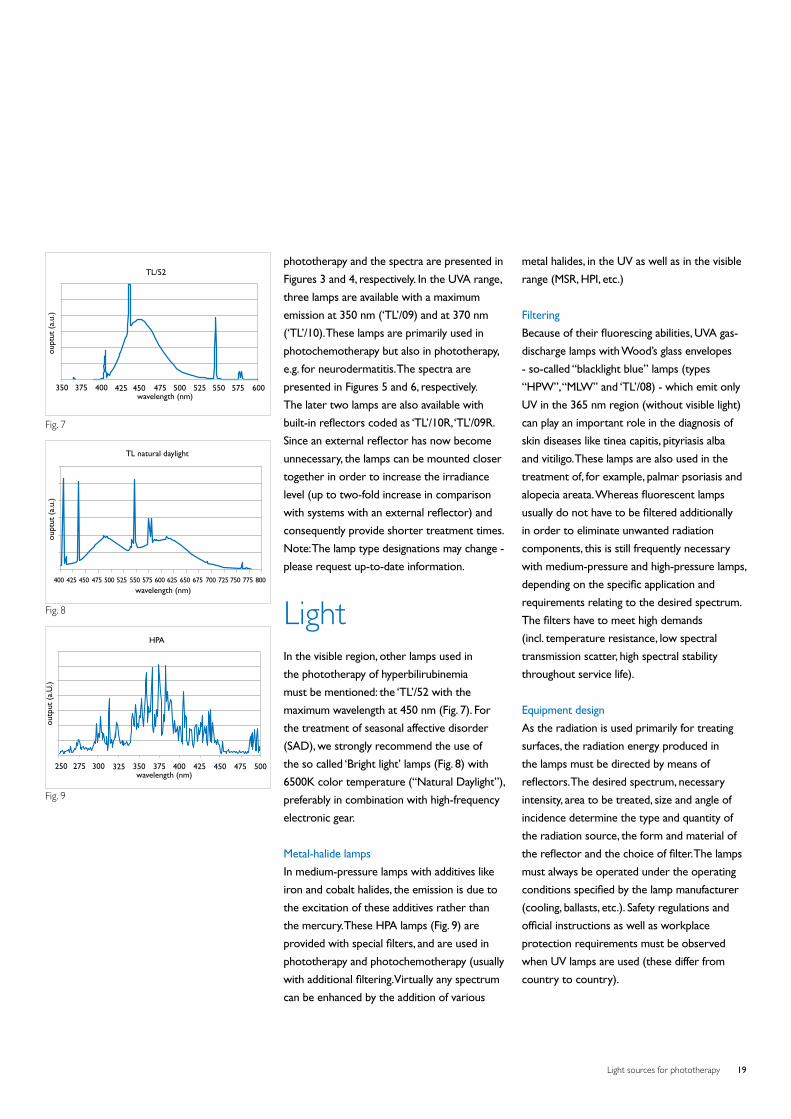
isomerization process(36). Figure 7 (page 19)
illustrates the spectrum of a blue lamp
TL/52, just emitting at the maximum
effective wavelength of 450 nm. The blue
light component in halogen dichroic mirror
lamps can also be used (UV and IR filtering
is necessary). There has also been research
showing the bilirubin content of the plasma
being lowered with the help of green light
(‘TL’/50). However, the results are still not
convincing enough to warrant a change from
blue light. The effective use of phototherapy
has eliminated the need for exchange
transfusion in almost all jaundiced infants.
Care must be taken to ensure effective
irradiance delivery, to maximize skin exposure
and to provide eye protection.
Photodynamic therapy
Photodynamic therapy (PDT) is a two-step
therapeutic technique in which the topical or
systemic delivery of photosensitizing drugs
is followed by irradiation with visible light.
Activated photosensitizers transfer energy to
molecular oxygen, generating reactive oxygen
species (ROS). The subsequent oxidation
of lipids, amino acids and proteins induces
cell necrosis and apoptosis. In addition,
ROS indirectly stimulate the transcription
and release of inflammatory mediators. The
photosensitizers are selective, in that they
penetrate and accumulate in tumor cells or
in the endothelium of newly formed vessels
while generally avoiding the surrounding
healthy tissue. The mechanisms of penetration
through the cell membrane and the pattern
of subcellular localization strongly influence
the type of cellular effect. The technique is
simple and effective: the topical application
of aminolaevulinic acid (ALA) and its methyl
ester (methyl aminolaevulinate, MAL)
followed by irradiation with broadband
The visible range of optical radiation
is now also used in prevention and
therapy of a number of diseases
and conditions.
• Phototherapy for hyperbilirubinemia
• Phototherapy for winter depression
(SAD), the jetlag syndrome and the
shift-worker syndrome
• Photodynamic therapy with red
light of skin precancer and certain
superficial types of skin cancer
• Prevention and rehabilitation
(practiced by physiotherapists)
Light sources for phototherapy 11
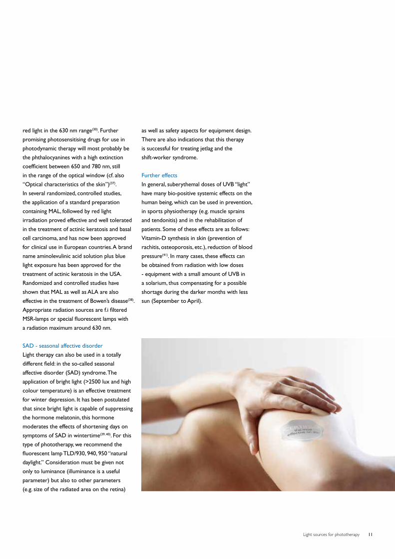
red light in the 630 nm range(30). Further
promising photosensitising drugs for use in
photodynamic therapy will most probably be
the phthalocyanines with a high extinction
coefficient between 650 and 780 nm, still
in the range of the optical window (cf. also
“Optical characteristics of the skin”)(37).
In several randomized, controlled studies,
the application of a standard preparation
containing MAL, followed by red light
irradiation proved effective and well tolerated
in the treatment of actinic keratosis and basal
cell carcinoma, and has now been approved
for clinical use in European countries. A brand
name aminolevulinic acid solution plus blue
light exposure has been approved for the
treatment of actinic keratosis in the USA.
Randomized and controlled studies have
shown that MAL as well as ALA are also
effective in the treatment of Bowen’s disease(38).
Appropriate radiation sources are f.i filtered
MSR-lamps or special fluorescent lamps with
a radiation maximum around 630 nm.
SAD - seasonal affective disorder
Light therapy can also be used in a totally
different field: in the so-called seasonal
affective disorder (SAD) syndrome. The
application of bright light (>2500 lux and high
colour temperature) is an effective treatment
for winter depression. It has been postulated
that since bright light is capable of suppressing
the hormone melatonin, this hormone
moderates the effects of shortening days on
symptoms of SAD in wintertime(39, 40). For this
type of phototherapy, we recommend the
fluorescent lamp TLD/930, 940, 950 “natural
daylight.” Consideration must be given not
only to luminance (illuminance is a useful
parameter) but also to other parameters
(e.g. size of the radiated area on the retina)
as well as safety aspects for equipment design.
There are also indications that this therapy
is successful for treating jetlag and the
shift-worker syndrome.
Further effects
In general, suberythemal doses of UVB “light”
have many bio-positive systemic effects on the
human being, which can be used in prevention,
in sports physiotherapy (e.g. muscle sprains
and tendonitis) and in the rehabilitation of
patients. Some of these effects are as follows:
Vitamin-D synthesis in skin (prevention of
rachitis, osteoporosis, etc.), reduction of blood
pressure(41). In many cases, these effects can
be obtained from radiation with low doses
- equipment with a small amount of UVB in
a solarium, thus compensating for a possible
shortage during the darker months with less
sun (September to April).
Light sources for phototherapy12
UVB broadbandEarly side effects of UVB broadband are
erythema and skin dryness. The maximum
erythema occurs 8-24 hours after irradiation.
The therapeutic effectiveness of UVB broadband
is the highest close to erythemogenic doses. This
means that erythema is likely to occur(9).
Chronic exposure to UV causes premature
aging of the skin. No evidence yet on differences
between the effect of UVA, UVB broadband
and UVB Narrowband on skin aging. In contrast
to the undisputed role of PUVA in skin tumor
induction, no significant increase in the risk of
developing squamous cell carcinoma or basal cell
carcinoma has been associated with long term
exposure to UVB even in combination with coal
tar over 25 years(9).
When used in humans, Narrowband UVB seems
not have a difference in the carcinogenic risk
compared to broadband UVB, but they both
have a clear significant lower risk compare
to PUVA therapy. A 10 year follow up study
of patient exposed to UVB broadband or
Narrowband showed no significantly increase
in the risk of skin cancer(35).
UVA (PUVA)Short-term side effects when using PUVA to
treat psoriasis include:
• Skin redness, headache, nausea,
itching, burns
• Disturbed liver functions
• The spread of psoriasis to skin that was
not affected before
• Nausea from the medication
• Squamous cell carcinoma or melanoma
Long-term side effects when using PUVA to
treat psoriasis include:
• Premature skin damage associated with
sun exposure
• Hypertrichosis
• Discolored spots on the skin
• Actinic keratosis
• Nonmelanoma skin cancer
• Cataracts (Cataracts may be avoided by
wearing goggles during treatments and UV
blocking sunglasses outdoors for the first
24 hours after treatment)
• Weakened immune system
Psoralens should not be used by:
• Children under age 12
• People who have diseases that make their
skin more sensitive to sunlight (such as lupus)
• Fertile men and women who do not
use birth control
• Pregnant women, because of possible effects
on developing foetuses
• In male patients the genitals should be
covered due to increased risk of skin cancer
Side effects
Since the late 1980’s PUVA therapy has
been largely replaced by Narrowband
UVB due to the many side effects(42,43).
Light sources for phototherapy 13
UVB NarrowbandEarly side effects of UVB Narrowband are
erythema and skin dryness. The maximum
erythema occurs 8-24 hours after irradiation.
However compared to broadband UVB
Narrowband UVB has been shown to be
effective is the sub-erythemogenic doses.
Therefore, erythema and DNA damage is
less likely to occur with UVB Narrowband
phototherapy(11). Chronic exposure to
UV causes premature aging of the skin.
No evidence yet on differences between
the effect of UVA, UVB broadband and UVB
Narrowband on skin aging.
In contrast to the undisputed role of PUVA in
skin tumor induction, no significant increase in
the risk of developing squamous cell carcinoma
or basal cell carcinoma has been associated with
long term exposure to UVB even in combination
with coal tar over 25 years(11).
When used in humans, Narrowband UVB seems
not have a difference in the carcinogenic risk
compared to broadband UVB, but they both
have a clear significant lower risk compare
to PUVA therapy. A 10 year follow up study
of patient exposed to UVB broadband or
Narrowband showed no significantly increase
in the risk of skin cancer(44).
Blue lightBlue light hazard is defined as the potential for
a photochemical induced retinal injury resulting
from radiation exposure at wavelengths
primarily between 400 nm and 500 nm(45).
For the purposes of this discussion, blue light
is defined as light within the wavelength range
of 400-480 nm, because over 88% of the
risk of photo-oxidative damage to the retina
from fluorescent lamps (cool white or ‘broad
spectrum”) is due to light wavelengths in the
range of 400-480 nm. The blue light hazard
peaks at 440 nm, and falls to 80% of peak at
460 and 415 nm. In contrast, green light of 500 nm
is only one-tenth as hazardous to the retina than
blue light with a wavelength of 440 nm(46).
The mechanisms for photochemical induced
retinal injury are caused by the absorption
of light by photoreceptors in the eye.
Under normal conditions when light hits a
photoreceptor, the cell bleaches and becomes
useless until it has recovered through a
metabolic process called the “visual cycle(47,48,49).
Absorption of blue light, however, has been
shown to cause a reversal of the process
where cells become unbleached and responsive
again to light before it is ready. This greatly
increases the potential for oxidative damage(50).
By this mechanism, some biological tissues such
as skin, the lens of the eye, and in particular the
retina may show irreversible changes induced
by prolonged exposure to moderate levels
of UV radiation and short-wavelength light.
According to some of these studies, blue light
waves may be especially toxic to those of us
who are prone to macular problems due to
genetics, nutrition, environment, health habits,
and aging(50,51,52).
Light sources for phototherapy14
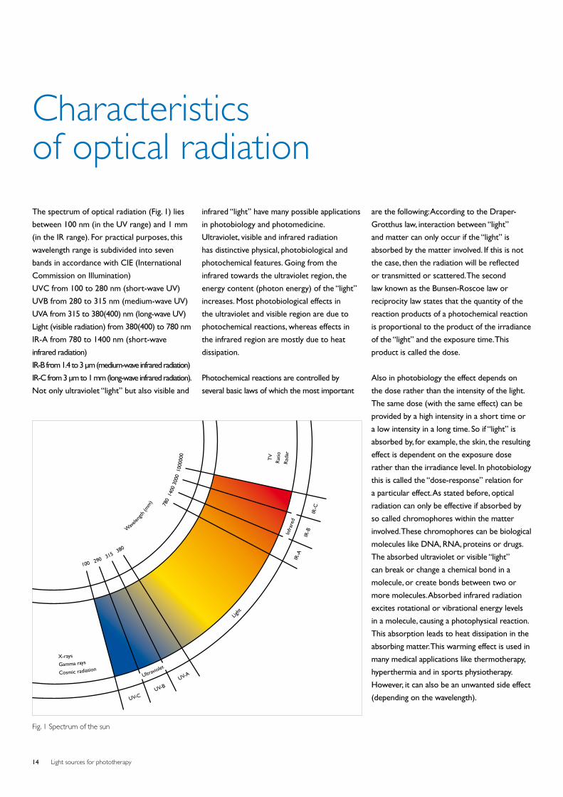
The spectrum of optical radiation (Fig. 1) lies
between 100 nm (in the UV range) and 1 mm
(in the IR range). For practical purposes, this
wavelength range is subdivided into seven
bands in accordance with CIE (International
Commission on Illumination)
UVC from 100 to 280 nm (short-wave UV)
UVB from 280 to 315 nm (medium-wave UV)
UVA from 315 to 380(400) nm (long-wave UV)
Light (visible radiation) from 380(400) to 780 nm
IR-A from 780 to 1400 nm (short-wave
infrared radiation)
IR-B from 1.4 to 3 μm (medium-wave infrared radiation)
IR-C from 3 μm to 1 mm (long-wave infrared radiation).
Not only ultraviolet “light” but also visible and
infrared “light” have many possible applications
in photobiology and photomedicine.
Ultraviolet, visible and infrared radiation
has distinctive physical, photobiological and
photochemical features. Going from the
infrared towards the ultraviolet region, the
energy content (photon energy) of the “light”
increases. Most photobiological effects in
the ultraviolet and visible region are due to
photochemical reactions, whereas effects in
the infrared region are mostly due to heat
dissipation.
Photochemical reactions are controlled by
several basic laws of which the most important
Characteristics of optical radiation
Wave
length
(mm
)
Cosmic radiation
R
adar
Gamma rays
Rat
io
X-rays
T
V
Ultraviolet
Ligh
t
Infr
ared
UV-C UV-B
UV-A
IR-A
IR
-B
IR-C
100 290 315 380
780
140
0 30
00 1
0000
00
Fig. 1 Spectrum of the sun
are the following: According to the Draper-
Grotthus law, interaction between “light”
and matter can only occur if the “light” is
absorbed by the matter involved. If this is not
the case, then the radiation will be reflected
or transmitted or scattered. The second
law known as the Bunsen-Roscoe law or
reciprocity law states that the quantity of the
reaction products of a photochemical reaction
is proportional to the product of the irradiance
of the “light” and the exposure time. This
product is called the dose.
Also in photobiology the effect depends on
the dose rather than the intensity of the light.
The same dose (with the same effect) can be
provided by a high intensity in a short time or
a low intensity in a long time. So if “light” is
absorbed by, for example, the skin, the resulting
effect is dependent on the exposure dose
rather than the irradiance level. In photobiology
this is called the “dose-response” relation for
a particular effect. As stated before, optical
radiation can only be effective if absorbed by
so called chromophores within the matter
involved. These chromophores can be biological
molecules like DNA, RNA, proteins or drugs.
The absorbed ultraviolet or visible “light”
can break or change a chemical bond in a
molecule, or create bonds between two or
more molecules. Absorbed infrared radiation
excites rotational or vibrational energy levels
in a molecule, causing a photophysical reaction.
This absorption leads to heat dissipation in the
absorbing matter. This warming effect is used in
many medical applications like thermotherapy,
hyperthermia and in sports physiotherapy.
However, it can also be an unwanted side effect
(depending on the wavelength).
Light sources for phototherapy 15
Light sources for phototherapy16
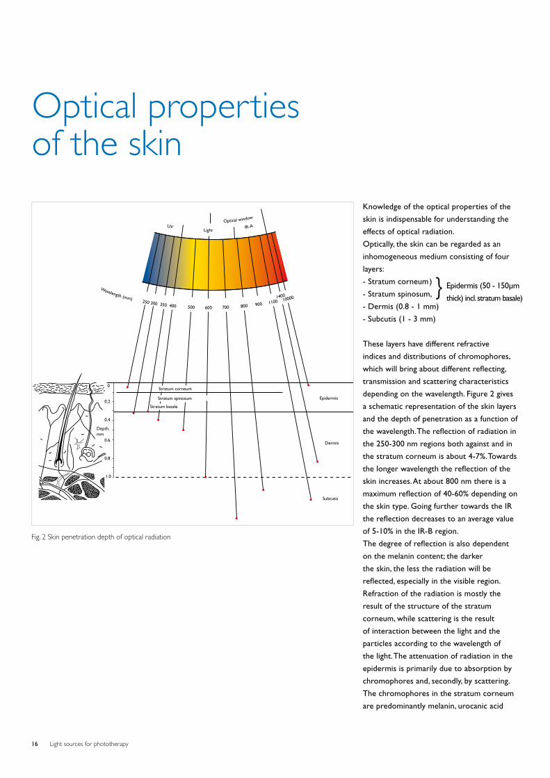
Knowledge of the optical properties of the
skin is indispensable for understanding the
effects of optical radiation.
Optically, the skin can be regarded as an
inhomogeneous medium consisting of four
layers:
- Stratum corneum )
- Stratum spinosum,
- Dermis (0.8 - 1 mm)
- Subcutis (1 - 3 mm)
These layers have different refractive
indices and distributions of chromophores,
which will bring about different reflecting,
transmission and scattering characteristics
depending on the wavelength. Figure 2 gives
a schematic representation of the skin layers
and the depth of penetration as a function of
the wavelength. The reflection of radiation in
the 250-300 nm regions both against and in
the stratum corneum is about 4-7%. Towards
the longer wavelength the reflection of the
skin increases. At about 800 nm there is a
maximum reflection of 40-60% depending on
the skin type. Going further towards the IR
the reflection decreases to an average value
of 5-10% in the IR-B region.
The degree of reflection is also dependent
on the melanin content; the darker
the skin, the less the radiation will be
reflected, especially in the visible region.
Refraction of the radiation is mostly the
result of the structure of the stratum
corneum, while scattering is the result
of interaction between the light and the
particles according to the wavelength of
the light. The attenuation of radiation in the
epidermis is primarily due to absorption by
chromophores and, secondly, by scattering.
The chromophores in the stratum corneum
are predominantly melanin, urocanic acid
Optical properties of the skin
Wavelength (mm) 250 300 350 400 500 600 700 800 900 1100 100001400
UV Light IR-A
Optical window
Epidermis
Dermis
Subcutis
Stratum corneum
Stratum spinosum
Stratum basale
0
0.2
0.4
0.6
0.8
1.0
Depth,mm
Fig. 2 Skin penetration depth of optical radiation
} Epidermis (50 - 150μm
thick) incl. stratum basale)
Light sources for phototherapy 17
and proteins consisting of aromatic amino
acids like tyrosine and tryptophan. The
stratum malpighi, (= stratum basale plus
statum spinosum), consisting of viable cells
(keratinocytes), has the same chromophores
as the stratum corneum, but here the
nucleic acids of DNA and RNA play an
important role in absorbing short-wave UV.
The absorption behavior and reaction of the
skin to UV exposure differs considerably
depending on the particular individual. Six
skin types (four light-skin, two dark-skin
types) have been defined in a commonly
used international classification based on
erythema formation and pigmentation
capability of the skin when exposed to
sunlight. The penetration of “light” into the
dermis, because of the vascularization, is also
influenced by the radiation absorbance of the
blood (haemoglobin and oxyhemoglobin) in
the 400-600 nm region and by the scattering
of light by collagen fibers. In Figure 2 it can
be seen that the greater part of UVC is
absorbed in the stratum corneum (90%)
and that 90% of the UVB is absorbed in the
epidermis but that a considerable part of the
UVA can reach the dermis which contains
blood vessels. The thin epidermis has no
blood vessels of its own, but receives what
it needs from the capillary blood vessels
immediately below the basal-cell layer of the
epidermis. Light with a wavelength between
600 and 1400 nm (red light, short-wave
infrared) can penetrate into the subcutis and
is therefore called the “optical window” of
the skin.
Light sources for phototherapy18
Artificial light sources can be divided
into the following groups:
Incandescent lamps:
- Normal lamps
- Halogen lamps
- Infrared lamps
Gas-discharge lamps:
- Low-pressure lamps
- Medium-pressure lamps
- High-pressure lamps
Although lasers, LEDs (light-emitting diodes)
and LCDs (liquid crystal displays) also
belong to the group of sources producing
optical radiation, they will not be described
in this brochure since their technology and
application are totally different from those of
“lamps”. The basic principles of lamps will be
briefly explained in the following (for detailed
technical information concerning light and
radiation sources and optimum application
thereof in equipment, please contact Philips
Lighting, addresses at the back of this brochure.
UltravioletGas-discharge lamps
A gas-discharge lamp is based on an electrical
discharge through a gas or vapor. In most cases
the discharge is sustained by the ionization of
mercury vapor or, in other cases, by inert gases
like Xe, Ar and Ne. Depending on the mercury
vapor pressure these lamps can be divided into:
- Low-pressure
- Medium-pressure
- High-pressure
The emitted spectrum of these lamps changes
from low- to high-pressure due to the
increased population of the higher energy
Artificial light sources
levels of mercury. In consequence, the emitted
energy distribution is shifted from the high
photon energy lines (185 and 254 nm) towards
the lower photon energy lines, i.e. towards the
UVA range and the visible part of the spectrum.
With the increasing mercury pressure we also
get a broadening of the emission lines due to
the influence of atoms or ions close to the
excited atom during its emission of radiation.
Low-pressure mercury lamps
(with special transmission glass and without
fluorescent layer). Tubular germicidal lamps
(“TUV”) belong to the group of the low-
pressure lamps. The germicidal “TUV” lamp
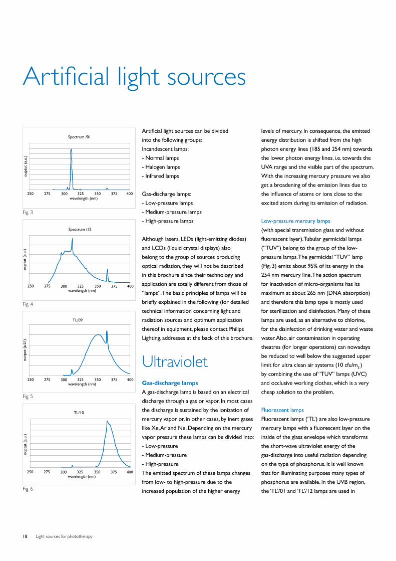
(Fig. 3) emits about 95% of its energy in the
254 nm mercury line. The action spectrum
for inactivation of micro-organisms has its
maximum at about 265 nm (DNA absorption)
and therefore this lamp type is mostly used
for sterilization and disinfection. Many of these
lamps are used, as an alternative to chlorine,
for the disinfection of drinking water and waste
water. Also, air contamination in operating
theatres (for longer operations) can nowadays
be reduced to well below the suggested upper
limit for ultra clean air systems (10 cfu/m3 )
by combining the use of “TUV” lamps (UVC)
and occlusive working clothes, which is a very
cheap solution to the problem.
Fluorescent lamps
Fluorescent lamps (‘TL’) are also low-pressure
mercury lamps with a fluorescent layer on the
inside of the glass envelope which transforms
the short-wave ultraviolet energy of the
gas-discharge into useful radiation depending
on the type of phosphorus. It is well known
that for illuminating purposes many types of
phosphorus are available. In the UVB region,
the ‘TL’/01 and ‘TL’/12 lamps are used in
Spectrum /01
wavelength (nm)250 275 300 325 350 375 400
oupt
ut (
a.u.
)
wavelength (nm)250 275 300 325 350 375 400
Spectrum /12
oupt
ut (
a.u.
)
wavelength (nm)250
outp
ut (
a.U
.)
275 300 325 350 375 400
TL/09
wavelength (nm)250 275 300 325 350 375 400
TL/10
oupt
ut (
a.u.
)
Fig. 3
Fig. 4
Fig. 6
Fig. 5
Light sources for phototherapy 19
phototherapy and the spectra are presented in
Figures 3 and 4, respectively. In the UVA range,
three lamps are available with a maximum
emission at 350 nm (‘TL’/09) and at 370 nm
(‘TL’/10). These lamps are primarily used in
photochemotherapy but also in phototherapy,
e.g. for neurodermatitis. The spectra are
presented in Figures 5 and 6, respectively.
The later two lamps are also available with
built-in reflectors coded as ‘TL’/10R, ‘TL’/09R.
Since an external reflector has now become
unnecessary, the lamps can be mounted closer
together in order to increase the irradiance
level (up to two-fold increase in comparison
with systems with an external reflector) and
consequently provide shorter treatment times.
Note: The lamp type designations may change -
please request up-to-date information.
LightIn the visible region, other lamps used in
the phototherapy of hyperbilirubinemia
must be mentioned: the ‘TL’/52 with the
maximum wavelength at 450 nm (Fig. 7). For
the treatment of seasonal affective disorder
(SAD), we strongly recommend the use of
the so called ‘Bright light’ lamps (Fig. 8) with
6500K color temperature (“Natural Daylight”),
preferably in combination with high-frequency
electronic gear.
Metal-halide lamps
In medium-pressure lamps with additives like
iron and cobalt halides, the emission is due to
the excitation of these additives rather than
the mercury. These HPA lamps (Fig. 9) are
provided with special filters, and are used in
phototherapy and photochemotherapy (usually
with additional filtering. Virtually any spectrum
can be enhanced by the addition of various
metal halides, in the UV as well as in the visible
range (MSR, HPI, etc.)
Filtering
Because of their fluorescing abilities, UVA gas-
discharge lamps with Wood’s glass envelopes
- so-called “blacklight blue” lamps (types
“HPW”, “MLW” and ‘TL’/08) - which emit only
UV in the 365 nm region (without visible light)
can play an important role in the diagnosis of
skin diseases like tinea capitis, pityriasis alba
and vitiligo. These lamps are also used in the
treatment of, for example, palmar psoriasis and
alopecia areata. Whereas fluorescent lamps
usually do not have to be filtered additionally
in order to eliminate unwanted radiation
components, this is still frequently necessary
with medium-pressure and high-pressure lamps,
depending on the specific application and
requirements relating to the desired spectrum.
The filters have to meet high demands
(incl. temperature resistance, low spectral
transmission scatter, high spectral stability
throughout service life).
Equipment design
As the radiation is used primarily for treating
surfaces, the radiation energy produced in
the lamps must be directed by means of
reflectors. The desired spectrum, necessary
intensity, area to be treated, size and angle of
incidence determine the type and quantity of
the radiation source, the form and material of
the reflector and the choice of filter. The lamps
must always be operated under the operating
conditions specified by the lamp manufacturer
(cooling, ballasts, etc.). Safety regulations and
official instructions as well as workplace
protection requirements must be observed
when UV lamps are used (these differ from
country to country).
wavelength (nm)350 375 400 425 450 475 500 525 550 575 600
TL/52
oupt
ut (
a.u.
)
Fig. 7
wavelength (nm)400 425 450 475 500 525 550 575 600 625 650 675 700 725 750 775 800
TL natural daylight
oupt
ut (
a.u.
)
Fig. 8
outp
ut (
a.U
.)
wavelength (nm)250 275 300 325 350 375 400 425 450 475 500
HPA
Fig. 9
Light sources for phototherapy20
1988-2000
• van Weelden H, De La Faille IIB, Young E, van
der Leun JC. A new development in UVB
phototherapy of psoriasis. Br J Dermatol. 1988
Jul; 119(1);11-9.
• Green C, Ferguson J, Lakshmipathi T, Johnson
BE. 311 nm UVB phototherapy - an effective
treatment for psoriasis. Br J Dermatol. 1988
Dec;119(6):691-6.
• Van Weelden H, Baart de la Faille H, Young E, van
der Leun JC. Comparison of Narrowband UVB
phototherapy and PUVA photochemotherapy in
the treatment of psoriasis. Acto Derm Venereol.
1990;70(3):212-5.
• Storbeck K, Ilolzle E, Schurer N, Lehmann P,
Plewig G. Narrowband UVB (311 nm) versus
conventional broadband UVB with and without
dithranol in phototherapy for psoriasis. J Am
Acad Dermatol. 1993 Feb;28 (2 Pt 1):227-31.
• Kerscher M, Volkenandt M, Plewig G, Lehmann
P. Combination phototherapy of psoriasis with
calcipotriol and Narrowband UVB. Lancet.
1993 Oct 9;342(8876):923.
• Ortel B, Perl S; Kinaciyan T, Calzavara-Pinton
PG, Honigsmann H. Comparison of
Narrowband (311 nm) UVB and broadband
UVA after oral or bath-water 8-methoxypsoralen
in the treatment of psoriasis. J Am Acad
Dermatol. 1993 Nov; 29 (5 Pt I):736-40.
• Collins P, Ferguson J. Narrowband UVB
(TL-01) phototherapy: an effective preventative
treatment for the photodermatoses. Br J
Dermatol. 1995 Jun;132(6):956-63.
• Hudson-Peacock MJ, Diffey BL, Farr PM.
Narrowband UVB phototherapy for severe
atopic dermatitis. Br J Dermatol. 1996
Aug;135(2):332.
• Lehmann P, Kerscher M. Combination
phototherapy of psoriasis with calcipotriene and
Narrowband (311 nm) UVB. J Am Acad Dermatol.
1997 Mar;36(3 Pt 1):501-2.
• el-Ghorr AA, Norval M. Biological effects of
Narrowband (331 nm TL-01) UVB irradiation:
a review. J Photochem Photobiol B,
1997 Apr;38(2-3):99-106. Review.
• de Berker DA, Sakuntabhai A, Diffey BL, Matthews
JN, Farr PM. Comparison of psoralen-UVB
and psoralen-UVA photochemotherapy in the
treatment of psoriasis. J Am Acad Dermatol.
1997 Apr;36(4):577-81.
• Coven TR, Burack LH, Gilleaudeau R, Keogh
M, Ozawa M, Krueger JG. Narrowband UVB
produces superior clinical and histopathological
resolution of moderate-to-severe psoriasis in
patients compared with broadband UVB. Arch
Dermatol. 1997 Dec;133(12):1514-22.
• de Rie MA, Out TA, Bos JD. Low-dose
Narrowband UVB phototherapy combined
with topical therapy is effective in psoriasis
and does not inhibit systemic T-cell activation.
Dermatology. 1998;196(4):412-7.
• Wishart JM. Narrowband UVB phototherapy:
nine months’ study in a New Zealand practice.
Clin Exp Dermatol. 1998 May;23(3):140-1.
• Dawe RS, Wainwright NJ, Cameron H, Ferguson
J. Narrowband (TL-01) ultraviolet B
phototherapy for chronic plaque psoriasis:
three times or five times weekly treatment? Br
J Dermatol. 1998 May; 138(5):833-9.
• Leenutaphong V, Sudtim S. A comparison
of erythema efficacy of ultraviolet B
irradiation from Philips TL12 and TL-01 lamps.
Photodermatol Photoimmunol Photomed.
1998 Jun-Aug;14(3-4):112-5.
• Warren LJ, George S. Erythropoietic
protoporphphyria treated with Narrowband
(TL-01) UVB phototherapy. Australas J
Dermatol. 1998 Aug;39(3):179-82. Review.
• Wainwright NJ, Dawe RS, Ferguson
J. Narrowband ultraviolet B (TL-01)
phototherapy for psoriasis which incremental
regimen? Br J Dermatol. 1998 Sep;139(3):410-4.
• Tanew A, Radakovic-Fijan S, Schemper
M, Honigsmann H. Narrowband UVB
phototherapy vs photochemotherapy in the
treatment of chronic plaque-type psoriasis: a
paired comparison study. Arch Dermatol, 1999
May;135(5):519-24.
• Grundmann-Kollmann M, Behrens S,
Podda M, Peter RU, Kaufmann R, Kerscher
M. Phototherapy for atopic eczema with
Narrowband UVB. J Am Acad Dermatol. 1999
Jun;40(6 Pt 1):995-7.
• Walters IB, Burack LH, Coven TR, Gilleaudeau
P, Krueger JG. Suberythemogenic Narrowband
UVB is markedly more effective than
conventional UVB in treatment of psoriasis
vulgaris. J Am Acad Dermatol. 1999 Jun;40
(6 Pt 1):893-900.
• Carrozza P, Hausermann P, Nestle FO, Burg
G, Boni R. Clinical efficacy of Narrowband
UVB (311 nm) combined with dithranol in
psoriasis. An open pilot study. Dermatology,
2000;200(1):35-9.
• Calzavara-Pinton PG, Zane C, Candiago
E, Facchetti F. Blisters on psoriatic lesions
treated with TL-01 lamps. Dermatology,
2000;200(2):115-9.
• Der-Petrossian M, Seeber A, Ilonigsmann H,
Tanew A. Half-side comparison study on the
efficacy of the 8-methoxypsoralen bath-PUVA
versus Narrowband ultraviolet B phototherapy
in patients with severe chronic atopic dermatitis.
Br J Dermatol. 2000 Jan;142(1):39-43.
• Njoo MD, Bos JD, Westerhof W. Treatment of
generalized vitiligo in children with Narrowband
(TL-01) UVB radiation therapy. J Am Acad
Dermatol. 2000 Feb;42(2 Pt 1):245-53.
• Behrens S, Grundmann-Kollmann M, Schiener
R, Peter RU, Kerscher M. Combination
phototherapy of psoriasis with Narrowband
UVB irradiation and topical tazarotene gel. J Am
Acad Dermatol. 2000 Mar;42(3):493-5.
• Clark C, Dawes RS, Evans AT, Lowe G, Ferguson
J. Narrowband TL-01 phototherapy for patch-
stage mycosis fungoides. Arch Dermatol. 2000
Jun;136(6):748-52.
• Guenther L. Tazarotene combination
treatments in psoriasis. J Am Acad Dermatol.
2000 Aug;43(2 Pt 3):S36-42. Review.
• Leenutaphong V, Nimkulrat P, Sudtim S.
Comparison of phototherapy two times
and four times a week with low doses of
Narrowband ultraviolet B in Asian patients
with psoriasis. Photodermatol Photoimmunol
Photomed. 2000 Oct;16(5):202-6.
• Pirkhammer D, Seeber A, Honigsmann H, Tanew
A. Narrowband UVB (ATL-01) phototherapy
Clinical references on usage of Philips UVB Narrowband (TL/01)
Light sources for phototherapy 21
is an effective and safe treatment option for
patients with severe seborrhoeic dermatitis. Br
J Dermatol. 2000 Nov;143(5):964-8.
2001
• Goodwin RG, Finlay AY, Anstey AV. Vitiligo
following Narrowband TL-01 phototherapy for
psoriasis. Br J Dermatol. 2001 Jun;144(6):1264-6.
• Reynolds NJ, Franklin V, Gray JC, Diffey BL, Farr
PM. Narrowband ultraviolet B and broadband
ultraviolet A phototherapy in aduly atopic eczema:
a randomized controlled trial. Lancet. 2001 Jun
23;357(9273):2012-6.
• Scherschun L, Kim JJ, Lim HW. Narrowband
ultraviolet B is a useful and well tolerated
treatment for vitiligo. J Am Acad Dermatol. 2001
Jun;44(6):999-1003.
• Carretero-Mangolis C, Lim HW. Correlation
between skin types and minimal erythema dose
in Narrowband UVB (TL-01) phototherapy.
Photodermatol Photoimmunol Photomed.
2001 Oct;17(5):244-6.
• Barbagallo J, Spann CT, Tutrone WD,
Weinberg JM. Narrowband UVB
phototherapy for the treatment of
psoriasis: a review and update. Cutis. 2001
Nov;68(5):345-7. Review.
• Hjerppe M, Hasan T, Saksala I, Reunala T.
Narrow-band UVB treatment in atopic
dermatitis. Acta Derm Venereol. 2001 Nov-
Dec;81(6):439-40.
• Holme SA, Mills CM. Crotamiton and
narrow-band UVB phototherapy: novel
approaches to alleviate pruritus of breast
carcinoma skin infiltration. J Pain Symptom
Manage. 2001 Oct;22(4):803-5.
• Reynolds NJ, Franklin V, Gray JC, Diffey BL,
Farr PM. Narrow-band ultraviolet B and
broad-band ultraviolet A phototherapy
in adult atopic eczema: a randomised
controlled trial. Lancet. 2001 Jun
23;357(9273):2012-6.
2002
• Macve JC, Norval M. The effects of UV
waveband and cis-urocanic acid on tumour
outgrowth in mice. Photochem Photobiol
Sci. 2002 Dec;1(12):1006-11.
• Cameron H, Yule S, Moseley H, Dawe RS,
Ferguson J. Taking treatment to the patient:
development of a home TL-01 ultraviolet B
phototherapy service. Br J Dermatol. 2002
Nov;147(5):957-65.
• Choe YB, Rim JH, Youn JI. Quantitative
assessment of narrow-band UVB induced
tanning during phototherapy in Korea.
Photodermatol Photoimmunol Photomed.
2002 Jun;18(3):127-30.
• Taneja A, Taylor CR. Narrow-band UVB for
lichen planus treatment. Int J Dermatol. 2002
May;41(5):282-3. Review.
2003
• Wong GA, Kaye SB, Parslew R. Reactivation
of herpes simplex keratitis during TL01
phototherapy for psoriasis. Clin Exp
Dermatol. 2003 Jul;28(4):453-4.
• Fesq H, Ring J, Abeck D. Management of
polymorphous light eruption : clinical course,
pathogenesis, diagnosis and intervention. Am
J Clin Dermatol. 2003;4(6):399-406. Review.
2004
• Wu CS, Yu CL, Wu CS, Lan CC, Yu HS.
Narrow-band ultraviolet-B stimulates
proliferation and migration of cultured
melanocytes. Exp Dermatol. 2004
Dec;13(12):755-63.
• Gudi VS, White MI. Progressive pigmented
purpura (Schamberg’s disease) responding
to TL01 ultraviolet B therapy. Clin Exp
Dermatol. 2004 Nov;29(6):683-4.
• Weischer M, Blum A, Eberhard F, Röcken M,
Berneburg M. No evidence for increased
skin cancer risk in psoriasis patients treated
with broadband or Narrowband UVB
phototherapy: a first retrospective study.
Acta Derm Venereol. 2004;84(5):370-4.
• Bandow GD, Koo JY. Narrow-band
ultraviolet B radiation: a review of the
current literature. Int J Dermatol. 2004
Aug;43(8):555-61. Review.
2005
• Kaya TI, Yazici AC, Tursen U, Ikizoglu G.
Idiopathic guttate hypomelanosis: idiopathic
or ultraviolet induced? Photodermatol
Photoimmunol Photomed. 2005
Oct;21(5):270-1.
• Penven K, Leroy D, Verneuil L, Faguer K,
Dompmartin A. Evaluation of vaseline oil
applied prior to UVB TL01 phototherapy in
the treatment of psoriasis.
Photodermatol Photoimmunol Photomed.
2005 Jun;21(3):138-41.
• Berneburg M, Röcken M, Benedix F.
Phototherapy with Narrowband vs
broadband UVB. Acta Derm Venereol.
2005;85(2):98-108. Review.
• Takata T, Ikeda M, Kodama H, Ohkuma
S. Regression of papular elastolytic giant
cell granuloma using narrow-band UVB
irradiation. Dermatology. 2006;212(1):77-9.
2006
• Kuwano Y, Watanabe R, Fujimoto M,
Komine M, Asahina A, Tsukada N, Tamaki K.
Treatment of HIV-associated eosinophilic
pustular folliculitis with narrow-band UVB.
Int J Dermatol. 2006 Oct;45(10):1265-7.
• Silva SH, Guedes AC, Gontijo B, Ramos AM,
Carmo LS, Farias LM, Nicoli JR.
Influence of narrow-band UVB phototherapy
on cutaneous microbiota of children with
atopic dermatitis. J Eur Acad Dermatol
Venereol. 2006 Oct;20(9):1114-20.
• Brazzelli V, Barbagallo T, Prestinari F, Vassallo
C, Agozzino M, Vailati F, Cespa M, Borroni
G. Keratoacanthoma in vitiligo lesion
after UVB Narrowband phototherapy.
Photodermatol Photoimmunol Photomed.
2006 Aug;22(4):211-3.
• Yones SS, Palmer RA, Garibaldinos TT,
Hawk JL. Randomized double-blind trial of
the treatment of chronic plaque psoriasis:
efficacy of psoralen-UV-A therapy vs
Narrowband UV-B therapy. Arch Dermatol.
2006 Jul;142(7):836-42.
• Lahiri K, Malakar S, Sarma N, Banerjee U.
Repigmentation of vitiligo with punch
grafting and narrow-band UV-B (311 nm)--a
prospective study. Int J Dermatol. 2006
Jun;45(6):649-55.
Light sources for phototherapy22
• Palmer RA, Aquilina S, Milligan PJ, Walker
SL, Hawk JL, Young AR. Photoadaptation
during Narrowband ultraviolet-B therapy
is independent of skin type: a study of
352 patients. J Invest Dermatol. 2006
Jun;126(6):1256-63
• Pavlotsky F, Baum S, Barzilai A, Shpiro D, Trau
H. UVB therapy of pityriasis lichenoides--
our experience with 29 patients. J Eur Acad
Dermatol Venereol. 2006 May;20(5):542-7.
• Gambichler T, Hyun J, Sommer A, Stücker
M, Altmeyer P, Kreuter A. A randomised
controlled trial on photo(chemo)therapy of
subacute prurigo. Clin Exp Dermatol. 2006
May;31(3):348-53.
2007
• Kunisada M, Kumimoto H, Ishizaki K, Sakumi
K, Nakabeppu Y, Nishigori C. Narrow-Band
UVB Induces More Carcinogenic Skin Tumors
than Broad-Band UVB through the Formation
of Cyclobutane Pyrimidine Dimer. J Invest
Dermatol. 2007 Dec;127(12):2865-2871.
• Lokitz ML, Wong HK. Bexarotene and
Narrowband ultraviolet B phototherapy
combination treatment for mycosis
fungoides. Photodermatol Photoimmunol
Photomed. 2007 Dec;23(6):255-7.
• Ahmad K, Rogers S, McNicholas PD,
Collins P. Narrowband UVB and PUVA
in the treatment of mycosis fungoides: a
retrospective study. Acta Derm Venereol.
2007;87(5):413-7.
• Brazzelli V, Antoninetti M, Palazzini S,
Barbagallo T, De Silvestri A, Borroni G.
Critical evaluation of the variants influencing
the clinical response of vitiligo: study of 60
cases treated with ultraviolet B narrow-band
phototherapy. J Eur Acad Dermatol Venereol.
2007 Nov;21(10):1369-74.
• Tamagawa-Mineoka R, Katoh N, Ueda E,
Kishimoto S. Narrow-band ultraviolet B
phototherapy in patients with recalcitrant
nodular prurigo. J Dermatol. 2007
Oct;34(10):691-5.
• Matsuoka Y, Yoneda K, Katsuura J, Moriue
T, Nakai K, Sadahira C, Yokoi I, Nibu N,
Demitsu T, Kubota Y. Successful treatment
of follicular cutaneous T-cell lymphoma
without mucinosis with narrow-band UVB
irradiation. J Eur Acad Dermatol Venereol.
2007 Sep;21(8):1121-2.
• Meduri NB, Vandergriff T, Rasmussen H,
Jacobe H. Phototherapy in the management
of atopic dermatitis: a systematic review.
Photodermatol Photoimmunol Photomed.
2007 Aug;23(4):106-12. Review.
• Sezer E, Erbil AH, Kurumlu Z, Taştan HB,
Etikan I. Comparison of the efficacy of
local Narrowband ultraviolet B (NB-
UVB) phototherapy versus psoralen plus
ultraviolet A (PUVA) paint for palmoplantar
psoriasis. J Dermatol. 2007 Jul;34(7):435-40.
• Yones SS, Palmer RA, Garibaldinos TM,
Hawk JL. Randomized double-blind trial of
treatment of vitiligo: efficacy of psoralen-
UV-A therapy vs Narrowband-UV-B therapy.
Arch Dermatol. 2007 May;143(5):578-84.
Erratum in: Arch Dermatol. 2007
Jul;143(7):906.
• Sezer E, Etikan I. Local Narrowband
UVB phototherapy vs. local PUVA in
the treatment of chronic hand eczema.
Photodermatol Photoimmunol Photomed.
2007 Feb;23(1):10-4.
• Do MO, Kim MJ, Kim SH, Myung KB,
Choi YW. Generalized lichen nitidus
successfully treated with narrow-band UVB
phototherapy: two cases report. J Korean
Med Sci. 2007 Feb;22(1):163-6.
• Wackernagel A, Legat FJ, Hofer A,
Quehenberger F, Kerl H, Wolf P. Psoralen
plus UVA vs. UVB-311 nm for the
treatment of lichen planus. Photodermatol
Photoimmunol Photomed. 2007
Feb;23(1):15-9.
• Brownell I, Soter NA, Franks AG Jr. Familial
linear scleroderma (en coup de sabre)
responsive to antimalarials and Narrowband
ultraviolet B therapy. Dermatol Online J.
2007 Jan 27;13(1):11.
• Clayton TH, Clark SM, Turner D, Goulden V.
The treatment of severe atopic dermatitis
in childhood with Narrowband ultraviolet
B phototherapy. Clin Exp Dermatol. 2007
Jan;32(1):28-33.
2008
• Youssef, Randa M.; Mahgoub, Doaa; Mashaly,
Heba M.; El-Nabarawy, Eman; Samir, Nesreen;
El-Mofty, Medhat. Different narrowband
UVB dosage regimens in dark skinned
psoriatics: a preliminary study. Department
of Dermatology, Faculty of Medicine, Cairo
University, Giza, Egypt. Photodermatology,
Photoimmunology & Photomedicine:Volume
24(5)October 2008p 256-259
• Vincenzo De Francesco, Giuseppe Stinco,
Sebastian Laspina, Maria Elena Parlangeli,
Laura Mariuzzi, Pasquale Patrone.
Immunohistochemical study before and after
narrow band (311 nm) UVB treatment in
vitiligo. European Journal of Dermatology.
Volume 18, Number 3, 292-6, May-June 2008,
Investigative report
• Ting Xiao, Li-Xin Xia, Zhen-Hai Yang,
Chun-Di He, Xing-Hua Gao, Hong-
Duo Chen. Narrow-band ultraviolet B
phototherapy for early stage mycosis
fungoides, Department of Dermatology,
No. 1 Hospital of China Medical University,
155 North Nanjing Street, Shenyang 110001,
China. European Journal of Dermatology.
Volume 18, Number 6, 660-2, November-
December 2008, Therapy
• Shintani, Yoichi; Yasuda, Yoko; Kobayashi,
Keiko; Maeda, Akira; Morita, Akimichi.
Narrowband ultraviolet B radiation
suppresses contact hypersensitivity.
Department of Geriatric and Environmental
Dermatology, Nagoya City University
Graduate School of Medical Sciences,
Nagoya, Japan. Photodermatology,
Photoimmunology & Photomedicine:Volume
24(1)February 2008p 32-37
• J. Kaur, V. K. Sharma, G. Sethuraman and
T. Tejasvi. Comparison of the efficacy of
psoralen ultraviolet A with narrowband
ultraviolet B phototherapy for the treatment
of chronic plaque psoriasis in patients
Light sources for phototherapy 23
with skin types IV and V. Department
of Dermatology and Venereology, All
India Institute of Medical Sciences, New
Delhi-110029, India. British Association of
Dermatologists, Volume 33 Issue 4, Pages
513 – 515, May 2008
• Namita Rath, HK Kar, Sunil Sabhnani. An
open labeled, comparative clinical study on
efficacy and tolerability of oral minipulse of
steroid (OMP) alone, OMP with PUVA and
broad / narrow band UVB phototherapy
in progressive vitiligo. Department of
Dermatology and STD, Dr. RML Hospital,
New Delhi, India. Indian Journal of
dermatology, Venereology and Leprology.
2008, Volume: 74, Issue: 4, Page: 357-360
• DeSilva, Bernadette; McKenzie, Roddie
C.; Hunter, John A. A.; Norval, Mary Local
effects of TL01 phototherapy in psoriasis.
Department of Dermatology, Department of
Biomedical Sciences, University of Edinburgh,
Edinburgh, UK. Photodermatology,
Photoimmunology & Photomedicine:Volume
24(5)October 2008p 268-269
• Kuhl, John T.; Davis, Mark D. P.; McEvoy,
Marian. Narrowband ultraviolet-B
phototherapy for hand and foot dermatoses.
Photodermatology, Photoimmunology &
Photomedicine. 24(3):152-153, June 2008.
• Pavlotsky, Felix; Nathansohn, Nir; Kriger,
Grigory; Shpiro, Dorit; Trau, Henri
Ultraviolet-B treatment for cutaneous
lichen planus: our experience with 50
patients. Department of Dermatology,
Phototherapy and Day Care Center, Chaim
Sheba Medical Center, Tel Hashomer, Israel,
and 2Sackler School of Medicine, Tel Aviv
University, Tel Aviv, Israel. Photodermatology,
Photoimmunology & Photomedicine:Volume
24(2)April 2008p 83-86
• Jain, Vijay Kumar M.D.; Bansal, Anu M.D.;
Aggarwal, Kamal M.D.; Jain, Kapil D.V.D.
Enhanced Response of Childhood Psoriasis
to Narrow-Band UV-B Phototherapy
with Preirradiation Use of Mineral Oil.
Department of Dermatology, Venereology
and Leprology, Pt. B.D. Sharma Post
Graduate Institute of Medical Sciences,
Rohtak. Pediatric Dermatology:Volume 25(5)
September/October 2008p 559-564
• Engin B, Ozdemir M, Balevi A, Mevlitoğlu
I. Treatment of chronic urticaria with
narrowband ultraviolet B phototherapy: a
randomized controlled trial. Department
of Dermatology, Meram Medical Faculty,
Selcuk University, Konya, Turkey. Acta Derm
Venereol. 2008; 88(3):247-51
• Ohtsuka T. Narrow band UVB phototherapy
for early stage mycosis fungoides. Eur J
Dermatol. 2008 Jul-Aug; 18(4):464-6. Epub
2008 Jun 23.
2009
• Wolf, P; Hofer, A; Legat, F. J; Bretterklieber, A;
Weger, W; Salmhofer, W; Kerl, H. Treatment
with 311-nm ultraviolet B accelerates and
improves the clearance of psoriatic lesions
in patients treated with etanercept.Research
Unit for Photodermatology and Department
of Dermatology, Medical University of Graz,
A-8036 Graz, Austria. British Journal of
Dermatology:Volume 160(1)January 2009p
186-189
• Olivier Dereure, Eric Picot, Christelle
Comte, Didier Bessis, Bernard Guillot.
Treatment of Early Stages of Mycosis
Fungoides with Narrowband Ultraviolet
B A Clinical, Histological and Molecular
Evaluation of Results. University of
Montpellier I, University Hospital of
Montpellier, Department of Dermatology,
Hôpital Saint Eloi, Montpellier, France.
Dermatology 2009;218:1-6
(DOI: 10.1159/000161114
Light sources for phototherapy24
1. N.R. Finsen, “Über die Bedeutung der
chemischen Strahlen des Lichtes fur
Medizin und Biologie”, Vogel, Leipzig (1899).
2. Fitzpatrick TB, Pathak MA. Historical
aspects of methoxsalen and other
furocoumarins. J Invest Dermatol
1959;31:229-31.
3. Smit NP, Vink AA, Kolb RM, Steenwinkel MJ,
van den Berg PT, van Nieuwpoort F, Roza
L, Pavel S. Melanin offers protection against
induction of cyclobutane pyrimidine dimers
and 6-4 photoproducts by UVB in cultured
human melanocytes. Photochem Photobiol.
2001;74:424-30.
4. Parrish JA, Fitzpatrick TB, Tanenbaum
L, Pathak MA. Photochemotherapy of
psoriasis with oral methoxsalen and
longwave ultraviolet light.
N Engl J Med. 1974;291:1207-11.
5. El-Gohrr AA, Norval M. “Biological
effects of Narrowband (311 nm TL/01)
UVB irradiation: a review”, J. Photochem.
Photobiol. B.: Biology 1997;38, 99-106.
6. Yones SS, Palmer RA, Garibaldinos TT,
Hawk JL. Randomized double-blind trial of
the treatment of chronic plaque psoriasis:
efficacy of psoralen-UVA therapy vs
Narrowband UVB therapy. Arch Dermatol.
2006;142:836-42.
7. Dawe RS, Cameron H, Yule S, Man I,
Wainwright NJ, Ibbotson SH, Ferguson
J. A randomized controlled trial of
Narrowband ultraviolet B vs bath-psoralen
plus ultraviolet A photochemotherapy for
psoriasis. Br J Dermatol. 2003;148:1194-
204.
8. van Weelden H, Baart de la Faille H, Young
E, van der Leun JC. A new development in
UVB phototherapy of psoriasis.
Brit. J. Dermatol. 1988;119:11-19.
9. B.E. Johnson, C. Green, T. Lakshmipathi
and J. Ferguson, “Ultraviolet Radiation
Phototherapy for Psoriasis: The use of a
new Narrowband UVB fluorescent lamp”,
Light in biology and medicine, p. 173,
Plenum Press, NY and London (1988).
10. Barbagallo J, Spann CT, Tutrone WD,
Weinberg JM. Narrowband UVB
phototherapy for the treatment of
psoriasis: a review and update. Cutis.
2001;68:345-7. Review.
11. Berneburg M, Roecken M, Benedix F.
Phototherapy with Narrowband vs
broadband UVB. Acta. Derm. Venerol. 2005;
85: 98-108.
12. Narrowband UVB phototherapy vs
photochemotherapy in the treatment of
chronic plaque-type psoriasis: a paired
comparison study. Arch Dermatol. 1999
May;135(5):519-24.
13. Mikula C. Balneo-phototherapy: a new
holistic approach to treating psoriasis. J Am
Acad Nurse Pract. 2003;15:253-9. Review.
14. Westerhof W and d’Ischia M. Vitiligo puzzle:
the pieces fall in place. Pigment Cell Res.
2007;20,;20:345-59.
15. Ongenae K, Van Geel N, De Schepper S,
Naeyaert JM. Effect of vitiligo on self-
reported health-related quality of life.
Br J Dermatol. 2005;152:1165-72.
16. Fahmy IR, Abu-Shady H. Ammi majus Linn:
pharmacognostical study and isolation of
a crystalline constituent, ammoidin.
Q J Pharmacol 1947;20:281-91.
17. Fahmy IR, Abu-Shady H, Schönberg A, Sina
A. A crystalline principle from Ammi majus
L. Nature 1947;160:468-9.
18. Fahmy IR, Abu-Shady H. Ammi majus Linn:
the isolation and properties of ammoidin,
ammidin and majudin, and their effect in
the treatment of leukoderma.
Q J Pharmacol 1948;21:499-503.
19. El-Mofty AM. Observations on the use of
Ammi majus Linn. In vitiligo. Br J Dermatol.
1952;64:431-41.
20. Kromayer ELF. Munch. Med. Woch.,
1906, No.10, p. 577,
21. Fulton JE, Leyden J, Papa C. Treatment
of vitiligo with topical methoxsalen and
blacklite. Arch Dermatol 1969;100:224-9.
23. Parrish JA, Fitzpatrick TB, Shea C, Pathak
MA. Photochemotherapy of vitiligo. Use of
orally administered psoralens and a high-
intensity long-wave ultraviolet light system.
Arch Dermatol. 1976;112:1531-4.
24. Hadi S, Tinio P, Al-Ghaithi K, Al-Qari H, Al-
Helalat M, Lebwohl M, Spencer J. Treatment
of vitiligo using the 308-nm excimer laser.
Photomed Laser Surg. 2006;24:354-7.
25. Boersma BR, Westerhof W, Bos JD.
Repigmentation in vitiligo vulgaris by
autologous minigrafting: results in nineteen
patients. J Am Acad Dermatol. 1995;33:990-
26. Krutmann J. Phototherapy for atopic
dermatitis. Clin Exp Dermatol.
2000;25:552-8. Review
27. Clayton TH, Clark SM, Turner D, Goulden V.
The treatment of severe atopic dermatitis
in childhood with Narrowband ultraviolet
B phototherapy. Clin Exp Dermatol. 2007
Jan;32(1):28-33.
28. Gambichler T, Breuckmann F, Boms S,
Altmeyer P, Kreuter A. Narrowband
UVB phototherapy in skin conditions
beyond psoriasis. J Am Acad Dermatol.
2005;52:660-70. Review.
Pertinent references
Light sources for phototherapy 25
29. Brawley OW, Barnes S, Parnes H.
FprostateThe future of prostate cancer
prevention. Ann N Y Acad Sci. 2001
Dec;952:145-52. Review.
30. Jimenez-Lara AM. Colorectal cancer:
potential therapeutic benefits of Vitamin D.
Int J Biochem Cell Biol. 2007;39(4):672-7.
31. Mernitz H, Smith DE, Wood RJ, Russell RM,
Wang XD. Inhibition of lung carcinogenesis
by 1alpha,25-dihydroxyvitamin D3 and
9-cis retinoic acid in the A/J mouse
model: evidence of retinoid mitigation of
vitamin D toxicity. Int J Cancer. 2007 Apr
1;120(7):1402-9.
32. Zinser GM, Suckow M, Welsh J. Vitamin D
receptor (VDR) ablation alters carcinogen-
induced tumorigenesis in mammary gland,
epidermis and lymphoid tissues. J Steroid
Biochem Mol Biol. 2005 Oct;97(1-2):153-64.
33. Kawara A. Acne phototherapy: A new
evolution for the treatment of acne
vulgaris. Expert Rev Dermatol 2007;2:1-3.
34. Papageorgiou P, Katsambas A, Chu A.
Phototherapy with blue (415 nm) and red
(660 nm) light in the treatment of acne
vulgaris. Br J Dermatol. 2000;142:973-8.
35. Elman M, Lebzelter J. Light therapy in the
treatment of acne vulgaris. Dermatol Surg.
2004;30(2 Pt 1):139-46. Review.
36. Stokowski LA. Fundamentals of
phototherapy for neonatal jaundice. Adv
Neonatal Care. 2006;6:303-12. Review.
37. Calzavara-Pinton PG, Venturini M, Sala R.
Photodynamic therapy: update 2006. Part 1:
Photochemistry and photobiology. J Eur Acad
Dermatol Venereol. 2007;21:293-302. Review
38. Calzavara-Pinton PG, Venturini M, Sala R.
Photodynamic therapy: update 2006. Part
2: Clinical results. J Eur Acad Dermatol
Venereol. 2007;21:439-51. Review.
39. Brainard GC, Sherry D, Skwerer RG,
Waxler M, Kelly K, Rosenthal NE. Effects of
different wavelengths in seasonal affective
disorder. J Affect Disord. 1990;20:209-16.
40. Hanifin JP, Brainard GC. Photoreception
for circadian, neuroendocrine, and
neurobehavioral regulation. J Physiol
Anthropol. 2007;26:87-94.
41. Krause R, Bühring M, Hopfenmüller W,
Holick MF, Sharma AM, “Ultraviolet B
and Blood Pressure”, The Lancet, Lancet.
1998;29;352(9129):709-10.
42. Abdullah AN, Keczkes K. Cutaneous
and ocular side-effects of PUVA
photochemotherapy-a 10-year follow-
up study. Clinical and Experimental
Dermatology 1989;14:421- 6.
43. Rampen FHJ. Hypertrichosis: A side-effect of
PUVA therapy. Archives of Dermatological
Research 1985;27:882– 3
44. Weischer M, Blum A, Eberhard F, Roecken
M, Berneburg M. No evidence for
increased skin cancer risk in psoriasis
patients treated with broadband or
Narrowband UVB phototherapy: a first
retrospective study. Acta Derm Venereol
2004; 84: 370–374.
45. American National Standard Institute/
Illuminating Engineering Society of
North America. “ANSI/IESNA RP-
27.1-05: Recommended Practice for
Photobiological Safety for Lamp and
Lamp Systems – General Requirements.”
Illuminating Engineering Society of North
America Web Store 10 June. 2007
<https://www.iesna.org/shop/>.
46. Guidelines on Limits of Exposure to
Broadband Incoherent Optical Radiation
(0.38 to 3µm). The International
Commission on Non-Ionizing Radiation
Protection. Health Physics Vol. 73, No 3,
pp 539-554, 1997. Table 1 Spectral hazard
weighting functions.
47. Williams TP, Howell WL. Action spectrum
of retinal light-damage in albino rats. Invest
Ophthalmol Vis Sci 1983;24:285–7.
48. Pautler EL, Morita M, Beezley D.
Hemoprotein(s) mediate blue light
damage in the retinal pigment epithelium.
Photochem Photobiol 1990;51:599–605.
49. Grimm C, et al. Rhodopsin-Mediated
Blue-Light Dam age to the rat Retina:
Effect of Photoreversal of Bleaching. Invest
Ophthalmol Vis Sci 2001 Feb;42(2):497-50.
50. Rozanowska et al. Blue light-induced singlet
oxygen generation by retinal lipofuscin
in ncin-polar media. Free Radic Biol Med.
1998 May;24(7-8):1 107-1 2.
51. Rozanowska M et al. Blue light-induced
reactivity of retinal age pigmerit. In vitro
generation of oxygen-reactive species. J Biol
Chem. 1995 Aug 11; 270(32): 18825-30.
52. Pawlak et al.Action spectra for the
photoconsumption of oxygen by human
ocular lipofuscin and lipofuscin extracts.
Arch Biochem Biophys. 2002
Jul 1;403(1):59-62.
53. Verma L, Venkatesh P, Tewari H. Phototoxic
retinopathy. Ophthalmology Clinics of
North America. 2001 ;14(4): 601-609.
54. Westerhof W, Nieuweboer-Krobotova L.
Treatment of vitiligo with UVB radiation vs
topical psoralen plus UVA. Arch Dermatol.
1997;133:1525-8.
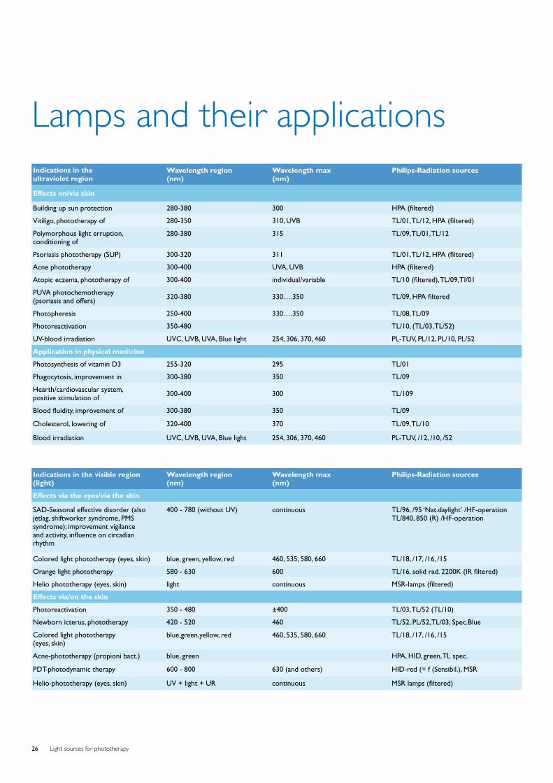
Light sources for phototherapy26
Lamps and their applicationsIndications in the ultraviolet region
Wavelength region (nm)
Wavelength max (nm)
Philips-Radiation sources
Effects on/via skin
Building up sun protection 280-380 300 HPA (filtered)
Vitiligo, phototherapy of 280-350 310, UVB TL/01, TL/12, HPA (filtered)
Polymorphous light erruption,conditioning of
280-380 315 TL/09, TL/01, TL/12
Psoriasis phototherapy (SUP) 300-320 311 TL/01, TL/12, HPA (filtered)
Acne phototherapy 300-400 UVA, UVB HPA (filtered)
Atopic eczema, phototherapy of 300-400 individual/variable TL/10 (filtered), TL/09, Tl/01
PUVA photochemotherapy (psoriasis and offers) 320-380 330….350 TL/09, HPA filtered
Photopheresis 250-400 330….350 TL/08, TL/09
Photoreactivation 350-480 TL/10, (TL/03, TL/52)
UV-blood irradiation UVC, UVB, UVA, Blue light 254, 306, 370, 460 PL-TUV, PL/12, PL/10, PL/52
Application in physical medicine
Photosynthesis of vitamin D3 255-320 295 TL/01
Phagocytosis, improvement in 300-380 350 TL/09
Hearth/cardiovascular system,positive stimulation of 300-400 300 TL/109
Blood fluidity, improvement of 300-380 350 TL/09
Cholesterol, lowering of 320-400 370 TL/09, TL/10
Blood irradiation UVC, UVB, UVA, Blue light 254, 306, 370, 460 PL-TUV, /12, /10, /52
Indications in the visible region (light)
Wavelength region (nm)
Wavelength max (nm)
Philips-Radiation sources
Effects via the eyes/via the skin
SAD-Seasonal effective disorder (also jetlag, shiftworker syndrome, PMS syndrome); improvement vigilanceand activity, influence on circadianrhythm
400 - 780 (without UV) continuous TL/96, /95 ‘Nat.daylight’ /HF-operationTL/840, 850 (R) /HF-operation
Colored light phototherapy (eyes, skin) blue, green, yellow, red 460, 535, 580, 660 TL/18, /17, /16, /15
Orange light phototherapy 580 - 630 600 TL/16, solid rad. 2200K (IR filtered)
Helio phototherapy (eyes, skin) light continuous MSR-lamps (filtered)
Effects via/on the skin
Photoreactivation 350 - 480 ±400 TL/03, TL/52 (TL/10)
Newborn icterus, phototherapy 420 - 520 460 TL/52, PL/52, TL/03, Spec.Blue
Colored light phototherapy (eyes, skin)
blue,green,yellow, red 460, 535, 580, 660 TL/18, /17, /16, /15
Acne-phototherapy (propioni bact.) blue, green HPA, HID, green, TL spec.
PDT-photodynamic therapy 600 - 800 630 (and others) HID-red (= f (Sensibil.), MSR
Helio-phototherapy (eyes, skin) UV + light + UR continuous MSR lamps (filtered)
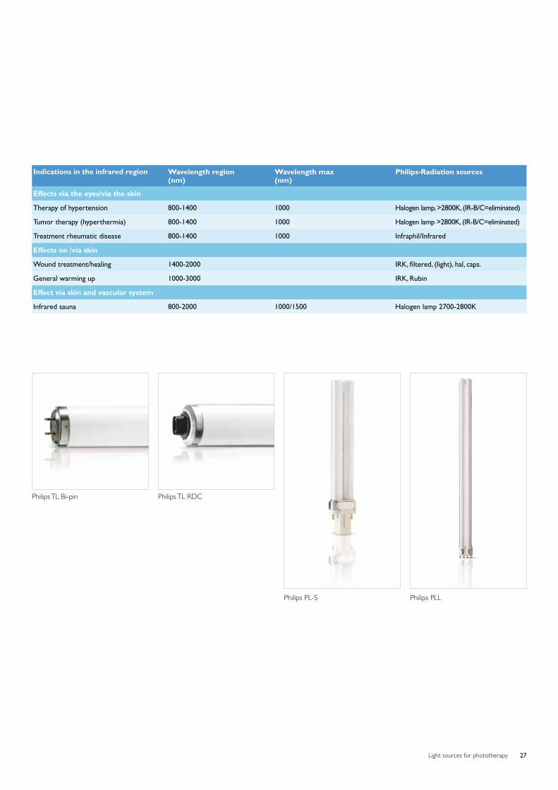
Light sources for phototherapy 27
Indications in the infrared region Wavelength region (nm)
Wavelength max (nm)
Philips-Radiation sources
Effects via the eyes/via the skin Therapy of hypertension 800-1400 1000 Halogen lamp, >2800K, (IR-B/C=eliminated)
Tumor therapy (hyperthermia) 800-1400 1000 Halogen lamp >2800K, (IR-B/C=eliminated)
Treatment rheumatic disease 800-1400 1000 Infraphil/Infrared
Effects on /via skin
Wound treatment/healing 1400-2000 IRK, filtered, (light), hal, caps.
General warming up 1000-3000 IRK, Rubin
Effect via skin and vascular system
Infrared sauna 800-2000 1000/1500 Halogen lamp 2700-2800K
Philips TL Bi-pin Philips TL RDC
Philips PL-S Philips PLL
©2009 Koninklijke Philips Electronics N.V.All rights reserved.
Document order number: 3222 635 49641
Business Line UV Health & Wellness Roosendaal Zwaanhoefstraat 2 4702 LC Roosendaal The Netherlands Tel +31 165 57 7169 Fax +31 165 57 7907
www.philips.com/phototherapy Benelux Boschdijk 525, bldg. VB-6 5600 PD Eindhoven The Netherlands Tel +31 6 130 44 765 Fax +31 40 27 82273
FranceRue Pierre RigaudVal de Marne 94856 FranceTel +33 1 49 87 64 57Fax +33 1 49 87 64 619 Germany Lübeckertordamm 5 D-20099 Hamburg Tel +49 40 2899 2788 Fax +49 40 2899 2205 Hungary Fehervari ut 84/A H-1119 Budapest Tel +36 1 3821 856 Fax +36 1 3821 851 Italy Via G. Casati 23 20052 Monza (MI) Tel +39 039 203 6085 Fax +39 039 203 6127 Poland UL. Kossaka 150 64-920 Pila Tel +48 67 351 32 62 Fax +48 67 351 31 04 Russia Usacheva str 35 A 119048 Moscow Tel +7 495 937 9350 Fax +7 495 937 9359 Spain / Portugal Philips Iberica S.A. Division Comercial Alumbrado Calle Martinez Villergas 49, planta 8 28027 Madrid Tel + 34 91 566 97 64 Fax + 34 91 566 92 42
Sweden/Norway Knarrarnasgatan 7 Kista S-16485 Stockholm Tel +46 8 5985 2000 Fax +46 8 5985 2797 Turkey Yukarı Dudullu Organize Sanayi Bolgesi 2. Cadde No: 22 34776 Umraniye Istanbul Tel +90 216 522 1844 Fax +90 216 522 1835
United Kingdom Philips Centre Guilford Business Park Guilford, Surrey GU2 8XH Tel +44 1483 293 156 Fax +44 1883 334 790 Ukraine Philips Ukraine LLC 4, Mykoly Grinchenka Sr. Horizon Park Business Center, Building N2, floor 4 03038 Kyiv Ukraine Tel +380 44 490 98 68 Fax +380 44 490 98 56 Argentina Vedia 3892 (C1430DAL) Buenos Aires Tel +54 11 4546 7651 Fax +54 11 4546 7672 Australia Philips House, 65 Epping Road North Ryde NSW, 2113 Tel +61 2 9947 0333 Fax +61 2 9947 0325 Brazil Philips Lighting Latin America Rua Verbo Divino 1400, 6 Andar Chacara Santo Antonio 04719-002 Sao Paulo Tel +55 11 2125 0746 Fax +55 11 2125 0580 U.S.A. 200 Franklin Square Drive Somerset N.J. 08875 6800 Tel +1 848 459 8629 Fax +1 847 885 7376 Canada 281 Hillmount Road Markham Ontario L6C 2S3 Tel +1 905 438 0817 Fax +1 416 915 6183
Asia Pacific Level 6, Three Pacific Place, 1 Queens Road East, Wanchai, Hong Kong Tel +86 21 5910 7475 Fax +86 21 5915 4024 Shanghai,China 1805 Hu Yi Road,MaLu Town, Jiading District Shanghai, China 201801 Tel: +86 21 5910 7047 Fax: +86 21 5915 4024 India Philips Electronics India Ltd9th Floor, Ashoka Estate24, Barakhamba Road New Delhi 110 001India Tel +91 11 43529800 Fax +91 11 41515221 Japan-domestic Philips Electronics Japan, Ltd. 13-37, Kohnan 2-chome Minato-ku Tokyo 108-8507 Tel +81 3 3740 4429 Fax +81 3 3740 5163 Korea Philips House P.O. Box 3680 260-199, Itaewon-Dong Yougsan-Ku Seoul 140-200 Tel +82 2 709 1345 Fax +82 2 709 1329 Mexico Norte 45 # 669 Col. Industrial Vallejo Distrito Federal México City Tel +52 55 52699137 Fax +52 55 52699101 Taiwan 14F, No.3-1, Yuan Qu Street Nan Gang District, Taipei 115,Taiwan, R.O.C. Tel +886 2 3789 2279 Fax +886 2 3789 2525