Embed Size (px)
Citation preview

HAL Id: mnhn-02299430https://hal-mnhn.archives-ouvertes.fr/mnhn-02299430
Submitted on 27 Sep 2019
HAL is a multi-disciplinary open accessarchive for the deposit and dissemination of sci-entific research documents, whether they are pub-lished or not. The documents may come fromteaching and research institutions in France orabroad, or from public or private research centers.
L’archive ouverte pluridisciplinaire HAL, estdestinée au dépôt et à la diffusion de documentsscientifiques de niveau recherche, publiés ou non,émanant des établissements d’enseignement et derecherche français ou étrangers, des laboratoirespublics ou privés.
Light stress in green and red Planktothrix strains: Theorange carotenoid protein and its related
photoprotective mechanismChakib Djediat, Kathleen Feilke, Arthur Brochard, Lucie Caramelle, Sandra
Tiam, Pierre Sétif, Theo Gauvrit, Claude Yéprémian, Adjélé Wilson, LéaTalbot, et al.
To cite this version:Chakib Djediat, Kathleen Feilke, Arthur Brochard, Lucie Caramelle, Sandra Tiam, et al.. Lightstress in green and red Planktothrix strains: The orange carotenoid protein and its related pho-toprotective mechanism. Biochimica biophysica acta (BBA) - Bioenergetics, Elsevier, In press,�10.1016/j.bbabio.2019.06.009�. �mnhn-02299430�
HAL Id: mnhn-02299430https://hal-mnhn.archives-ouvertes.fr/mnhn-02299430
Submitted on 27 Sep 2019
HAL is a multi-disciplinary open accessarchive for the deposit and dissemination of sci-entific research documents, whether they are pub-lished or not. The documents may come fromteaching and research institutions in France orabroad, or from public or private research centers.
L’archive ouverte pluridisciplinaire HAL, estdestinée au dépôt et à la diffusion de documentsscientifiques de niveau recherche, publiés ou non,émanant des établissements d’enseignement et derecherche français ou étrangers, des laboratoirespublics ou privés.
Light stress in green and red Planktothrix strains: Theorange carotenoid protein and its related
photoprotective mechanismChakib Djediat, Kathleen Feilke, Arthur Brochard, Lucie Caramelle, Sandra
Tiam, Pierre Sétif, Theo Gauvrit, Claude Yéprémian, Adjélé Wilson, LéaTalbot, et al.
To cite this version:Chakib Djediat, Kathleen Feilke, Arthur Brochard, Lucie Caramelle, Sandra Tiam, et al..Light stress in green and red Planktothrix strains: The orange carotenoid protein and its re-lated photoprotective mechanism. Biochimica et Biophysica Acta (BBA) - Bioenergetics, 2019,�10.1016/j.bbabio.2019.06.009�. �mnhn-02299430�
Light stress in green and red Planktothrix strains: The orange carotenoidprotein and its related photoprotective mechanism
Chakib Djediata,b,1, Kathleen Feilkec,1, Arthur Brocharda,b, Lucie Caramellea,b, Sandra Kim Tiamb,Pierre Sétifc, Theo Gauvritb, Claude Yéprémianb, Adjélé Wilsonc, Léa Talbotc, Benjamin Marieb,Diana Kirilovskyc,⁎, Cécile Bernardb,⁎⁎
a Electron Microscopy Platform, Muséum National d'Histoire Naturelle, CP 39, 12 rue Buffon, F-75231 Paris Cedex 05, FrancebUMR 7245 MCAM, Muséum National d'Histoire Naturelle - CNRS, Paris, 12 rue Buffon, CP 39, 75231 Paris Cedex 05, Francec Institute for Integrative Biology of the Cell (I2BC), CNRS, Commissariat à l'Energie Atomique et aux Energies Alternatives, Université Paris-Sud, Université Paris-Saclay,91198 Gif sur Yvette, France
A R T I C L E I N F O
Keywords:CyanobacteriaPlanktothrixOrange carotenoid proteinMicrocystinFluorescence
A B S T R A C T
Photosynthetic organisms need to sense and respond to fluctuating environmental conditions, to perform effi-cient photosynthesis and avoid the formation of harmful reactive oxygen species. Cyanobacteria have developeda photoprotective mechanism that decreases the energy arriving at the reaction centers by increasing thermalenergy dissipation at the level of the phycobilisome, the extramembranal light-harvesting antenna. This me-chanism is triggered by the photoactive orange carotenoid protein (OCP). In this study, we characterized OCPand the related photoprotective mechanism in non-stressed and light-stressed cells of three different strains ofPlanktothrix that can form impressive blooms. In addition to changing lake ecosystemic functions and biodi-versity, Planktothrix blooms can have adverse effects on human and animal health as they produce toxins (e.g.,microcystins). Three Planktothrix strains were selected: two green strains, PCC 10110 (microcystin producer) andPCC 7805 (non-microcystin producer), and one red strain, PCC 7821. The green strains colonize shallow lakeswith higher light intensities while red strains proliferate in deep lakes. Our study allowed us to conclude thatthere is a correlation between the ecological niche in which these strains proliferate and the rates of inductionand recovery of OCP-related photoprotection. However, differences in the resistance to prolonged high-lightstress were correlated to a better replacement of damaged D1 protein and not to differences in OCP photo-protection. Finally, microcystins do not seem to be involved in photoprotection as was previously suggested.
1. Introduction
Photosynthetic organisms have to cope with the impossibility tocontrol incoming flux of light. Indeed while light is essential for pho-tosynthetic organisms, full sunlight is harmful for them. Therefore, theyhave developed several photoprotective mechanisms to protect them-selves from excess light. One mechanism involves the reduction of ex-citation energy funneled from the antenna into the photochemicalcenters (for a review see [1]). This decreases the accumulation of high-energy reactive oxygen species (ROS; singlet oxygen and oxygen radi-cals) generated during secondary reactions that occur when the pho-tosynthetic electron transport chain is over-reduced. In cyanobacteria,
the orange carotenoid protein (OCP), by interacting with the cyano-bacterial antenna, the phycobilisome (PBS), induces thermal dissipationof the excess energy absorbed at the level of the PBS [2–4].
OCP is a photoactive soluble protein formed by two globular do-mains, the α helix N-terminal domain (NTD) and α-helix/β-sheet C-terminal domain (CTD) [5]. A ketocarotenoid, 3′ hydroxyechinenone, isshared by the two domains and is essential for OCP photoactivation[5–7]. Two other ketocarotenoids, echinenone and canthaxanthin, areable to bind to the OCP and to allow photoactivation [6,8]. The car-bonyl group of the carotenoid is hydrogen-bound to Tyr201 and Trp288in the CTD [5]. Upon illumination with strong blue light, OCP photo-converts from an orange closed inactive form (OCPO) to a red open
⁎ Correspondence to: D. Kirilovsky, Institute for Integrative Biology of the Cell (I2BC), CEA, CNRS, Université Paris-Sud, Université Paris-Saclay, 91198 Gif surYvette, France.
⁎⁎ Correspondence to: C. Bernard, UMR 7245 MCAM, Muséum National d'Histoire Naturelle - CNRS, Paris, 12 rue Buffon, CP 39, 75231 Paris Cedex 05, France.E-mail addresses: [email protected] (D. Kirilovsky), [email protected] (C. Bernard).
1 These authors contributed equally to this work.
BBA - Bioenergetics xxx (xxxx) xxx–xxx
Please cite this article as: Chakib Djediat, et al., BBA - Bioenergetics, https://doi.org/10.1016/j.bbabio.2019.06.009
active form (OCPR) [7]. Upon light absorption by the carotenoid, the Hbonds between the carotenoid and protein are broken, and the car-otenoid translocates into the NTD by undergoing several intermediarysteps [9–11]. Then, the interactions between the domains are disrupted,and the domains separate from each other [10,12]. Only OCPR is able tointeract with PBS [7,13]. The NTD of OCPR binds to the core of the PBSand induces thermal dissipation of excess absorbed energy [13,14]. Thetwo forms of the OCP are also efficient quenchers of singlet oxygen[5,15].
OCP is highly conserved among PBS-containing cyanobacteria[16,17]. At least three paralogous families of OCP exist: OCP1, OCP2,and OCPX [16]. The photoactivity and role of OCP have been mostlystudied using OCP1s, especially that of Synechocystis PCC 6803. Re-cently, an OCP from another family, OCP2 from Tolypothrix PCC 7601(also known as Fremyella diplosiphon), was characterized [16,18].
To recover the full capacity of the antenna, another protein is es-sential: the Fluorescence Recovery Protein (FRP) [19]. This proteinhelps in the detachment of the OCP1 from the PBSs and accelerates theconversion of OCPR to OCPO [19–21]. By contrast, OCP2 does not in-teract with FRP. The recovery reaction from OCPR to OCPO is fast inOCP2, even in the absence of FRP [16,18]. In addition, OCP2 does notneed FRP to detach from the PBS [16,18].
OCP1 is ubiquitous and present in the cells even under low lightconditions [22], but its concentration varies under different growthconditions. For example, the concentration of OCP1 depends on theintensity of growth light ([23] and references inside). General tran-scriptomic studies have shown that ocp transcription increases underdifferent stress conditions ([23] and references inside). Almost all thesestudies were performed using the model organisms Synechocystis PCC6803 (Synechococcales) and Anabaena PCC 7120 (Nostocales). Thephotoprotective mechanism in the filamentous strains that belong toOscillatoriales, widely distributed in natural environments, is still un-clear. In this study, we decided to characterize OCP and its relatedphotoprotective mechanism in non-stressed and light-stressed cells ofthree different Planktothrix strains.
Planktothrix species are of particular environmental interest becausethey are able to form impressive blooms that strongly impact the uses ofwater for drinking water or recreational activities (see review [24]).One of the challenges related to the global expansion of cyanobacterialblooms is to understand the species adaptation to environmentalchanges. The adaptation mechanisms of cyanobacterial species tostressful environments related to human activities that cause warming,CO2 increase, water pollution, and acidification, which decrease thephytoplankton biodiversity in aquatic ecosystems, have not yet beenelucidated. All these changes have been demonstrated to have a largeinfluence on cyanobacterial bloom formations. In addition to changes inlake functioning and biodiversity, these blooms can have adverse ef-fects on human and animal health because of cyanotoxin-producingcyanobacteria (e.g., microcystins [MCs]). MCs are cyclic heptapeptidessynthesized non-ribosomally by large multifunctional enzyme com-plexes containing both non-ribosomal peptide synthetase and polyke-tide synthase domains encoded by the mcy gene cluster [25]. Amongbloom-forming cyanobacteria, filamentous Planktothrix species aredistributed to the greatest extent in freshwater bodies worldwide, andthey are common bloom-forming species [24]. Two main ecology-genotypes have been described within Planktothrix: a red pigmentedecotype (formerly named Planktothrix rubescens) that proliferates indeep lakes under low light intensity and temperature conditions [26]and a green pigmented ecotype (formerly named Planktothrix agardhii)that colonizes shallow lakes with higher intensities of light and tem-perature [26, 27]. To date, all red Planktothrix strains studied containthe mcy operon, indicating they are MC producers and suggesting thatMCs could have an essential role in the survival of these strains [28].Some green Planktothrix strains are MC producers, and others are not[29]. Previous results on green Planktothrix strains have shown that thenon-MC genotypes are dominant under favorable growth conditions,
whereas the MC genotypes grow better under stress conditions [30].Therefore, MCs could have a protective role against oxidative and lightstresses [28, 31].
Three Planktothrix strains were selected: two green strains PCC10110 (MC producer) [32] and PCC 7805 (non-MC producer) [33], andone red strain, PCC 7821 (MC producer) [33]. We checked whetherthere is a correlation between the ecological niches in which thesestrains proliferate (shallow lakes with high light-temperate temperaturefor the green Planktothrix versus deep lakes with low light-low tem-perature for the red Planktothrix) and amplitude and kinetics of theOCP-related mechanism. We also studied the relationship among re-sistance to light stress, amplitude of the photoprotective mechanism,and/or presence of MCs. We used a multidisciplinary approach ofbiophysical (fluorescence measurements), molecular biology (mRNAquantification), and structural (transmission electron microscopy[TEM] and immunolocalization) studies to evaluate the changes thatoccur in Photosystem II (PSII) activity, OCP expression and con-centration, related photoprotective mechanism, and MC concentrationduring light stress.
2. Material and methods
2.1. Strains and culture conditions
For the characterization of OCP and related photoprotection me-chanism in Planktothrix strains, independent experiments were per-formed under controlled light conditions. The cells were grown in BG11medium [34], maintained in a rotary shaker (40 rpm) at 22 °C, and il-luminated by fluorescence white lamps (20 μmol photons m−2 s−1).
For the big high light experiment, pre-culture of 1 L of each strainwas grown in 2 L bottles for four weeks at 20 °C in a thermostatic roomwith a photon flux density of 6 μmol·m−2·s−1 and a 13:11 h light:darkcycle; agitation was provided by magnetic stirring bars. After fourweeks, the cultures reached an OD750nm of about 0.2–0.3, and the highlight experiment was conducted. T0 was sampled after 1 h of illumi-nation at 6 μmol photons m−2 s−1. Then, the cells were illuminatedwith strong white light (150 μmol photons m−2 s−1) for 4 h (T4,240min). After this stress, the cells were transferred to control lightconditions (6 μmol photons m−2 s−1) for 6 h (T10, 600min). Then thecells were transferred to dark during 11 h and illuminated again for 1 hwith 6 μmol photons m−2 s−1 (T24 of Fig. 8). Some cultures weremaintained at 6 μmol photons m−2 s−1 (control conditions, control).The experiment was performed in three different weeks, in triplicates,leading to nine biological replicates at each studied time.
In Fig. 3C and D, results of an independent high light stress ex-periment are shown. In this experiment, Planktothrix cells grown at20 μmol photons m−2 s−1 were illuminated by 4 h (240min) with250 μmol photons m−2 s−1 of white light at a concentration of 0.4OD800 in a PSI multi-cultivator (Photon System Instrument, Czech Re-public). Three biological independent experiments were realized.
2.2. Chlorophyll a quantification and cell number and biovolumemeasurements
The cell abundance was estimated in triplicate by using theMallassez-based method [35]. For chlorophyll a analysis, 5 mL of eachculture was used and analyzed as described previously [36]. The cellvolume of the Planktothrix strains was measured on the basis of theirsizes [37].
2.3. Sample fixation and immunogold labelling
For TEM analysis, the cells were harvested by centrifugation andfixed with a mixture of 2% glutaraldehyde, 2% formaldehyde, 0.18Msucrose, 0.1% picric acid in 0.1 M, pH 7.4, and Sorensen phosphatebuffer (SPB) for 1 h. The cells were then washed three times with SPB.
C. Djediat, et al. BBA - Bioenergetics xxx (xxxx) xxx–xxx
2

Subsequently, they were washed with distilled water before dehydra-tion in a graded ethanol series (30%, 50%, 70%, 90%, and 100%), withagitation and centrifugation. The samples were embedded in a non-antigenic Unicryl resin and sectioned (60 nm) using an ultra-microtome(Reichert-Jung Ultracut, Germany) with a diamond knife and trans-ferred onto 150-mesh gold grids (for immunogold). For OCP localiza-tion in the Planktothrix cells, a polyclonal primary antibody againstSynechocystis OCP (Covance) was used. To localize MCs, the monoclonalanti-MC antibody AD4G2 (Enzo), which is directed against the Addagroup of the peptide, was used [38]. The sections were incubated withthe primary antibodies (we used a dilution 1:250 for both antibodies)and then with the secondary antibodies (we used a dilution 1:500 forboth antibodies) coupled to nanogold particles (6 nm ø for OCP and10 nm ø for MCs). The prepared sections were stained with a saturatedsolution of uranyl acetate/50% ethanol for 15min and washed threetimes in 50% distilled water/50% ethanol and finally twice in distilledwater. The gold grids were then dried and examined using a trans-mission electron microscope (Hitachi HT-7700, Japan), and imageswere obtained using a digital camera (Hamamatsu, Japan).
2.4. Quantification and statistical analysis of immunogold labelling
To quantify the number of OCP and MC gold particles, the imageswere optimized before counting by using the Paint Shop Pro 8 softwareto eliminate elements that could interfere with the counting of the goldnanobeads. The counting was performed using the Icy Freeware SpotDetector plug-in parameterized with the appropriate settings to obtainexact detection (scale, detection threshold, and size filter). To obtain anumber of detections per unit area (μm2), the surfaces analyzed withIcy were calculated using the freeware Image J (NIH).
The statistical analysis was performed using the R Studio software.Because the data distribution was not normal (did not follow a Gaussiandistribution), the following non-parametric tests were performed: theWilcoxon test and Kruskal–Wallis test.
2.5. PAM fluorometer measurements
For characterization of the OCP-related photoprotection mechanismin Planktothrix, changes in the fluorescence levels were measured using101/102/103-PAM (Walz, Effeltrich, Germany) with 650 nm detectinglight. The cells were at a concentration of 2.5 μg chlorophyll a in a1× 1 cm2 stirred cuvette, and the experiments were performed at20 °C. The dark-adapted cells (5 min) were illuminated with85 μmol photons m−2 s−1 of blue-green light (halogen white light fil-tered with a Corion cut-off 550-nm filter; 400 to 500 nm). Then, theywere illuminated with strong blue light (blue-green,1200 μmol photons m−2 s−1). For fluorescence recovery, the cells wereilluminated again with low intensities of blue-green light. Saturatingflashes (400ms×5000 μmol photons m−2 s−1 of white light) wereprovided to probe the maximum fluorescence level.
For the high light experiment, changes in the fluorescence levelswere measured using a multicolor PAM (Walz, Effeltrich, Germany)with 590 nm detecting light. The cells were measured at a OD750nm ofabout 0.2–0.3. The dark-adapted cells (5 min) were illuminated withdiode blue light (440 nm, 53 μmol photons m−2 s−1). Then, they wereilluminated with strong blue light (440 nm, 2160 μmol pho-tons m−2 s−1).
Saturating flashes (400ms×12,000 μmol photons m−2 s−1) wereprovided to probe the maximum fluorescence level. The fluorescenceparameters used in the analysis were as follows: F0, basal fluorescence;Fm′, maximum fluorescence in blue light; Fv= variablefluorescence= Fm− F0.
2.6. Gel electrophoresis and western blotting
Total proteins and membrane-bound PBS proteins were analyzed
with SDS-PAGE on 15% polyacrylamide/2M urea gel in a Tris/MESsystem [39]. In each slot, a sample containing 3 μg of chlorophyll a wasloaded. The OCP band was detected using an antibody against Sy-nechocystis OCP (Covance).
2.7. RNA extraction and quantification by using quantitative real-time PCR
The culture (50mL) was filtered through a polycarbonate mem-brane (diameter, 47mm; pore size, 3 μm; Whatman Nucleopore), andthe filter was transferred into 1.5 mL of RNAlater (Sigma) and stored at4 °C overnight and then at −80 °C. For RNA extraction, the filter wascut into small pieces, and 850 μL of lysis buffer (ML lysis buffer,Macherey Nagel) was added. Extraction was performed using high-in-tensity ultrasonication (Ultrasonic Processor VCX 130; SONICS) twicefor 30 s at 20% of the maximal power. The sample was vortexed aftereach 30 s pulse. After 5min of incubation at room temperature, 700 μLof the sample was transferred into a NucleoSpin Filter with a silicamembrane (Macherey Nagel) and centrifuged for 1min; then, 600 μL ofthe supernatant was transferred, and 225 μL of ethanol was added. Thetotal RNA was extracted using the NucleoSpin miRNA kit (MachereyNagel), according to manufacturer's instructions. A large RNA fraction(> 200 nucleotides) was obtained and stored at −80 °C. Reversetranscription into cDNA and qPCR analyses were performed by the“Plateforme de PCR Quantitative à Haut Débit Genomic Paris Centre,Ecole Normale Supérieure.” The primers used for frp and ocp expressionstudies are listed in Table S1. Relative quantification of each gene ex-pression level was normalized according to rpoC, gyrB, and rpsL ex-pression. The relative mRNA expression was evaluated using the 2−ΔΔC
T
method [40].
2.8. Intracellular MC concentration
Ten milliliters of the MC-producing Planktothrix strains was cen-trifuged (4000 rpm, 10min), and the pellet was freeze-dried. The lyo-philized cells were weighed and then sonicated for 2min in 80% me-thanol with a constant ratio of 100 μL of solvent to 1mg of driedbiomass and centrifuged at 4 °C (12,000× g; 5 min). Two microliters ofthe supernatant representing the metabolite extracts was then analyzedin triplicate by using ultra high-performance liquid chromatography(UHPLC Ultimate 3000; ThermoFisher Scientific) and a Polar AdvancesII 2.5 pore C18 column (Thermo) at 300 μL·min−1
flow rate and a lineargradient of acetonitrile in 0.1% formic acid (5 to 90% in 21min) cou-pled with a high-resolution mass spectrometer. The eluted metabolitecontents were analyzed using an electrospray ionization hybrid quad-rupole time-of-flight (ESI-QqTOF) high-resolution mass spectrometer(Maxis II ETD, Bruker) on positive autoMS/MS mode with information-dependent acquisition (IDA) between 2 and 16 Hz speed, according tothe relative intensity of the parent ions, in consecutive cycle times of2.5 s and active exclusion of previously analyzed parents. The data wereanalyzed with the DataAnalysis 4.4 software for internal recalibration(< 0.5 ppm), and MGF export was generated from the MS/MS spectrabetween 1 and 15min. Microcystin annotation was attempted ac-cording to the precise mass of the molecules and their respective MS/MS fragmentation patterns. The most intense MC variants were semi-quantified according to similar LC-MS/MS analyses performed on po-sitive broad-band CID fragmentation mode (bbCID) at 2 Hz, and the rawdata were searched with the TASQ 1.3 software (Bruker, Germany) fortargeted screening and quantification according to a list of targetedmost-intense analytes (based on the accuracy of their mass, isotopicpattern, retention time, and detection of qualifying fragment ions). Theeffects of high light on MC concentrations were tested using multi-factorial analysis of variance (two-way ANOVA), and multiple com-parisons were conducted with Tukey-HSD tests. Normality and homo-geneity of variance were checked prior to the data analysis.
C. Djediat, et al. BBA - Bioenergetics xxx (xxxx) xxx–xxx
3
3. Results and discussion
3.1. Planktothrix strain characteristics under control and high lightconditions
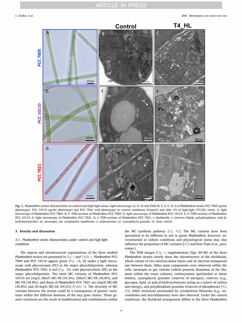
The aspects and ultrastructural organizations of the three studiedPlanktothrix strains are presented in Fig. 1 and Table 1. Planktothrix PCC7805 and PCC 10110 appear green (Fig. 1A, D) under a light micro-scope with phycocyanin (PC) as the major phycobiliprotein, whereasPlanktothrix PCC 7821 is red (Fig. 1G) with phycoerythrin (PE) as themajor phycobiliprotein. The main MC variants of Planktothrix PCC10110 are [Asp3, Dha7] MC-YR (34.3%), [Dha7] MC-YR (30.8%), andMC-YR (34.8%), and those of Planktothrix PCC 7821 are (Asp3) MC-RR(45.8%) and (D-Asp3) MC-LR (54.2%) (Table 1). The diversity of MCvariants between the strains could be a consequence of genetic varia-tions within the different domains of the mcy gene cluster. These ge-netic variations are the result of modifications and combinations within
the MC synthesis pathway [31, 41]. The MC variants have beenspeculated to be different in red or green Planktothrix; however, en-vironmental or culture conditions and physiological status may alsoinfluence the proportion of MC variants ([41] and Kim Tiam et al., pers.comm.).
The TEM images (Fig. 1; supplementary Figs. S4–S6) of the threePlanktothrix strains clearly show the ultrastructure of the thylakoids,which consist of two electron-dense layers and an electron-transparentone between them. Other main components were observed within thecells: aerotopes or gas vesicles (which promote floatation of the fila-ment within the water column), carboxysomes (polyhedral or densebodies), cyanophycin granules (reserves of nitrogen), reserves (e.g.,glycogen, lipid, or poly-β-hydroxybutyrate acting as a source of carbonand energy), and polyphosphate granules (reserves of phosphorus) [42,43]. Other inclusions surrounded by cytoskeleton filaments (e.g., mi-crotubules and microfilaments) were also observed. Under the controlconditions, the thylakoid arrangement differs in the three Planktothrix
Fig. 1. Planktothrix strains characteristics in control and high-light stress. Light microscopy (A, D, G) and TEM (B, C, E, F, H, I) of Planktothrix strains PCC 7805 (greenphenotype), PCC 10110 (green phenotype) and PCC 7821 (red phenotype) in control conditions (Control) and after 4 h of high-light (T4_HL) stress. A: lightmicroscopy of Planktothrix PCC 7805. B, C: TEM sections of Planktothrix PCC 7805. D: light microscopy of Planktothrix PCC 10110. E, F: TEM sections of PlanktothrixPCC 10110. G: light microscopy of Planktothrix PCC 7821. H, I: TEM sections of Planktothrix PCC 7821. t: thylakoids, r: reserves (lipids, polyphosphates, poly-β-hydroxybutyrate), ae: aerotopes, cm: cytoplasmic membrane, c: carboxysome, cy: cyanophycin granule, vs: clear vesicle.
C. Djediat, et al. BBA - Bioenergetics xxx (xxxx) xxx–xxx
4
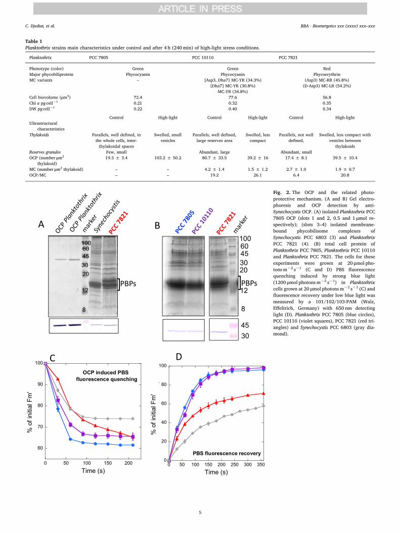
Table 1Planktothrix strains main characteristics under control and after 4 h (240min) of high-light stress conditions.
Planktothrix PCC 7805 PCC 10110 PCC 7821
Phenotype (color) Green Green RedMajor phycobiliprotein Phycocyanin Phycocyanin PhycoerythrinMC variants – [Asp3, Dha7] MC-YR (34.3%)
[Dha7] MC-YR (30.8%)MC-YR (34.8%)
(Asp3) MC-RR (45.8%)(D-Asp3) MC-LR (54.2%)
Cell biovolume (μm3) 72.4 77.6 56.8Chl a pg·cell−1 0.21 0.32 0.35DW pg·cell−1 0.22 0.40 0.34
Control High-light Control High-light Control High-lightUltrastructural
characteristicsThylakoids Parallels, well defined, in
the whole cells, inter-thylakoidal spaces
Swelled, smallvesicles
Parallels, well defined,large reserves area
Swelled, lesscompact
Parallels, not welldefined,
Swelled, less compact withvesicles between
thylakoidsReserves granules Few, small Abundant, large Abundant, smallOCP (number μm2
thylakoid)19.3 ± 3.4 103.2 ± 50.2 80.7 ± 33.5 39.2 ± 16 17.4 ± 8.1 39.5 ± 10.4
MC (number μm2 thylakoid) – – 4.2 ± 1.4 1.5 ± 1.2 2.7 ± 1.0 1.9 ± 0.7OCP/MC – – 19.2 26.1 6.4 20.8
Fig. 2. The OCP and the related photo-protective mechanism. (A and B) Gel electro-phoresis and OCP detection by anti-Synechocystis OCP. (A) isolated Planktothrix PCC7805 OCP (slots 1 and 2, 0.5 and 1 μmol re-spectively); (slots 3–4) isolated membrane-bound phycobilisome complexes ofSynechocystis PCC 6803 (3) and PlanktothrixPCC 7821 (4). (B) total cell protein ofPlanktothrix PCC 7805, Planktothrix PCC 10110and Planktothrix PCC 7821. The cells for theseexperiments were grown at 20 μmol pho-tons m−2 s−1 (C and D) PBS fluorescencequenching induced by strong blue light(1200 μmol photons m−2 s−1) in Planktothrixcells grown at 20 μmol photons m−2 s−1 (C) andfluorescence recovery under low blue light wasmeasured by a 101/102/103-PAM (Walz,Effeltrich, Germany) with 650 nm detectinglight (D). Planktothrix PCC 7805 (blue circles),PCC 10110 (violet squares), PCC 7821 (red tri-angles) and Synechocystis PCC 6803 (gray dia-mond).
C. Djediat, et al. BBA - Bioenergetics xxx (xxxx) xxx–xxx
5

strains. In Planktothrix PCC 7805, the thylakoids are well-defined, ingroups of> 6 units, and distributed in the whole cell (Fig. 1B, supp. Fig.S4). Several interthylakoidal spaces are observed in this strain. InPlanktothrix PCC 10110, the thylakoids are also arranged in parallellines; however, they are less compact, with numerous interthylakoidalspaces and a remarkably large “white” area, suspected to be poly-β-hydroxybutyrate reserves (Fig. 1E, supp. Fig. S5). Finally, in the redPlanktothrix PCC 7821, the thylakoids seem to be shorter and dis-tributed all over the cell with smaller fragmented vesicles and reservegranules (Fig. 1H, supp. Fig. S6).
Cell exposure to high light induced important ultrastructural mod-ifications (Fig. 1C, F, I; supp. Figs. S4–S6). In all the strains, the thy-lakoids were swollen (less compact) after 4 h of light stress. In addition,Planktothrix PCC 7805 and PCC 7821 showed a large increase in clearelectron vesicles between the thylakoids (Fig. 1C and I; supp. Figs. S4and S6). According to [42], these small vesicles could contain poly-β-hydroxybutyrate. In Planktothrix PCC 10110, the large reserve area(Fig. 1F, gray areas in the image) appears to be smaller after light stress.These ultrastructural responses of Planktothrix to high light stress havealready been described in other cyanobacteria (e.g., Anabaena variabilisCALU 458) [43]; the authors showed that the thylakoids swelled, losttheir parallel orientation, and formed vesicles under high light in-tensity. The authors suggested there could be a correlation among theorganization of the thylakoid system, chlorophyll content, and photo-synthesis rate as well as between the ultrastructure and chemicalcomposition of the thylakoids under various illumination conditions[43].
3.2. OCP and related photoprotective mechanism in the Planktothrix strains
The genomes of the three strains and of all the Planktothrix strainssequenced to date have only one ocp gene and one frp gene [16]. ThePlanktothrix OCP belongs to the OCP1 clade [16]. The OCP proteins arehighly conserved among the three studied Planktothrix strains(97.5–98.7% similarity); only a variation of nine amino acids was ob-served between the three protein sequences (Supplementary Fig. S1).
Preliminary independent experiments were performed to confirmthe presence of the OCP and the OCP-related NPQ mechanism in thethree studied Planktothrix strains. The cells for these experiments weregrown in 250mL Erlenmeyer's maintained in a rotary shaker (40 rpm)at 22 °C, and illuminated by fluorescence white lamps (20 μmol pho-tons m−2 s−1).
The presence of OCP in the three strains was detected using gelelectrophoresis and western blot analysis with the OCP antibodyagainst Synechocystis OCP. This antibody identified the isolatedPlanktothrix OCP (Fig. 2). In Fig. 2A, the presence of OCP in membrane-bound phycobilisome (MP) complexes isolated from Synechocystis andPlanktothrix PCC 7821 is shown. In Fig. 2B, the presence of OCP isdetected in total cell protein of the three Planktothrix strains. In eachslot of the gel, the same quantity of chlorophyll a (3 μg), was loaded.Although the method was not quantitative because the specificity of theantibody could differ between strains, we always observed a strongerOCP band from the total cell protein extracted from Planktothrix PCC7821.
The OCP-related photoprotective mechanism, induced by strong
Fig. 3. PSII activity decrease during high-lightstress. (A) Scheme of the high-light stress and re-covery experiment. Cells grown at 6 μmol pho-tons m−2 s−1 were exposed to 150 μmol pho-tons m−2 s−1 (B) The decrease and increase of PSIIactivity during the experiment was followed bymeasuring dark Fv measured using a multicolor-PAM. The graph shows the mean of three in-dependent biological experiments with technicaltriplicates (C and D) Independent experiments inwhich the cells grown at 20 μmol photons m−2 s−1
(at OD800= 0.4) were light stressed(250 μmol photons m−2 s−1 white light) in the pre-sence (C) or absence (D) of lyncomycin (0.4mg/mL)that inhibits PSII repair. Planktothrix PCC 7805(blue circles), PCC 10110 (violet squares), PCC 7821(red triangles). The graph shows the mean of threebiological independent experiments. The error barsrepresent SD.
C. Djediat, et al. BBA - Bioenergetics xxx (xxxx) xxx–xxx
6
blue-green or white light, increases the thermal dissipation of excessenergy absorbed by the PBS. This process is accompanied by a decreasein PBS fluorescence that was measured by a 101/102/103 PAM (Walz,Effeltrich, Germany) with 650 nm detecting light. The dark-adaptedPlanktothrix cells were first illuminated with low intensities of blue-green light that induced an increase in fluorescence due to transition toState I; then, they were illuminated with strong blue-green light to in-duce the OCP-related mechanism (example of PAM traces in supp. Fig.S2). A decrease in the maximal fluorescence (Fm′) under strong light inthe three Planktothrix strains was observed (Fig. 2C). When the cellswere then illuminated again with low intensities of blue-green light, anincrease in maximal fluorescence was detected related to the recoveryof full antenna capacity (Fig. 2D). The rate and amplitude of fluores-cence quenching under strong light as well as the rate of fluorescencerecovery under dim light observed in the green PCC 7805 and PCC10110 strains were similar (Fig. 2C and D). In contrast, the rate offluorescence quenching was slower in the red PCC 7821 strain, al-though the final amplitude of the quenching was equal to those ob-served in the green strains (Fig. 2C). This could be due, at least par-tially, to the presence of PE in the PBS of PCC 7821. PE absorbs greenlight and reduces the light available for photoactivation of OCP.However when the OCP-related NPQ mechanism was compared indifferent marine Synechococcus strains containing only PC or PC anddifferent types of PE, it was shown that the presence of PE is not alwaysrelated to lower OCP-related photoprotection [44]. One PE containingstrain, presented larger fluorescence quenching than the blue-greenstrain, other one the same and the third one less fluorescencequenching. Thus, most probably, the difference in the OCP-related NPQbetween green and red Planktothrix strains is related to their ecological
niches. The green strains, which live in shallow lakes and can be ex-posed to the highest light intensities, are able to induce a fast quenchingof excess absorbed energy; the quenching is much faster than that bythe red strain which in nature grows in deep lakes under low lightconditions. The rate of fluorescence recovery was also slower in the redstrain (Fig. 2D).
The blue-green light induced PBS fluorescence quenching, and thefluorescence recovery was compared to those of Synechocystis PCC 6803cells, which is the most studied cyanobacterial strain (Fig. 2C and D).The amplitude of the fluorescence quenching was larger in the Plank-tothrix strains than in Synechocystis, and the fluorescence recovery wasfaster. The rate and amplitude of fluorescence quenching depend on theconcentration of photoactivated OCPR, which depends on the totalconcentration of OCP, light intensity, and ratio of OCP to FRP. Theaffinity of OCP for the PBS of Synechocystis PCC 6803 also plays animportant role in the rate and amplitude of OCP-induced quenching. Allthese factors could be different in the red and green Planktothrix andSynechocystis, leading to different quenching amplitudes and recoveryrates even when they seem to have similar OCP to chlorophyll a ratios.Also Arthrospira maxima and Synechocystis PCC 6803 strains presenteddifferent amplitudes of the OCP related photoprotection although theOCP concentrations were similar [45].
3.3. PSII activity and ocp and frp mRNA levels during high-light stress andrecovery
Once characterized the OCP-related NPQ mechanism in thePlanktothrix strains, we studied the high light sensitivity of these strainsand followed the behavior of this mechanism during a light stress and
Fig. 4. Relative expression of ocp and frp genes under high light treatment and recovery. (A) Relative mRNA expression level at T0 normalized against three referencegenes (rpoC, gyrB and rpsL) for Planktothrix PCC 7805 (blue), PCC 10110 (violet), PCC 7821 (red). (B and C) Relative expression compared T0 of ocp (B) and frp (C)normalized against three reference genes (rpoC, gyrB and rpsL), generated using the 2-ΔΔCT method, during the high-light stress experiment for the three Planktothrixstrains. Planktothrix PCC 7805 (blue circles), PCC 10110 (violet squares), PCC 7821 (red triangles). The graph present the mean of three biological independentexperiments.
C. Djediat, et al. BBA - Bioenergetics xxx (xxxx) xxx–xxx
7

recovery. For this, the three strains were grown in a light:dark (13:11 h)regimen. The T0 was sampled after 1 h of illumination at6 μmol photons m−2 s−1. Then, the cells were illuminated with strongwhite light (150 μmol photons m−2 s−1) during 4 h (T4, 240min). Afterthis stress, the cells were transferred to low light conditions(6 μmol photons m−2 s−1) during 6 h. Control cells were maintainedunder low light conditions during the period of light stress. PSII activitywas evaluated by measuring the decrease in dark variable fluorescence(Fv) during the high light-stress experimentation with a multicolor PAMfluorometer with 590 nm detecting light (Fig. 3, examples of PAMtraces in supplemental Fig. S3). Planktothrix PCC 7805 lost only 60% ofthe initial dark Fv after 4 h (240min) of high-light stress; the other two
lost 90%, indicating that this strain is more resistant to photoinhibition(Fig. 3B). During the 6 h of recovery under low intensities of whitelight, only Planktothrix PCC 7805 recovered almost all the lost dark Fv;the other two recovered only 30% (600min in Fig. 3B).
To elucidate whether the higher resistance to high light inPlanktothrix PCC 7805 was related to less PSII photodamage or betterrecovery, the three Planktothrix strains were photoinhibited in the ab-sence or presence of lyncomycin in independent experiments.Lyncomycin is an inhibitor of protein synthesis, and it inhibits the re-placement of damaged proteins, especially D1 protein. In these ex-periments, Planktothrix cells grown at 20 photons m−2 s−1 were illu-minated during 4 h (240min) with 250 μmol photons m−2 s−1 in at a
Fig. 5. Immunogold labelling of OCP (6 nm gold particles) in control and high-light stress. TEM micrographs of Planktothrix PCC 7805 (A, B), PCC 10110 (C, D) andPCC 7821 (E, F) in control conditions (Control) and after 4 h of high-light (T4_HL) stress with a clear and specific OCP localization on the thylakoids membranes. t:thylakoids, r: reserves (lipids, polyphosphates, poly-β-hydroxybutyrate …), vs: clear vesicle.
C. Djediat, et al. BBA - Bioenergetics xxx (xxxx) xxx–xxx
8

concentration of 0.4 OD800 in a PSI multi-cultivator (Photon SystemInstrument, Czech Republic). In the presence of lyncomycin, the de-crease in PSII activity was similar among the three strains (Fig. 3C). Inthe absence of lyncomycin, PSII activity decreased slower in Plankto-thrix PCC 7805 (Fig. 3D), indicating that this strain possesses a betterrecovery machinery.
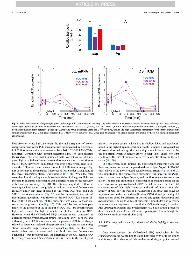
The quantification of ocp and frp mRNAs by using quantitative real-time PCR showed that the concentration of ocp mRNAs is largely higherthan that of frp mRNAs at T0 (Fig. 4). This is in accordance with otherstudies showing that OCP concentration is largely higher than that ofFRP in Synechocystis cells [19, 20]. This is essential for the photo-protective mechanism: a high concentration of FRP completely inhibits
Fig. 6. Immunogold labelling of OCP (6 nm gold particles) and MC (10 nm gold particles) in control and high-light stress. TEM micrographs of Planktothrix PCC 7805(A, B), PCC 10110 (C, D) and PCC 7821 (E, F) with co-immunolocalization of OCP (6 nm, orange dot) and MC (10 nm, red dot) in control conditions (Control) after4 h of high-light (T4_HL) stress with a clear localization of OCP and MC on the thylakoids membranes for the two MC-producing strains PCC 10110 and 7821. t:thylakoids, r: reserves (lipids, polyphosphates, poly-β-hydroxybutyrate …), vs: clear vesicle.
C. Djediat, et al. BBA - Bioenergetics xxx (xxxx) xxx–xxx
9
the photoprotective mechanism by rapidly reconverting the OCPR toOCPO before it can attach to the PBS [20].
Our study revealed that ocp mRNA concentration increased in thethree strains during the 4 h (240min) of high-light stress (Fig. 4B). Thekinetics and amplitude of this increase were different in each strain. InPCC 10110 and PCC 7821, the maximum concentration of ocp mRNAwas observed after 4 h (240min). In PCC 7805, the maximum con-centration was detected at 1 h (60min). The increase was higher inPlanktothrix PCC 10110 than in the other two strains (2.5-fold versus1.5-fold). During the recovery process, the concentration of ocp mRNAdecreased to lower than that at the beginning of the experiments. Theocp expression also increases under high light stress in Synechocystis andTolypothrix cells [16, 46]; by contrast, there is limited information onthe regulation of frp because of its low mRNA concentration. ocp and frppossess their own promoters and could be regulated independently[20]. Nevertheless, changes in the concentration of frp mRNA followedthe same pattern as that of ocp mRNA although unexpectedly, the in-crease in frp mRNA was higher than that of ocp mRNA (Fig. 4C). InPlanktothrix PCC 10110 and PCC 7821, the frp mRNA concentrationlargely increased during the 4 h of high-light stress, and it then de-creased during the recovery time under control light conditions. InPlanktothrix PCC 7805, the frpmRNA concentration was maximum after1 h of high-light stress, and it then decreased.
In conclusion, our results demonstrated that, in Planktothrix cells,ocp and frp are co-regulated: both increase during light stress and thendecrease during the recovery.
3.4. TEM and detection of OCP and MC by using immunogold
For OCP and MC localization in the Planktothrix cells, a polyclonal
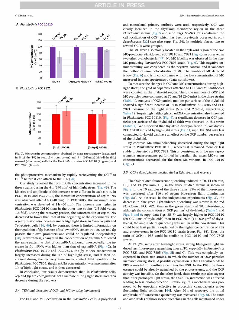
and monoclonal primary antibody were used, respectively. OCP wasclearly localized in the thylakoid membrane region in the threePlanktothrix strains (Fig. 5 and supp. Figs. S5–S7). This confirmed thecell localization of OCP, which has been previously observed in onlySynechocystis [22] (see also supp. Fig. S4). In multiple places, two orseveral OCPs were grouped.
The MC were also mainly located in the thylakoid region of the twoMC-producing Planktothrix PCC 10110 and 7821 (Fig. 6), as observed intwo other cyanobacteria [47]. No MC labeling was observed in the non-MC-producing Planktothrix PCC 7805 strain (Fig. 6). This negative im-munostaining was considered as the negative control, and it validatesthe method of immunolocalization of MC. The number of MC detectedis low (Fig. 6) and is in concordance with the low concentration of MCmeasured in mass spectrometry (data not shown).
To measure the changes in OCP and MC concentrations during high-light stress, the gold nanoparticles attached to OCP and MC antibodieswere counted in the thylakoid region. Then, the numbers of OCP andMC particles were compared at T0 and T4 (240min) in the three strains(Table 1). Analysis of OCP particle number per surface of the thylakoidshowed a significant increase at T4 in Planktothrix PCC 7805 and PCC7821 because of the light stress (5.3- and 2.3-fold, respectively;Table 1). Surprisingly, although ocpmRNA concentration also increasedin Planktothrix PCC 10110, (Fig. 4), a significant decrease in OCP par-ticles per surface of the thylakoid (2-fold) was observed in this strain(Table 1). We suspected that thylakoid disorganization in PlanktothrixPCC 10110 induced by high-light stress (Fig. 1I; supp. Fig. S6) with lesscompacted thylakoid can have an effect on the OCP number per surfaceof the thylakoid.
By contrast, MC immunolabeling decreased during the high-lightstress in Planktothrix PCC 10110, whereas it remained more or lessstable in Planktothrix PCC 7821. This is consistent with the mass spec-trometry measurements performed in parallel; the mean MC-variantconcentrations decreased, for the three MC-variants, in PCC 10110(Fig. 7).
3.5. OCP-related photoprotection during light stress and recovery
The OCP-related fluorescence quenching induced in T0, T1 (60min,HL), and T4 (240min, HL) in the three studied strains is shown inFig. 8. In the T0 samples of the three strains, 35% of the fluorescencewas decreased after 110 s of strong blue-green light illumination(Fig. 8A). As observed in the independent experiments (Fig. 3), thedecrease in blue-green light-induced quenching was slower in the redPlanktothrix PCC 7821 than in the green strains at T0. Interestingly,although the concentration of OCP per μm2 of thylakoids (Table 1 andFigs. 5 and 6; supp. data Figs. S5–7) was largely higher in PCC 10110(80 OCP μm2 of thylakoids) than in PCC 7805 (17 OCP μm2 of thyla-koids), the amplitude of quenching was similar in the two strains. Thiscould be at least partially explained by the higher concentration of PBSand photosystems in the PCC 10110 strain (supp. Fig. S8). Thus, theratio of OCP to PBS could be similar in PCC 10110 and PCC 7805strains.
At T4 (240min) after high-light stress, strong blue-green light in-duced less fluorescence quenching than at T0, especially in PlanktothrixPCC 7821 and PCC 7805 (Fig. 8B and C). This was completely un-expected in these two strains, in which the number of OCP particlesincreased during stress. A possible explanation is that OCP also binds toPBS connected to non-fluorescent inactive PSII. In the PBS, the fluor-escence could be already quenched by the photosystems, and the OCPactivity was invisible. On the other hand, these results can also suggestthat, after prolonged light stress, the OCP-PBS interaction was affectedleading to less photoprotection. Previously, this mechanism was pro-posed to be especially effective in protecting cyanobacteria underfluctuating light conditions [44]. After 20 h of recovery, the initialamplitude of fluorescence quenching was recovered (Fig. 8). The ratesand amplitudes of fluorescence quenching in the cells maintained under
Fig. 7. Microcystin concentrations obtained by mass spectrometry (calculatedin % of the T0) in control (strong colors) and 4 h (240min) high-light (HL)stressed (dim colors) cells for the Planktothrix strains PCC 10110 (A, green) andPCC 7821 (B, red).
C. Djediat, et al. BBA - Bioenergetics xxx (xxxx) xxx–xxx
10
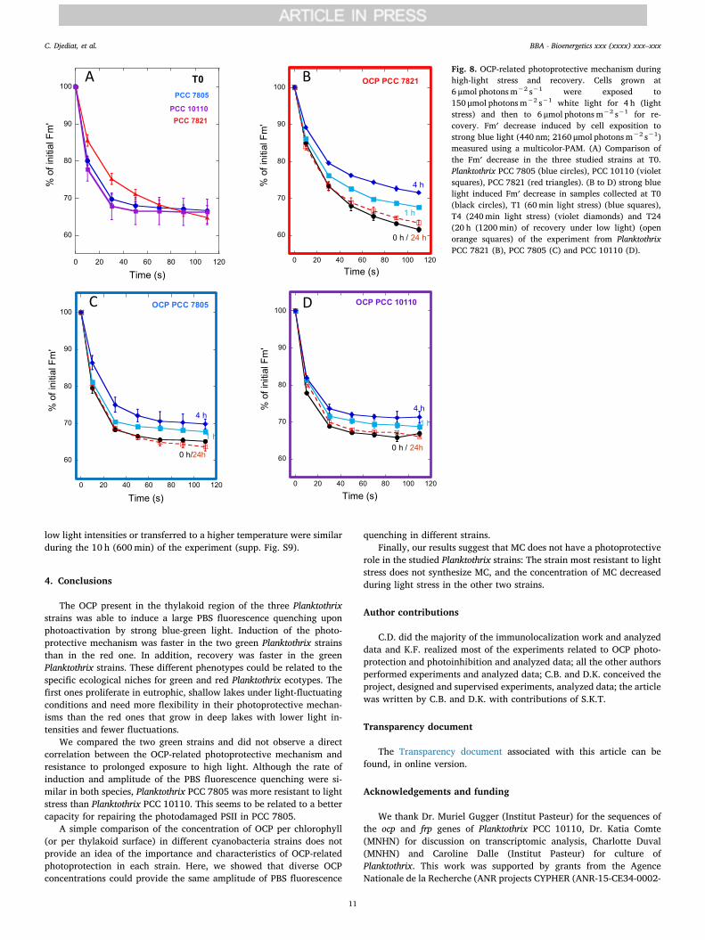
low light intensities or transferred to a higher temperature were similarduring the 10 h (600min) of the experiment (supp. Fig. S9).
4. Conclusions
The OCP present in the thylakoid region of the three Planktothrixstrains was able to induce a large PBS fluorescence quenching uponphotoactivation by strong blue-green light. Induction of the photo-protective mechanism was faster in the two green Planktothrix strainsthan in the red one. In addition, recovery was faster in the greenPlanktothrix strains. These different phenotypes could be related to thespecific ecological niches for green and red Planktothrix ecotypes. Thefirst ones proliferate in eutrophic, shallow lakes under light-fluctuatingconditions and need more flexibility in their photoprotective mechan-isms than the red ones that grow in deep lakes with lower light in-tensities and fewer fluctuations.
We compared the two green strains and did not observe a directcorrelation between the OCP-related photoprotective mechanism andresistance to prolonged exposure to high light. Although the rate ofinduction and amplitude of the PBS fluorescence quenching were si-milar in both species, Planktothrix PCC 7805 was more resistant to lightstress than Planktothrix PCC 10110. This seems to be related to a bettercapacity for repairing the photodamaged PSII in PCC 7805.
A simple comparison of the concentration of OCP per chlorophyll(or per thylakoid surface) in different cyanobacteria strains does notprovide an idea of the importance and characteristics of OCP-relatedphotoprotection in each strain. Here, we showed that diverse OCPconcentrations could provide the same amplitude of PBS fluorescence
quenching in different strains.Finally, our results suggest that MC does not have a photoprotective
role in the studied Planktothrix strains: The strain most resistant to lightstress does not synthesize MC, and the concentration of MC decreasedduring light stress in the other two strains.
Author contributions
C.D. did the majority of the immunolocalization work and analyzeddata and K.F. realized most of the experiments related to OCP photo-protection and photoinhibition and analyzed data; all the other authorsperformed experiments and analyzed data; C.B. and D.K. conceived theproject, designed and supervised experiments, analyzed data; the articlewas written by C.B. and D.K. with contributions of S.K.T.
Transparency document
The Transparency document associated with this article can befound, in online version.
Acknowledgements and funding
We thank Dr. Muriel Gugger (Institut Pasteur) for the sequences ofthe ocp and frp genes of Planktothrix PCC 10110, Dr. Katia Comte(MNHN) for discussion on transcriptomic analysis, Charlotte Duval(MNHN) and Caroline Dalle (Institut Pasteur) for culture ofPlanktothrix. This work was supported by grants from the AgenceNationale de la Recherche (ANR projects CYPHER (ANR-15-CE34-0002-
Fig. 8. OCP-related photoprotective mechanism duringhigh-light stress and recovery. Cells grown at6 μmol photons m−2 s−1 were exposed to150 μmol photons m−2 s−1 white light for 4 h (lightstress) and then to 6 μmol photons m−2 s−1 for re-covery. Fm′ decrease induced by cell exposition tostrong blue light (440 nm; 2160 μmol photons m−2 s−1)measured using a multicolor-PAM. (A) Comparison ofthe Fm′ decrease in the three studied strains at T0.Planktothrix PCC 7805 (blue circles), PCC 10110 (violetsquares), PCC 7821 (red triangles). (B to D) strong bluelight induced Fm′ decrease in samples collected at T0(black circles), T1 (60min light stress) (blue squares),T4 (240min light stress) (violet diamonds) and T24(20 h (1200min) of recovery under low light) (openorange squares) of the experiment from PlanktothrixPCC 7821 (B), PCC 7805 (C) and PCC 10110 (D).
C. Djediat, et al. BBA - Bioenergetics xxx (xxxx) xxx–xxx
11
01)). The research is also supported by the Centre National de laRecherche Scientifique (CNRS, France), the Commissariat à l'EnergieAtomique (CEA, France) and the Muséum National d'Histoire Naturelle(MNHN, France). The French Infrastructure for Integrated StructuralBiology (FRISBI, France) ANR-10-INBS-05 also partially supported thisresearch. We would like to thank Editage (www.editage.com) forEnglish language editing.
Appendix A. Supplementary data
Supplementary data to this article can be found online at https://doi.org/10.1016/j.bbabio.2019.06.009.
References
[1] K.K. Niyogi, T.B. Truong, Evolution of flexible non-photochemical quenching me-chanisms that regulate light harvesting in oxygenic photosynthesis, Curr. Opin.Plant Biol. 16 (2013) 307–314.
[2] C.A. Kerfeld, M.R. Melnicki, M. Sutter, M.A. Dominguez-Martin, Structure, functionand evolution of the cyanobacterial orange carotenoid protein and its homologs,New Phytol. 215 (2017) 937–951.
[3] D. Kirilovsky, C.A. Kerfeld, Cyanobacterial photoprotection by the orange car-otenoid protein, Nat. Plants 2 (2016) 16180.
[4] N.N. Sluchanko, Y.B. Slonimskiy, E.G. Maksimov, Features of protein-protein in-teractions in the cyanobacterial photoprotection mechanism, Biochemistry 82(2017) 1592–1614.
[5] C.A. Kerfeld, M.R. Sawaya, V. Brahmandam, D. Cascio, K.K. Ho, C.C. Trevithick-Sutton, D.W. Krogmann, T.O. Yeates, The crystal structure of a cyanobacterialwater-soluble carotenoid binding protein, Structure 11 (2003) 55–65.
[6] C. Punginelli, A. Wilson, J.M. Routaboul, D. Kirilovsky, Influence of zeaxanthin andechinenone binding on the activity of the Orange carotenoid protein, Biochim.Biophys. Acta 1787 (2009) 280–288.
[7] A. Wilson, C. Punginelli, A. Gall, C. Bonetti, M. Alexandre, J.M. Routaboul,C.A. Kerfeld, R. van Grondelle, B. Robert, J.T. Kennis, D. Kirilovsky, A photoactivecarotenoid protein acting as light intensity sensor, Proc. Natl. Acad. Sci. U. S. A. 105(2008) 12075–12080.
[8] C. Bourcier de Carbon, A. Thurotte, A. Wilson, F. Perreau, D. Kirilovsky,Biosynthesis of soluble carotenoid holoproteins in Escherichia coli, Sci. Rep. 5(2015) 9085.
[9] P.E. Konold, I.H.M. van Stokkum, F. Muzzopappa, A. Wilson, M.L. Groot,D. Kirilovsky, J.T.M. Kennis, Photoactivation mechanism, timing of protein sec-ondary structure dynamics and carotenoid translocation in the orange carotenoidprotein, J. Am. Chem. Soc. 141 (2018) 520–530.
[10] R.L. Leverenz, M. Sutter, A. Wilson, S. Gupta, A. Thurotte, C. Bourcier de Carbon,C.J. Petzold, C. Ralston, F. Perreau, D. Kirilovsky, C.A. Kerfeld, PHOTOSYNTHESIS.A 12 a carotenoid translocation in a photoswitch associated with cyanobacterialphotoprotection, Science 348 (2015) 1463–1466.
[11] E.G. Maksimov, N.N. Sluchanko, Y.B. Slonimskiy, E.A. Slutskaya, A.V. Stepanov,A.M. Argentova-Stevens, E.A. Shirshin, G.V. Tsoraev, K.E. Klementiev,O.V. Slatinskaya, E.P. Lukashev, T. Friedrich, V.Z. Paschenko, A.B. Rubin, Thephotocycle of orange carotenoid protein conceals distinct intermediates and asyn-chronous changes in the carotenoid and protein components, Sci. Rep. 7 (2017)15548.
[12] S. Gupta, M. Guttman, R.L. Leverenz, K. Zhumadilova, E.G. Pawlowski, C.J. Petzold,K.K. Lee, C.Y. Ralston, C.A. Kerfeld, Local and global structural drivers for thephotoactivation of the orange carotenoid protein, Proc. Natl. Acad. Sci. U. S. A. 112(2015) E5567–E5574.
[13] M. Gwizdala, A. Wilson, D. Kirilovsky, In vitro reconstitution of the cyanobacterialphotoprotective mechanism mediated by the orange carotenoid protein inSynechocystis PCC 6803, Plant Cell 23 (2011) 2631–2643.
[14] D. Harris, O. Tal, D. Jallet, A. Wilson, D. Kirilovsky, N. Adir, Orange carotenoidprotein burrows into the phycobilisome to provide photoprotection, Proc. Natl.Acad. Sci. U. S. A. 113 (2016) E1655–E1662.
[15] A. Sedoud, R. Lopez-Igual, A. Ur Rehman, A. Wilson, F. Perreau, C. Boulay, I. Vass,A. Krieger-Liszkay, D. Kirilovsky, The cyanobacterial photoactive orange carotenoidprotein is an excellent singlet oxygen quencher, Plant Cell 26 (2014) 1781–1791.
[16] H. Bao, M.R. Melnicki, E.G. Pawlowski, M. Sutter, M. Agostoni, S. Lechno-Yossef,F. Cai, B.L. Montgomery, C.A. Kerfeld, Additional families of orange carotenoidproteins in the photoprotective system of cyanobacteria, Nat. Plants 3 (2017)17089.
[17] D. Kirilovsky, C.A. Kerfeld, The orange carotenoid protein: a blue-green lightphotoactive protein, Photochem. Photobiol. Sci. 12 (2013) 1135–1143.
[18] S. Lechno-Yossef, M.R. Melnicki, H. Bao, B.L. Montgomery, C.A. Kerfeld, SyntheticOCP heterodimers are photoactive and recapitulate the fusion of two primitivecarotenoproteins in the evolution of cyanobacterial photoprotection, Plant J. 91(2017) 646–656.
[19] C. Boulay, A. Wilson, S. D'Haene, D. Kirilovsky, Identification of a protein requiredfor recovery of full antenna capacity in OCP-related photoprotective mechanism incyanobacteria, Proc. Natl. Acad. Sci. U. S. A. 107 (2010) 11620–11625.
[20] M. Gwizdala, A. Wilson, A. Omairi-Nasser, D. Kirilovsky, Characterization of theSynechocystis PCC 6803 fluorescence recovery protein involved in photoprotection,Biochim. Biophys. Acta 1827 (2013) 348–354.
[21] M. Sutter, A. Wilson, R.L. Leverenz, R. Lopez-Igual, A. Thurotte, A.E. Salmeen,D. Kirilovsky, C.A. Kerfeld, Crystal structure of the FRP and identification of the
active site for modulation of OCP-mediated photoprotection in cyanobacteria, Proc.Natl. Acad. Sci. U. S. A. 110 (2013) 10022–10027.
[22] A. Wilson, G. Ajlani, J.M. Verbavatz, I. Vass, C.A. Kerfeld, D. Kirilovsky, A solublecarotenoid protein involved in phycobilisome-related energy dissipation in cyano-bacteria, Plant Cell 18 (2006) 992–1007.
[23] D. Kirilovsky, R. Kana, O. Prasil, Mechanisms modulating energy arriving at reac-tion centers in cyanobacteria, in: B. Demmig-Adams, G. Garab, W. Adams,Govindjee (Eds.) Non-Photochemical Quenching and energy dissipation in plants,algae and cyanobacteria, Springer Dordrecht Heidelberg New York London, 2014,pp. 471–501.
[24] J. Huisman, G.A. Codd, H.W. Paerl, B.W. Ibelings, J.M.H. Verspagen, P.M. Visser,Cyanobacterial blooms, Nat. Rev. Microbiol. 16 (2018) 471–483.
[25] E. Dittmann, T. Borner, Genetic contributions to the risk assessment of microcystinin the environment, Toxicol. Appl. Pharmacol. 203 (2005) 192–200.
[26] E. Briand, M. Gugger, J.C. Francois, C. Bernard, J.F. Humbert, C. Quiblier, Temporalvariations in the dynamics of potentially microcystin-producing strains in a bloom-forming Planktothrix agardhii (cyanobacterium) population, Appl. Environ.Microbiol. 74 (2008) 3839–3848.
[27] E. Briand, N. Escoffier, C. Straub, M. Sabart, C. Quiblier, J.F. Humbert,Spatiotemporal changes in the genetic diversity of a bloom-forming Microcystisaeruginosa (cyanobacteria) population, ISME J. 3 (2009) 419–429.
[28] C. Tran Du, C. Bernard, M. Ammar, S. Chaouch, K. Comte, Heat shock transcrip-tional responses in an MC-producing cyanobacterium (Planktothrix agardhii) andits MC-deficient mutant under high light conditions, PLoS One 8 (2013) e73198.
[29] C. Yepremian, M.F. Gugger, E. Briand, A. Catherine, C. Berger, C. Quiblier,C. Bernard, Microcystin ecotypes in a perennial Planktothrix agardhii bloom, WaterRes. 41 (2007) 4446–4456.
[30] E. Briand, C. Yepremian, J.F. Humbert, C. Quiblier, Competition between micro-cystin- and non-microcystin-producing Planktothrix agardhii (cyanobacteria)strains under different environmental conditions, Environ. Microbiol. 10 (2008)3337–3348.
[31] R. Kurmayer, L. Deng, E. Entfellner, Role of toxic and bioactive secondary meta-bolites in colonization and bloom formation by filamentous cyanobacteriaPlanktothrix, Harmful Algae 54 (2016) 69–86.
[32] V. Gaget, M. Welker, R. Rippka, N.T. de Marsac, A polyphasic approach leading tothe revision of the genus Planktothrix (cyanobacteria) and its type species, P.agardhii, and proposal for integrating the emended valid botanical taxa, as well asthree new species, Planktothrix paucivesiculata sp. nov.ICNP, Planktothrix tepidasp. nov.ICNP, and Planktothrix serta sp. nov.ICNP, as genus and species names withnomenclatural standing under the ICNP, Syst. Appl. Microbiol. 38 (2015) 141–158.
[33] C. Pancrace, M.A. Barny, R. Ueoka, A. Calteau, T. Scalvenzi, J. Pedron, V. Barbe,J. Piel, J.F. Humbert, M. Gugger, Insights into the Planktothrix genus: genomic andmetabolic comparison of benthic and planktic strains, Sci. Rep. 7 (2017) 41181.
[34] R.Y. Stanier, R. Kunisawa, M. Mandel, G. Cohen-Bazire, Purification and propertiesof unicellular blue-green algae (order Chroococcales), Bacteriol. Rev. 35 (1971)171–205.
[35] A. Catherine, S. Maloufi, R. Congestri, E. Viaggiu, R. Pilkaityte, Cyanobacterialsamples: preservation, enumeration, and biovolume measurements, in:J. Meriluoto, L. Spoof, G. Codd (Eds.), Handbook of Cyanobacterial Monitoring andCyanotoxin Analysis, Wiley, 2017, pp. 333–334.
[36] C. Yéprémian, A. Catherine, R. Congestri, C. Bernard, T. Elersek, R. Pilkaityte,Chlorophyll a extraction and determination, in: J. Meriluoto, L. Spoof, G. Codd(Eds.), Handbook of Cyanobacterial Monitoring and Cyanotoxin Analysis, Wiley,2017, pp. 315–330.
[37] J. Sun, D. Liu, Geometric models for calculating cell biovolume and surface area forphytoplankton, J. Plankton Res. 25 (2003) 1331–1346.
[38] B. Marie, H. Huet, A. Marie, C. Djediat, S. Puiseux-Dao, A. Catherine, I. Trinchet,M. Edery, Effects of a toxic cyanobacterial bloom (Planktothrix agardhii) on fish:insights from histopathological and quantitative proteomic assessments followingthe oral exposure of medaka fish (Oryzias latipes), Aquat. Toxicol. 114-115 (2012)39–48.
[39] Y. Kashino, H. Koike, K. Satoh, An improved sodium dodecyl sulfate-poly-acrylamide gel electrophoresis system for the analysis of membrane protein com-plexes, Electrophoresis 22 (2001) 1004–1007.
[40] K.J. Livak, T.D. Schmittgen, Analysis of relative gene expression data using real-time quantitative PCR and the 2(-Delta Delta C(T)) method, Methods 25 (2001)402–408.
[41] E.M. Janssen, Cyanobacterial peptides beyond microcystins - a review on co-oc-currence, toxicity, and challenges for risk assessment, Water Res. 151 (2019)488–499.
[42] A. Canini, S. Pellegrini, M. Grilli Caiola, Ultrastructural variations in Microcystisaeruginosa (Chroococcales, Cyanophyta) during a surface bloom induced by highincident light irradiance, Plant Biosyst. 137 (2003) 235–248.
[43] O. Baulina, Ultrastructural Plasticity of Cyanobacteria, Springer-Verlag, BerlinHeidelberg, 2012.
[44] C. Boulay, L. Abasova, C. Six, I. Vass, D. Kirilovsky, Occurrence and function of theorange carotenoid protein in photoprotective mechanisms in various cyanobacteria,Biochim. Biophys. Acta 1777 (2008) 1344–1354.
[45] D. Jallet, A. Thurotte, R.L. Leverenz, F. Perreau, C.A. Kerfeld, D. Kirilovsky,Specificity of the cyanobacterial orange carotenoid protein: influences of orangecarotenoid protein and phycobilisome structures, Plant Physiol. 164 (2014)790–804.
[46] Y. Hihara, A. Kamei, M. Kanehisa, A. Kaplan, M. Ikeuchi, DNA microarray analysisof cyanobacterial gene expression during acclimation to high light, Plant Cell 13(2001) 793–806.
[47] F.M. Young, C. Thomson, J.S. Metcalf, J.M. Lucocq, G.A. Codd, Immunogold loca-lisation of microcystins in cryosectioned cells of Microcystis, J. Struct. Biol. 151(2005) 208–214.
C. Djediat, et al. BBA - Bioenergetics xxx (xxxx) xxx–xxx
12


![RESIDUAL STRESS MEASUREMENTS BY NEUTRON …3 Residual stress measurements by neutron diffraction at the IBR-2 pulsed reactor 493 strains along different [hkl] directions simultaneously,](https://img.pdfslide.net/doc/110x75/60de95aa68163e53d2609032/residual-stress-measurements-by-neutron-3-residual-stress-measurements-by-neutron.jpg)