Embed Size (px)
Citation preview

123
Biochimica et Biophysica Acta, 504 (1978) 123--135 © Elsevier /Nor th-Hol land Biomedical Press
BBA 47564
LIGHT/DARK LABELING DIFFERENCES IN CHLOROPLAST MEMBRANE POLYPEPTIDES ASSOCIATED WITH CHLOROPLAST COUPLING FACTOR 0
JAMES L. ELLEN S ON *, DEBORAH J. PHEASANT, and R.P. LEVINE
The Biological Laboratories, Harvard University, 16 Divinity Ave., Cambridge, Mass. 02138 (U.S.A.)
(Received March 10th, 1978)
Summary
The fluorogenic reagent fluorescamine has been used to determine the labeling patterns of Type C spinach chloroplast membrane polypeptides. Membrane polypeptides labeled with fluorescamine were detected by scanning high resolution sodium dodecyl sulfate polyacrylamide gradient slab gels for fluorescence emission.
Three membrane polypeptides show a decrease in the extent of labeling when chloroplast membranes are labeled in the light compared to when they are labeled in the dark. These polypeptides have apparent molecular weights of 32 000, 23 000 and 15 000.
The decrease in labeling observed in the light is abolished or reduced by treatments which inactivate the light-generated transmembrane pH gradient. CF~-depleted chloroplasts show neither a light-activated pH gradient nor a light/dark difference in labeling of these three polypeptides. Both a light- activated pH gradient and light/dark differences in labeling are observed in CF,-depleted chloroplasts which have been treated with N,N'-dicyclohexylcar- bodiimide.
The same ammonium sulfate fractions of a 2% sodium cholate extract, which are believed to be enriched in the membrane-bound sector of the chloroplast ATPase (CFo) are also found to be enriched in the 32 000, 23 000 and 15 000 molecular weight polypeptides. The three polypeptides are believed to be com- ponents of CFo, and the light/dark labeling differences may indicate confor- mational changes within CFo. Such conformational changes may reflect a mechanism which couples light-generated proton gradients to ATP synthesis.
* To whom all correspondence should be addressed. Abbreviations: CFI, chloroplast coupling factor 1; CF o, hydrophobic portion from chloroplast ATPase', DCCD, N,N'-dicyclohexylcarbodiimide; DCMU, 3-(3,5-diehlorophenyl)-l,l-dimethylurea; FCCP, cas- bonylcyanide p-trifluoromethoxyphenylhydrazone; SDS, sodium d o d e c y l sulfate.

1 2 4
Introduction
The complex responsible for ATP synthesis in chloroplast membranes consists of a soluble portion, CF1, 'which in solution can act as an ATPase [1,2] and an intrinsic membrane portion, CFo *, the membrane binding site of CF1 vchich apparently contains the membrane proton pore [3]. Removal of CFI from chloroplast membranes abolishes the light-generated pH gradient [4]. This gradient can be restored by incubating CFl-depleted membranes with DCCD [4].
Although the subunit composition and many of the functional properties of CFl are known (for a recent review see ref. 5), the nature of CF o is less well characterized. The extraction of chloroplasts with sodium cholate by Younis and Winget [6] yields a fraction containing several polypeptides which, when reconstituted in liposomes together with bacteriorhodopsin and purified CFI, ca ta lyze a light-dependent, N,N'-dicyclohexylcarbodiimide (DCCD)- sensitive ATP synthesis. This particular fraction thus apparently contains the component(s) constituting CFo. The exact interaction between CFo and CF1 which couples a light-driven pH gradient to ATP synthesis is not known. Light-induced changes in labeling patterns [7--11] and nucleotide binding affinities [12--16] have, however, been interpreted as evidence that CF~ can undergo conformational changes in response to membrane energization. Evidence implying that CFo also undergoes conformational changes has not, to our knowledge, been reported. During the course of investigations of label- ing chloroplast membranes in the light and in the dark with the fluorogenic reagent fluorescamine [ 17], we have observed light/dark labeling differences in the labeling of three membrane polypeptides having molecular weights of approximately 32 000, 23 000 and 15 000. These polypeptides are identical with three major polypeptides of the sodium cholate extract which reconstitutes light-sensitive ATP synthesis. The differences in labeling which we describe here are associated with the chloroplast membrane proton gradient, and they may reflect conformational changes in proteins that participate in light-induced proton movement.
Methods
Chloroplast isolation. Type C [18] chloroplast membranes, those lacking the outer limiting membrane, were prepared from spinach by the method of Avron [19]. Final suspension was either in a medium consisting of 0.4 M sucrose/0.05 M Tricine (pH 8.0)/0.01 M KC1/2 mM MgC12, or in either of two low-salt media: 5 mM phosphate or 5 mM Tricine, pH 8.0. The chloroplasts were suspended to a final chlorophyll concentration of 100 pg/ml. Chlorophyll determinations were performed according to a modification [20] of the method of MacKinney [21].
Fluorescamine labeling and sample preparation. The labeling procedure con- sisted of adding, while vortexing, enough 0.4% (w/v) fluorescamine (Pierce}
* We cons ide r ' C F o ' to cons i s t o f the m e m b r a n e sec to r o f the DCCD-sens i t i ve ATPase c o m p l e x w h i c h n o t on ly inc ludes the ac tua l DCCD-sens i t ive s i te , b u t also the C F l b i n d i n g s i te .
125
in acetone to a suspension of chloroplasts at 20--22°C to achieve a final acetone concentrat ion of 0.5% (v/v). Chloroplasts to be labeled in the light were illuminated with white light ( 1 . 1 0 6 erg/cm 2" s -1) at least 5 s before the addition of fluorescamine. For labeling in the dark, chloroplasts were kept in the dark for at least 5 min before addition of the labeling reagent. The labeled chloroplast membranes were centrifuged for 10 min at 10 000 X g. The resulting pellet was solubilized to a final protein concentrat ion of 2 mg/ml in a pH 8.4 buffer consisting of 6 M urea/0.5% SDS/0.01 M EDTA/0.1 M Tris. Protein concentrations were determined by the method of Schaffner and Weissmann [22]. Prior to electrophoresis, mercaptoethanol was added to 1% (v/v) to the solubilized membranes; this solution was heated for 15 s in boiling water before an aliquot was loaded onto the gel.
Cholate extraction. Chloroplasts isolated and labeled in the dark or in the light, as described above, were extracted with sodium cholate according to a modification of the procedure of Younis and Winget [6]: instead of a 1 h, 229 000 X g centrifugation of the initial 2% cholate extract, a 20 min, 20 000 X g centrifugation was used. Fractions precipitating between 33--39% and 39--49% saturation (at 20°C) in ammonium sulfate were centrifuged for 10 min at 20 000 X g and solubilized for electrophoresis.
Electrophoresis. Linear 7.5--15% polyacrylamide gradient slab gels (14 X 18 X 0.07 cm) topped with a 1.5 cm, 5% polyacrylamide stacking gel were made according to the method of Laemmli [23]. Solubilized samples contain- ing 25--60 pg of protein were loaded into each well, and electrophoresis proceeded overnight at 5--8°C with a constant current setting between 7.5 and 11 mA. Gels were stained with Coomassie Blue and destained according to Fairbanks et al. [24].
Fluorescence detection. Different lanes of the processed gel were sliced with the aid of a circular pizza cutter. Each gel slice was t r immed to 13 cm to fit a rectangular glass cuvette, and the slices were scanned for fluorescence using a fluorescence gel scanner constructed in part from the gel transport and monochrometer units of a Zeiss M4QIII gel scanning spectrophotometer . Excitation light from a 100 W tungsten projection lamp powered by an Aminco regulated DC power supply was modulated at 300 Hz with a chopping wheel. A Corning 7-51 filter placed at the exit slit of the monochrometer blocked unwanted stray light from passing through the detect ion system. Broad band emission originating f rom the gel passed through a Coming 4-64 and Wratten No. 4 filter and was detected by a photomultiplier tube (EMI 9558 operated at 900 V). The modulated componen t of the detected signal was amplified using a Princeton Applied Research HR-8 Lock-in amplifier. Recordings were made with an Esterline Angus Speed Servo Recorder.
Results
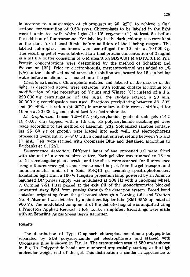
The distribution of Type C spinach chloroplast membrane polypeptides separated by SDS polyacrylamide gel electrophoresis and stained with Coomassie Blue is shown in Fig. la . The transmission scan at 550 nm is shown in Fig. lb . Polypeptide bands are numbered sequentially starting at the high molecular weight end of the gel. This distribution is similar in appearance to

126
s )
I , I J I I 1 .I .2 .3 .4
TT 5ir y °Vi I I i I I I I , i , i
.5 .4 .7 .8 .9 1.0
T r a n s m i s s i o n ~ I
Fluorescence I s3
I , I , I , I , I , I , I , I , I , I • ! .3 ,3 .4 .S ~ .7 ,0 ,9 t~
RF
Fig. 1. (a) Coomass ie s ta ining p a t t e r n o f ch lo rop las t m e m b r a n e p o l y p e p t i d e s s epa ra t ed by SDS po lyac ry l - a m i d e e lec t rophores i s . Major p o l y p e p t i d e bands have been n u m b e r e d in o rd e r of decreas ing mo lecu l a r weight . T he pos i t ions o f the five subun i t s of CF 1 have been iden t i f i ed as c~, fl, ~/, 6, an d e and c o r r e s p o n d to m o l e c u l a r weights of 62 000 , 57 000 , 38 000 , 18 0 0 0 and 14 0 0 0 [ 2 9 ] , respec t ive ly . (b) Transmiss ion scan at 550 n m of the gel slice in (a). (c) F luorescence scan of an uns t a ined gel slice sh o win g the fluo- rescence label ing p a t t e r n o f p o l y p e p t i d e s or ig ina t ing f r o m ch lo rop las t m e m b r a n e s labeled wi th fiuo- r e s cam i ne in the dark . (d) Same as (c), b u t wi th f luorescen t labeling p a t t e r n of p o l y p e p t i d e s or ig inat ing f r o m i l l umina t ed chloroplas ts , A r r o w s m a r k the posi t ions wh e re b an d s 11 and 23 wo u ld appear . (e) F luo rescence prof i le of ch lo rop las t m e m b r a n e p o l y p e p t i d e s labeled wi th f iuo rescamine a f te r solubfliza- t ion of the m e m b r a n e s in 1% SDS a t pH 8.0. The ho r i zon t a l d imens ion , RF , for (a) - - (e) is m e a s u r e d as the re la t ive r e t e n t i o n f ac to r of p o l y p e p t i d e s a long the length of the gel slice re la t ive to the b u f f e r f ront . The gel slices have been t r u n c a t e d to fit a 13 c m scanning cuve t t e . Th e ver t ical axis in (b) measures relat ive t r ansmiss ion a t 550 n m , and ver t ical axes in (c)---(d) m e a s u r e f luorescence in tens i ty in a rb i t r a ry units . The m e t h o d o l o g y for m e m b r a n e p repa ra t i on , labeling, so lubi l iza t ion and gel e lec t rophores i s and scanning is given in Methods .

127
that reported by Chua et al. [25] and Henriques et al. [26]. In order to determine which polypeptides of Type C chloroplast membrane are accessible to and can react with fluorescamine, membranes were incubated with the reagent either in the light or in the dark, pelleted, solubilized in SDS, separated by SDS polyacrylamide gel electrophoresis, and scanned for fluorescence as described in Methods.
Light/dark fluorescamine labeling patterns. Fig. l c shows a fluorescence profile of chloroplast membrane polypept ides labeled after an aliquot of fluo- rescamine was added to a suspension of chloroplasts in the dark, while Fig. l d is the pattern obtained when fluorescamine was added in the light. Both traces can be compared to the fluorescence pattern in Fig. l e obtained when chloroplast membranes were solubilized in SDS before fluorescamine was added to the sample.
The major labeled polypeptides obtained from intact membranes include the cluster of bands 13--16, two of which (band 13 and 15} are subunits of the chlorophyll a/b light-harvesting protein complex [27,28]. Several major Coom- assie staining bands including the ~ and ~ subunits of CF, band 5, the 57 000 molecular weight major subunit of ribulose 1,5-diphosphate carboxylase [29] and band 3, the 68 000 molecular weight polypept ide which derives from the chlorophyl l , protein complex of Photosystem I [28], are not significantly labeled.
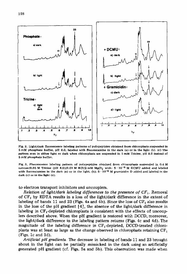
It can be seen from Figs. l c and l d that the labeling of bands 11 and 23 is greater in the dark than in the light. This difference in labeling pattern is seen in chloroplasts suspended either in the normal isolation medium (the condi- tions for Figs. l c and ld ) or a low-salt medium such as 5 mM phosphate buffer (Figs. 2a and 2b). In contrast, when chloroplasts are suspended in 5 mM Tricine buffer, the pattern of labeling in the light is identical to that seen in the dark (Fig. 2c) and is similar to the labeling pattern in Fig. 2a, except that the label- ing of band 11 has increased relative to that of band 23. A labeling pattern sim- ilar to Figs. 2a and 2b was obtained when chloroplast.s were labeled after addi- t ion of either millimolar levels of divalent cations (Ca 2+, Mn 2+, or Mg 2÷ ) or deci- molar levels of K ÷ or Na ÷ to the 5 mM Tricine.
Dependence of labeling pattern on photosynthetic activity. Although the addition of electron acceptors such as ferricyanide may slightly increase the degree of light/dark !abeling of bands 11 and 23, the phenomenon is easily observed with basa l electron transport activity. The inhibition of electron transport with DCMU (3-(3,5-dichlorophenyl)- l , l -dimethylurea) diminishes the light/dark difference in labeling (Figs. 3a and 3b). The addition of such agents as FCCP (carbonylcyanide p- t r i f luoromethoxyphenylhydrazone) , nigeticin in the presence of KC1, gramicidin (Figs. 3c and 3d), and the divalent cation iono- phore A23187, all of which uncouple photophosphoryla t ion, inhibit the light- induced pH gradient and abolish the difference in the light/dark labeling levels of bands 11 and 23. In contrast, phloridzin, an uncoupler of photophosphoryl- ation but not of light ' induced pH gradients [30], had no noticeable effect on the labeling pattern.
Although we have often noted that all of the bands are labeled to a lesser degree (up to 30%) in the light than in the dark, only bands 11 and 23 show a complete loss of labeling in the light. It is this latter change which is sensitive
128
2 3
b) l i g h t ~
T,,c no." J ° ' 'or' I
[ I [ i I I I , I I I I I I I J I J 1 ,1 . 2 .3 .4 . $ .6 .7 .8 ,9 1.0
R F
23
+ G r a m i c i d i n : ~ ~ ~ / VVV
t , I , I , I , I , I I , I , I J I J .2 .3 .4 .8 .6 .? .8 .9 t0
RF
Fig. 2. L i g h t / d a r k f luorescence label ing p a t t e r n s of p o l y p e p t i d e s o b t a i n e d f r o m ch lorop las t s su spended in 5 m M p h o s p h a t e buf fe r , p H 8.0, labeled wi th f l uo re scamine in the da rk (a) or in the l ight (b). (c) T h e p a t t e r n seen in e i the r l ight or da rk w h e n ch lo rop las t s a re su sp en d ed in 5 m M Tricine, p H 8.0 ins tead of 5 m M p h o s p h a t e bu f fe r .
Fig. 3. F luo rescence label ing p a t t e r n of p o l y p e p t i d e s o b t a i n e d f r o m ch lorop las t s su spended in 0.4 M s u c r o s e / 0 . 0 5 M Tric ine (pH 8 .0 ) /0 .01 M KCI/2 m M MgCI 2 wi th : 5 • 10 -6 M DCMU a d d e d an d labeled wi th f luo rescamine in the da rk (a) or in the l ight , (b); 5 • 10 -6 M s ramic id in D a d d e d and labeled in the d a r k (c) or in t he l ight (d).
to electron transport inhibitors and uncouplers. Relation of light/dark labeling differences to the presence of CF1. Removal
of CF1 by EDTA results in a loss of the light/dark difference in the extent of labeling of bands 11 and 23 (Figs. 4a and 4b). Since the loss of CF~ also results in the loss of the pH gradient [4], the absence o f the light/dark difference in labeling in CF,-depleted chloroplasts is consistent with the effects of uncoup- lers described above. When the pH gradient is restored with DCCD, moreover, the light/dark difference in the labeling pattern returns (Figs. 4c and 4d). The magnitude of the labeling difference in CFrdeple ted , DCCD-treated chloro- plasts was at least as large as the change observed in chloroplasts retaining CFI (Figs. l c and ld) .
Artificial pH gradients. The decrease in labeling of bands 11 and 23 brought about in the light can be partially mimicked in the dark using an artificially generated pH gradient (cf. Figs. 5a and 5b). This observation was made when

129
-CFI= 1 , ~
1
- C F 1 + D C C D :
I , I , I , I , I , I , I , i l l i J ,2 .3 A ,$ .8 .T .8 .g 1.0
RF
Fig. 4 . F l u o r e s c e n c e l a b e l i n g p a t t e r n s o b t a i n e d f r o m c h l o r o p l a s t s deple ted in C F 1 a n d labeled with f l u o r e s c a m i n e in t h e d a r k (a) o r in the Hght (b) ; C F l - d e p l e t e d c h l o r o p l a s t s i n c u b a t e d w i t h 7 .5 • 1 0 -5 M D C C D and labeled in the d a r k (c) o r in the l i g h t (d) . C F l - d e p l e t e d c h l o r o p l a s t s w e r e p r e p a r e d a c c o r d i n g t o t h e p r o c e d u r e o f M c C a r t y a n d R a c k e r [ 5 6 ] .
the labeling reagent was added immediately after the addition of the base phase (Fig. 5b). However, when the labeling reagent was added to acid-base treated chloroplasts between 5 and 30 s after the addition of the base phase (Fig. 5a) the labeling of bands 11 and 23 had returned to a higer level characteristic of chloroplasts labeled in the dark.
Relation of labeling pattern to CFo. Noting that the light/dark labeling dif- ference of bands 11 and 23 apparently depends upon the generation of a pH gradient and not the presence of CF~, we turned our at tention to the relation of labeling patterns to membrane detergent fractions containing components which apparently include CF o [6,31,32]. Chloroplast membranes which had been labeled either in the light or in the dark were extracted with 2% cholate according to the method of Winget et al. [32]. In order to examine the perti- nent ammonium sulfate fractions analyzed by Winget et al. (33--39% [32]), and Younis and Winget (35--50% [6]), we looked at two fractions: 33--39% and 39--49% saturation in ammonium sulfate.
The 33--39% fraction is similar to that shown by Winget et al. [33] ; it con- tains a considerable amount of CF1, whose a, fl, 7, 5, and e subunits are identi- fied in Fig. 6. In addition, three major bands, 10, 11, and 16, similar to those

1 3 0
a ) t b = 3 0 s e c _2S,, b ) t b = l s e c ^ f ~
I i I i I I I I I I I i I i l i i I i I .1 .2 ..1 .4 .iS .ll .7 .Ill 1.0
RF
Figl 5. Labe l ing p a t t e r n of p o l y p e p t i d e s o b t a i n e d f r o m ch lo rop las t m e m b r a n e s sub jec ted to an art if icial p H - j u m p and labe led (a) 30 s a f t e r add i t i on of base a nd (b) 1 s a f t e r the add i t ion of base. Chloroplas ts (100 # g / m l ch lo rophy l l ) were init ial ly suspended in 0.4 M suc rose /0 .01 KCI /2 m M MGC12/3 m M succinic acid , pH. 5. An a l iquo t of 0,1 M K O H was a d d e d wi th vo r t ex in g to b r ing the final p H up to 9.5. Fluo- r e s cami ne was a d d e d a t a t i m e t b a f t e r the add i t i on of base.
3 3 - 3 9 ~ Fract ion 23
I L I , I , I , I , I , I , I , I , I ,I ,2 .3 A .5 ,e .7 .8 .9 1.0
RF
Fig. 6. T he Coomass ie s ta in ing p a t t e r n a nd f luorescence prof i les of ch lo rop las t m e m b r a n e p o l y p e p t i d e s e x t r a c t e d wi th 2% s o d i u m cho la t e a nd p r e c i p i t a t e d b e t w e e n 33 and 39% sa tu ra t i on in a m m o n i u m sulfate . Chloroplas t s we re l abe l ed e i ther in the d a r k ( u p p e r cu rve ) or t h e in t h e l ight ( lower cu rve ) b e f o r e ext rac- t ion wi th cho la te . T he Coomass ie s ta ining p a t t e r n for b o t h samples was ident ica l . N u m b e r s above the Coomass ie p ic tu re i den t i fy r e l evan t p o l y p e p t i d e s discussed in the t ex t . Th e a , ~, T, ~, an d e subuni t s of CF 1 have also b e e n ident i f ied .

131
3 9 - 4 9 ~ Fract ion
2 3
5 1011 17 2 3 2 5
I I I I I I
i ~ i , I , i , i , [ , i , l , i , i .! .2 .3 A .5 .6 .7 .8 .9 1.0
R F
Fig. 7. T h e C o o m a s s i e Pattern and f luorescence pro f i l e o f c h l o r o p l a s t m e m b r a n e po lypept ides extracted w i t h 2% s o d i u m c h o l a t e and precipitated b e t w e e n 39 and 49% saturat ion in a m m o n i u m su l f a t e . C h l o r o - p la s t s w e r e l a b e l e d in t h e d a r k ( u p p e r c u r v e ) o r in t h e l i g h t ( l ower c u r v e ) p r i o r t o extract ion .
shown by Winget et al. [32] are evident. These three bands have apparent molecular weights of 35 000, 32 000 and 25 000, respectively. However, our estimated molecular weight of 35 000 for band 10 differs from the 42 000 value for the corresponding major polypeptide reported by Younis and Winget [6]. In contrast to their results, at least 10 minor bands, including band 23, are also observed, most probably reflecting the inherently greater resolution of a gradient slab gel system over a uniform tube gel system.
In contrast to the two bands (11 and 23) showing light/dark labeling differ- ences in the whole membrane preparation (e.g., Fig. 1), three light/dark label- ing differences in the fluorescence labeling pattern can be observed among the polypeptides recovered in the 33--39% fraction. Two of these correspond to the familiar bands 11 and 23, while a third, band 17, having an apparent molecular weight of 23 000, presumably corresponds to band 8 of Younis and Winger [6]. The light/dark labeling difference of this polypeptide is difficult to discern in whole membrane preparations (see Figs. lc and ld, for example).
Six major Coomassie staining bands appear in the 39--49% ammonium sul- fate fraction (Fig. 7). Two of these, bands 5 and 25, having apparent molec- ular weights of 54 000 and 13 000, are the major and minor subunits of ribu- lose 1,5-diphosphate carboxylase [29]. The other four bands, 10, 11, 17, and 23 are all apparently hydrophobic components of the chloroplast membrane and may in part be identical to CFo. Of these, 11, 17, and 23 show light/dark labeling differences.

132
Discussion
Fluorescamine has been used to label the proteins of intact cell membranes [34,35], and although some results have been interpreted as the labeling of external proteins [34,35] , recent evidence [36] indicates that, because of its hydrophobic nature, fluorescamine partitions into the interior of lipid bilayers. We have found, for example, that polypept ide subunits of chloroplast mem- brane proteins such as CFI and ribulose 1,5-diphosphate carboxylase which are known to be externally exposed [37--40] are poorly labeled. On the other hand, we find extensive labeling of both of the polypept ide subunits of the light-harvesting chlorophyll protein, a complex which is believed to be buried within the membrane bilayer [ 4 1 J 4 4 ] . Thus, fluorescamine does not appear to be a reliable reagent for detecting proteins that are exposed on the surface of chloroplast membranes.
Fluorescamine has, however, proved to be useful for studying the properties of chloroplast proteins for which the rrumber of groups available to react with the reagent vary depending on whether the membranes are in the dark or the light. When spinach chloroplast membranes are incubated in the light in the presence of fluorescamine, $DS polyacrylamide gel electrophoresis reveals that three membrane polypeptides, numbers 11, 17, and 23, having molecular weights of 32 000, 23 000 and 15 000, respectively show little or no fluores- cence compared to that seen when incubation occurs in the dark.
The increased labeling in the dark can be mimicked in the light if CF1 is removed prior to the addition of fluorescamine, and a similar result is obtained in the light when photosynthet ic electron transport is inhibited with DCMU. Incubation in the light with uncouplers of photosynthet ic phosphorylat ion which abolish the light-induced pH change also results in an increase in the fluorescamine labeling of these polypeptides. Furthermore, the addition of DCCD to CFl-depleted chloroplast membranes restores the light/dark difference in labeling of the polypeptides, and the difference can be obtained in the dark when an artificial pH gradient is established across the chloroplast membranes. It appears, therefore, that the light/dark difference in the reactiv- ity of polypept ides 11, 17, and 23 with fluorescamine is correlated with the occurrence of proton movement across the chloroplast membrane, but that the polypeptides involved are not derived from CF~. •
Bands 11, 17, and 23 appear in the same ammonium sulfate fractions of 2% cholate extracts o f chloroplast membranes that reconsti tute light-dependent ATP synthesis [6,32]. The molecular weights of these polypept ides also bear a striking resemblance to those of three polypeptides (molecular weights of 32 000, 23 000 and 11 000 [45]) which are major consti tuents of a cholate fraction containing the proton-translocating ATPase of bovine heart mitochon- dria [45]. It has been infered that these three polypeptides are a part of the membrane-bound sector of the mitochondrial, DCCD sensitive ATPase [45]. The polypept ide composi t ion of the chloroplast DCCD-sensitive ATPase is less well-defined. A s ingle proteolipid of molecular weight 8000 active in proton transport has been isolated from chloroplasts and identified as the site of DCCD sensitivity [46]. However, the nature of the CFI binding site and the total polypept ide composi t ion o f CFo are not necessarily identical with

133
that of the DCCD-sensitive proteolipid. In view of the nature of the energy- dependent labeling differences and the detergent isolation properties of bands 11, 17, and 23, these polypeptides may be associated with CF o.
The differences in the degree of light/dark labeling of bands 11, 17, and 23 may arise from two sources: (1) a conversion in the light of reactive -NH2 groups to non-reactive -NH3 groups and (2) a change in the light of steric conditions such that the ability of fluorescamine to react with previously exposed groups is blocked. With regard to the first possibility it is known that light activation of the chloroplast membranes results in a pH increase on the external side of the membrane [44], and therefore it is unlikely that any non- reactive -NH 3 groups would be formed on that side. On the other hand, either a pH decrease leading to increased formation of -NH3 groups located at the inner surface of the membrane or the protonat ion of amino groups associated with a hydrophobic region of a protein could result in decreased labeling. Although it is not ye t possible to distinguish whether the reduced degree of labeling in the light of bands 11, 17, and 23 is due to a protonat ion or steric blocking effect, there is a high probabil i ty that some form of conformational change occurs. Even in the case of a protonat ion event, the accommodat ion of a new charge on, or especially within, a protein will most likely cause some rearrangement in conformation.
A great deal of evidence suggests that CF1 Undergoes conformational changes in response to membrane energization [7--16,47]. The possibility that CFo may also undergo some sort of conformational alterations is therefore very intriguing. If it can be shown that CF o undergoes conformational changes related to the formation of proton gradients, a mechanical coupling of CF1 to CFo through their known binding interaction [6,32] could translate confor- mational changes of CFo into alterations in conformations and enzymatic properties of CF~. Such conformational changes in CF o are compatible with the proton dependent conformational hypothesis of Boyer [48--52] and Slater [53], in which conformational changes and transmembrane proton gradients are coupled to ATP synthesis. We therefore propose that the hypothesis includes the possibility that conformational changes in polypeptides associated with CF o play an important role in energy transduction. Accordingly, we suggest that the conversion of proton gradients into ATP synthesis includes the following steps:
1. Light-activated transmembrane proton transport leading to the localiza- tion of the proton gradient at CFo (i.e., protonat ion and deprotonat ion within CFo).
2. A change in the conformat ion of CFo, inducing the capability of proton movement through the membrane proton channel (as for example, across a series of membrane-spanning hydrogen bonds [ 54] ).
3. A change in the conformat ion of CF~ coupled to that of CF o resulting in a change in the enzymatic and nucleotide binding properties of CF,.
4. Restorat ion of CFo and CF~ Conformations to original states after utiliza- tion of protons, either in the synthesis of ATP or other proton binding events.
The necessary components for this hypothesis include a pro ton pump driven by photosynthet ic electron transport, CFo as the primary transducer of ApH into mechanochemical energy, the coupled interaction between CF o and CF~,
1 3 4
and the dependence of ATP synthesis on the conformation of CF,. This proposal is very similar to the model proposed by Boyer [49,50]
but differs in part by our suggestion that conformational coupling could occur in association with CFo. We also emphasize the point that these conformational changes in CFo may occur whether or not CFI is present. Whether the important detail that conformational changes in CFo are responsible for the observed conformational changes in CF1 remains to be proven by future experi- mentation.
We would also like to note in passing that the anomalous behavior of low concentrations of Tricine toward fluorescamine labeling of band 11 {Fig. 2c) may be related to the observation by Gross [55] that similar concentrations of Tricine result in the uncoupling of photophosphorylation from electron transport.
Acknowledgements
We wish to thank Professors A. Jagendorf and E. Racker of Cornell Univer- sity for their helpful discussion and criticisms. This work was supported in part by the Cabot Foundation of Harvard University and by Grant No. PCM74- 31661 from the National Science Foundation.
References
1 Vambutas, V.K. and Racker, E. (1965) J. Biol. Chem. 240, 2660--2667 2 Nelson, N., Nelson, H. and Racker, E. (1972) J. Biol. Chem. 247, 6506---6510 3 Hauska, G. and Trebst, A. (1977) Curt. Topics in Bioenerg. 6, 151--220 4 McCarty, R.E. and Racker, E. (1966) Brookhaven Syrup. Biol. 19, 202--214 5 Nelson, N. (1976) Biochim. Biophys. Acta 456, 314--338 6 Yotmis, H.M. and Winget, C.D. (1977) Biochem. Biophys. Res. Commun. 77, 168--174 7 Ryrie, I.J. and Jagendorf, A.T. (1972) J. Biol. Chem. 247, 4453--4459 8 Ryrie, I.J. and Jagendorf, A.T. (1971) J. Biol. Chem. 246, 3771--3774 9 Magnusson, R.P. and McCarty, R.E. (1975) J. Biol. Chem. 250, 2593--2598
10 Kraayenhof, R. and Slater, E.C. (1975) in Proceedings of the Third Internat ional Conference on Photosynthes i s , Rehovot, Israel (Avron, M., ed.), pp. 985--996, Elsevier, Amsterdam
11 Oliver, D. and Jagcndorf, A. (1976) J. Biol. Chem. 251, 7168--7175 12 Harris, D.A. and Slater, E.C. (1975) Biochim. BioPhys. Acta 387, 335--348 13 Roy, H. and Moudrianakis, E.N. (1971) Proc. Natl. Acad. Sci. U.S. 68, 2720---2724 14 Bachofen, R., Beyeler0 W. and Pflugshaupt, C. (1975) in Electron Transfer Chains and Oxidative
Phosphoryla t ion (Quagliarielio, E., Papa, S., Slater, E.C., Palmieri, F. and Siliprandi, N., eds.), pp. 167--172, North-Holland Publishing Co., Amsterdam
15 McCarty, E.A. and Racker, E. (1968) J. Biol. Chem. 243, 129--137 16 Stotmann, H., Bickel, S. and Huchzermeyer, B. (1976) FEBS Lett. 61 , 194 - -198 17 Weigele, M., DeBcrnardo, S.L., Tengi, J.P. and Leimgruber, W. (1972) J. Am. Chem. Soc. 94,
5927--5928 18 H a l l D.O. (1972) Nat. New Biol. 235, 125--126 19 Avron , M. (1961) Anal. Biochem. 2, 535--543 20 Arnon, D.I. (1949) Plant Physiol. 24° 1--15 21 MacKinney, G. (1941) J, BioLChem. 140, 315--322 22 Schaffner, W. and Wels~mann, C. (1973) Anal. Biochem. 56, 502--514 23 Laemmli, U.K. (1970) Nature 227, 680--685 24 Faixbanks, G., Steck, T.L. and Wallach, D.F.H. (1971) Biochemistry 10, 2606--2617 25 Chua, N.-H., Matlin, K. and Bennoun, P. (1975) J. Cell Biol. 67, 361--377 26 Henriques, F., Vaughan, W. and Park, R. (1975) Plant Physiol. 55, 338--339 27 Apel, K., Bogorad, L. and Woodcock, C.L.F. (1975) Biochim. Biophys. Acta 387, 568--579 28 Anderson, J.M. and Levine, R.P. (1974) Biochim. Biophys. Acta 357, 118--126 29 Sugiyama, T., Ito, T. and Akazawa. T. (1971) Biochemistry 10, 3406--3411

1 3 5
30 Crofts, A,R. (1966) Biochem. Biophys. Res. Commun. 24, 725--731 31 Carmeli, C. and Racker, E. (1973) J. Biol. Chem. 23, 8281--8287 32 Winget, G.D., Kanner, N. and Racker, E. (1977) Biochim. Biophys. Acta 460, 490--499 33 Hawkes, S., Meehan, T. and Bissell, M. (1976) Biochem. Biophys. Res. Commun. 68, 1226--1233 34 Nakaya, K., Yabuta, M., I inuma, F., Kinoshita, T. and Nakamura, Y. (1975) Biochem. Biophys. Res.
Commun. 6 7 , 7 6 0 - - 7 6 6 35 Cross, J.W. and Briggs, W.R. (1977) Biochim. Biophys. Acta 471, 67--77 36 Howell, S.H. and Moudrianakis, E.N. (1967) J. Mol. Biol. 27, 323--333 37 Howell, S.H. and Moudrianakis, E.N. (1967) Proc. Natl. Aead. Sei. U.S. 58, 1261--1268 38 Garber, M.P. and Steponkus, P.L. (1974) J. Cell Biol. 63, 24--34 39 Miller, K.R. and Staehelin, L.A. (1976) J. Cell Biol. 68, 30---47 40 Machold, O. (1975) Biochim. Biophys. Acta 3 8 2 , 4 9 4 - - 5 0 5 41 Sfiss, K.-H., Schmidt, O. and Machold, O. (1976) Biochim. Biophys. Acta 448, 103--113 42 Apel, K. (1977) Biochim. Biophys. Acta 462, 390--402 43 APel, K., Miller, K.R., Bogorad, L. and Miller, G. (1976) J. Cell Biology 71 ,876- -893 44 Jagendorf, A.T. and Hind, G. (1963) N.A.S.-N.R.C. Publ. 1145, 509 45 Serrano, R., Kanner, B.I. and Racker, E. (1976) J. Biol. Chem. 251, 2453--2461 46 Nelson, N., Eytan, E., Notsani, B.E., Sigrist, H., Sigrist-Nelson, K. and Gitler, C., (1977) Proc. Natl.
Acad. Sci. U.S. 74, 2375--2378 47 Gr~/ber, P., Schlodder, E. and Witt, H.T. (1977) Biochim. Biophys. Acta 4 6 1 , 4 2 6 - - 4 4 0 48 Boyer, P.D., Cross, R.L. and Momsen, W. (1973) Proc. Natl. Acad. Sci. U.S. 70, 2837--2839 49 Boyer, P.D. (1975) FEBS Lett. 50, 91--93 50 Boyer, P,D. (1977) Trends Biochem. Sci. 2, 38--41 51 Boyer, P,D. (1975) FEBS Lett . 58, 1--6 52 Boyer, P.D., Smith, D.J., Rosing, J. and Kayais.r, C. (1975) in Electron Transfer Chains and Oxidative
Phosphoryla t ion (Quagliariello, E., Papa, S., Palmieri, F., Slater, E.C. and Sillprandi, N., eds.), pp. 361--372, North-Holland Publishing Co., Amsterdam
53 Slater, E.C. (1974) in Dynamics of Energy Transducing Membranes (Ernster, L., Estabrook, R.W. and Slater, E.C., eds.), pp. 289--301, Elsevier, Amsterdam
54 Nagle, J.F. and Morowitz, H.J. (1978) Proc. Natl. Acad. Sci. U.S. 75, 298--302 55 Gross, E. (1971) Arch. Biochem. Biophys. 147, 77--84 56 MeCarty, R.E. and Racker, E. (1967) J. Biol. Chem. 242, 3435--3439