Embed Size (px)
Citation preview
Limbic system
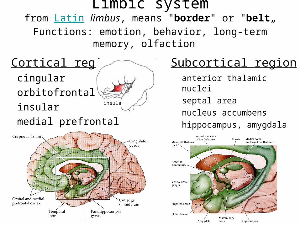
Limbic systemfrom Latin limbus, means "border" or "belt„
Functions: emotion, behavior, long-term memory, olfaction
Cortical regioncingular
orbitofrontal
insular
medial prefrontal
Subcortical regionanterior thalamic nuclei
septal area
nucleus accumbens
hippocampus, amygdala
mammilary body
insula
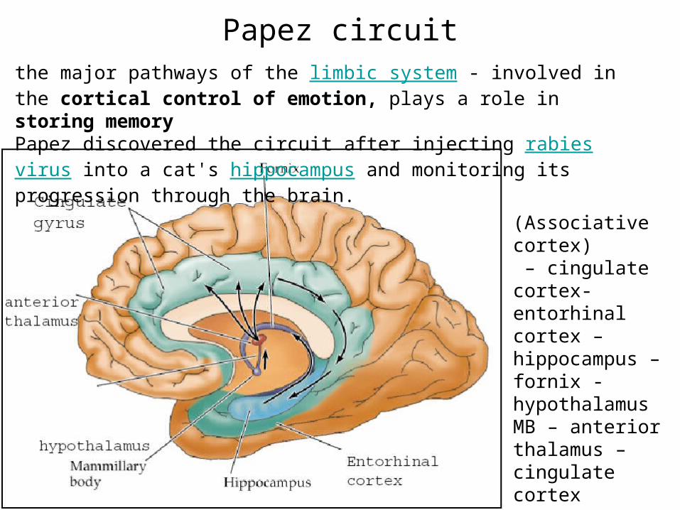
Papez circuit
(Associative cortex) – cingulate cortex- entorhinal cortex – hippocampus – fornix - hypothalamus MB – anterior thalamus – cingulate cortex
the major pathways of the limbic system - involved in the cortical control of emotion, plays a role in storing memory Papez discovered the circuit after injecting rabies virus into a cat's hippocampus and monitoring its progression through the brain.
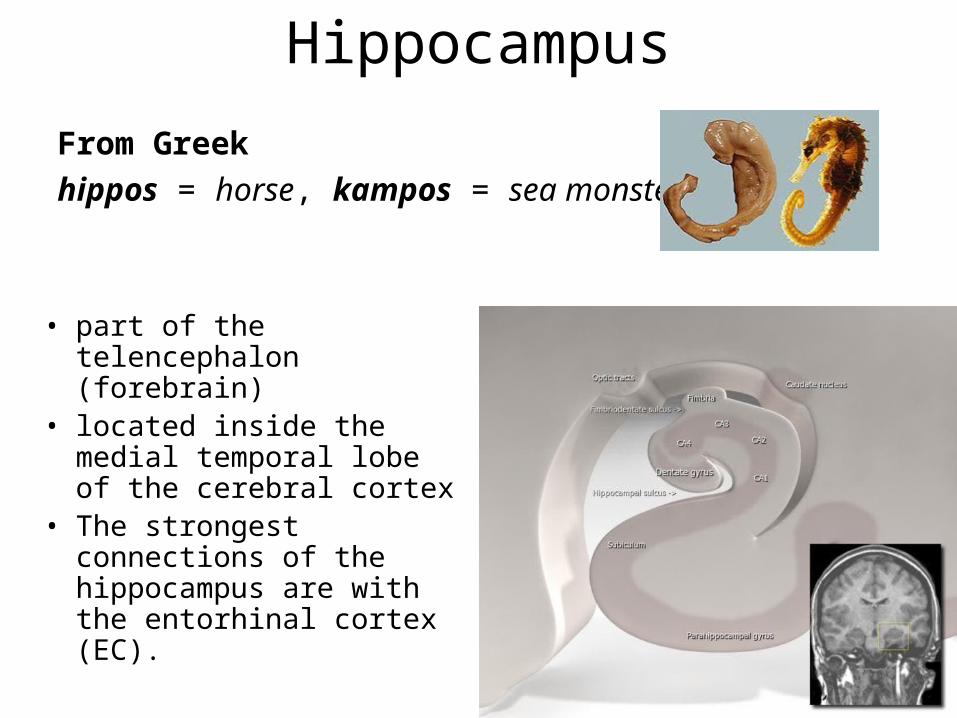
Hippocampus
From Greek
hippos = horse, kampos = sea monster)
• part of the telencephalon (forebrain)
• located inside the medial temporal lobe of the cerebral cortex
• The strongest connections of the hippocampus are with the entorhinal cortex (EC).
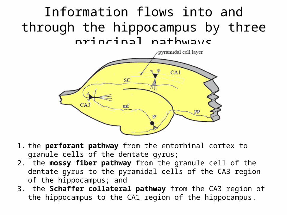
Information flows into and through the hippocampus by three principal pathways
1. the perforant pathway from the entorhinal cortex to granule cells of the dentate gyrus;
2. the mossy fiber pathway from the granule cell of the dentate gyrus to the pyramidal cells of the CA3 region of the hippocampus; and
3. the Schaffer collateral pathway from the CA3 region of the hippocampus to the CA1 region of the hippocampus.
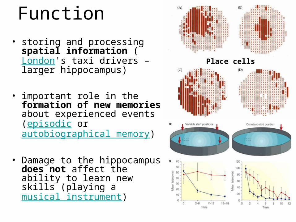
Function
• storing and processing spatial information (London's taxi drivers – larger hippocampus)
• important role in the formation of new memories about experienced events (episodic or autobiographical memory)
• Damage to the hippocampus does not affect the ability to learn new skills (playing a musical instrument)
Place cells
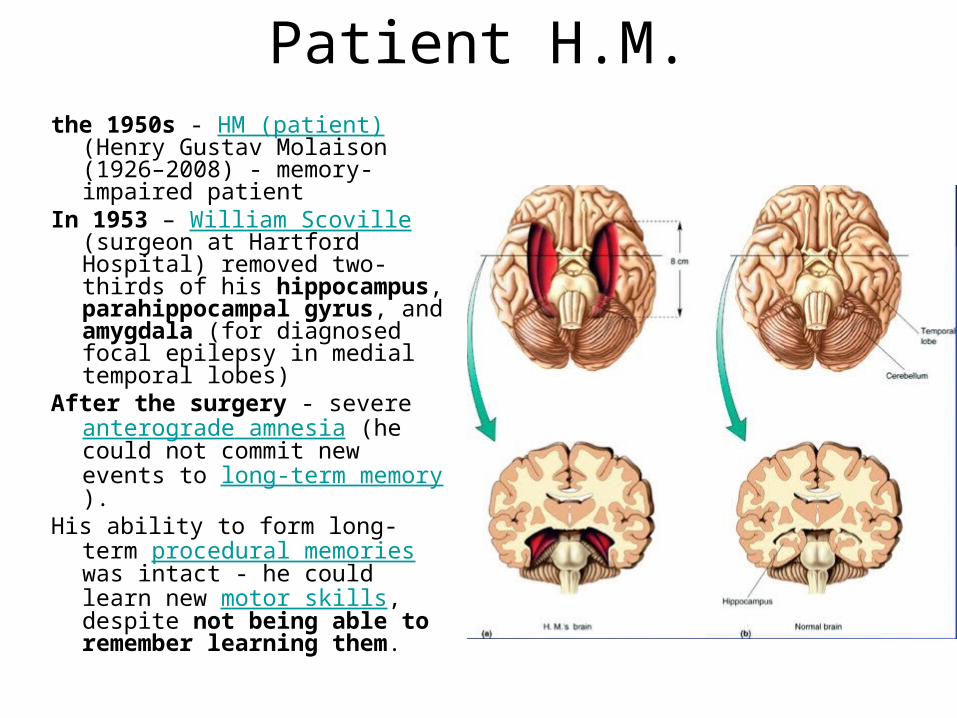
Patient H.M.the 1950s - HM (patient) (Henry
Gustav Molaison (1926–2008) - memory-impaired patient
In 1953 – William Scoville (surgeon at Hartford Hospital) removed two-thirds of his hippocampus, parahippocampal gyrus, and amygdala (for diagnosed focal epilepsy in medial temporal lobes)
After the surgery - severe anterograde amnesia (he could not commit new events to long-term memory).
His ability to form long-term procedural memories was intact - he could learn new motor skills, despite not being able to remember learning them.

LTPLong Term Potentiation (LTP)
• long-lasting improvement in communication between two neurons that results from stimulating them simultaneously
• one of the major cellular mechanisms that underlies learning and memory
• electrical stimulation to a fiber of the perforant pathway caused an excitatory postsynaptic potential (EPSP) in a cell of the dentate gyrus
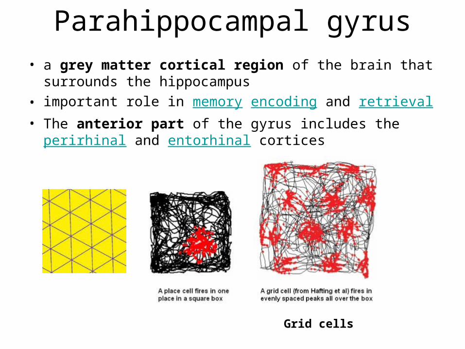
Parahippocampal gyrus
• a grey matter cortical region of the brain that surrounds the hippocampus
• important role in memory encoding and retrieval• The anterior part of the gyrus includes the perirhinal
and entorhinal cortices
Grid cells
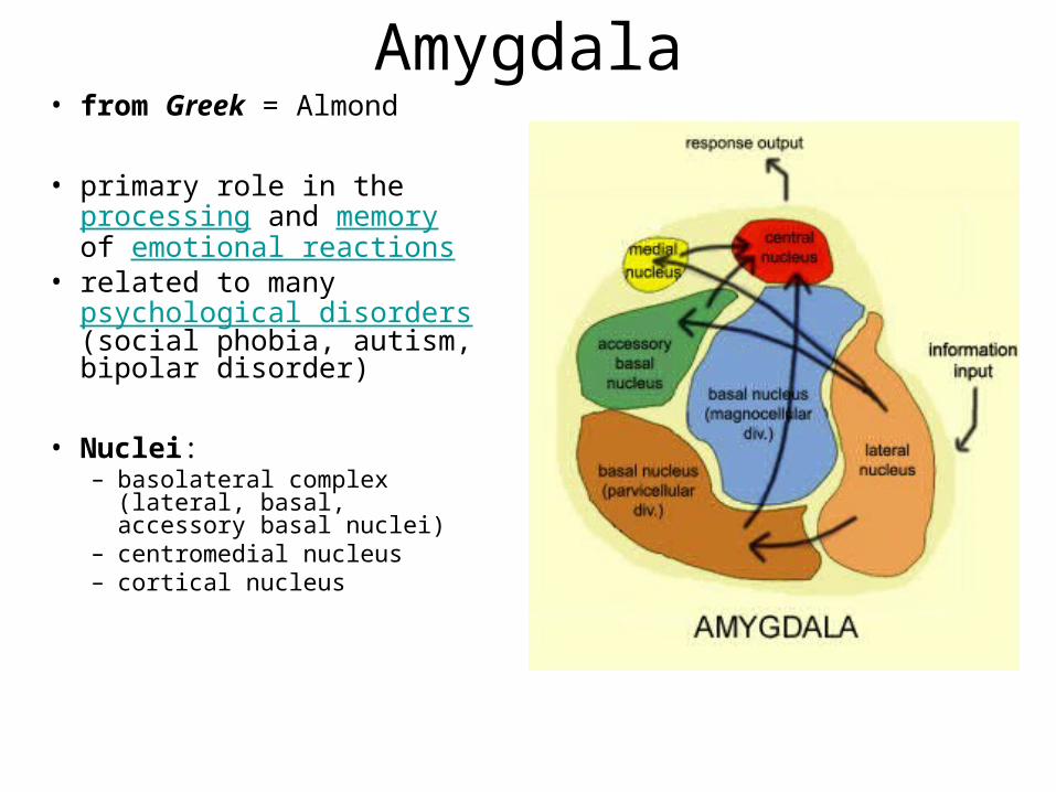
Amygdala• from Greek = Almond
• primary role in the processing and memory of emotional reactions
• related to many psychological disorders (social phobia, autism, bipolar disorder)
• Nuclei: – basolateral complex (lateral,
basal, accessory basal nuclei) – centromedial nucleus– cortical nucleus
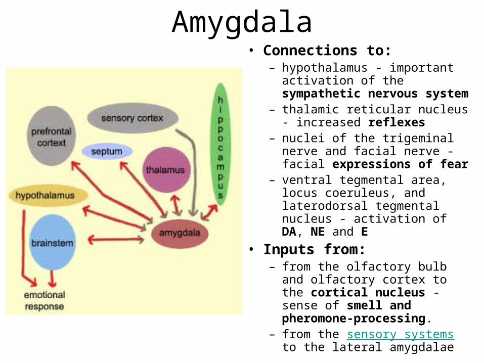
Amygdala• Connections to:
– hypothalamus - important activation of the sympathetic nervous system
– thalamic reticular nucleus - increased reflexes
– nuclei of the trigeminal nerve and facial nerve - facial expressions of fear
– ventral tegmental area, locus coeruleus, and laterodorsal tegmental nucleus - activation of DA, NE and E
• Inputs from:– from the olfactory bulb and
olfactory cortex to the cortical nucleus - sense of smell and pheromone-processing.
– from the sensory systems to the lateral amygdalae
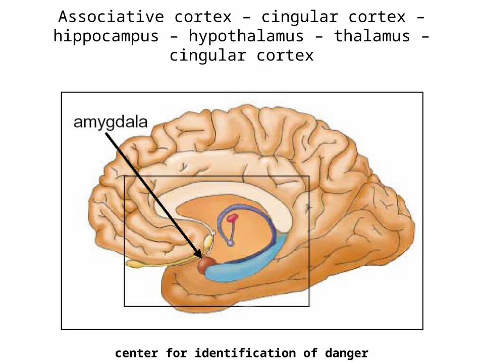
Associative cortex – cingular cortex – hippocampus – hypothalamus – thalamus – cingular cortex
center for identification of danger
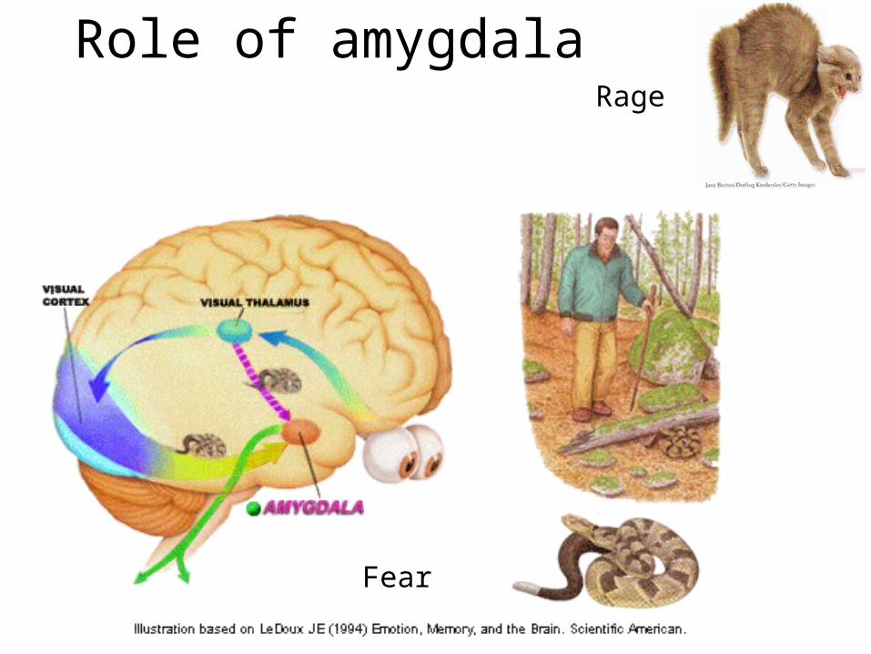
Role of amygdala
Fear
Rage
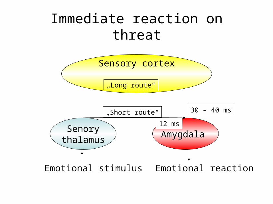
Immediate reaction on threat
Senorythalamus
Amygdala
Emotional stimulus Emotional reaction
Sensory cortex
„Long route“
„Short route“ 30 – 40 ms
12 ms

Conditioned fear
(emotional
learning) memory is stored in
amygdala nuclei
Medina et al. 2002
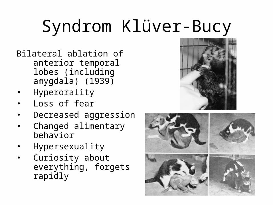
Syndrom Klüver-Bucy
Bilateral ablation of anterior temporal lobes (including amygdala) (1939)
• Hyperorality• Loss of fear• Decreased aggression• Changed alimentary
behavior• Hypersexuality• Curiosity about
everything, forgets rapidly
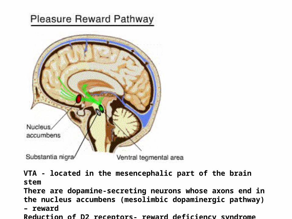
VTA - located in the mesencephalic part of the brain stemThere are dopamine-secreting neurons whose axons end in the nucleus accumbens (mesolimbic dopaminergic pathway) – rewardReduction of D2 receptors- reward deficiency syndrome
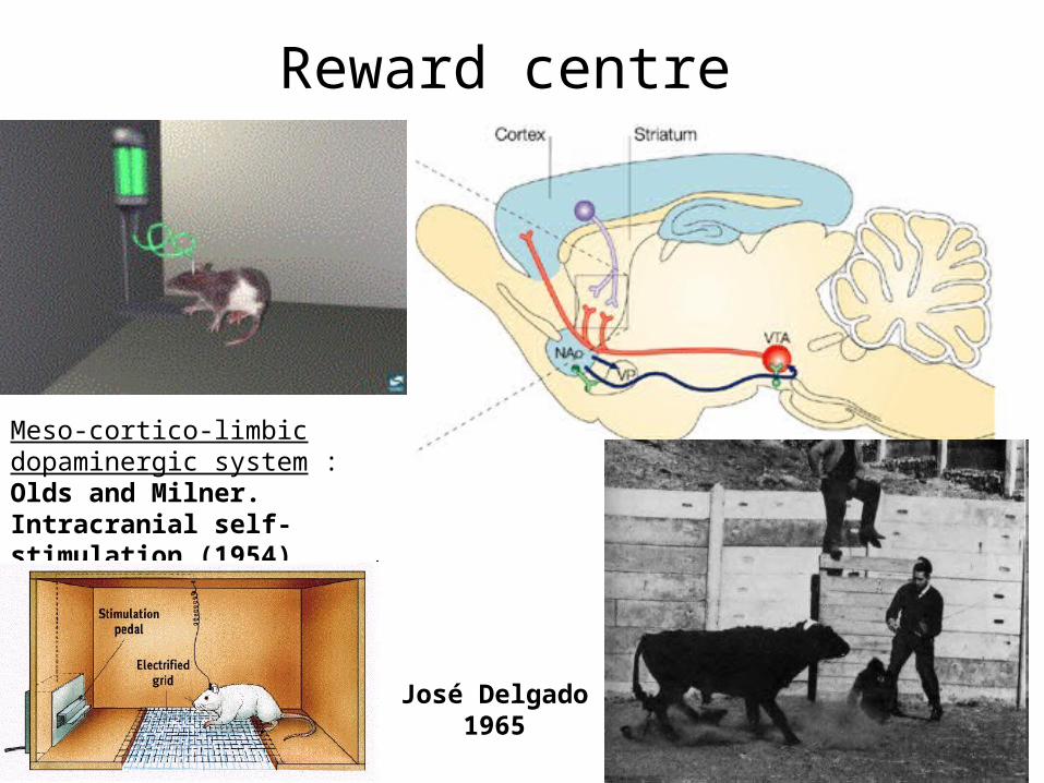
Reward centre
Meso-cortico-limbic dopaminergic system :Olds and Milner. Intracranial self-stimulation (1954)
José Delgado1965
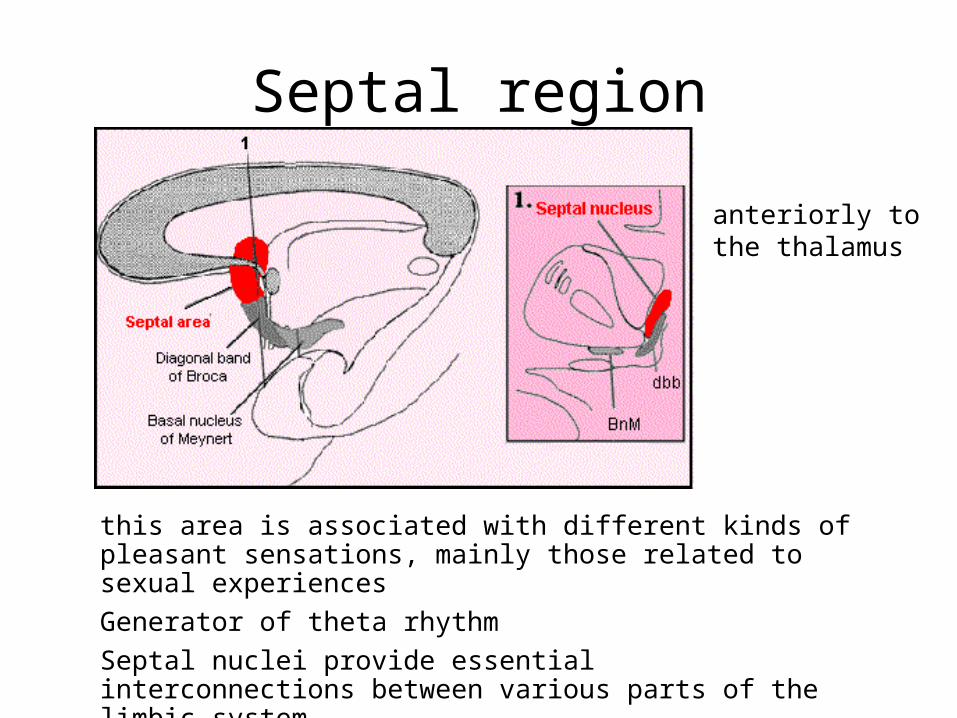
Septal region
this area is associated with different kinds of pleasant sensations, mainly those related to sexual experiences
Generator of theta rhythm
Septal nuclei provide essential interconnections between various parts of the limbic system
anteriorly to the thalamus

NTS: from the vagus (blood pressure and gut distension) Reticular formation: from
the spinal cord (skin temperature)
Retina: from retinohypothalamic tract to SCN (photoperiod)
circumventricular organs: lack a blood-brain barrier (osmolarity, toxins)
amygdala, hippocampus, olfactory cortex
H
Hypothalamus
Endocrine system
Autonomic nervous system
Limbic system
A division of the diencephalon
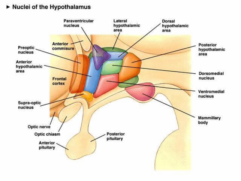
Hypothalamus 12 important nuclei:
• MEDIAL PREOPTIC NUCLEUS– Regulates the release of gonadotropic hormones from the
Adenohypophysis• SUPRACHIASMIC NUCLEUS
– Receives input directly form the retina. – Plays a role in regulating circadian rhythm
• ANTERIOR NUCLEUS– Important in temperature regulation– Stimulates PNS– It’s destruction results in hyperthermia
• PARAVENTRICULAR NUCLEUS– Synthesizes ADH- and thus regulates water balance– Releases oxytocin– Synthesizes CRH - stress
• SUPRAOPTIC NUCLEUS– Synthesizes ADH- and thus regulates water balance– Releases oxytocin
• DORSOMEDIAL NUCLEUS– When stimulated in animals, causes savage behavior!
• VENTROMEDIAL NUCLEUS– Is the satiety center- this means that once it is stimulates, it inhibits the
urge to eat• LATERAL HYPOTHALAMIC NUCLEUS
– Induces eating• ARCUATE (INFUNDIBULAR) NUCLEUS
– Contains neurons that produce factors that stimulate or inhibit action of hypothalamus
– Contains neurons that produce Dopamine• MAMILLARY NUCLEUS
– Lesions (Korsakoff syndrome) are associated with thiamine deficiency and alcoholism – anterograde amnesia
• POSTERIOR HYPOTHALAMIC NUCLEUS– Plays a role in thermoregulation– Lesion results in poikilothermia
• VENTROLATERAL PREOPTIC NUCLEUS (VLPO) - regulation of sleep and wakefulness
Hypothalamus - functions• AUTONOMIC
– Stimulation of the ANTERIOR HYPOTHALAMUS: excitatory effect on parasympathetic system (trophotropic)
– Stimulation of POSTERIOR HYPOTHALAMUS: excitatory effect of sympathetic system (ergotropic)
• THERMOREGULATION– Stimulation of ANTERIOR HYPOTHALAMUS: regulates and maintains
temperature– Stimulation of POSTERIOR HYPOTHAMUS: produces and conserves
heat
• WATER BALANCE– Paraventricular (Supraoptic) nuclei synthesize ADH and control kidney
water excretion
• FOOD INTAKE– Stimulation of VENTROMEDIAL NUCLEUS inhibits the urge to eat– Stimulation of LATERAL HYPOTHALAMIC NUCLEUS induces the
urge to eat
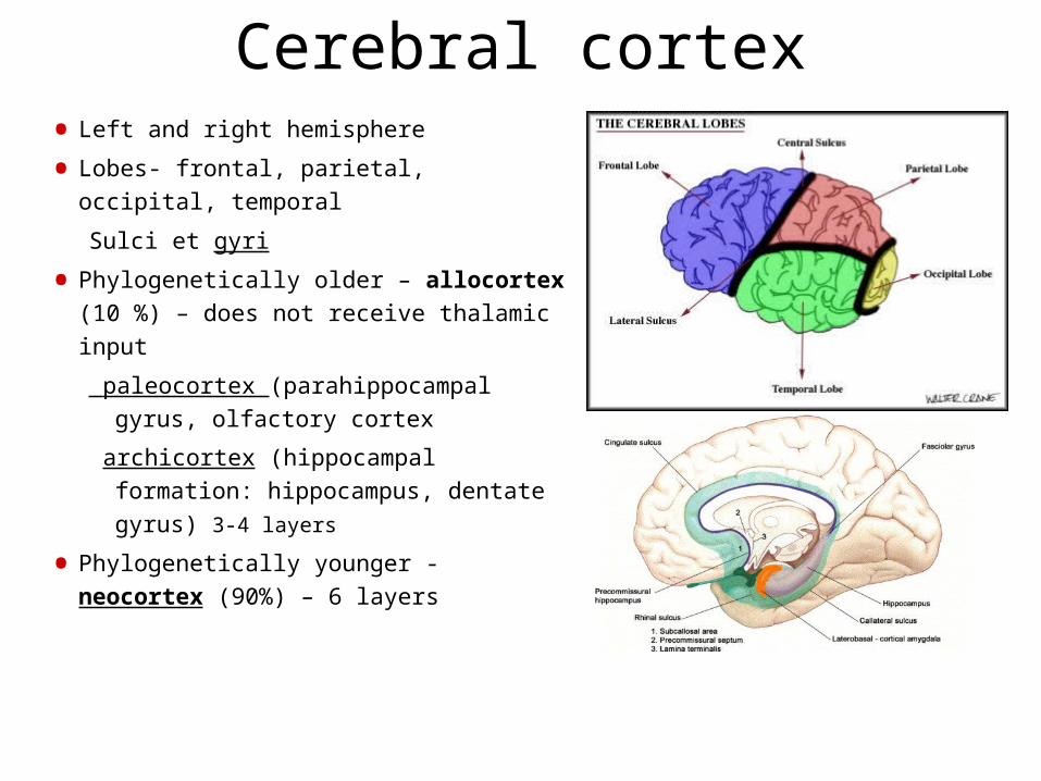
Cerebral cortex• Left and right hemisphere
• Lobes- frontal, parietal, occipital, temporal
Sulci et gyri
• Phylogenetically older – allocortex (10
%) – does not receive thalamic input
paleocortex (parahippocampal gyrus,
olfactory cortex
archicortex (hippocampal formation:
hippocampus, dentate gyrus) 3-4
layers
• Phylogenetically younger - neocortex
(90%) – 6 layers
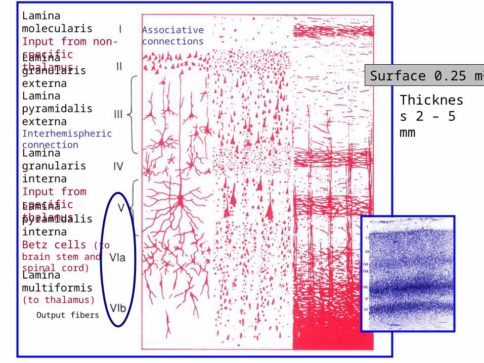
Lamina molecularisInput from non-specific thalamus
Lamina granularisexterna
Lamina pyramidalisexterna Interhemispheric connection
Lamina granularisinterna Input from specific thalamusLamina pyramidalisinternaBetz cells (to brain stem and spinal cord)
Lamina multiformis(to thalamus)
Surface 0.25 m2
Thickness 2 – 5 mm
Output fibers
Associativeconnections
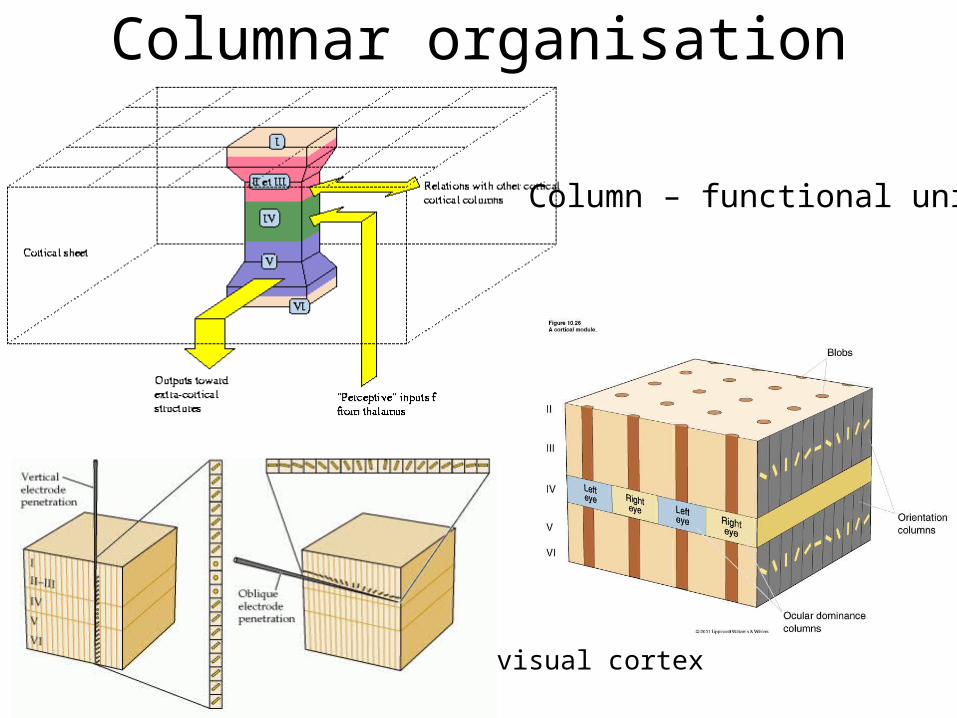
Columnar organisation
Column – functional unite
visual cortex
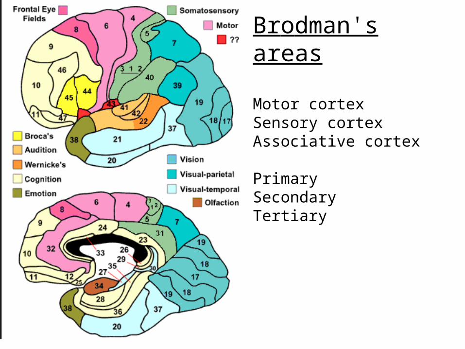
Brodman's areas
Motor cortexSensory cortexAssociative cortex
PrimarySecondary Tertiary
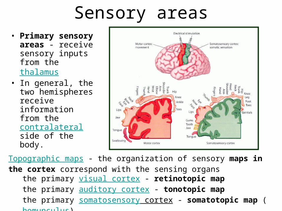
Sensory areas• Primary
sensory areas - receive sensory inputs from the thalamus
• In general, the two hemispheres receive information from the contralateral side of the body.
Topographic maps - the organization of sensory maps in the cortex correspond with the sensing organs
the primary visual cortex - retinotopic mapthe primary auditory cortex - tonotopic mapthe primary somatosensory cortex - somatotopic map (homunculus)
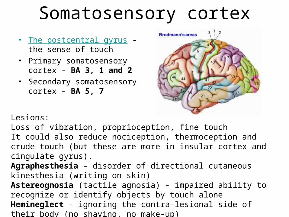
Somatosensory cortex• The postcentral gyrus - the
sense of touch • Primary somatosensory cortex
- BA 3, 1 and 2 • Secondary somatosensory
cortex – BA 5, 7
Lesions:Loss of vibration, proprioception, fine touchIt could also reduce nociception, thermoception and crude touch (but these are more in insular cortex and cingulate gyrus).Agraphesthesia - disorder of directional cutaneous kinesthesia (writing on skin)Astereognosia (tactile agnosia) - impaired ability to recognize or identify objects by touch alone Hemineglect - ignoring the contra-lesional side of their body (no shaving, no make-up)
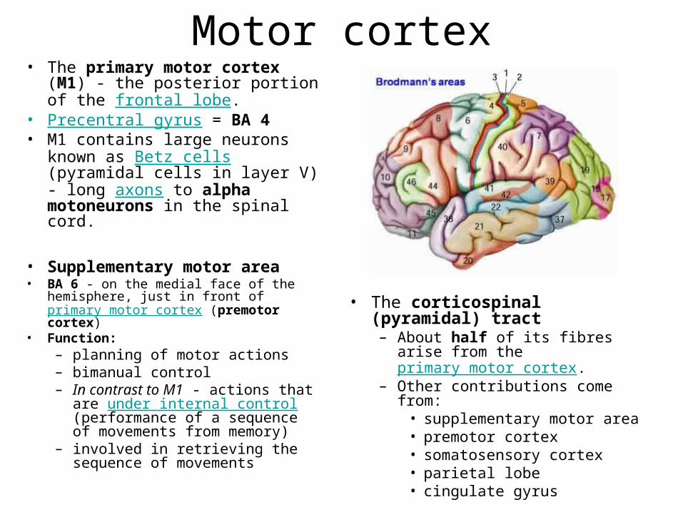
Motor cortex• The primary motor cortex (M1)
- the posterior portion of the frontal lobe.
• Precentral gyrus = BA 4 • M1 contains large neurons
known as Betz cells (pyramidal cells in layer V) - long axons to alpha motoneurons in the spinal cord.
• Supplementary motor area• BA 6 - on the medial face of the
hemisphere, just in front of primary motor cortex (premotor cortex)
• Function:– planning of motor actions– bimanual control – In contrast to M1 - actions that
are under internal control (performance of a sequence of movements from memory)
– involved in retrieving the sequence of movements
• The corticospinal (pyramidal) tract – About half of its fibres arise
from the primary motor cortex. – Other contributions come from:
• supplementary motor area• premotor cortex• somatosensory cortex• parietal lobe• cingulate gyrus
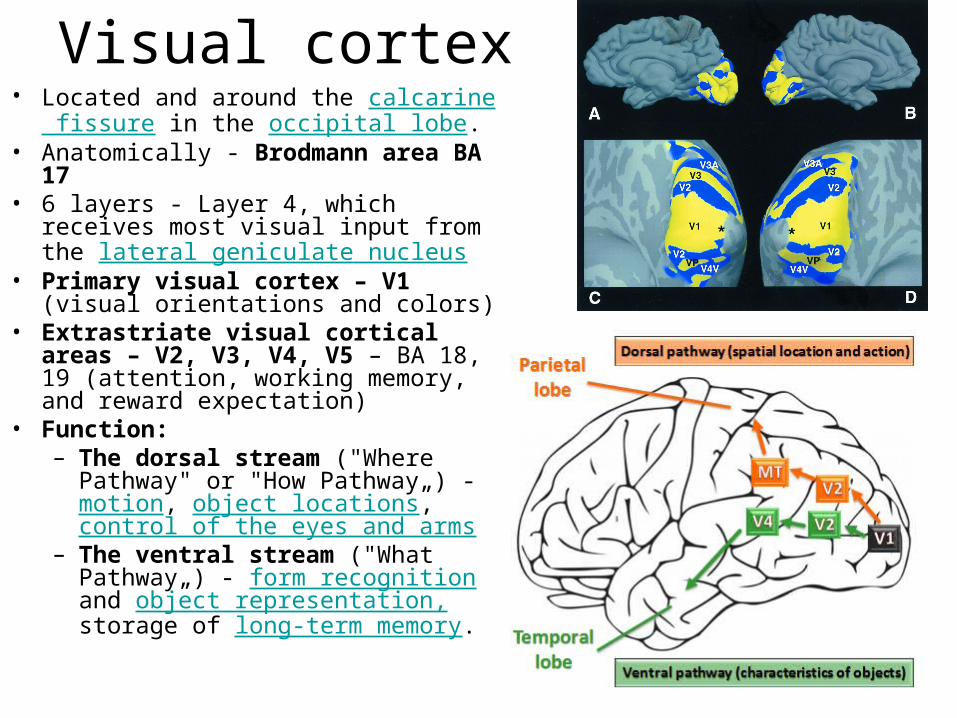
Visual cortex• Located and around the calcarine
fissure in the occipital lobe.• Anatomically - Brodmann area BA 17• 6 layers - Layer 4, which receives
most visual input from the lateral geniculate nucleus
• Primary visual cortex – V1 (visual orientations and colors)
• Extrastriate visual cortical areas – V2, V3, V4, V5 – BA 18, 19 (attention, working memory, and reward expectation)
• Function:– The dorsal stream ("Where
Pathway" or "How Pathway„) - motion, object locations, control of the eyes and arms
– The ventral stream ("What Pathway„) - form recognition and object representation, storage of long-term memory.
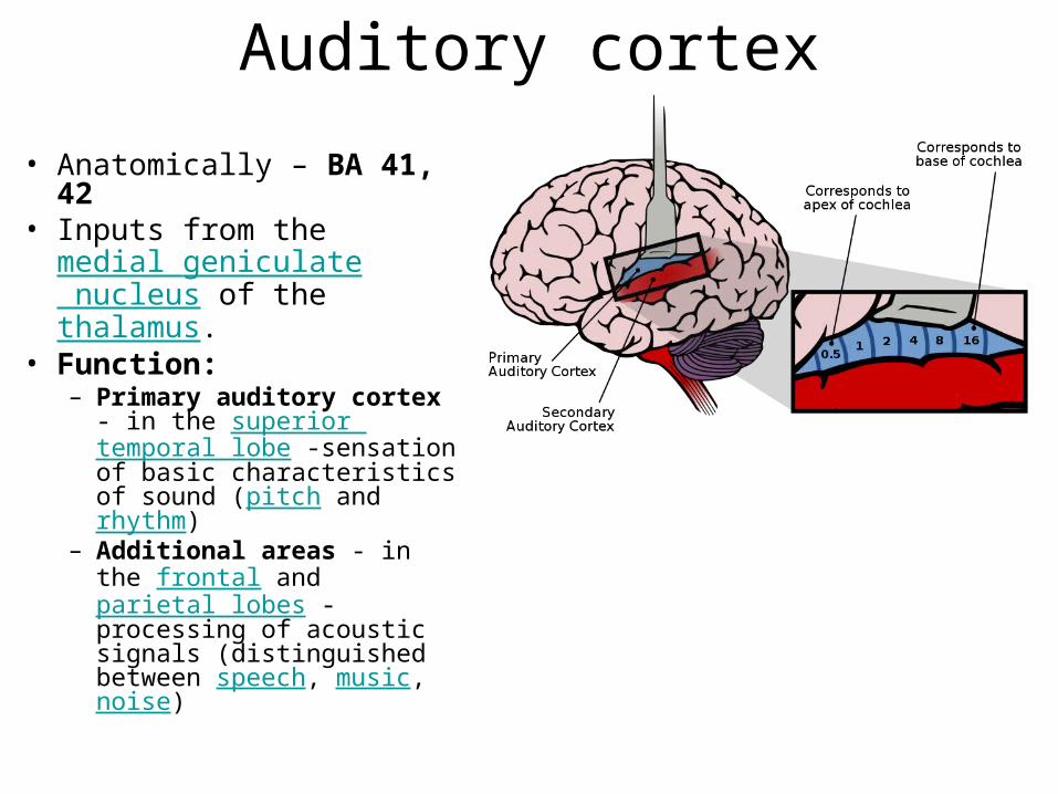
Auditory cortex
• Anatomically – BA 41, 42
• Inputs from the medial geniculate nucleus of the thalamus.
• Function:– Primary auditory cortex -
in the superior temporal lobe -sensation of basic characteristics of sound (pitch and rhythm)
– Additional areas - in the frontal and parietal lobes - processing of acoustic signals (distinguished between speech, music, noise)
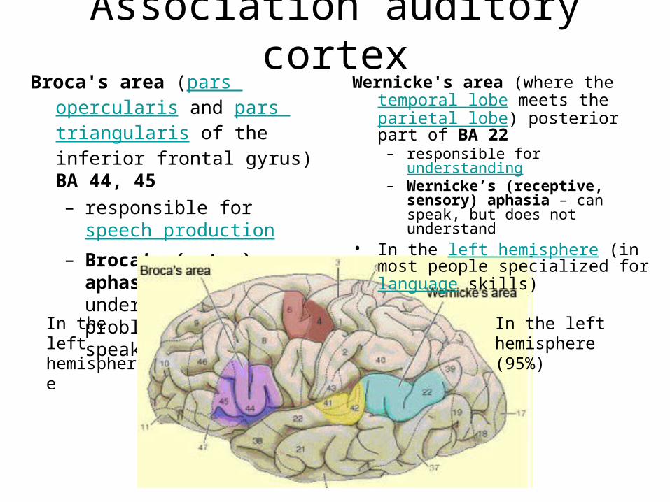
Association auditory cortexBroca's area (pars opercularis and
pars triangularis of the inferior frontal gyrus) BA 44, 45 – responsible for
speech production – Broca’s (motor) aphasia –
understands, but problems with fluent speaking
Wernicke's area (where the temporal lobe meets the parietal lobe) posterior part of BA 22
– responsible for understanding– Wernicke’s (receptive, sensory)
aphasia – can speak, but does not understand
• In the left hemisphere (in most people specialized for language skills)
In the left hemisphere (95%)
In the left hemisphere
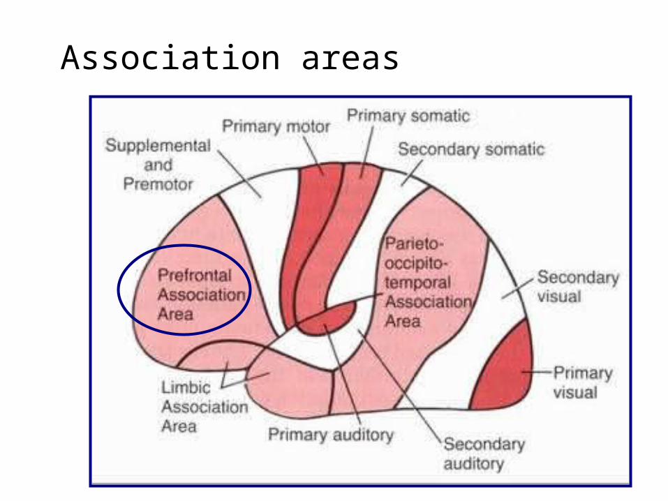
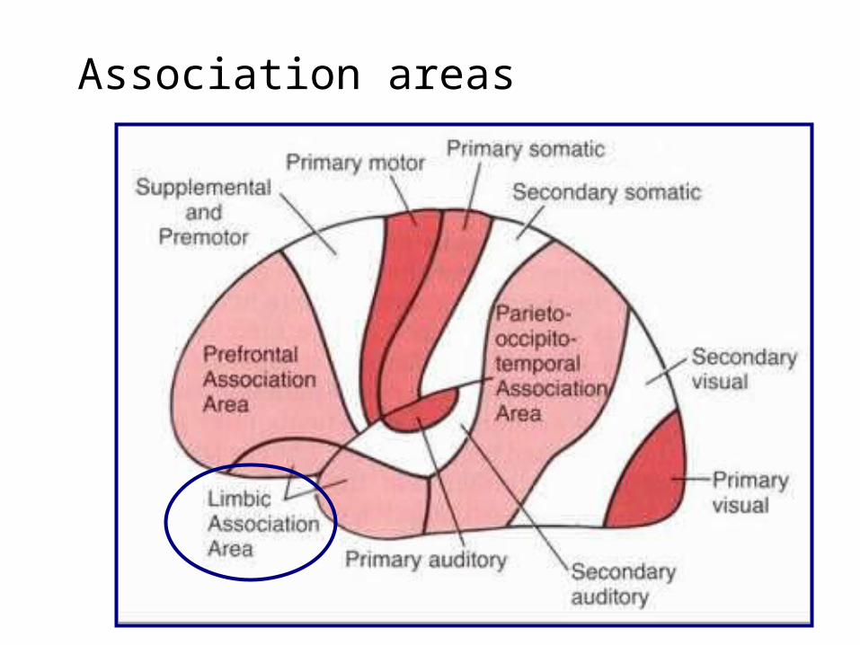
Association areas
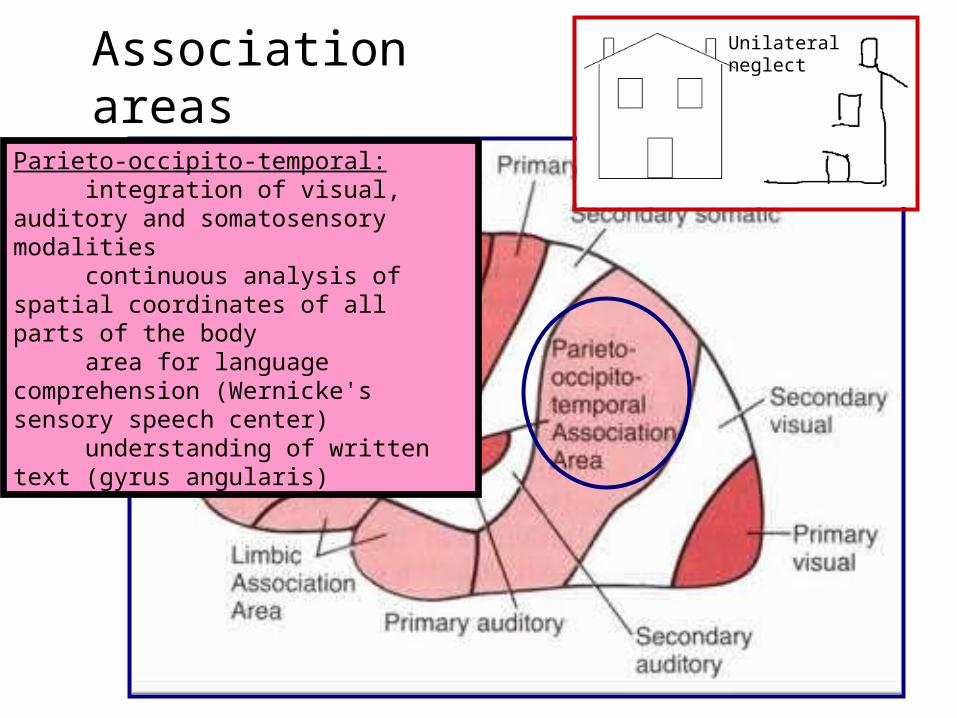
Parieto-occipito-temporal: integration of visual, auditory and somatosensory modalities continuous analysis of spatial coordinates of all parts of the body area for language comprehension (Wernicke's sensory speech center) understanding of written text (gyrus angularis)
Unilateralneglect
Association areas
Frontal lobe
• Reaches full maturity around age 25 – increased myelin in the frontal lobe white matter of young
adults compared to that of teens – A typical onset of schizophrenia in early adult years correlates
with poorly myelinated (inefficient) connections between cells in the fore-brain.
• The frontal lobe contains most of the dopamine-sensitive neurons in the cerebral cortex.
• Functions (involved in higher mental functions):– to recognize future consequences resulting from current
actions– to choose between good and bad actions– override and suppress unacceptable social responses– determine similarities and differences between things or
events
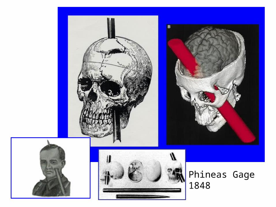
Phineas Gage1848

Damage of frontal lobes
• Impaired mental flexibility and spontaneity, but IQ is not reduced.
• Talking may increase or decrease dramatically. • Increase of risk taking behavior. • Socialization can diminish or increase. • Orbital frontal lobe damage can result in
perverse sexual habits. • Diminished creativity and problem solving
skills. • Frequent distractions.

Prefrontal cortex• the anterior part of the frontal lobes • defined by the presence of an internal granular layer IV
(in contrast to the agranular premotor cortex)• Parts:
– orbitofrontal (OFC) and ventromedial areas (vm-PFC) – dorsolateral prefrontal cortex (dl-PFC) – anterior and ventral cingulate cortex– ventrolateral cortex (vl-PFC)– medial prefrontal cortex (m-PFC)– anterior prefrontal cortex (a-PFC).
• Function:– planning complex cognitive behaviors– personality expression– decision making – moderating correct social behavior
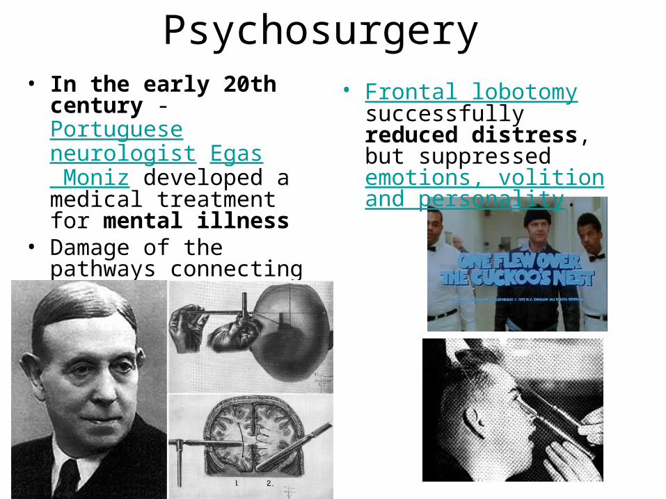
Psychosurgery • In the early 20th century
- Portuguese neurologist Egas Moniz developed a medical treatment for mental illness
• Damage of the pathways connecting the frontal lobe and the limbic system
• Frontal lobotomy successfully reduced distress, but suppressed emotions, volition and personality.
Association areas
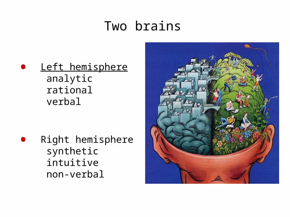
Two brains
• Left hemisphereanalyticrationalverbal
• Right hemispheresyntheticintuitivenon-verbal

![RESEARCH ARTICLE Open Access Abnormalities of cortical ......campus [18], and basal ganglia [19]. These abnormal brain regions are predominantly located in the limbic-cortical-striatal-pallidal-thalamic](https://img.pdfslide.net/doc/110x75/60c2168295781709ed2436d2/research-article-open-access-abnormalities-of-cortical-campus-18-and.jpg)